Communication

Long-Term Tumor-Targeting Effect of E. coli as a Drug Delivery System

1
Department of Nuclear Medicine, Dongnam Institute of Radiological and Medical Sciences, Busan 46033, Republic of Korea
2
Department of Advanced Materials Chemistry, Dongguk University, Gyeongju 38066, Republic of Korea
3
Department of Medical Biotechnology, Dongguk University, Gyeongju 38066, Republic of Korea
*
Authors to whom correspondence should be addressed.
Pharmaceuticals 2024, 17(4), 421; https://doi.org/10.3390/ph17040421
Submission received: 27 February 2024 / Revised: 21 March 2024 / Accepted: 22 March 2024 / Published: 26 March 2024
(This article belongs to the Special Issue Recent Advancements in Radiochemistry and PET Radiotracer Development)

Abstract

:
To overcome the limitations of current nano/micro-scale drug delivery systems, an Escherichia coli (E. coli)-based drug delivery system could be a potential alternative, and an effective tumor-targeting delivery system can be developed by attempting to perform chemical binding to the primary amine group of a cell membrane protein. In addition, positron emission tomography (PET) is a representative non-invasive imaging technology and is actively used in the field of drug delivery along with radioisotopes capable of long-term tracking, such as zirconium-89 (89Zr). The membrane proteins were labeled with 89Zr using chelate (DFO), and not only was the long-term biodistribution in tumors and major organs evaluated in the body, but the labeling stability of 89Zr conjugated to the membrane proteins was also evaluated through continuous tracking. E. coli accumulated at high levels in the tumor within 5 min (initial time) after tail intravenous injection, and when observed after 6 days, 89Zr-DFO on the surface of E. coli was found to be stable for a long period of time in the body. In this study, we demonstrated the long-term biodistribution and tumor-targeting effect of an E. coli-based drug delivery system and verified the in vivo stability of radioisotopes labeled on the surface of E. coli.

1. Introduction

Over recent decades, a range of systems for the delivery of therapeutic agents, including polymeric micro/nanoparticles, liposomes, and magnetic-based nanoparticles, have been developed and assessed [1,2,3]. These systems, which rely on either active or passive targeting mechanisms, have undergone rigorous testing in animal models and clinical trials. However, a persistent issue is their nonspecific distribution throughout the bloodstream, which can lead to significant toxicity and undesirable side effects [4]. Hence, it is crucial to develop advanced drug delivery systems that can bypass physical and biological barriers, ensuring that cancerous tissues receive adequately high drug concentrations. Bacteria utilize their cellular chemical energy to drive their helical, flagellum-fueled bio-motors, enabling fluidic propulsion. These living, motile bacteria, equipped with surface appendages like flagella and pili, display a variety of movement behaviors. These include swimming, swarming, twitching, gliding, and sliding, which permit them to navigate both liquid and semi-solid environments [5,6,7]. The process of bacterial cells actively moving toward more favorable conditions is known as bacterial taxis. This autonomous response to environmental stimuli can be tactically leveraged to direct them to specified locations within the body, a crucial aspect in designing drug delivery systems [8,9,10]. The capacity of bacterial cells to navigate through oxygen gradients is an integral attribute that aids in identifying bacteria with tumor-targeting potential. For instance, studies have revealed that Escherichia coli (E. coli) selectively colonizes and proliferates in tumor regions due to its anaerobic properties upon introduction into the body [11,12]. Concurrently, E. coli present in normal tissues is progressively eliminated by the immune system over time. For bacterial-based drug delivery, chemotherapeutic, radiation therapeutic, photothermal therapeutic, and immunotherapeutic drugs are loaded to bacteria. They can bind to the bacterial surface using chemical linkers such as cis-aconitic anhydride or bacterial-binding antibody interactions, and the drug can be contained inside the bacteria using bacterial metabolism. In addition, drug-loaded nanoparticles can be chemically bound to the bacterial surface or can conjugate through surface charge and complementary noncovalent interactions to enhance drug loading efficiency [6]. E. coli DH5α is a genetically modified non-invasive strain with the unique capability to absorb exogenous gene products and an immunity to hydrolysis by endonucleases. These attributes render E. coli DH5α as an excellent candidate for genetic modifications and targeted delivery [13,14]. The methodology we propose hinges on the ability of primary amine groups present on the surface of Gram-negative bacterial cells, such as E. coli, to form covalent bonds with various compounds that contain carboxyl or isothiocyanate groups [15]. This feature lays the foundation for the development of a robust targeted delivery system. Non-invasive imaging techniques such as positron emission tomography (PET) have made significant strides and play a pivotal role in the field of personalized medicine [16,17]. These advancements offer practical solutions to a range of currently incurable diseases, including cancer. Recently, there has been growing interest in the use of emerging radioactive isotopes including zirconium-89 (89Zr, half-life = 78.4 h) [18,19,20], copper-64 (64Cu, half-life = 12.7 h) [21,22], yttrium-86 (86Y, half-life = 14.7 h) [23,24], and gallium-68 (68Ga, half-life = 67.6 m) [25,26] for PET. The long-term in vivo tracking of drug carriers, roughly the size of a bacterium, warrants investigation. Of these radioisotopes (Ris), 89Zr has been thoroughly studied and shows promise for long-term tumor diagnostic studies and in vivo distribution research [27,28]. This is attributed to the ease of its relatively long-term production, its optimal nuclear decay characteristics, and its suitable chemical binding properties. In this study, we labeled E. coli membrane proteins with 89Zr and confirmed its long-term in vivo biodistribution in tumors and major organs (Scheme 1). In addition, by continuously tracking 89Zr, we verified the labeling stability of 89Zr chemically bound to E. coli membrane proteins and we report the excellent in vivo stability of 89Zr-labeled E. coli.

2. Results and Discussion
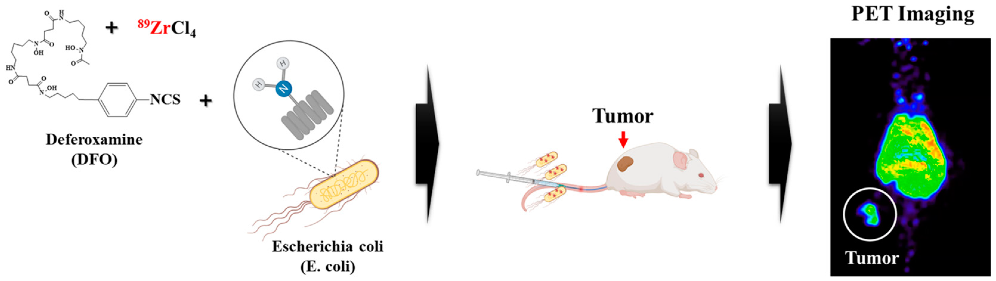
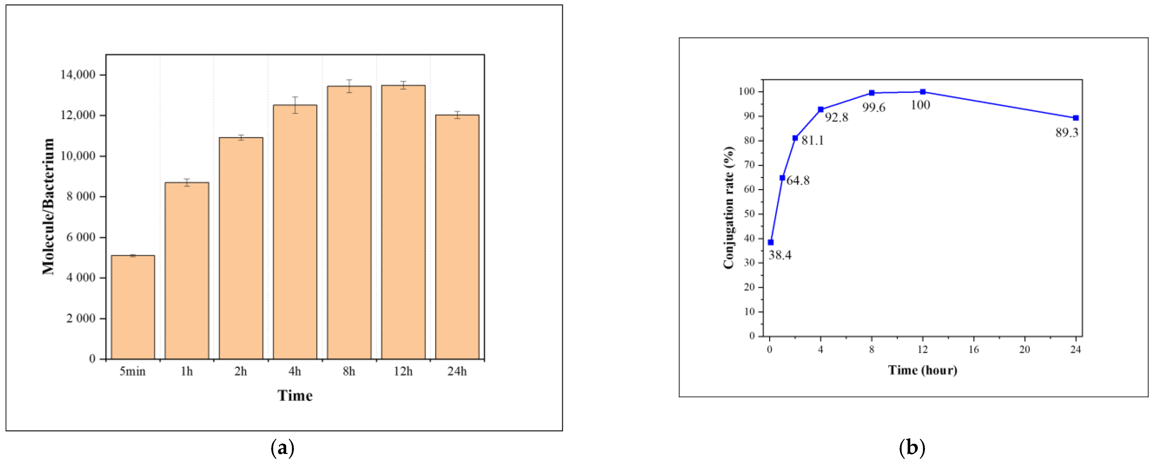
2.1. Synthesis

The binding conditions between the primary amine groups of E. coli membrane proteins and p-NCS-Bz-DFO were analyzed through quantitative analysis using the fluorescent molecule FITC, which has an isothiocyanate (-NCS) group at its terminal. As a result of analyzing the reaction time, as shown in Figure 1, the highest binding rate was observed when the reaction was performed for 8 h. Nevertheless, we adopted a 1 h reaction time as this was advantageous for preserving the radioactivity of 89Zr and minimizing stress on the chemical reaction of E. coli. The FITC and E. coli were confirmed to bind to 65% of the maximum binding amount after 1 h.
The morphological damage and viability of E. coli were evaluated after sequential reactions of p-NCS-Bz-DFO and Zr. As shown in Figure 2, E. coli that did not participate in the chemical reaction (Figure 2a) and Zr-DFO-conjugated E. coli (Figure 2b) were morphologically identical. Since Zr-DFO-conjugated E. coli did not shrink or tear morphologically, the chemical reactions of the two steps did not affect the morphology of E. coli. Additionally, it was confirmed, as seen in Figure 3a, that more than 75% of Zr-DFO-conjugated E. coli survived.
In Figure 4a, as a result of evaluating the radiochemical purity of 89Zr-labeled E. coli purified by means of centrifugation after 89Zr labeling using a radio-thin-layer chromatography (TLC) scanner, a high radiochemical purity of 99.9% was confirmed. This high radiochemical purity was also confirmed when the instant thin-layer chromatography (i-TLC) plate was imaged using a PET scan (Figure 4b).
In the in vitro stability evaluation conducted using PBS and HS, high stability of 95% or more was confirmed for 7 days. This high stability of 89Zr-labeled E. coli was confirmed through in vitro stability evaluation, and it is expected that long-term stability in blood can be secured, meaning that it can be used for tumor tracking and evaluating major organ distribution (Figure 3b).

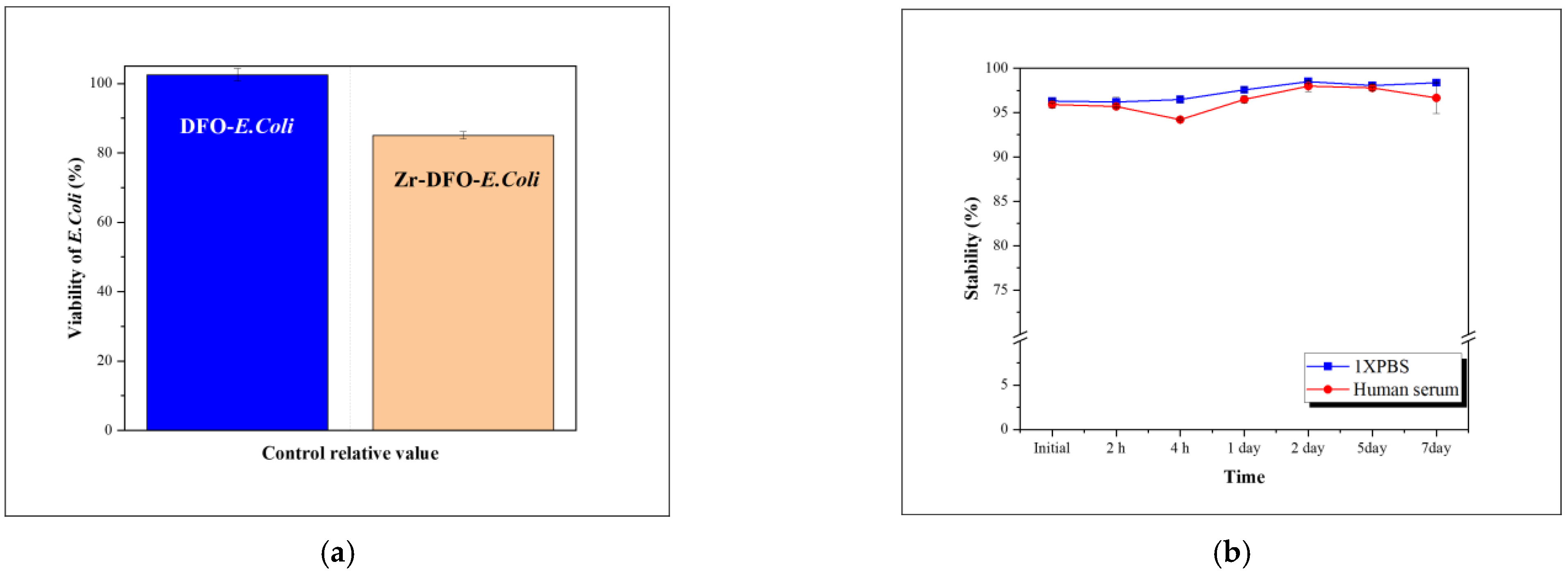
2.2. Cellular Uptake
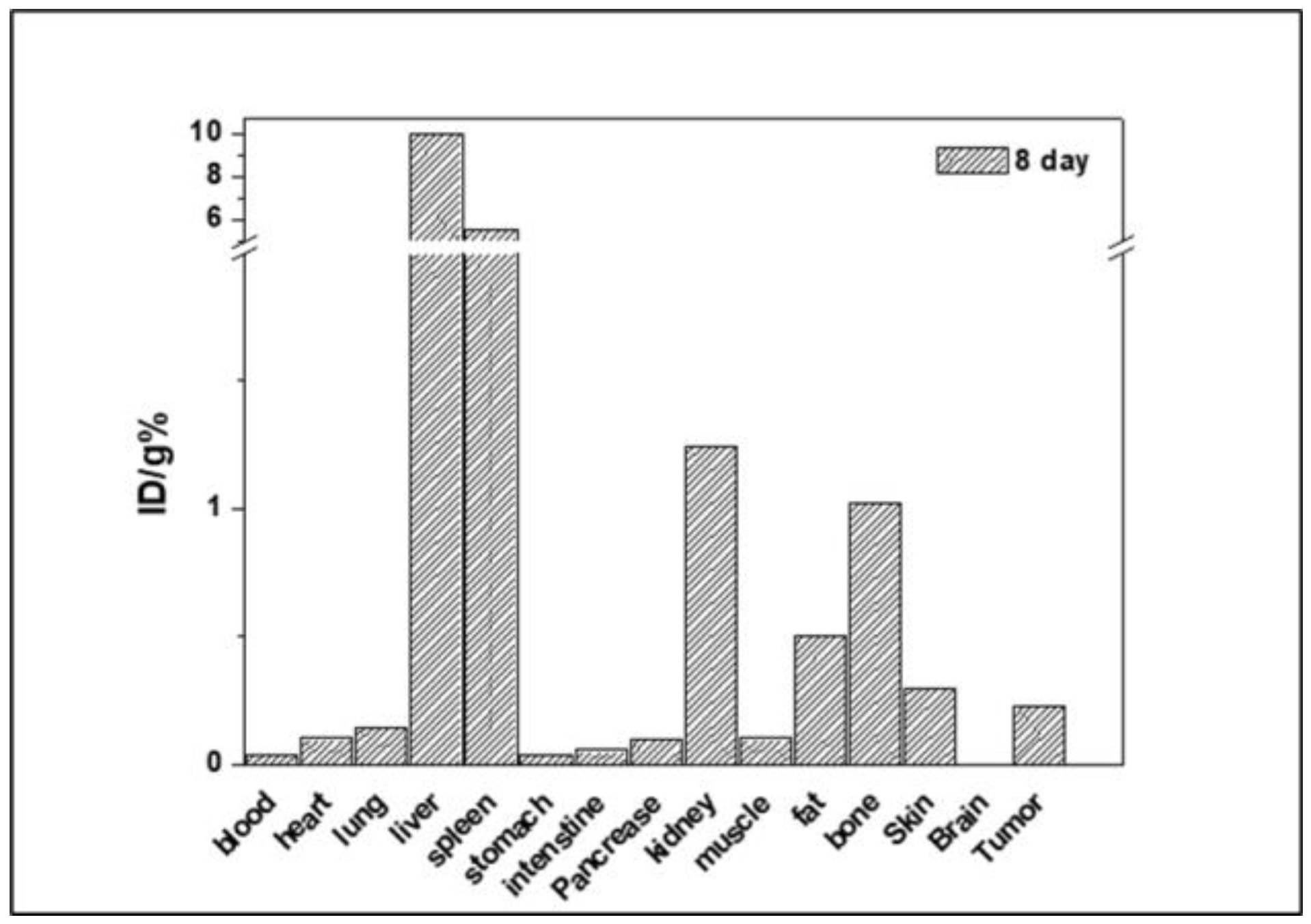
In the evaluation of cellular uptake for CT-26 and A549, the uptake of 89Zr-labeled E. coli into cancer cells was confirmed. CT-26 is a mouse colon cancer cell line and A549 is a human lung cancer cell line. An analysis of both cancer cells confirmed that the uptake was increased in a time-dependent manner; in the case of A549, the uptake rate was close to 25% after 24 h, and in the case of CT-26, it was confirmed that the uptake rate was more than 15%. Although there was a difference in the amount of uptake of E. coli depending on the cancer cell line, it was consistent that the uptake of E. coli continuously increased for 24 h. Although further studies are needed, this suggests that E. coli-based drug delivery can be effective in various cancer cells and confirms the possibility of continuous accumulation in tumors in the body (Figure 5).

2.3. PET Studies

PET was performed to evaluate the drug delivery effect of 89Zr-labeled E. coli in the body. 89Zr-labeled E. coli cells were infused via tail vein injection and were evaluated for 6 days (Figure 6). It was confirmed that E. coli cells accumulated in the cancer tissue at the initial time after tail intravenous injection, and it was found that the accumulation was maintained for 6 days. The representative reticuloendothelial system (RES), including the liver and spleen, showed high accumulation, but as shown in Figure 7, as a result of tracking the regions of interest (ROIs) of the liver, bladder, and tumor for 6 days, time-dependent clearance of 89Zr-labeled E. coli was confirmed. As no increase in excretion-related organ- and bone-specific uptake was observed over time, it is believed that there was no decomposition of E. coli by fasting cells of the liver and spleen. The clearance of E. coli to the RES in the future is worth studying intensively. The high accumulation in the liver and spleen needs to be improved to increase drug delivery efficiency. Studies that reduce toxicity to normal cells by introducing polyethylene glycol (PEG) on the bacterial surface can be referred to, and as the introduction of PEG reduces the accumulation of the RES of nanoparticles, it needs to be studied in E. coli-based drug delivery systems [29]. The radioactivity observed in the bladder over a period of 1 day appears to be related to the clearance of 89Zr-DFO dissociated from E. coli, considering the diameter of E. coli cells (1–2 µm) [30,31]. In the case of tumors, 89Zr-labeled E. coli accumulation was observed to increase, and the tumor/liver ratio was also confirmed to continuously increase for 6 days. Dissociated 89Zr has been reported to accumulate in bones in the body [31,32,33]. Nevertheless, bone accumulation by 89Zr was not observed, as seen in Figure 7, so it is not dissociated from E. coli after intravenous injection in the body, and this result is associated with high in vitro labeling stability (Figure 4b). In addition, E. coli has high potential as a drug delivery system that accumulates in tumor tissues at a very fast rate and is maintained for a long time. Eight days after intravenous injection, the mice were sacrificed to confirm the biodistribution in major organs. As shown in Figure 8, there was low accumulation in major organs except for the liver and spleen, suggesting that E. coli-based drug carriers can reduce side effects on normal organs.

3. Materials and Methods

3.1. Materials

Fluorescein isothiocyanate (FITC), diethylenetriaminepentaacetic acid (DTPA), dimethyl sulfoxide (DMSO), zirconium (IV) chloride, and hydrogen chloride (HCl) were purchased from Merck (Darmstadt, Germany). N1-hydroxy-N1-(5-(4-(hydroxy(5-(3-(4-isothiocyanatophenyl)thioureido)pentyl)amino)-4-oxobutanamido)pentyl)-N4-(5-(N-hydroxyacetamido)pentyl)succinamide (p-NCS-Bz-DFO) was purchased from Chematech (Dijon, France). Dulbecco’s modified Eagle’s medium (DMEM), phosphate-buffered saline (PBS), HEPES buffer solution, human serum (HS), and fetal bovine serum (FBS) were purchased from Gibco BRL Life Technologies (Waltham, MA, USA). QMA Plus Short Cartridge was purchased from Waters (Worcester County, MA, USA). Bacterial Viability Kit (LIVE/DEAD™ BacLight™ Bacterial Viability Kit, Middlesex County, MA, USA) was purchased from Thermo Fisher Scientific (Middlesex County, MA, USA). CT-26 and A549 cells were procured from the Korean cell line bank (Seoul, Republic of Korea). Balb/c mice (20 ± 1.5 g, female) were purchased from Orient Bio (Seongnam, Republic of Korea). Zirconium-89 was procured from the Korea Atomic Energy Research Institute (KAERI, Jeongeup, Republic of Korea) and produced using RFT-30 (30 MeV cyclotron). Quantitative analysis of FITC was performed using a fluorescence spectrometer (Hitachi, Tokyo, Japan). Morphological data of E. coli were obtained using a field-emission scanning electron microscope (FE-SEM) (Hitachi, Tokyo, Japan). Radiochemical yield was assessed using an AR-2000 radio-TLC imaging scanner (Bioscan, Santa Barbara, CA, USA). The in vitro stability of the radiolabeled sample was measured using a Wizard-1470 automatic gamma counter (Perkin Elmer, Waltham, MA, USA). Small animal PET imaging was carried out using a Genesis 4 (Sofie Biosciences, Culver City, CA, USA).

3.2. Preparation of FITC-Conjugated E. coli

One milligram of FITC was placed in a 10 mL vial and dispersed in 0.1 mL of DMSO. Then, 9.9 mL of 0.1 M HEPES (pH 8) was added into the vial and stirred for 10 min. After stirring, E. coli (1.0 × 10⁹ cells) was added and incubated for 1 h at 25 °C. The solvent and FITC-labeled E. coli were separated by means of centrifugation and washed two times with physiological saline to remove unreacted FITC. Centrifugation was carried out at 500 rcf for 5 min. FITC-conjugated E. coli was measured with a fluorescence spectrometer and the calibration curve was used to quantify FITC bound to the E. coli surface.

3.3. Preparation of Zr-Conjugated E. coli

One milliliter of E. coli (2 × 10⁸/mL in 0.1 M HEPES) was reacted with 80 nmol of p-NCS-Bz-DFO for 1 h, followed by washing twice with PBS. Then, 1 mL of 0.1 M HEPES (pH 8) was reacted with 80 nmol of ZrCl4 for 1 h. After the reaction, E. coli bacteria were washed twice with 50 mM DTPA to remove free Zr. Centrifugation was carried out at 500 rcf for 5 min each time. For the viability evaluation of E. coli after the chemical reaction, the activity of E. coli was measured using a bacterial viability kit.

3.4. Preparation of 89Zr-Labeled E. coli

89Zr(ox)2 was exchanged for ZrCl4 using a QMA cartridge and 1M HCl aqueous solution was used as the elution solvent. The reaction was performed at room temperature for 1 h by adding 89ZrCl4 of 37 MBq/10 μL to a 2 mL tube containing 1 μg of p-NCS-Bz-NCS. Then, a 1 mL E. coli dispersion (1 × 10⁹/mL in 0.1 M HEPES, pH8) was added to the reaction vial. After the completion of the reaction, the solvent and 89Zr-labeled E. coli were separated using centrifugation, washed with a 50 mM DTPA (pH 7) solution to remove free 89Zr, and washed two more times with PBS. Centrifugation was performed at 500 rcf for 5 min each time.

3.5. In Vitro Stability

89Zr-labeled E. coli bacteria were dispersed in PBS to test their in vitro stability. Then, 100 μL of E. coli, labeled with 37 MBq/mL, was added to 1 mL of 10% HS and 10% FBS. The mixture in the tube was carefully stirred at 37 °C for 7 days. The in vitro stability was assessed at specific time intervals (1, 2, and 4 h and 1, 2, 5, and 7 days) using a radio-TLC scanner. For the chromatographic process, the mobile phase utilized 50 mM of DTPA, while the stationary phase employed i-TLC-SG with dimensions of 1.0 × 10 cm.

3.6. Cellular Uptake

For cellular uptake, 1.0 × 10⁵ cancer cells (CT-26 and A549) were seeded into 24-well plates and grown in DMEM medium containing 10% (v/v) FBS at 37 °C for 24 h. 89Zr-labeled E. coli was added with 190 kBq/well to cancer cells and incubated for 48 h at 37 °C. The sample was washed two times with PBS and cell-associated radioactivity was detected using a gamma ray counter at 0.5, 1, 4, and 24 h.

3.7. PET Studies and Ex Vivo Biodistribution

PET studies were conducted in accordance with animal testing guidelines and ethics and were approved by the Korea Atomic Energy Research Institute (IACUC KAERI-2023-002). PET images were taken to determine the in vivo distribution of 89Zr-labeled E. coli. CT-26 cells were dispersed in saline and injected subcutaneously into the thighs of Balb/c mice (5.0 × 10⁶ per mouse). After the length of tumors had grown to 120 mm, 89Zr-labeled E. coli was injected intravenously into the tail of each mouse (3.7 MBq/100 µL per mouse). As for the injection dose, 3.7 MBq was selected to effectively track Zr-89 for more than 6 days. The mice were anesthetized with 2% isoflurane and whole-animal imaging was measured using PET for 6 days. Mice were measured by designating a time point of 30 min, 1, 2, 4, 6, and 24 h, and 2, 5, and 6 days, and the time required for the measurement was approximately 5 min. Then, the mice were sacrificed and ex vivo biodistribution in major organs was measured using PET and a gamma counter. PET studies were analyzed using Amide’s Medical Image Data Examiner (AMIDE, open-source software tool, version 1.0.6). Using PET image reconstruction and quantification conditions, the maximum threshold was 10 and the minimum threshold was 0. The voxel size was 0.45 × 0.45 × 0.45 mm. The quantitative values were calculated using a maximum SUV.

4. Conclusions

In this study, we confirmed the remarkable effectiveness and in vivo stability of an E. coli-based drug carrier for cancer diagnosis and treatment using the radioisotope 89Zr. E. coli accumulated in the tumor quickly (within 5 min) after intravenous injection and remained accumulated for 6 days. Additionally, 89Zr-labeled E. coli cells were confirmed to be stable for 7 days in human serum. Since the accumulation of free 89Zr in bone was not observed when it was administered, it was confirmed that 89Zr was specifically bound to the chelate (DFO). Considering that clearance through the bladder did not increase after 24 h, 89Zr-DFO on the surface of E. coli was stable for a long period of time in the body. In conclusion, this study can contribute to research on the in vivo tracking of various Gram-negative bacteria with tumor therapeutic effects and to the development of a tumor-targeting drug delivery system that can overcome physical and biological barriers.

Author Contributions

Conceptualization, G.G.K. and S.W.K.; data curation, G.G.K., D.B.J. and J.-S.S.; formal analysis, D.B.J.; funding acquisition, H.L. and S.W.K.; methodology, G.G.K.; project administration, J.-S.S.; validation, D.B.J.; writing—original draft, G.G.K.; writing—review and editing, H.L., J.-S.S. and S.W.K. All authors have read and agreed to the published version of the manuscript.

Funding

This work was supported by the Dongnam Institute of Radiological & Medical Sciences (DIRAMS) grant funded by the Korean government (MSIT) (50594-2024) and the National Research Foundation of Korea (NRF) grant funded by the Korean government (MSIT) (2021R1I1A3059257).

Institutional Review Board Statement

This study was conducted according to the guidelines of the Declaration of Helsinki and approved by the Institutional Review Ethics Committee of the Korea Atomic Energy Research Institute (IACUC KAERI-2023-002, 25 January 2023) and Dongguk University (IACUC-2023-22, 7 November 2023).

Informed Consent Statement

Not applicable.

Data Availability Statement

The data presented in this study are available upon request from the corresponding author.

Acknowledgments

We would like to thank the Korea Atomic Energy Research Institute Cyclotron application section for supporting our research and allowing us to use the RFT-30 cyclotron facility.

Conflicts of Interest

The authors declare no conflicts of interest.

References

  1. Blanco, E.; Shen, H.; Ferrari, M. Principles of nanoparticle design for overcoming biological barriers to drug delivery. Nat. Biotechnol. 2015, 33, 941–951. [Google Scholar] [CrossRef] [PubMed]
  2. Kou, L.; Bhutia, Y.D.; Yao, Q.; He, Z.; Sun, J.; Ganapathy, V. Transporter-Guided Delivery of Nanoparticles to Improve Drug Permeation across Cellular Barriers and Drug Exposure to Selective Cell Types. Front. Pharmacol. 2018, 9, 27. [Google Scholar] [CrossRef] [PubMed]
  3. Patra, J.K.; Das, G.; Fraceto, L.F.; Campos, E.V.R.; Rodriguez-Torres, M.D.P.; Acosta-Torres, L.S.; Diaz-Torres, L.A.; Grillo, R.; Swamy, M.K.; Sharma, S.; et al. Nano based drug delivery systems: Recent developments and future prospects. J. Nanobiotechnol. 2018, 16, 71. [Google Scholar] [CrossRef] [PubMed]
  4. Hua, S.; de Matos, M.B.C.; Metselaar, J.M.; Storm, G. Current Trends and Challenges in the Clinical Translation of Nanoparticulate Nanomedicines: Pathways for Translational Development and Commercialization. Front. Pharmacol. 2018, 9, 790. [Google Scholar] [CrossRef]
  5. Hosseinidoust, Z.; Mostaghaci, B.; Yasa, O.; Park, B.W.; Singh, A.V.; Sitti, M. Bioengineered and biohybrid bacteria-based systems for drug delivery. Adv. Drug Deliv. Rev. 2016, 106, 27–44. [Google Scholar] [CrossRef] [PubMed]
  6. Cao, Z.; Liu, J. Bacteria and bacterial derivatives as drug carriers for cancer therapy. J. Control. Release 2020, 326, 396–407. [Google Scholar] [CrossRef] [PubMed]
  7. Zhao, X.; Xie, N.; Zhang, H.; Zhou, W.; Ding, J. Bacterial Drug Delivery Systems for Cancer Therapy: “Why” and “How”. Pharmaceutics 2023, 15, 2214. [Google Scholar] [CrossRef] [PubMed]
  8. Claesen, J.; Fischbach, M.A. Synthetic microbes as drug delivery systems. ACS Synth. Biol. 2015, 4, 358–364. [Google Scholar] [CrossRef] [PubMed]
  9. Yu, X.; Lin, C.; Yu, J.; Qi, Q.; Wang, Q. Bioengineered Escherichia coli Nissle 1917 for tumour-targeting therapy. Microb. Biotechnol. 2020, 13, 629–636. [Google Scholar] [CrossRef]
  10. Zhan, Y.; Fergusson, A.; McNally, L.R.; Davis, R.M.; Behkam, B. Robust and Repeatable Biofabrication of Bacteria-Mediated Drug Delivery Systems: Effect of Conjugation Chemistry, Assembly Process Parameters, and Nanoparticle Size. Adv. Intell. Syst. 2021, 4, 2100135. [Google Scholar] [CrossRef]
  11. Fan, J.X.; Li, Z.H.; Liu, X.H.; Zheng, D.W.; Chen, Y.; Zhang, X.Z. Bacteria-Mediated Tumor Therapy Utilizing Photothermally-Controlled TNF-alpha Expression via Oral Administration. Nano Lett. 2018, 18, 2373–2380. [Google Scholar] [CrossRef] [PubMed]
  12. Xiao, S.; Shi, H.; Zhang, Y.; Fan, Y.; Wang, L.; Xiang, L.; Liu, Y.; Zhao, L.; Fu, S. Bacteria-driven hypoxia targeting delivery of chemotherapeutic drug proving outcome of breast cancer. J. Nanobiotechnol. 2022, 20, 178. [Google Scholar] [CrossRef] [PubMed]
  13. Zhang, H.Y.; Man, J.H.; Liang, B.; Zhou, T.; Wang, C.H.; Li, T.; Li, H.Y.; Li, W.H.; Jin, B.F.; Zhang, P.J.; et al. Tumor-targeted delivery of biologically active TRAIL protein. Cancer Gene Ther. 2010, 17, 334–343. [Google Scholar] [CrossRef] [PubMed]
  14. Deng, X.; Yang, W.; Shao, Z.; Zhao, Y. Genetically modified bacteria for targeted phototherapy of tumor. Biomaterials 2021, 272, 120809. [Google Scholar] [CrossRef] [PubMed]
  15. Bansal, A.; Pandey, M.K.; Demirhan, Y.E.; Nesbitt, J.J.; Crespo-Diaz, R.J.; Terzic, A.; Behfar, A.; DeGrado, T.R. Novel 89Zr cell labeling approach for PET-based cell trafficking studies. EJNMMI Res. 2015, 5, 19. [Google Scholar] [CrossRef] [PubMed]
  16. Chakravarty, R.; Hong, H.; Cai, W. Positron emission tomography image-guided drug delivery: Current status and future perspectives. Mol. Pharm. 2014, 11, 3777–3797. [Google Scholar] [CrossRef] [PubMed]
  17. Ghosh, K.K.; Padmanabhan, P.; Yang, C.T.; Ng, D.C.E.; Palanivel, M.; Mishra, S.; Halldin, C.; Gulyas, B. Positron emission tomographic imaging in drug discovery. Drug Discov. Today 2022, 27, 280–291. [Google Scholar] [CrossRef]
  18. Lee, J.Y.; Vyas, C.K.; Kim, G.G.; Choi, P.S.; Hur, M.G.; Yang, S.D.; Kong, Y.B.; Lee, E.J.; Park, J.H. Red Blood Cell Membrane Bioengineered Zr-89 Labelled Hollow Mesoporous Silica Nanosphere for Overcoming Phagocytosis. Sci. Rep. 2019, 9, 7419. [Google Scholar] [CrossRef]
  19. Giesen, D.; Hooge, M.N.L.; Nijland, M.; Heyerdahl, H.; Dahle, J.; de Vries, E.G.E.; Pool, M. 89Zr-PET imaging to predict tumor uptake of 177Lu-NNV003 anti-CD37 radioimmunotherapy in mouse models of B cell lymphoma. Sci. Rep. 2022, 12, 6286. [Google Scholar] [CrossRef]
  20. Rosar, F.; Schaefer-Schuler, A.; Bartholoma, M.; Maus, S.; Petto, S.; Burgard, C.; Prive, B.M.; Franssen, G.M.; Derks, Y.H.W.; Nagarajah, J.; et al. [89Zr]Zr-PSMA-617 PET/CT in biochemical recurrence of prostate cancer: First clinical experience from a pilot study including biodistribution and dose estimates. Eur. J. Nucl. Med. Mol. Imaging 2022, 49, 4736–4747. [Google Scholar] [CrossRef]
  21. Shi, S.; Li, T.; Wen, X.; Wu, S.Y.; Xiong, C.; Zhao, J.; Lincha, V.R.; Chow, D.S.; Liu, Y.; Sood, A.K.; et al. Copper-64 Labeled PEGylated Exosomes for In Vivo Positron Emission Tomography and Enhanced Tumor Retention. Bioconjug Chem. 2019, 30, 2675–2683. [Google Scholar] [CrossRef] [PubMed]
  22. Lee, J.Y.; Chae, J.H.; Hur, M.G.; Yang, S.D.; Kong, Y.B.; Lee, J.; Ju, J.S.; Choi, P.S.; Park, J.H. Theragnostic 64Cu/67Cu Radioisotopes Production With RFT-30 Cyclotron. Front. Med. 2022, 9, 889640. [Google Scholar] [CrossRef] [PubMed]
  23. Sadeghi, M.; Aboudzadeh, M.; Zali, A.; Zeinali, B. 86Y production via 86Sr(p,n) for PET imaging at a cyclotron. Appl. Radiat. Isot. 2009, 67, 1392–1396. [Google Scholar] [CrossRef] [PubMed]
  24. Brechbiel, T.K.N.a.M.W. 86Y Based PET Radiopharmaceuticals: Radiochemistry and Biological. Applications. Med. Chem. 2011, 7, 380–388. [Google Scholar] [CrossRef] [PubMed]
  25. Eder, M.; Neels, O.; Muller, M.; Bauder-Wust, U.; Remde, Y.; Schafer, M.; Hennrich, U.; Eisenhut, M.; Afshar-Oromieh, A.; Haberkorn, U.; et al. Novel Preclinical and Radiopharmaceutical Aspects of [68Ga]Ga-PSMA-HBED-CC: A New PET Tracer for Imaging of Prostate Cancer. Pharmaceuticals 2014, 7, 779–796. [Google Scholar] [CrossRef] [PubMed]
  26. Rodnick, M.E.; Sollert, C.; Stark, D.; Clark, M.; Katsifis, A.; Hockley, B.G.; Parr, D.C.; Frigell, J.; Henderson, B.D.; Abghari-Gerst, M.; et al. Cyclotron-based production of 68Ga, [68Ga]GaCl3, and [68Ga]Ga-PSMA-11 from a liquid target. EJNMMI Radiopharm. Chem. 2020, 5, 25. [Google Scholar] [CrossRef] [PubMed]
  27. Cornelis, F.H.; Durack, J.C.; Pandit-Taskar, N.; Ulaner, G.A.; Lewis, J.S.; Morris, M.J.; Solomon, S.B. Long-Half-Life 89Zr-Labeled Radiotracers Can Guide Percutaneous Biopsy Within the PET/CT Suite Without Reinjection of Radiotracer. J. Nucl. Med. 2018, 59, 399–402. [Google Scholar] [CrossRef] [PubMed]
  28. Choi, P.S.; Lee, J.Y.; Yang, S.D.; Park, J.H. Biological behavior of nanoparticles with Zr-89 for cancer targeting based on their distinct surface composition. J. Mater. Chem. B 2021, 9, 8237–8245. [Google Scholar] [CrossRef]
  29. Chaturvedi, R.; Kang, Y.; Eom, Y.; Torati, S.R.; Kim, C. Functionalization of Biotinylated Polyethylene Glycol on Live Magnetotactic Bacteria Carriers for Improved Stealth Properties. Biology 2021, 10, 993. [Google Scholar] [CrossRef]
  30. Chomet, M.; Schreurs, M.; Bolijn, M.J.; Verlaan, M.; Beaino, W.; Brown, K.; Poot, A.J.; Windhorst, A.D.; Gill, H.; Marik, J.; et al. Head-to-head comparison of DFO* and DFO chelators: Selection of the best candidate for clinical 89Zr-immuno-PET. Eur. J. Nucl. Med. Mol. Imaging 2021, 48, 694–707. [Google Scholar] [CrossRef]
  31. Friberger, I.; Nilsson, J.N.; Lu, L.; Siikanen, J.; Ardenfors, O.; Milton, S.; Samen, E.; Goos, J.; Carlsten, M.; Holmin, S.; et al. Comparative in vivo biodistribution of cells labelled with [89Zr]Zr-(oxinate)4 or [89Zr]Zr-DFO-NCS using PET. EJNMMI Res. 2023, 13, 73. [Google Scholar] [CrossRef] [PubMed]
  32. Kim, G.G.; Lee, J.Y.; Choi, P.S.; Kim, S.W.; Park, J.H. Tumor Targeting Effect of Triphenylphosphonium Cations and Folic Acid Coated with Zr-89-Labeled Silica Nanoparticles. Molecules 2020, 25, 2922. [Google Scholar] [CrossRef] [PubMed]
  33. Khan, A.A.; Man, F.; Faruqu, F.N.; Kim, J.; Al-Salemee, F.; Carrascal-Minino, A.; Volpe, A.; Liam-Or, R.; Simpson, P.; Fruhwirth, G.O.; et al. PET Imaging of Small Extracellular Vesicles via [89Zr]Zr(oxinate)4 Direct Radiolabeling. Bioconjug. Chem. 2022, 33, 473–485. [Google Scholar] [CrossRef] [PubMed]
Scheme 1. Total scheme of biological evaluation for 89Zr-labeled E. coli.
Pharmaceuticals 17 00421 sch001
Figure 1. Analysis of primary amine reaction conditions of membrane proteins using FITC: (a) binding amount according to reaction time and (b) binding ratio to maximum binding amount.
Pharmaceuticals 17 00421 g001
Figure 2. Morphological analysis of E. coli after the chemical reaction using SEM measurement. Morphological images of (a) control and (b) Zr-DFO-conjugated E. coli (scale bar: 1 µm).
Pharmaceuticals 17 00421 g002
Figure 3. (a) Viability of E. coli after the chemical reaction compared to the control group and (b) the in vitro stability of 89Zr-labeled E. coli (n = 3).
Pharmaceuticals 17 00421 g003
Figure 4. Radiochemical purity of 89Zr-labeled E. coli using (a) a radio-TLC scan and (b) a PET scan; mobile phase: 50 mM DTPA (n = 3).
Pharmaceuticals 17 00421 g004
Figure 5. Cellular uptake of 89Zr-labeled E. coli. Evaluation using CT-26 (murine colorectal cancer cell) and A549 (human lung cancer cell) (n = 3).
Pharmaceuticals 17 00421 g005
Figure 6. Positron emission tomography (PET) study of 89Zr-labeled E. coli; CT-26 xenograft mice were scanned for a 5 min static image at time points. T: tumor, L: liver, S; spleen; 5-week-old female Balb/c mice.
Pharmaceuticals 17 00421 g006
Figure 7. Region of interest (ROI) and tumor/liver ratio analysis using PET scan. CT-26 xenograft mice were scanned for a 5 min static image at time points; 5-week-old female Balb/c mice.
Pharmaceuticals 17 00421 g007
Figure 8. Ex vivo biodistribution of 89Zr-labeled E. coli; CT-26 xenograft 5-week-old female Balb/c mouse.
Pharmaceuticals 17 00421 g008
Disclaimer/Publisher’s Note: The statements, opinions and data contained in all publications are solely those of the individual author(s) and contributor(s) and not of MDPI and/or the editor(s). MDPI and/or the editor(s) disclaim responsibility for any injury to people or property resulting from any ideas, methods, instructions or products referred to in the content.

