Article

The Xanthine Derivative KMUP-1 Inhibits Hypoxia-Induced TRPC1 Expression and Store-Operated Ca2+ Entry in Pulmonary Arterial Smooth Muscle Cells

1
Department of Pediatrics, School of Medicine, College of Medicine, Kaohsiung Medical University, Kaohsiung 807, Taiwan
2
Division of Pediatric Cardiology and Pulmonology, Department of Pediatrics, Kaohsiung Medical University Hospital, Kaohsiung 807, Taiwan
3
Department of Pharmacology, Graduate Institute of Medicine, College of Medicine, Kaohsiung Medical University, Kaohsiung 807, Taiwan
4
Department of Pharmacy, Kaohsiung Medical University Hospital, Kaohsiung 807, Taiwan
5
Department of Medical Research, Kaohsiung Medical University Hospital, Kaohsiung 807, Taiwan
*
Author to whom correspondence should be addressed.
Pharmaceuticals 2024, 17(4), 440; https://doi.org/10.3390/ph17040440
Submission received: 23 February 2024 / Revised: 17 March 2024 / Accepted: 28 March 2024 / Published: 29 March 2024
(This article belongs to the Section Pharmacology)

Abstract

:
Exposure to hypoxia results in the development of pulmonary arterial hypertension (PAH). An increase in the intracellular Ca2+ concentration ([Ca2+]i) in pulmonary artery smooth muscle cells (PASMCs) is a major trigger for pulmonary vasoconstriction and proliferation. This study investigated the mechanism by which KMUP-1, a xanthine derivative with phosphodiesterase inhibitory activity, inhibits hypoxia-induced canonical transient receptor potential channel 1 (TRPC1) protein overexpression and regulates [Ca2+]i through store-operated calcium channels (SOCs). Ex vivo PASMCs were cultured from Sprague-Dawley rats in a modular incubator chamber under 1% O2/5% CO2 for 24 h to elucidate TRPC1 overexpression and observe the Ca2+ release and entry. KMUP-1 (1 μM) inhibited hypoxia-induced TRPC family protein encoded for SOC overexpression, particularly TRPC1. KMUP-1 inhibition of TRPC1 protein was restored by the protein kinase G (PKG) inhibitor KT5823 (1 μM) and the protein kinase A (PKA) inhibitor KT5720 (1 μM). KMUP-1 attenuated protein kinase C (PKC) activator phorbol 12-myristate 13-acetate (PMA, 1 μM)-upregulated TRPC1. We suggest that the effects of KMUP-1 on TRPC1 might involve activating the cyclic guanosine monophosphate (cGMP)/PKG and cyclic adenosine monophosphate (cAMP)/PKA pathways and inhibiting the PKC pathway. We also used Fura 2-acetoxymethyl ester (Fura 2-AM, 5 μM) to measure the stored calcium release from the sarcoplasmic reticulum (SR) and calcium entry through SOCs in hypoxic PASMCs under treatment with thapsigargin (1 μM) and nifedipine (5 μM). In hypoxic conditions, store-operated calcium entry (SOCE) activity was enhanced in PASMCs, and KMUP-1 diminished this activity. In conclusion, KMUP-1 inhibited the expression of TRPC1 protein and the activity of SOC-mediated Ca2+ entry upon SR Ca2+ depletion in hypoxic PASMCs.

1. Introduction

Pulmonary arterial hypertension (PAH) is characterized by progressive pulmonary arterial resistance and elevation of pulmonary artery pressure (PAP). The increased pulmonary vascular resistance contributes to right ventricular failure, poor prognosis, and impaired quality of life [1]. The pathogenesis and pathophysiology of PAH remain elusive. They include a rare dyspnea-fatigue syndrome due to a progressive increase in pulmonary vascular resistance (PVR) and eventually right ventricular (RV) failure [2]. The vascular lesions in PAH are characterized by vasocontraction, smooth muscle and endothelial cell proliferation, and thrombus formation [1,2]. The imbalance in vasoactive mediators is critical in developing and progressing the obstructive proliferative pathological changes of the distal pulmonary arteries [3]. When untreated, it leads to heart failure and premature death [4]. The precise mechanism that contributes to the development, exacerbation, and treatment of PAH remains to be investigated.
A definitive diagnosis of PAH necessitates ruling out any underlying cardiac, pulmonary, or thromboembolic factors that may lead to an increase in PAP and then conducting a right heart catheterization showing a mean PAP higher than 20 mmHg, pulmonary capillary wedge pressure (PCWP) lower than or equal to 15 mmHg, and PVR higher than or equal to 3 Wood units [1,5,6]. The sharp increase of PAP caused by hypoxia-induced vascular constriction is one of the most critical pathogeneses of high-altitude pulmonary edema [7,8]. Due to limited research on human beings, relevant animal and cell models are needed to investigate the molecular mechanisms of pulmonary vascular remodeling processes.
In PAH, disruption of the homeostasis of intracellular Ca2+ ([Ca2+]i) can partly describe the dysfunction of pulmonary artery smooth muscle cells (PASMCs). One of the most essential players in modulating Ca2+ homeostasis is store-operated Ca2+ channels (SOCs) that mediate store-operated Ca2+ entry (SOCE) [9]. Generally, people recognize that SOCs regulate vascular smooth muscle (VSM) contraction and cell proliferation in the resistant vasculature [9,10]. Similarly, SOCs have also been demonstrated to modulate the pulmonary vascular tone and PASMC proliferation [11]. Indeed, people are interested in SOCs regulated in PASMCs for severe pulmonary arteriopathies [12]. Until now, research on PASMCs has apparently emphasized the role of PIP2, IP3, and other lipid products in modulating those channels. Surprisingly, only certain pulmonary studies have gone beyond SOC regulation and investigated the role of protein kinase C (PKC), protein kinase A, and protein kinase G. In PASMCs, SOCs are shown to be stimulated by PKC activation and PKA and PKG inhibition [13].
SOCs are calcium-selective cation channels representing a major pathway for calcium signaling throughout the body. The essential function of SOCs in maintaining calcium homeostasis is directly tied to their physical and functional association with the endoplasmic reticulum (ER), which triggers calcium influx in response to ER Ca2+ depletion [14]. Upon depletion of Ca2+ in the ER/SR, SOCs in the plasma membrane are activated, causing capacitative Ca2+ entry (CCE). This exceptional Ca2+ influx mechanism supports the maintenance of elevated cytoplasmic Ca2+ concentration ([Ca2+]cyt) and is also crucial for refilling Ca2+ storage [15].
Canonical transient receptor potential channel 1 (TRPC1) is a non-selective cation channel permeable to monovalent and divalent cations. TRPC1 has a role in activating SOCE that is not direct but requires the interplay between stromal interaction molecule 1 (STIM1) and Orai1 [16,17,18]. SOCE is an essential mechanism of calcium influx expressed in excitable and non-excitable cells. TRPC1-based SOCE contributes to VSM contraction, proliferation, and migration, a potential target for cardiovascular diseases [14,17]. SOCE is also responsible for the development and progression of PAH [17,19,20]. Transient receptor potential (TRP) channels have gained considerable attention, focusing on the role of TRPC1 and other TRPC channels as candidates for conducting Ca2+ influx during SOCE [14]. It is generally agreed that SOCE is one of the critical factors leading to pulmonary vascular smooth muscle functional and structural changes during hypoxia [14,21,22].
PAH remains incurable despite therapeutic advances during recent decades [23]. New therapies and preventative strategies to lower the impact of PAH are significantly needed. Thus, we are searching for and developing new agents that can target PAH. Our previous findings showed that the xanthine derivative KMUP-1 (7-[2-[4-(2-chlorobenzene)piperazinyl]ethyl]-1,3-dimethylxanthine, Figure 1A) is recognized to elevate PKA and PKG and stimulate K+ channels, leading to the relaxation of smooth muscles. KMUP-1 decreases cardiac hypertrophy via the NO/cGMP/PKG pathway and prevents PAH via K+-channel activation and Rho kinase inhibition. It has also been demonstrated to improve monocrotaline-induced PAH by modulating Ca2+ sensitization and K+-channel [24]. In rat basilar arteries, KMUP-1 was shown to activate large-conductance Ca2+-activated K+ (BKCa) channels and inhibit L-type calcium channels (LTCCs). In addition to BKCa channel activation, KMUP-1 depresses the transient and late sodium current (INa) components in GH3 pituitary cells [25]. Furthermore, KMUP-1 has been confirmed to prevent subarachnoid hemorrhage-induced cerebral vasospasm in rats, attributed to its stimulation in K+ channels [26]. Emerging evidence displays that the activation of the K+-channel opening is considered to be associated with antinociceptive actions [27]. KMUP-1 was proven to restore the peripheral nerve injury-induced BKCa channel inhibition in dorsal root ganglia [28]. This study aims to investigate how the K+ channel activator KMUP-1 modulates protein kinases to protect against hypoxic PASMCs that mimic PAH.

2. Results

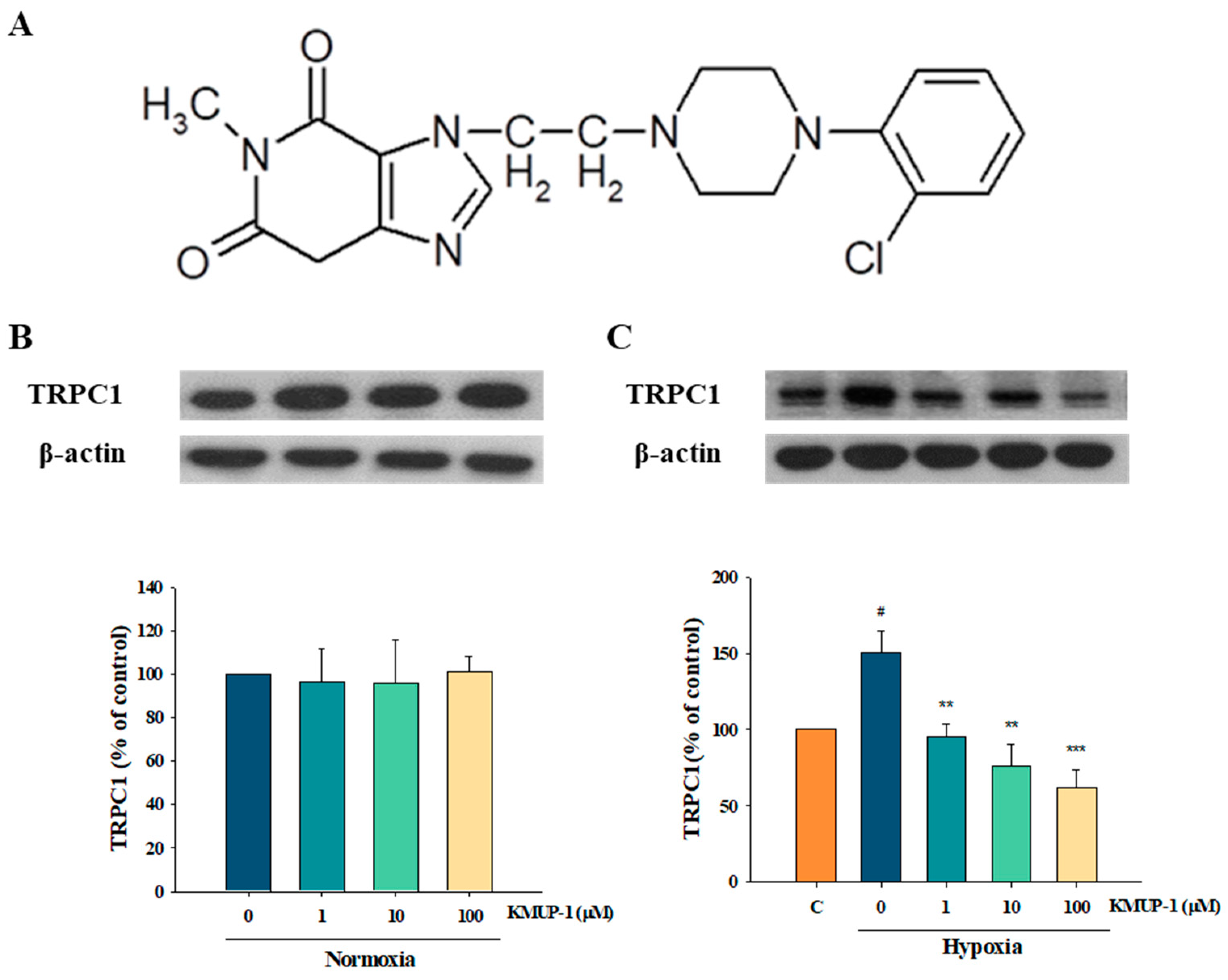
2.1. Effects of KMUP-1 in Normoxic and Hypoxic PASMCs

The expression of the TRPC family is encoded for store-operated calcium channels in hypoxic pulmonary arterial smooth muscle cells. A previous report showed that TRPC1 is an important Ca2+-permeable channel that mediates pulmonary vasoconstriction when PASMC intracellular Ca2+ stores are depleted [15,21]. Thus, we investigated the effects of KMUP-1 incubated in normoxia and hypoxia mediums for 24 h to observe the expression of TRPC1. TRPC1 protein was markedly increased under hypoxic conditions, but there were no changes with normoxia. KMUP-1 (1, 10, 100 μM) did not influence TRPC1 under normoxia (Figure 1B) but significantly attenuated hypoxia-enhanced TRPC1 (Figure 1C).

2.2. KMUP-1 Prevented Hypoxia-Enhanced TRPC1 Expression through cGMP/PKG Activation

As shown in Figure 2, the PKG inhibitor KT5823 increased the expression of TRPC-1 in a hypoxic medium. KMUP-1 attenuated hypoxia-induced TRPC1 protein, and the effect was restored by KT5823. A membrane-permeable analog of cGMP, 8-Br-cGMP, is a PKG activator that also suppresses hypoxia-enhanced TRPC1 protein, whereas KMUP-1 combined with 8-Br-cGMP appears not to affect this response significantly. Thus, we suggest that the effects of KMUP-1 could be due to the activation of the cGMP/PKG pathway to attenuate hypoxia-enhanced TRPC1 protein.

2.3. KMUP-1 Prevented Hypoxia-Enhanced TRPC1 Expression through cAMP/PKA Activation

Like PKA5823, PKA inhibitor KT5720 increased the expression of TRPC-1 in the hypoxia state. KMUP-1 lessens hypoxia-induced TRPC1 protein, and it can be restored by KT5720. The PKA activator 8-Br-cAMP weakened hypoxia-enhanced TRPC1 protein expression, while KMUP-1 combined with 8-Br-cAMP did not markedly influence this effect (Figure 3). The data show that KMUP-1 reduction of hypoxia-enhanced TRPC1 protein could be involved in activating the cAMP/PKA pathway.

2.4. KMUP-1 Prevented Hypoxia-Enhanced TRPC1 Expression through PKC Inhibition

A PKC inhibitor, chelerythrine, suppressed the expression of TRPC-1 under hypoxic conditions. KMUP-1 reduced hypoxia-induced TRPC1 protein, which was restored by a PKC activator, PMA. A PKC inhibitor, chelerythrine, suppressed hypoxia-enhanced TRPC1 protein, and KMUP-1 combined with chelerythrine had no significant effects on this (Figure 4). The data indicate that KMUP-1-retarded hypoxia-enhanced TRPC1 protein could be involved in inhibiting the PKC pathway.

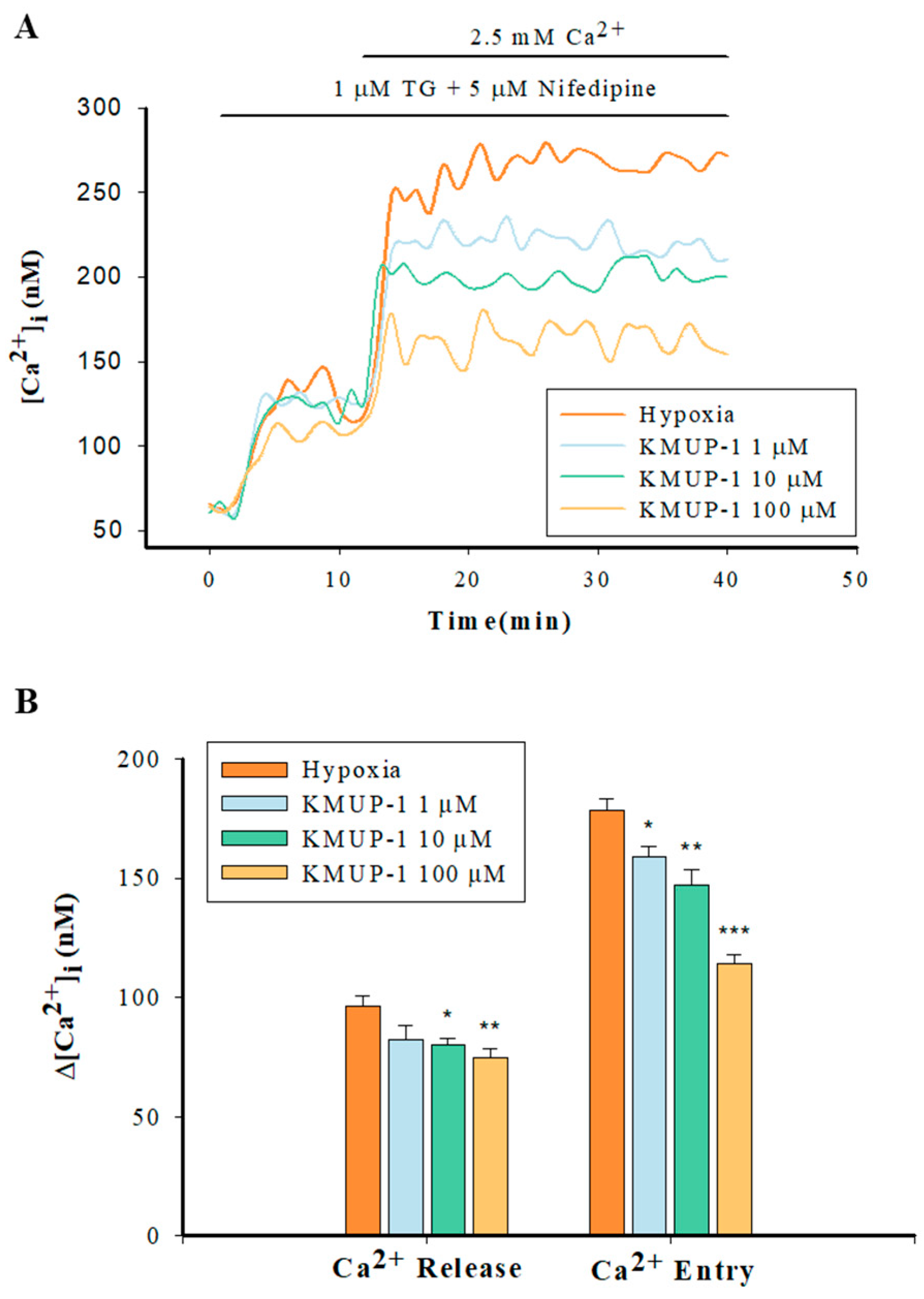
2.5. Enhanced Capacitative Ca2+ Entry (CCE) in Hypoxic PASMCs

PASMCs were loaded with 5 µM Fura-2-AM for 30 min in a Ca2+-free PBS solution. Next, we added 1 µM SERCA inhibitor thapsigargin (TG) and 5 µM VOCC inhibitor nifedipine to deplete intracellular Ca2+ stores and block the L-type Ca2+ channels, and then 2.5 mM Ca2+ was applied to Fura-2-loaded PASMCs. As Figure 5 shows, hypoxia did not affect the sarcoplasmic reticulum (SR) Ca2+ release but significantly increased the capacitative Ca2+ entry (CCE) via the SOCs in PASMCs.

2.6. KMUP-1 Attenuated SR Ca2+ Release and SOCs-Mediated CCE in Hypoxic PASMCs

In the hypoxic Ca2+-free PBS solution, Fura-2-loaded PASMCs were incubated with 1 µM TG and 5 µM nifedipine in the presence or absence of KMUP-1 (1, 10, 100 μM). KMUP-1 produced markedly dose-dependent (10, 100 μM) decreases in the SR-mediated Ca2+ release except at 1 μM. After that, 2.5 mM Ca2+ was added to induce the CCE via the SOCs. KMUP-1 (1, 10, 100 μM) significantly attenuated the CCE in a dose-dependent manner (Figure 6).

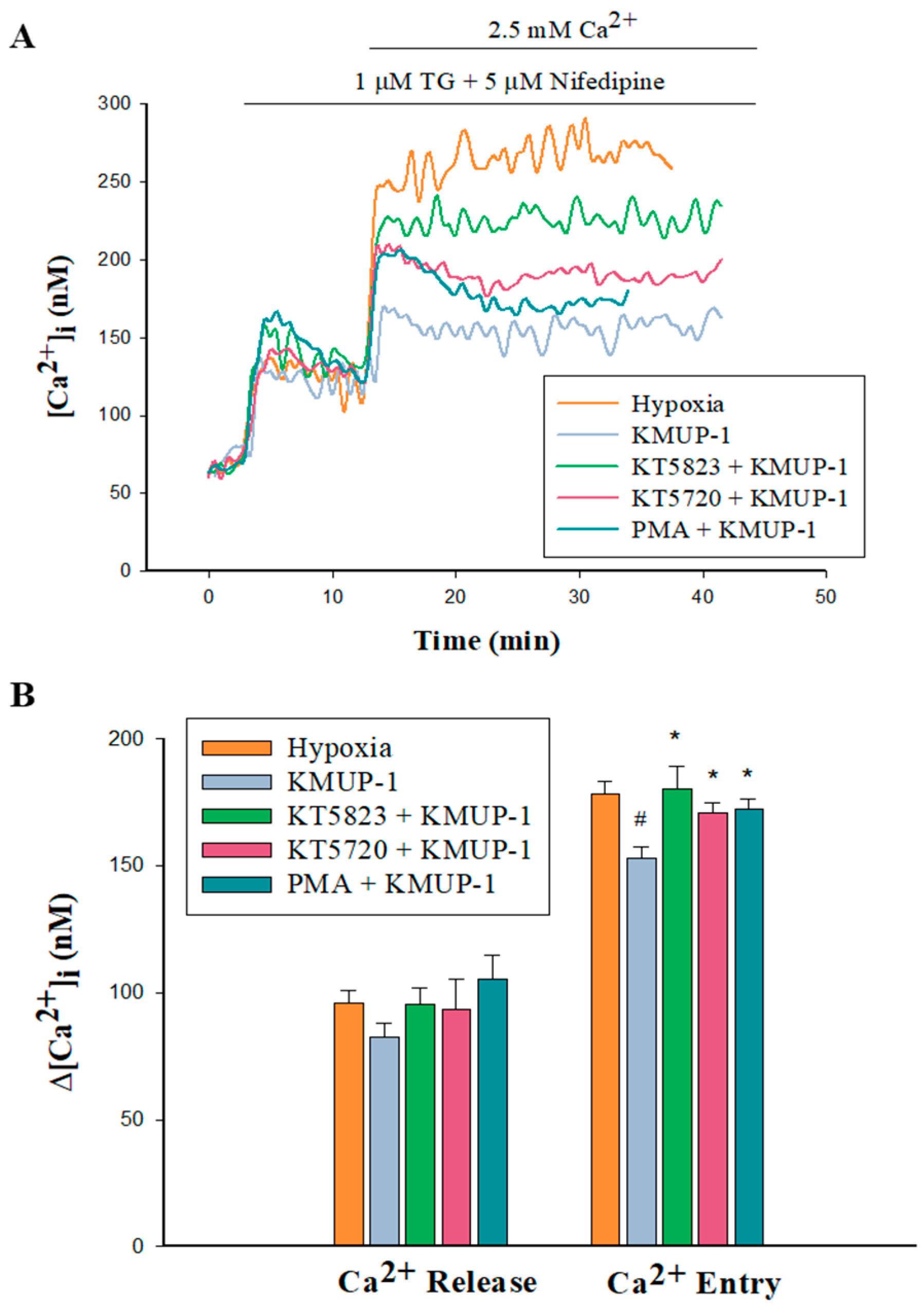
2.7. PKA/PKG/PKC Involvement in KMUP-1-Attenuated SOCs-Mediated CCE in Hypoxic PASMCs
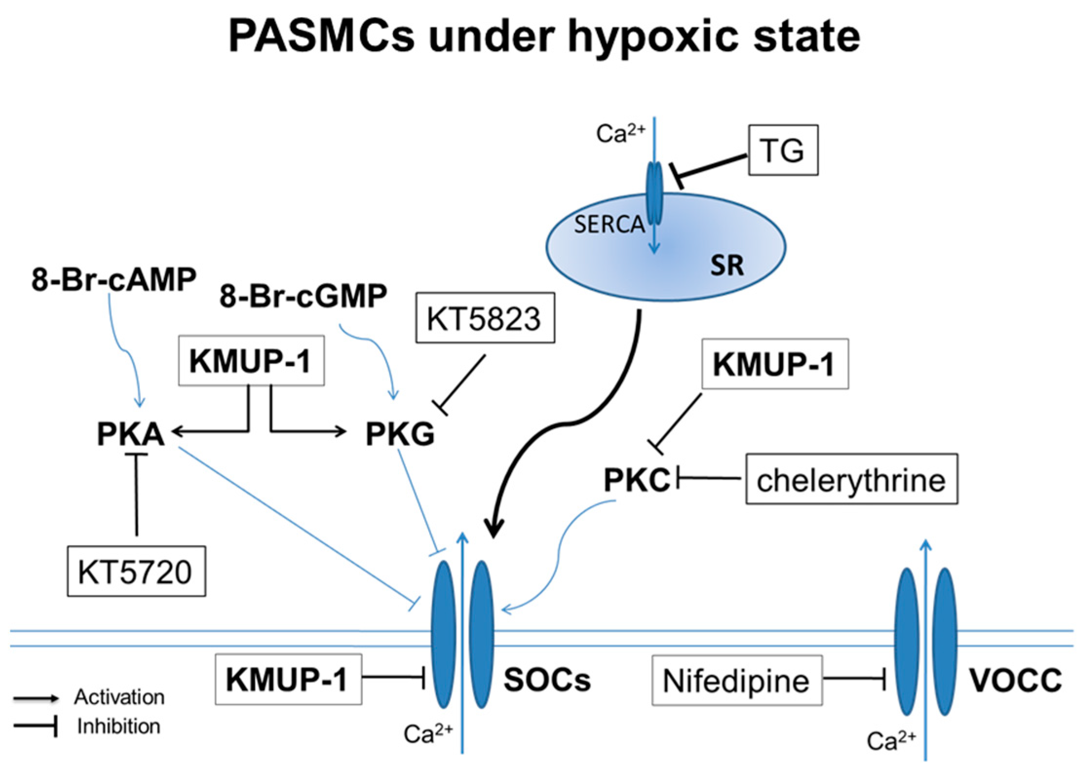
As Figure 7 shows, 1 μM KMUP-1 did not influence the SR Ca2+ release but significantly attenuated the Ca2+ entry via the SOCs in hypoxic PASMCs. Subsequently, KMUP-1 was co-incubated with the PKA inhibitor KT520 (1 μM), PKG inhibitor KT5823 (1 μM), and PKA activator PMA (1 μM). Those three agents did not affect the SR Ca2+ release but significantly restored Ca2+ entry via the SOCs. Those data further suggested that KMUP-1 retarded CCE through the SOCs involved in the PKA and PKG activation and PKC inhibition.

3. Discussion

The SOCE in PASMCs has gained substantial attention from research on hypoxic pulmonary vasoconstriction and pulmonary vascular remodeling. TRPC1 mainly contributes to SOCs, mediating SOCE induced by agonists or hypoxia [14,21,29]. In this study using hypoxic PASMCs, we first demonstrated that the xanthine derivative KMUP-1 inhibited TRPC1 expression and SOCs-mediated Ca2+ entry. This finding is consistent with previous findings [17,19,20] that KMUP-1 might be able to control cardiovascular diseases, particularly for the prevention or treatment of PAH.
A previous report showed that the levels of TRPC1 protein in PASMCs increased with the duration of hypoxia and reached a maximal level at 24 h [21]. We also found that the SR Ca2+ release and Ca2+ entry via the SOCs were significantly elevated under 24 h hypoxia incubations. Thus, we used the same conditions to observe changes in TRPC1 and Ca2+ entry via the SOCs by treatment with KMUP-1 in a hypoxia chamber for 24 h. Kunichika et al. [15] have demonstrated that TRPC1 in PASMC is an essential canonical TRP isoform that forms native SOC to regulate pulmonary vascular contractility. Our results also showed the relationship between TRPC1 and SOCE under hypoxic rat PASMCs ex vivo. Therefore, the data obtained from this study suggested that KMUP-1 hindered hypoxia-stimulating the expression of TRPC1 protein and associated Ca2+ entry via the SOCs, which could involve the PKA and PKG activation and the PKC inhibition. However, we cannot exclude that other factors and signaling pathways are involved in the effects of KMUP-1 in this rat model of hypoxic PASMCs.
SOCs are essential in controlling Ca2+ influx, arterial tone development, and smooth muscle cell growth in the pulmonary vasculature [10,11,12,13,30]. Few pulmonary VSM investigations have ventured beyond SOC modulation and explored the role of protein kinases [13,17,31]. Likewise, one of our previous reports demonstrated that PKA, PKG, and PKC regulate the activation of SOCs in pulmonary artery myocytes. PKA/PKG and PKC activation inhibited and stimulated SOCs, respectively [13]. As previously reported, KMUP-1 can modulate protein kinases A, G, and C [13]. Thus, we used PKA/PKG/PKC activators and inhibitors to study the effects of KMUP-1 on SR Ca2+ release and Ca2+ entry via the SOCs. In this study, we further confirmed that KMUP-1 attenuated TRPC1-dependent Ca2+ entry through the SOCs, which can be attributed to PKA/PKG activation and PKC inhibition. Notably, this finding can vary between different cell types and even within different subsets of smooth muscle cells. So, the actions of KMUP-1 on various smooth muscle cells under normoxic and hypoxic conditions need further investigation.
The cAMP/PKA signaling pathway is essential in several physiological processes, including vascular tone and cell cycle progression. Like PKA, cGMP/PKG relaxes vascular smooth muscle partly due to the inhibition of SOCs [13,30,31]. In contrast to PKA/PKG activation, PKC activation is strongly linked to increased SOC activity in vascular smooth muscle [13,31]. In physiological states, PKC activation would be more critical than PKA/PKG inhibition to induce vasoconstriction in PASMCs, stimulating the development of PAH. This study provides evidence that KMUP-1 is effective in preventing hypoxia-augmented TRPC1 expression and SOC-mediated Ca2+ entry in rat PASMCs (Figure 8).
Here, we want to address some of the limitations of this study. In general, the findings of this study were obtained from the SD rats and cannot be directly applicable to other species or human subjects. In addition, ex vivo PASMCs may not fully replicate the complex in vivo conditions within the PAs. The absence of interactions with neighboring cells, the lack of blood flow, and the absence of an extracellular matrix can influence cell behaviors. In other words, the behavior of PASMCs in culture may differ from their behavior in the intact PA due to alterations in the cellular microenvironment.
On the other hand, we have shown that KMUP-1 reduced the expression of the TRPC-1 protein in hypoxic conditions but did not measure its channel inhibitory activity on this current, which can be performed by patch-clamp electrophysiology in fresh PASMCs. In what way KMUP-1 modulates the SOCs-mediated Ca2+ influx upon SR Ca2+ depletion in hypoxic conditions, and the SOC channel inhibition by KMUP-1, still need to be considered. Moreover, TRPC1 activation of SOCE is not direct but needs the interaction of STIM1 and Orai1, which remains to be investigated by KMUP-1.

4. Materials and Methods

4.1. Animal Procedures and Tissue Preparation

The Kaohsiung Medical University Animal Care and Use Committee approved (IACUC No. 110140, 1 August 2022 to 31 July 2023) all procedures and protocols in compliance with the Guide for the Care and Use of Laboratory Animals published by the US National Institutes of Health. In brief, male Sprague-Dawley (SD) rats (13–15 weeks old) were euthanized by intraperitoneal injection (i.p.) of an overdose of pentobarbital sodium (130 mg/kg). The lungs and heart were cut by en bloc resection and put in a cold physiological salt solution (PSS) (in mM): 119 NaCl, 4.8 KCl, 1.7 KH2PO4, 20 NaHCO3, 10 Glucose, 1.2 CaCl2, and 1.2 MgSO4 (pH 7.4). Extralobar pulmonary arteries (PAs) were removed and free of the surrounding tissue.

4.2. Primary Culture of PASMCs

Extralobar PAs were dissected carefully and prepared for tissue culture. Ex vivo explant cultures were executed according to our previous reports. The vascular endothelium was removed using a sterile cotton swab with mild rubbing. Then, we carefully removed the tunica adventitia and the superficial part of the tunica media. The remaining pieces were cut into small pieces and then transferred to culture flasks for further cultivation. The ex vivo explants were incubated in Dulbecco’s Modified Eagle Medium (DMEM; Gibco Laboratories, Gaithersburg, MD, USA) with 10% fetal bovine serum and 1% penicillin-streptomycin amphotericin B (Biological Industries, Kibbutz Beit Haemek, Israel) to promote cell growth. The incubator was set to 37 °C with a humidified 5% CO2 environment. PASMCs began to proliferate from explants after culture for 7 days. The growth of PASMCs was arrested by substituting the media with FBS-free DMEM. Then, the cells were incubated in the absence and presence of test agents under normoxia (20% O2) or hypoxia (1% O2) for 24 h at 37 °C [21]. No more than five passages of primary cultures were used in the subsequent experiments. The immunofluorescent staining of α-actin was used to confirm the purity of PASMCs.

4.3. Western Blot Analysis

The expression of TRPC1 was determined by Western blotting. Briefly, PASMCs were lysed in M-PER (Mammalian Protein Extraction Reagent, 78501, Thermo Fisher Scientific, Waltham, MA, USA) comprising EDTA-free protease inhibitor cocktail and PhosSTOP phosphatase inhibitor (Roche Diagnostics, Mannheim, Germany). Next, the supernatants were collected after centrifugation at 15,000 rpm for 20 min at 4 °C. The Bradford assay was used to estimate the protein concentration. The protein was separated by sodium dodecyl sulfate–polyacrylamide gel electrophoresis (SDS-PAGE) and transferred to a polyvinylidene difluoride (PVDF) membrane. Subsequently, the membranes were blocked for 2 h at room temperature with 5% nonfat milk in Tris-buffered saline (20 mM Tris-HCl, 150 mM NaCl) with 0.1% Tween-20 (TBST), then incubated with primary antibodies against TRPC1 (1:200; #ACC-010, Alomone, Jerusalem, Israel) at 4 °C overnight. After TBST buffer washing three times, the membranes were incubated with horseradish peroxidase-conjugated secondary antibody. β-actin (1:5000, A5441; Sigma-Aldrich, St. Louis, MO, USA) was used as an internal control. Signals were detected with enhanced chemiluminescence (ECL) (Amersham Life Sciences Inc., Arlington Heights, IL, USA) HRP substrate reagent and quantized by densitometry with ImageJ software (version 1.53j; National Institutes of Health, Bethesda, MD, USA).

4.4. Measurement of Capacitative Ca2+ Entry

The intracellular Ca2+ concentrations ([Ca2+]i) were estimated using the fluorescent indicator Fura 2-AM (F0888, Sigma-Aldrich, St. Louis, MO, USA) and recorded by spectrofluorophotometer (Shimadzu, RF-5301PC, Kyoto, Japan). The preparation of PASMCs was excited at 340 and 380 nm to obtain the Fura 2 emission, and the resulting fluorescence emission at 510 nm was monitored. The ratio of emissions at 510 nm (F340/F380) was recorded every 2 sec based on the equation [Ca2+]i = Kd × (Sf2/Sb2) × (R − Rmin)/(Rmax − R). Depletion of Ca2+ from the ER/SR triggered the SOCs opening in the plasma membrane, so-called capacitative Ca2+ entry (CCE) [15]. For measuring CCE, PASMCs were loaded with 5 µM Fura 2-AM for 30 min at 37 °C in the dark and then washed with PSS twice to eliminate the extracellular Fura 2. Then, the PASMCs were put in Ca2+-free PSS and supplied with 1 µM sarco/endoplasmic reticulum Ca2+-ATPase (SERCA) inhibitor thapsigargin (TG) to deplete intracellular Ca2+ stores and 5 µM nifedipine to block the voltage-operated Ca2+ channels (VOCC), followed by extracellular Ca2+ (2.5 mM) to initiate CCE.

4.5. Chemicals

Buffer reagents, 8-Br-cAMP, 8-Br-cGMP, chelerythrine, Fura 2-AM, KT5823, KT5720, phorbol 12-myristate 13-acetate (PMA), and thapsigargin were purchased from Sigma-Aldrich Chemical Co. (St. Louis, MO, USA). Unless specified otherwise, all drugs and reagents were dissolved in distilled water. Fura 2-AM, KT5720, KT5823, PMA, and thapsigargin were dissolved in DMSO at 10 mM; KMUP-1 was dissolved in 10% absolute alcohol, 10% propylene glycol, and 2% 1N HCl at 10 mM. Serial dilutions were performed in phosphate buffer solution, with the final solvent concentration < 0.01%.

4.6. Statistical Analysis

All data are shown as the mean ± SE. To identify statistical variances, independent and paired Student’s t-tests were conducted on unpaired and paired samples. The one-way analysis of variance (ANOVA) was employed when multiple treated groups were compared to a control group. When the ANOVA manifested a statistical difference, a Tukey–Kramer pairwise comparison was used for post hoc analysis in cases where applicable. A probability value (p-value) less than 0.05 was considered significant.

5. Conclusions

KMUP-1 attenuated TRPC1 protein and SOC-dependent Ca2+ entry in this hypoxic PASMCs model, which could be attributed to its PKA and PKG activation and PKC inhibition. In addition to the benefits of monocrotaline-induced PAH by KMUP-1 [25,26], it also could cause PA smooth muscle relaxation in the model of hypoxia-induced PAH. Finally, we suggest that KMUP-1 can be a potential therapeutic candidate for targeting hypoxia-induced PAH.

Author Contributions

Z.-K.D. and Y.-C.C. executed the experiments and data analysis and wrote the article. S.-L.H., J.-L.Y. and J.-H.H. designed the study and data interpretation and helped to write the manuscript. B.-N.W. directed and conceived the experiments, interpreted the data, and revised the manuscript. All authors have read and agreed to the published version of the manuscript.

Funding

This study was supported by grants from the National Science and Technology Council, Taiwan (NSTC 112-2320-B-037-020-MY3 and NSTC 111-2314-B-037-076), and the Kaohsiung Medical University Hospital Research Foundation, Taiwan (KMUH110-0R48 and KMUH111-1R43).

Institutional Review Board Statement

Not applicable.

Informed Consent Statement

Not applicable.

Data Availability Statement

The data supporting this study’s findings are available from the corresponding author upon request.

Acknowledgments

The authors thank the Center for Laboratory Animals at Kaohsiung Medical University for the animal care. We thank Li-Mei An for her excellent technical assistance as well.

Conflicts of Interest

The authors declare no conflicts of interest.

Abbreviations

cAMP, cyclic adenosine monophosphate; CCE, capacitative Ca2+ entry; cGMP, cyclic guanosine monophosphate; ER, endoplasmic reticulum; PAs, pulmonary arteries; PAH, pulmonary arterial hypertension; PAP, pulmonary artery pressure; PASMCs, pulmonary artery smooth muscle cells; PKA, protein kinase A; PKC, protein kinase C; PKG, protein kinase G; PMA, phorbol 12-myristate 13-acetate; PSS, physiological salt solution; PVR, pulmonary vascular resistance; TG, thapsigargin; TRP channels, transient receptor potential channels; TRPC1, canonical transient receptor potential channel 1; SERCA, sarco/endoplasmic reticulum Ca2+-ATPase; SOCs, store-operated calcium channels; SOCE, store-operated calcium entry; SR, sarcoplasmic reticulum; stromal interaction molecule 1, STIM1; VOCC, voltage-operated Ca2+ channels; VSM, vascular smooth muscle.

References

  1. Otani, N.; Tomoe, T.; Kawabe, A.; Sugiyama, T.; Horie, Y.; Sugimura, H.; Yasu, T.; Nakamoto, T. Recent Advances in the Treatment of Pulmonary Arterial Hypertension. Pharmaceuticals 2022, 15, 1277. [Google Scholar] [CrossRef] [PubMed]
  2. Naeije, R.; Richter, M.J.; Rubin, L.J. The physiologic basis of pulmonary arterial hypertension. Eur. Respir. J. 2022, 59, 2102334. [Google Scholar] [CrossRef] [PubMed]
  3. Humbert, M.; Guignabert, C.; Bonnet, S.; Dorfmüller, P.; Klinger, J.R.; Nicolls, M.R.; Olschewski, A.J.; Pullamsetti, S.S.; Schermuly, R.T.; Stenmark, K.R.; et al. Pathology and pathobiology of pulmonary hypertension: State of the art and research perspectives. Eur. Respir. J. 2019, 53, 1801887. [Google Scholar] [CrossRef] [PubMed]
  4. Vonk Noordegraaf, A.; Chin, K.M.; Haddad, F.; Hassoun, P.M.; Hemnes, A.R.; Hopkins, S.R.; Kawut, S.M.; Langleben, D.; Lumens, J.; Naeije, R. Pathophysiology of the right ventricle and of the pulmonary circulation in pulmonary hypertension: An update. Eur. Respir. J. 2019, 53, 1801900. [Google Scholar] [CrossRef] [PubMed]
  5. Simonneau, G.; Montani, D.; Celermajer, D.S.; Denton, C.P.; Gatzoulis, M.A.; Krowka, M.; Williams, P.G.; Souza, R. Haemodynamic definitions and updated clinical classification of pulmonary hypertension. Eur. Respir. J. 2019, 53, 1801913. [Google Scholar] [CrossRef]
  6. Ruaro, B.; Salton, F.; Baratella, E.; Confalonieri, P.; Geri, P.; Pozzan, R.; Torregiani, C.; Bulla, R.; Confalonieri, M.; Matucci-Cerinic, M.; et al. An Overview of Different Techniques for Improving the Treatment of Pulmonary Hypertension Secondary in Systemic Sclerosis Patients. Diagnostics 2022, 12, 616. [Google Scholar] [CrossRef] [PubMed]
  7. Stenmark, K.R.; Fagan, K.A.; Frid, M.G. Hypoxia-induced pulmonary vascular remodeling: Cellular and molecular mechanisms. Circ. Res. 2006, 99, 675–691. [Google Scholar] [CrossRef] [PubMed]
  8. Ball, M.K.; Waypa, G.B.; Mungai, P.T.; Nielsen, J.M.; Czech, L.; Dudley, V.J.; Beussink, L.; Dettman, R.W.; Berkelhamer, S.K.; Steinhorn, R.H.; et al. Regulation of hypoxia-induced pulmonary hypertension by vascular smooth muscle hypoxia-inducible factor-1α. Am. J. Respir. Crit. Care Med. 2014, 189, 314–324. [Google Scholar] [CrossRef] [PubMed]
  9. Masson, B.; Montani, D.; Humbert, M.; Capuano, V.; Antigny, F. Role of Store-Operated Ca2+ Entry in the Pulmonary Vascular Remodeling Occurring in Pulmonary Arterial Hypertension. Biomolecules 2021, 11, 1781. [Google Scholar] [CrossRef]
  10. Smani, T.; Zakharov, S.I.; Csutora, P.; Leno, E.; Trepakova, E.S.; Bolotina, V.M. A novel mechanism for the store-operated calcium influx pathway. Nat. Cell Biol. 2004, 6, 113–120. [Google Scholar] [CrossRef]
  11. Landsberg, J.W.; Yuan, J.X. Calcium and TRP channels in pulmonary vascular smooth muscle cell proliferation. News Physiol. Sci. 2004, 19, 44–50. [Google Scholar] [CrossRef] [PubMed]
  12. Lu, W.; Ran, P.; Zhang, D.; Peng, G.; Li, B.; Zhong, N.; Wang, J. Sildenafil inhibits chronically hypoxic upregulation of canonical transient receptor potential expression in rat pulmonary arterial smooth muscle. Am. J. Physiol. Cell Physiol. 2010, 298, C114–C123. [Google Scholar] [CrossRef] [PubMed]
  13. Chen, I.S.; Dai, Z.K.; Welsh, D.G.; Chen, I.J.; Wu, B.N. Protein kinases modulate store-operated channels in pulmonary artery smooth muscle cells. J. Biomed. Sci. 2011, 18, 2. [Google Scholar] [CrossRef] [PubMed]
  14. Lopez, J.J.; Jardin, I.; Sanchez-Collado, J.; Salido, G.M.; Smani, T.; Rosado, J.A. TRPC Channels in the SOCE Scenario. Cells 2020, 9, 126. [Google Scholar] [CrossRef] [PubMed]
  15. Kunichika, N.; Yu, Y.; Remillard, C.V.; Platoshyn, O.; Zhang, S.; Yuan, J.X. Overexpression of TRPC1 enhances pulmonary vasoconstriction induced by capacitative Ca2+ entry. Am. J. Physiol. Lung Cell. Mol. Physiol. 2004, 287, L962–L969. [Google Scholar] [CrossRef] [PubMed]
  16. Martín-Bórnez, M.; Galeano-Otero, I.; Del Toro, R.; Smani, T. TRPC and TRPV Channels’ Role in Vascular Remodeling and Disease. Int. J. Mol. Sci. 2020, 21, 6125. [Google Scholar] [CrossRef]
  17. Lu, T.; Zhang, Y.; Su, Y.; Zhou, D.; Xu, Q. Role of store-operated Ca2+ entry in cardiovascular disease. Cell Commun. Signal. 2022, 20, 33. [Google Scholar] [CrossRef] [PubMed]
  18. Qu, Y.Y.; Wang, L.M.; Zhong, H.; Liu, Y.M.; Tang, N.; Zhu, L.P.; He, F.; Hu, Q.H. TRPC1 stimulates calcium-sensing receptor-induced store-operated Ca2+ entry and nitric oxide production in endothelial cells. Mol. Med. Rep. 2017, 16, 4613–4619. [Google Scholar] [CrossRef] [PubMed]
  19. Zhou, C.; Townsley, M.I.; Alexeyev, M.; Voelkel, N.F.; Stevens, T. Endothelial hyperpermeability in severe pulmonary arterial hypertension: Role of store-operated calcium entry. Am. J. Physiol. Lung Cell. Mol. Physiol. 2016, 311, L560–L569. [Google Scholar] [CrossRef]
  20. Miao, R.; Wan, J.; Liu, J.; Yuan, J.X.; Wang, J.; Xie, W.; Zhai, Z.; Wang, C. Bone Marrow-Derived Endothelial Progenitor Cells Contribute to Monocrotaline-Induced Pulmonary Arterial Hypertension in Rats via Inhibition of Store-Operated Ca2+ Channels. BioMed Res. Int. 2018, 2018, 4892349. [Google Scholar] [CrossRef]
  21. Chen, T.X.; Xu, X.Y.; Zhao, Z.; Zhao, F.Y.; Gao, Y.M.; Yan, X.H.; Wan, Y. Hydrogen peroxide is a critical regulator of the hypoxia-induced alterations of store-operated Ca2+ entry into rat pulmonary arterial smooth muscle cells. Am. J. Physiol. Lung Cell. Mol. Physiol. 2017, 312, L477–L487. [Google Scholar] [CrossRef] [PubMed]
  22. Wang, Y.X.; Zheng, Y.M. Role of ROS signaling in differential hypoxic Ca2+ and contractile responses in pulmonary and systemic vascular smooth muscle cells. Respir. Physiol. Neurobiol. 2010, 174, 192–200. [Google Scholar] [CrossRef] [PubMed]
  23. Galiè, N.; Channick, R.N.; Frantz, R.P.; Grünig, E.; Jing, Z.C.; Moiseeva, O.; Preston, I.R.; Pulido, T.; Safdar, Z.; Tamura, Y.; et al. Risk stratification and medical therapy of pulmonary arterial hypertension. Eur. Respir. J. 2019, 53, 1801889. [Google Scholar] [CrossRef] [PubMed]
  24. Dai, Z.K.; Cheng, Y.J.; Chung, H.H.; Wu, J.R.; Chen, I.J.; Wu, B.N. KMUP-1 ameliorates monocrotaline-induced pulmonary arterial hypertension through the modulation of Ca2+ sensitization and K+-channel. Life Sci. 2010, 86, 747–755. [Google Scholar] [CrossRef] [PubMed]
  25. Lo, Y.C.; Tseng, Y.T.; Liu, C.M.; Wu, B.N.; Wu, S.N. Actions of KMUP-1, a xanthine and piperazine derivative, on voltage-gated Na+ and Ca2+ -activated K+ currents in GH3 pituitary tumour cells. Br. J. Pharmacol. 2015, 172, 5110–5122. [Google Scholar] [CrossRef] [PubMed]
  26. Chen, J.Y.; Cheng, K.I.; Tsai, Y.L.; Hong, Y.R.; Howng, S.L.; Kwan, A.L.; Chen, I.J.; Wu, B.N. Potassium-channel openers KMUP-1 and pinacidil prevent subarachnoid hemorrhage-induced vasospasm by restoring the BKCa-channel activity. Shock 2012, 38, 203–212. [Google Scholar] [CrossRef] [PubMed]
  27. Ocaña, M.; Cendán, C.M.; Cobos, E.J.; Entrena, J.M.; Baeyens, J.M. Potassium channels and pain: Present realities and future opportunities. Eur. J. Pharmacol. 2004, 500, 203–219. [Google Scholar] [CrossRef]
  28. Cheng, K.I.; Yang, K.T.; Kung, C.L.; Cheng, Y.C.; Yeh, J.L.; Dai, Z.K.; Wu, B.N. BK(Ca) Channel Inhibition by Peripheral Nerve Injury Is Restored by the Xanthine Derivative KMUP-1 in Dorsal Root Ganglia. Cells 2021, 10, 949. [Google Scholar] [CrossRef]
  29. Ambudkar, I.S.; de Souza, L.B.; Ong, H.L. TRPC1, Orai1, and STIM1 in SOCE: Friends in tight spaces. Cell Calcium 2017, 63, 33–39. [Google Scholar] [CrossRef]
  30. Albert, A.P.; Large, W.A. Store-operated Ca2+-permeable non-selective cation channels in smooth muscle cells. Cell Calcium 2003, 33, 345–356. [Google Scholar] [CrossRef]
  31. Albert, A.P.; Saleh, S.N.; Peppiatt-Wildman, C.M.; Large, W.A. Multiple activation mechanisms of store-operated TRPC channels in smooth muscle cells. J. Physiol. 2007, 583, 25–36. [Google Scholar] [CrossRef] [PubMed]
Figure 1. Effects of KMUP-1 in normoxic and hypoxic pulmonary arterial smooth muscle cells (PASMCs). (A) Structure of KMUP-1. (B) TRPC1 protein showed no significant differences in KMUP-1 (1, 10, 100 μM)-treated PASMCs under normoxic conditions. (C) Various concentrations of KMUP-1 (1, 10, 100 μM) inhibited hypoxia-induced TRPC1 protein overexpression. The quantitation of this protein is shown in the lower panel. Results were presented as the mean ± SE, n = 7. # p < 0.05 compared with control (normoxia), r = 0.66; ** p < 0.01, *** p <0.001 compared with hypoxia, r = 0.81. C: control represents normoxia. r: correlation coefficient.
Pharmaceuticals 17 00440 g001
Figure 2. KMUP-1 inhibited hypoxia-enhanced TRPC1 expression via the cGMP/PKG pathway. PASMCs pretreated with KMUP-1 (1 μM), KT5823 (1 μM), 8-Br-cGMP (100 μM), KT5823+KMUP-1, and 8-Br-cGMP+KMUP-1 under hypoxic states. The quantitation of these proteins is shown in the lower panel. Results are presented as the mean ± SE, n = 6. # p < 0.05 compared with normoxia, r = 0.47; ** p < 0.01, *** p < 0.001 compared with hypoxia, r = 0.72;  p < 0.05 compared with KMUP-1 group, r = 0.56. 8-Br-cGMP: membrane-permeable analog of cGMP. r: correlation coefficient.
Pharmaceuticals 17 00440 g002
Figure 3. KMUP-1 inhibited hypoxia-enhanced TRPC1 expression via the cAMP/PKA pathway. PASMCs pretreated with KMUP-1 (1 μM), KT5720 (1 μM), 8-Br-cAMP (100 μM), KT5720+KMUP-1, and 8-Br-cAMP+KMUP-1 under hypoxic states. The quantitation of these proteins is shown in the lower panel. Results are presented as the mean ± SE, n = 6. # p < 0.05 compared with normoxia, r = 0.59; ** p < 0.01, *** p < 0.001 compared with hypoxia, r = 0.68; p < 0.05 compared with KMUP-1 group, r = 0.61. 8-Br-cAMP: membrane-permeable analog of cAMP. r: correlation coefficient.
Pharmaceuticals 17 00440 g003
Figure 4. KMUP-1 inhibited hypoxia-enhanced TRPC1 expression via PKC pathway. PASMCs pretreated with KMUP-1 (1 μM), PMA (1 μM), chelerythrine (1 μM), PMA+KMUP-1, and chelerythrine+KMUP-1 under hypoxic states. The quantitation of these proteins is shown in the lower panel. Results are presented as the mean ± SE, n = 6. # p < 0.05 compared with normoxia, r = 0.48; ** p < 0.01, *** p < 0.001 compared with hypoxia, r = 0.70;  p < 0.05 compared with the PMA group, r = 0.52. PMA: phorbol 12-myristate 13-acetate. r: correlation coefficient.
Pharmaceuticals 17 00440 g004
Figure 5. Effects of exposure to hypoxia on capacitative calcium entry in PASMCs. (A) Change in [Ca2+]i in pulmonary arterial smooth muscle cells (PASMCs) from control or hypoxia and subjected to the restoration of extracellular Ca2+ following store depletion with TG (1 μM). (B) Bar graph illustrates mean ± SE change in [Ca2+]i (∆[Ca2+]i) in response to TG and Ca2+ restoration. All experiments were performed in the presence of nifedipine, n = 12 for control and n = 13 for hypoxia. *** p < 0.001 compared with control (normoxia). TG: thapsigargin.
Pharmaceuticals 17 00440 g005
Figure 6. Effects of KMUP-1 on capacitative calcium entry in PASMCs. (A) Change in [Ca2+]i in pulmonary arterial smooth muscle cells from hypoxic or pretreated KMUP-1 groups and subjected to the restoration of extracellular Ca2+ following store depletion with TG (1 μM). (B) Bar graph illustrates mean ± SEM change in [Ca2+]i (∆[Ca2+]i) in response to TG and Ca2+ restoration. All experiments were performed in the presence of nifedipine, n = 7–13 of independent experiments. * p < 0.05, ** p < 0.01, *** p < 0.001 compared with hypoxic group. TG: thapsigargin.
Pharmaceuticals 17 00440 g006
Figure 7. Effects of KT5823, KT5720, and PMA on KMUP-1-inhibited capacitative calcium entry in hypoxic PASMCs. (A): Change in [Ca2+]i in pulmonary arterial smooth muscle cells (PASMCs) from pretreated KMUP-1 (1 μM), KT5823 (1 μM) with KMUP-1, KT5720 (1 μM) with KMUP-1, PMA (1 μM) with KMUP-1 and then subjected to the restoration of extracellular Ca2+ following store depletion with TG (1 μM). (B): Bar graph illustrates mean ± SE change in [Ca2+]i (∆[Ca2+]i) in response to TG and Ca2+ restoration. All experiments were performed in the presence of nifedipine, n = 6 of independent experiments. # p < 0.05 compared with hypoxic group; * p < 0.05 compared with KMUP-1 group in hypoxic PASMCs. PMA: phorbol 12-myristate 13-acetate; TG: thapsigargin.
Pharmaceuticals 17 00440 g007
Figure 8. Diagram summarizing the actions of KMUP-1 on the store-operated calcium channel (SOCs) in hypoxic PASMCs. Data suggest that KMUP-1 inhibits TRPC1 encodes for SOCs, activates the PKA/PKG pathway, and inhibits the PKC pathway. SOCs: store-operated calcium channels; SR: sarcoplasmic reticulum; SERCA: sarco/endoplasmic reticulum Ca2+-ATPase; TG: thapsigargin; PMA: phorbol 12-myristate 13-acetate; 8-Br-cAMP: membrane-permeable analog of cAMP; 8-Br-cGMP: membrane-permeable analog of cGMP; VOCC: voltage-operated calcium channels.
Pharmaceuticals 17 00440 g008
Disclaimer/Publisher’s Note: The statements, opinions and data contained in all publications are solely those of the individual author(s) and contributor(s) and not of MDPI and/or the editor(s). MDPI and/or the editor(s) disclaim responsibility for any injury to people or property resulting from any ideas, methods, instructions or products referred to in the content.

