Review

Exploring the Comprehensive Neuroprotective and Anticancer Potential of Afzelin

1
Department of Molecular Biotechnology and Genetics, Faculty of Biology and Environmental Protection, University of Lodz, 12/16 Banacha St., 90-237 Lodz, Poland
2
Chitkara College of Pharmacy, Chitkara University, Rajpura 140401, Punjab, India
3
M. M. College of Pharmacy, Maharishi Markandeshwar (Deemed to be) University, Mullana, Ambala 133207, Haryana, India
4
Swami Vivekanand College of Pharmacy, Ramnagar, Banur 140601, Punjab, India
5
Department of Pharmacology, Central University of Punjab, Bathinda 151401, Punjab, India
6
Doctoral School of Exact and Natural Sciences, University of Lodz, 90-237 Lodz, Poland
*
Authors to whom correspondence should be addressed.
These authors contributed equally to this work.
Pharmaceuticals 2024, 17(6), 701; https://doi.org/10.3390/ph17060701
Submission received: 30 April 2024 / Revised: 18 May 2024 / Accepted: 23 May 2024 / Published: 28 May 2024
(This article belongs to the Special Issue Bioactive Compounds Derived from Plants and Their Medicinal Potential)

Abstract

:
Neurodegenerative diseases (Alzheimer’s disease, Parkinson’s disease, Huntington’s disease, and others) and cancer, seemingly disparate in their etiology and manifestation, exhibit intriguing associations in certain cellular and molecular processes. Both cancer and neurodegenerative diseases involve the deregulation of cellular processes such as apoptosis, proliferation, and DNA repair and pose a significant global health challenge. Afzelin (kaempferol 3-O-rhamnoside) is a flavonoid compound abundant in various plant sources. Afzelin exhibits a diverse range of biological activities, offering promising prospects for the treatment of diseases hallmarked by oxidative stress and deregulation of cell death pathways. Its protective potential against oxidative stress is also promising for alleviating the side effects of chemotherapy. This review explores the potential therapeutic implications of afzelin, including its capacity to mitigate oxidative stress, modulate inflammation, and promote cellular regeneration in neurodegenerative and cancer diseases.

Graphical Abstract

1. Introduction

Neurodegenerative diseases are complex conditions that share the characteristics of cell death and malfunctioning of neurons. These conditions, sometimes referred to as geriatric diseases, are primarily characterized by a gradual degeneration of neurons, ultimately leading to the decline of crucial cognitive, motor, and sensory skills [1,2,3]. The defining features of Alzheimer’s disease, a prevalent neurodegenerative disorder, involve the development of aberrant protein aggregates in the brain, including beta-amyloid plaques and tau tangles. The presence of these aggregates hinders the transmission of brain signals and induces an inflammatory response, both of which are implicated in the development of cognitive dysfunction [4,5]. Similarly, Parkinson’s disease is defined by the degeneration of dopaminergic neurons in the substantia nigra, which ultimately leads to the death of these neurons. The depletion of neurons leads to motor dysfunctions characterized by tremors, rigidity, and bradykinesia [6]. In contrast, Huntington’s disease arises as a consequence of a genetic mutation that results in the misfolding of proteins. This phenomenon results in a progressive deterioration of cognitive and physical functions. The neuronal cell loss implicated in amyotrophic lateral sclerosis (ALS), colloquially referred to as Lou Gehrig’s disease, is accountable for muscle atrophy, culminating in eventual paralysis [7]. Various types of neurodegeneration, including multiple sclerosis (MS) [8] and frontotemporal dementia (FTD) [9], are characterized by specific patterns of neuronal degeneration and exhibit diverse manifestations across the brain and spinal cord [10]. Despite their intended objectives of symptom relief and disease progression cessation, conventional therapies often fail to provide comprehensive solutions. The examination of diverse natural compounds has arisen as a feasible avenue to harness the potential of nature’s abundant medicinal properties to treat these intricate disorders. Natural chemicals are derived from diverse sources, encompassing plants, herbs, fungi, and marine life [11]. Within the research area of neurodegenerative disorder treatments, some naturally occurring compounds have garnered interest because of their inherent neuroprotective, anti-inflammatory, antioxidative, and modulatory properties. For example, curcumin (a compound extracted from turmeric), resveratrol (derived from red grapes), and flavonoids (isolated from citrus fruits) have all demonstrated neuroprotective properties in various studies. These compounds are believed to provide neuroprotection due to their impact on inflammation and toxic protein aggregation-induced neuronal injury. Furthermore, it was shown that naturally derived compounds possess the capacity to regulate cellular pathways that play a critical role in the survival and proliferation of cells [12,13,14].
The increasing prevalence of cancer and the limitations of conventional therapies have spurred a growing interest in exploring alternative treatment modalities, particularly the use of natural products. Bioactive compounds such as polyphenols, alkaloids, terpenoids, and flavonoids have exhibited potent anti-proliferative, anti-angiogenic, and pro-apoptotic effects across a spectrum of cancer types. Moreover, the potential synergistic interactions between natural products and conventional anti-cancer therapies are explored. Evidence suggests that natural products can enhance the efficacy of chemotherapy and radiotherapy while mitigating their adverse effects. The modulation of drug resistance mechanisms and the promotion of sensitization to standard therapies underscore the promising role of natural products in combination regimens [15].
Oxidative stress, which is caused by an imbalance between reactive oxygen species (ROS) and antioxidants, is also an essential factor in the pathophysiology of neurodegenerative diseases [16,17,18,19] and cancer [20,21,22]. Natural compounds, such as vitamin E [23,24], vitamin C [25,26], or flavonoids, can scavenge dangerous free radicals, thereby minimizing the amount of oxidative damage that is caused to neurons. This antioxidative capability goes beyond only protecting cells; it also has the potential to influence the signaling cascades and gene expression inside the cells [27,28].
Flavonoids represent a class of polyphenolic compounds that are widely distributed across the plant kingdom. Afzelin (Figure 1), also known as kaempferol 3-O-rhamnoside, is a flavonoid compound that can be found in high concentrations in a wide variety of plant sources. The diverse range of biological activities, including the antioxidative properties exhibited by afzelin, holds a significant translational potential of this compound in the prevention and therapy of cancer and neurodegenerative diseases manifested by oxidative stress and deregulation of cell death pathways [29,30,31,32]. Moreover, intrinsic links have been discovered between oxidative stress and inflammation. Therefore, the ROS-scavenging effects of afzelin could mitigate neuroinflammation, which constitutes a characteristic feature of neurodegenerative disorders such as Alzheimer’s disease and Parkinson’s disease [33,34].

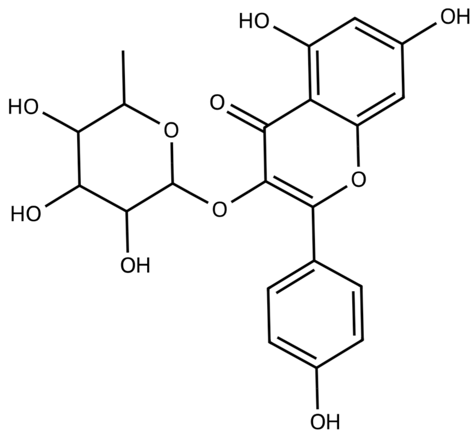
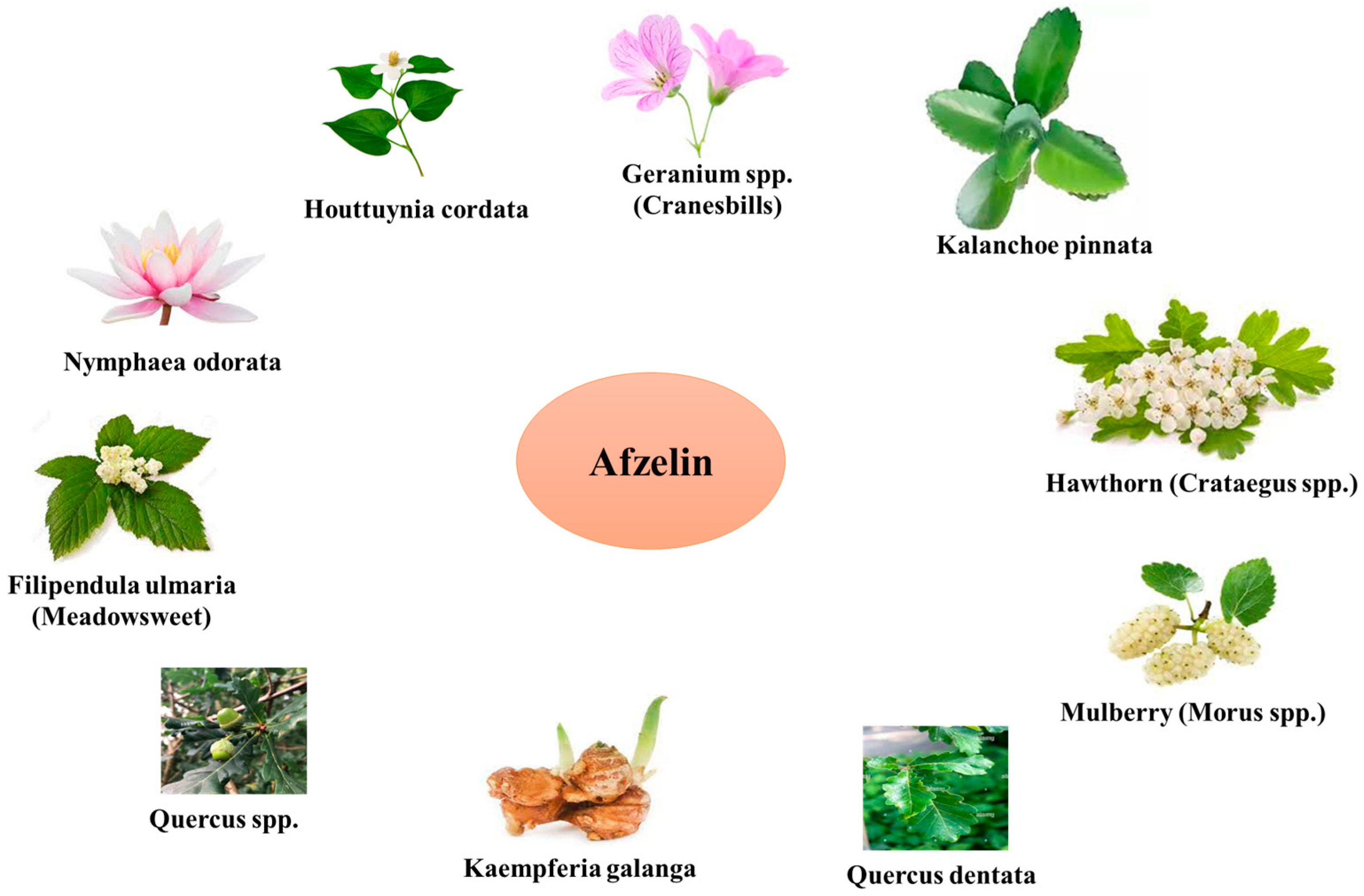
2. Sources and Pharmacological Profile of Afzelin

The anti-bacterial, anti-inflammatory, antioxidant, and cytoprotective effects of afzelin have been supported by numerous studies [29,30,31,32].
Afzelin come from a variety of different places, including but not limited to the following:
Houttuynia cordata: The plant under consideration is native to Southeast Asia and possesses a rich historical background in its utilization within traditional medicinal practices. Its applications encompass a diverse array of diseases, such as cancer, inflammation, infection, and fever. The main flavonoids found in the Houttuynia cordata plant include afzelin, quercetin, and hyperoside [35,36].
Nymphaea odorata: The plant is native to North America and is alternatively referred to as the American white waterlily. This botanical remedy has a long-standing medicinal history, as it was historically used for the treatment of skin conditions, wounds, inflammation, and cancer. Afzelin, kaempferol, quercetin, myricetin, and gallic acid represent abundant phenolic compounds found in this plant [37,38].
Kalanchoe pinnata: This plant is commonly known as an ‘air plant’ or ‘miracle leaf’ and is native to regions in Africa and Asia. The Kalanchoe pinnata plant has been employed in traditional medicinal practices for the treatment of various diseases, including ulcers, asthma, coughing, and diarrhea. Afzelin represents one of the flavonoids present in this plant. Others include quercitrin, quercetin, kaempferol, and rutin. The hepatoprotective benefits of afzelin, derived from Kalanchoe pinnata, have been demonstrated, indicating its potential to mitigate liver damage induced by carbon tetrachloride [39,40,41,42].
Quercus spp. (Oak Trees): Oak trees are renowned for their impressive stature in woodland areas and natural environments. Additionally, they offer a wide array of biologically active secondary metabolites, including afzelin. Afzelin is present in several anatomical components of oak trees, encompassing their foliage, bark, and reproductive structures, such as acorns. Quercus species exhibit a wide range of climatic and geographical distributions. Variability in the concentration of afzelin in Quercus plants can be attributed to several factors, including species differences, the conditions under which the plant is growing, and the age of the plant [43,44].
Filipendula ulmaria (Meadowsweet): This perennial herbaceous plant is native to both Europe and Asia. It has a rich history of application in the practice of herbal medicine. Afzelin found in meadowsweet is responsible for the pain-relieving and anti-inflammatory effects of the herb [45].
Hawthorn (Crataegus spp.): This plant belongs to the Rosaceae family and is well-known for its beneficial effects on the cardiovascular system. The antioxidative and vasorelaxant properties of hawthorn can be partially attributed to the presence of afzelin in the plant [46,47].
The natural sources of afzelin were summarized in Figure 2.

3. Biological Activity of Afzelin

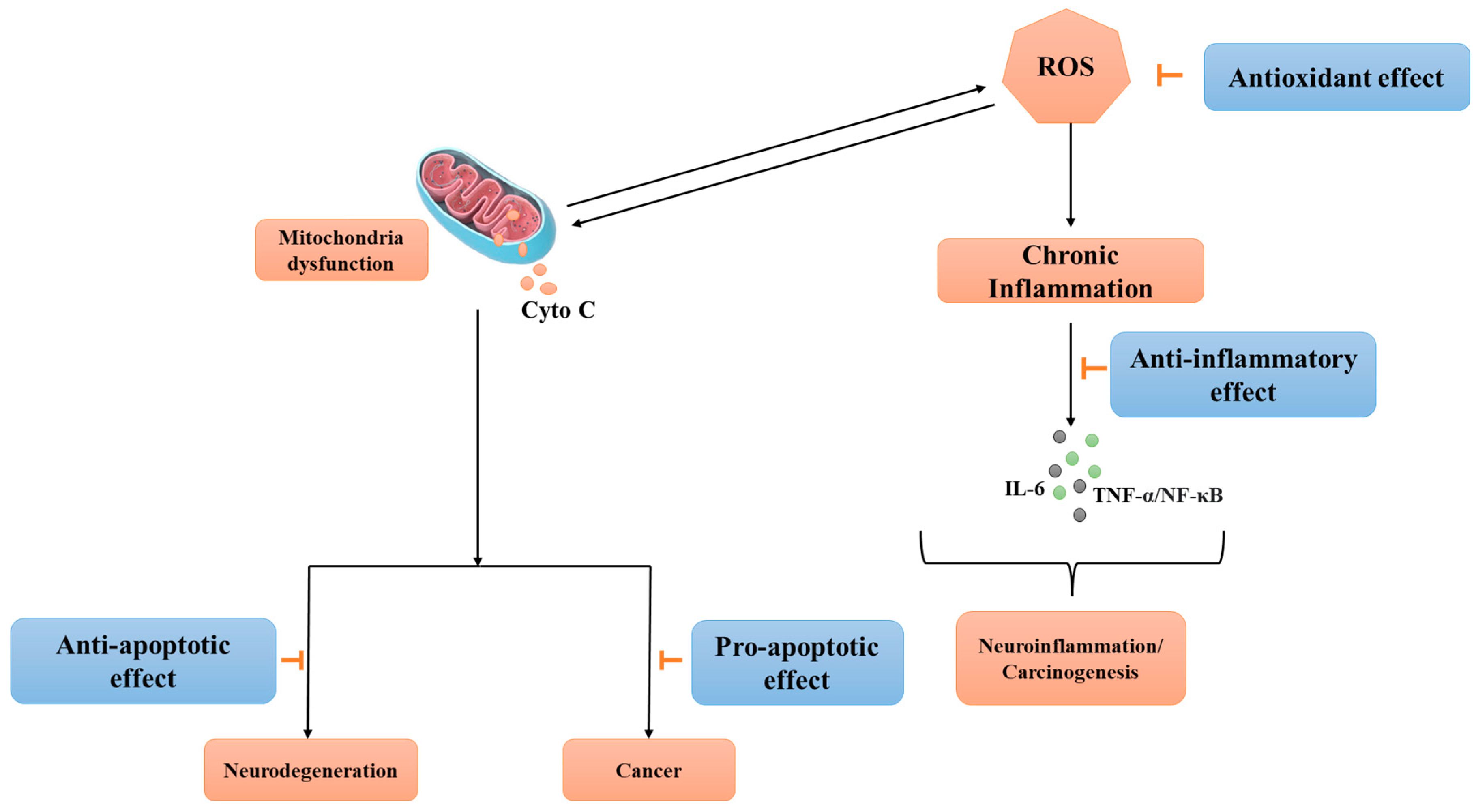
3.1. Antioxidative Effects of Afzelin

Oxidative stress arises from a state of disequilibrium between the generation and buildup of ROS within cells and tissues, as well as the capacity of a biological system to effectively neutralize these reactive byproducts. ROS fulfill various physiological functions, such as cell signaling, and are typically produced as byproducts of oxygen metabolism. However, exposure to environmental stressors like UV radiation, ionizing radiation, pollutants, and heavy metals, as well as the presence of xenobiotics, significantly enhance ROS production. Consequently, this imbalance results in oxidative stress, leading to cellular and tissue damage [48]. Afzelin’s potential therapeutic applications in the context of neurodegenerative illnesses and cancer could be mainly attributed to the chemical’s ability to alter redox signaling pathways and prevent oxidative stress [30,49,50,51] through the following:
  • Scavenging of free radicals: Highly reactive chemicals, such as free radicals and ROS, can oxidatively damage biological components like DNA, proteins, and lipids [52]. Afzelin has been found to possess ROS-scavenging characteristics, suggesting it can neutralize the effects of dangerous compounds through interaction with them [53,54].
  • Metal chelation: Afzelin also possesses metal chelation capabilities, making it possible for it to bind to metal ions such as those of iron and copper. These metal ions are well-known to have a role in the generation of free radicals via the Fenton and Haber-Weiss reactions. Afzelin is effective in blocking the generation of ROS because of its binding affinity for these metals [55,56,57].
  • Activation of nuclear factor erythroid 2-related factor 2 (NRF2) pathway: The NRF2 pathway plays a significant role in the regulation of the antioxidant response in cells via transcriptional control of many target genes involved in the maintenance of oxidation-reduction homeostasis inside the cell. The antioxidative actions of kaempferol and its derivatives, including afzelin, extend to the protection of cellular components like cell membranes and mitochondria from the destructive effects of oxidation. Thereby, afzelin may contribute to the maintenance of proper cellular function by preserving the structural integrity of these components [58].
Mitochondria are crucial for the various biochemical activities that are fundamental to the survival of cells. The morphology of mitochondria undergoes alterations in response to external stimuli and metabolic fluctuations through the processes of fission and fusion, collectively known as mitochondrial dynamics. The elimination of impaired mitochondria is facilitated through a cellular mechanism referred to as mitophagy, wherein these damaged organelles undergo breakdown via a distinct autophagosomal pathway. In recent years, significant endeavors have been undertaken to examine the influence of mitochondrial dysfunction manifested by excessive production of ROS, disturbances in mitochondrial calcium homeostasis, depletion of adenosine triphosphate (ATP), impairments in mitochondrial dynamics and transport, and deficiencies in mitophagy on the development of Alzheimer’s disease [59,60], Parkinson’s disease [61,62], other neurodegenerative diseases [63], and cancer [64,65].
Lee et al. investigated the cytoprotective mechanisms of afzelin against D-galactosamine (GalN)/lipopolysaccharide (LPS)-induced fulminant hepatic failure (FHF), with a specific emphasis on mitochondrial quality control and dynamics. The findings of the study demonstrated that the administration of the flavonoid compound resulted in enhanced survival rates and decreased levels of alanine aminotransferase and pro-inflammatory cytokines in mice that were treated with GalN/LPS. Afzelin contributed to a reduction in mitochondrial damage in mice treated with GalN/LPS, as evidenced by a decrease in mitochondrial swelling and a reduction in the mitochondrial glutamate dehydrogenase activity. The administration of afzelin resulted in an augmentation of mitochondrial biogenesis, as evidenced by elevated levels of PPAR-γ coactivator 1α (PGC-1α), nuclear respiratory factor 1 (NRF1), and mitochondrial transcription factor A (TFAM) [31].
PGC-1α has been shown to exert a protective effect on neuroblastoma cells by mitigating neuronal mortality and neuroinflammation produced by amyloid-beta (Aβ). Additionally, the neuroprotective impact of PGC-1α is governed by the nuclear factor kappa B (NF-κB) pathway [66]. There is evidence to suggest that PGC-1α plays a crucial role in the preservation of neuronal viability and the facilitation of synaptic transmission. Changes in the levels of PGC-1α expression were observed in patients with neurodegenerative disorders, including Alzheimer’s [67]. The down-regulation of PGC-1α in various brain regions, such as the hippocampus, substantia nigra (SN), cortex, striatum, and spinal cord, has been observed in animal and cellular models of neurodegenerative diseases. This down-regulation is associated with neurological damage, including oxidative stress, neuronal loss, and motor disorders. The existing body of evidence suggests that the overexpression of PGC-1α may offer a promising therapeutic strategy in mitigating the onset and advancement of neuronal damage [68]. Changes and adjustments in cellular metabolism are distinctive characteristics of cancer cells; therefore, it is unsurprising that PGC-1α has a function in cancer. Recent papers have demonstrated that the expression of PGC-1α is changed in metastatic tumors because of alterations in cellular metabolism, as recently reviewed [69].
TFAM is an important protein for genome maintenance as it binds to and stabilizes mitochondrial DNA (mtDNA). Multiple neurodegenerative diseases exhibit alterations in mtDNA and TFAM protein levels. In studies of neurodegenerative models, it has been observed that the levels of mtDNA exhibit a reduction of approximately 30%. In both cellular and animal models of neurodegeneration, the overexpression of TFAM and the administration of TFAM replacement therapy demonstrated significantly enhanced brain function and composition [70].
Furthermore, afzelin use was associated with the reduction in the expression of mitophagy-associated proteins, namely parkin (PARKIN) and PTEN-induced putative kinase 1 (PINK1) [31]. The signaling pathways involving PINK1 and PARKIN are crucial in regulating mitophagy. PINK1 exhibits an accumulation at the outer mitochondrial membrane (OMM) as a result of a decrease in mitochondrial membrane potential (ΔΨm) due to injury or malfunction. Consequently, this process induces the translocation of PARKIN from the cytosol to the OMM, where its E3 enzymatic activity facilitates mitophagy by ubiquitinating mitochondrial proteins, ultimately resulting in the degeneration of mitochondria. Neurodegenerative disorders such as Alzheimer’s disease and Parkinson’s disease [71] or cancer [72,73] often exhibit the presence of defective mitophagy and aberrant PINK1/PARKIN signaling pathways. In addition, it was observed that the administration of GalN/LPS resulted in a substantial elevation in the expression of dynamin-related protein 1 (DNM1L), a protein associated with fission processes, while concurrently causing a reduction in the expression of mitofusin 2 (MFN2), a protein involved in fusion processes. However, the impact of GalN/LPS on these protein levels was mitigated by the presence of afzelin [31].
It has been shown that kaempferol-3-O-β-d-glucuronate or afzelin inhibits neuroinflammation by preventing the phosphorylation of mitogen-activated protein kinases (MAPKs) [74,75] and also boosts antioxidant defenses by increasing the activity of the NRF2/heme oxygenase-1 (HO-1) signaling pathway possibly via glycogen synthase kinase-3β (GSK-3β) inhibition [50]. NRF2 constitutes a pivotal transcription factor that is of significant importance in the cellular response to oxidative stress and the maintenance of redox equilibrium in cells. The fundamental role of this NRF2 is to control the activation of several antioxidant and detoxification proteins in reaction to oxidative stress and other stressors present in the environment. NRF2 facilitates the transcriptional activation of a diverse array of genes that encode antioxidant enzymes, phase II detoxification enzymes, and other proteins that safeguard cellular integrity. The genes encompassed in this set include HO-1, NAD(P)H quinone oxidoreductase 1 (NQO1), glutathione S-transferases (GSTs), and superoxide dismutase (SOD). Several studies have shown that activation of the NRF2 signaling pathway can protect neuronal cells against oxidative stress, as reviewed by many authors [76,77,78], and its activation could constitute an important approach to the treatment of Alzheimer’s disease [79], Parkinson’s disease [78], ALS [80], and other movement disorders [81]. On the other hand, the NRF2 signaling pathway can act as a double-edged sword in cancer. The NRF2 pathway protects against exogenous chemical-induced oxidative damage. It regulates downstream cytoprotective genes to preserve redox equilibrium and has anti-inflammatory and anti-cancer actions, ensuring cell survival. Aberrant NRF2 activity is related to poor prognosis in cancer patients. Constitutive NRF2 activation in malignancies increases cancer cell proliferation by metabolic reprogramming, suppression of apoptosis, and enhanced self-renewal potential of cancer stem cells. Furthermore, NRF2 is linked to cancer cell chemoresistance and radioresistance [82]. HO-1 is an enzyme that performs a pivotal function in the process of heme catabolism, which entails the degradation of heme into biliverdin, carbon monoxide (CO), and iron [83]. HO-1 is a member of the heme oxygenase enzyme family, alongside another isoform—heme oxygenase-2 (HO-2). However, HO-2 differs from HO-1 in terms of its tissue distribution and regulatory function [84]. Biliverdin undergoes conversion into bilirubin by the catalytic action of an enzyme known as biliverdin reductase. Bilirubin possesses antioxidant capabilities that contribute to cellular protection against oxidative stress through its ability to scavenge free radicals [85]. HO-1 also plays a crucial role in the modulation of the immune response and the regulation of inflammation. It can inhibit the synthesis of pro-inflammatory molecules and enhance the synthesis of anti-inflammatory cytokines, thereby facilitating the mitigation of inflammation [86,87]. The phenomenon of HO-1 overexpression has been linked to the recovery of numerous ailments, such as cardiovascular diseases, neurodegenerative disorders, and different inflammatory conditions. The stimulation of HO-1 activity has the potential to ameliorate tissue damage and facilitate tissue regeneration. The potential therapeutic targeting of HO-1 and its associated targets have been extensively investigated due to their protective and anti-inflammatory effects across a range of disorders. Efforts are currently undertaken to pharmacologically stimulate the expression of HO-1 or regulate its functionality as a potential therapeutic approach for illnesses that are characterized by the presence of oxidative stress and inflammation [88,89,90,91].

Anti-Inflammatory Effects of Afzelin

Neuroinflammation, a complex immune response that occurs within the central nervous system (CNS), is an underlying factor that is shared by many of these conditions. Neuroinflammation plays a significant role in neuronal damage and the unrelenting progression of these diseases when it is dysregulated. The activation of glial cells, namely microglia and astrocytes, in pathological states such as Alzheimer’s disease, Parkinson’s disease, and MS initiates an intricate cascade of events leading to the secretion of pro-inflammatory mediators, cytokines, chemokines, and ROS. The aberrant immune response contributes to the perpetuation of neurotoxicity and intensifies neuronal cell death. This topic was covered in excellent review papers by multiple authors and will not be discussed here in detail [92,93,94,95,96,97]. Chronic inflammation has emerged as a pivotal contributor to cancer initiation, progression, and metastasis, representing a dynamic and intricate interplay between the immune system and tumorigenesis. Inflammatory responses, intended as protective mechanisms against infections or tissue damage, can become persistently activated, fostering a microenvironment conducive to carcinogenesis [98].
Focusing on afzelin, its anti-inflammatory properties are believed to stem from its interactions at the molecular level with several cellular processes and components associated with the inflammatory response [30,35,99]. Elevated concentrations of nitric oxide (NO), mostly generated by the synthesis of inducible nitric oxide synthase (iNOS) in glial cells, have been discovered to induce neuronal cell death by inhibiting neuronal mitochondrial cytochrome oxidase. Therefore, targeting NO signaling with natural compounds can constitute a feasible approach. Afzelin was shown to act as an inhibitor of NO production with an IC50 value of 42.8 µg/mL with comparable efficacy to nitric oxide inhibitor L-NMMA used in the experiment as a positive control (IC50 of 42.1 µg/mL) [100]. Furthermore, interleukin-1 beta (IL-1), interleukin-6 (IL-6), and tumor necrosis factor-alpha (TNF-α) production were shown to be down-regulated by afzelin treatment suggesting that this compound can dampen the inflammation in the human skin for effective prevention of skin cancers [101,102]. The administration of flavonoids, specifically quercitrin, isoquercitrin, and afzelin, to human neuroblastoma SH-SY5Y cells have demonstrated advantageous outcomes via modulating inflammation, apoptosis (through inhibition of caspase activation), and ROS-scavenging [103,104]. In contrast, the pro-apoptotic properties of afzelin in cancer cells may account for its anti-cancer effects [105,106], as described in the following sections of this work (Figure 3).

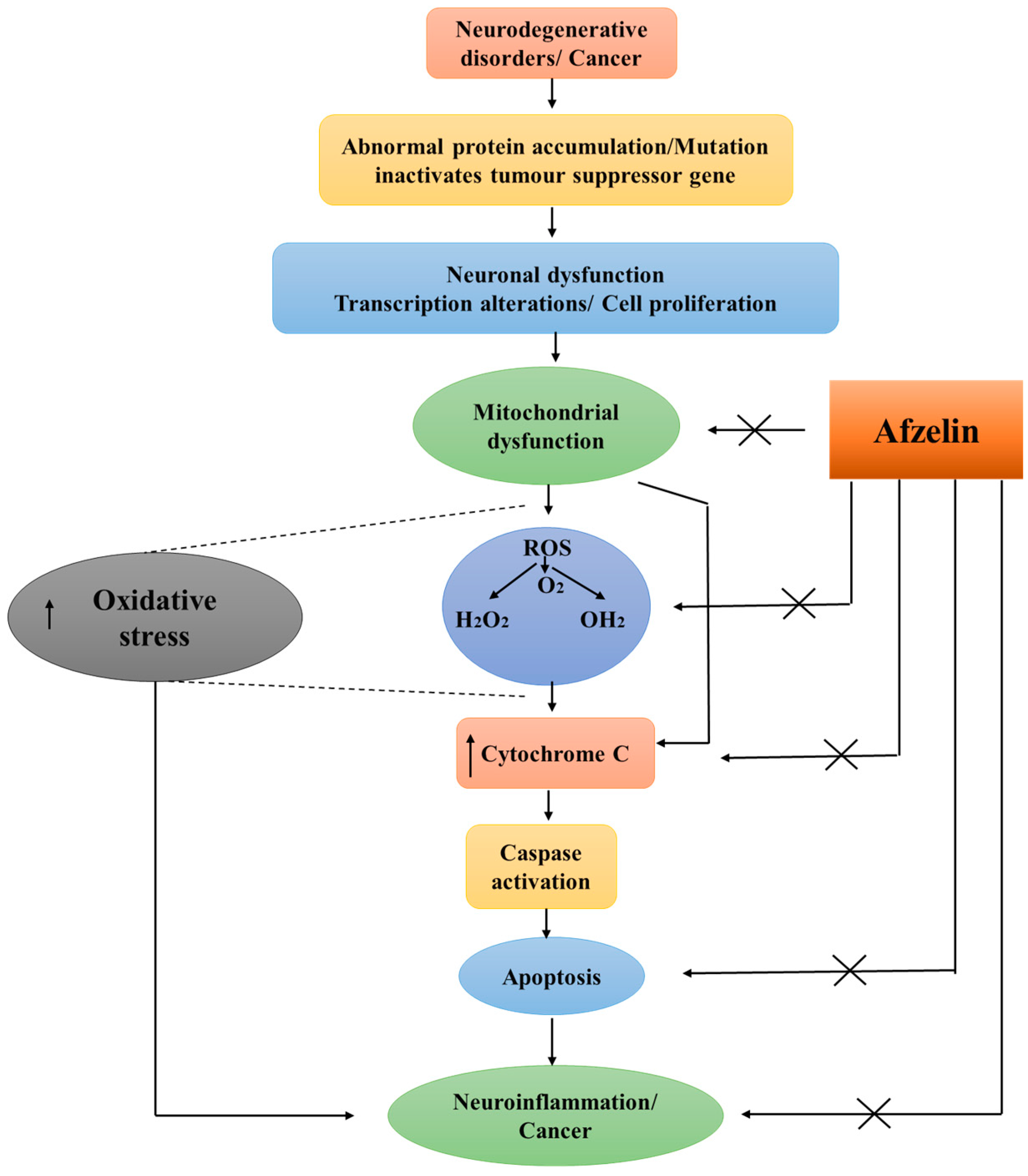
3.2. Neuroprotective and Neurogenic Effects

Brain-derived neurotrophic factor (BDNF) is a member of the neurotrophin protein family and works as a pivotal growth factor required for the integrity, individuality, and adaptability of neuronal populations [107]. BDNF is broadly expressed in the mammalian brain, with especially high levels in the hippocampus and parahippocampal regions, both of which are involved in memory formation. Its expression is modulated dynamically in response to external cues and internal states, which at the molecular level involves the cooperation of multiple events, including transcriptional control, epigenetic processes, and post-translational modifications. BDNF interacts with its receptors, tropomyosin kinase B (TRKB) and P75 neurotrophin receptor (P75NTR), to play an important role in synaptic transmission and activity-dependent plasticity [108]. By interacting with these proteins, BDNF triggers a cascade of signaling events that control gene expression, synaptic strength, neuronal shape, and neurogenesis. In addition, BDNF is involved in a wide variety of physiological and pathological processes, including nutritional control, stress response, aging, neurodegeneration, and neuropsychiatric diseases. Changes in BDNF expression levels are associated with Alzheimer’s disease, Parkinson’s disease, Huntington’s disease, and other forms of neurodegeneration [109,110]. As a consequence of this, BDNF is being investigated as a potential therapeutic target for neurological conditions. An increase in the synthesis of BDNF has been suggested as a therapeutic avenue for neurodegenerative disease treatment [109,111].
The introduction of afzelin into the mice brains on a chronic schedule resulted in the improvement of synaptic plasticity and cognitive/memory functions in animals that were administered scopolamine. Research conducted on the hippocampi of mice has demonstrated that the restorative effects observed in the cholinergic systems and molecular signal transduction through cyclic AMP response element-binding protein (CREB)-BDNF pathways can be attributed to the response elicited by afzelin. It was demonstrated that the administration of afzelin resulted in enhanced neurocognitive and neuroprotective effects on synaptic plasticity and behaviors, partially attributed to the up-regulation of CREB-BDNF signaling [112]. Moreover, the increased BDNF production may promote neuroprotective activity in hippocampal neurons through the stimulation of phosphatidylinositol 3-kinase (PI3K) and RAS/MAPK pathways and consequent abrogation of apoptotic cell death as evidenced by reduced caspase activity and increased B-cell lymphoma 2 (BCL2) protein expression [113]
Aldose reductase (AR) is an enzymatic protein responsible for the conversion of glucose into sorbitol within the NADPH-dependent polyol pathway of glucose metabolism. AR plays a pivotal role in the activation of microglia and the progression of retinal neurodegeneration. The activation of surveillance microglia occurs with the application or deposition of β-amyloid, subsequently leading to the formation of ROS, increased release of TNF-α, and enhanced phagocytosis. These processes collectively contribute to the death of neurons. The enhanced migratory capacity of microglia may result in cerebral injury. The inhibition of AR activity has the potential to impede the activation and migration of microglia, hence offering a potential therapeutic strategy for mitigating neuronal cell death and decelerating the progression of Alzheimer’s disease [114]. Afzelin was shown to act as an AR inhibitor with an IC50 concentration of 1.91 μM compared with 12.87 μM estimated for kaempferol and could constitute one of the underlying causes of down-regulation of inflammatory mediators expression observed during the study [115], as shown for other AR inhibitors. These effects can be attributed to the modulation of MAPK and NF-κB-mediated pathways [116].
The areas inside the brain that are responsible for the production of significant quantities of neuromelanin (NM), a dark cytoplasmic pigment, are limited to the SN and locus coeruleus (LC). Evidence suggests that NM serves an initial protective function by isolating toxic catecholamine metabolites and heavy metals. However, it is posited that this protective role may transition into a destructive one during the aging process, especially in the context of Parkinson’s disease. This transition occurs as the aforementioned toxic substances accumulate to a point where they overpower cellular recovery machinery and are subsequently released during the process of neurodegeneration. The accumulation of NM was linked to the occurrence of neurodegeneration, as well as a strong neuroinflammatory response [117]. The process of synthesizing human NM is considered to be analogous to the generation of melanin in melanocytes. In the skin, melanin synthesis occurs through the conversion of DOPAquinone (DQ) by the enzyme tyrosinase. On the other hand, NM synthesis in neurons takes place through the conversion of dopamine quinone (DAQ) by the enzymes tyrosine hydroxylase (TH) and aromatic L-amino acid decarboxylase (AADC). Dopamine within the cytoplasm exhibits a high level of reactivity and is thought to undergo spontaneous oxidation or be oxidized by an undiscovered tyrosinase enzyme, resulting in the formation of DAQ. Subsequently, DAQ is converted into NM. The buildup of intracellular NM over a certain threshold has been documented to be linked to the death of dopaminergic neurons and the manifestation of symptoms associated with Parkinson’s disease. Therefore, tyrosinase inhibition could constitute a potential therapeutic approach in this neurodegenerative disease [118].
Rho et al. aimed to assess the tyrosinase inhibitory activity of the ethanolic extract derived from the leaves of kenaf (Hibiscus cannabinus L.) both before and after exposure to far-infrared (FIR) irradiation. No inhibitory activity of the extract was seen in a tyrosinase enzymatic activity experiment before FIR irradiation. Nevertheless, when subjecting it to FIR, a noteworthy degree of tyrosinase inhibitory activity (IC50 = 3500 ppm) was detected. The high-performance liquid chromatography (HPLC) examination revealed the presence of derhamnosylation products, specifically kaempferol, afzelin, and α-rhamnoisorobin. The observed inhibitory activity could perhaps be attributed to the presence of derhamnosylation products [119].
Afzelin is one of 28 metabolites revealed by liquid chromatography–tandem mass spectrometry (LC-MS/MS) in the ethanolic extract of Polygonum minus. Studies of neuroprotection against H2O2-induced oxidative stress performed on differentiated SH-SY5Y cells revealed activation of NRF2/antioxidant response element (ARE), NF-κB/IκB, and MAPK signaling pathways. A 48 h pre-incubation of cells with PMEE extract resulted in a significant up-regulation of NRF2, NQO1, SOD1, SOD2, and catalase gene expression levels compared to both PMEE extract and H2O2 treatments. A similar effect was observed for MAPK8 (JNK), MAPK14 (p38), and PPP2CA (PP2A) genes. Also, there are various in silico and molecular docking studies that showed that afzelin was one of the compounds present in the Polygonum minus extract with the highest stability and docking score [120].
Acetylcholine (ACH) is a neurotransmitter that may play a positive role in modulating the cellular response to oxidative stress by regulating antioxidant processes, mitochondrial function, and protection against negative effects resulting from excess ROS. In some neurodegenerative diseases like Alzheimer’s disease, low levels of ACH are observed. Acetylcholinesterase (AChE) is an enzyme responsible for metabolizing the ACH into acetic acid and choline, which allow for the termination of the signal transmission at the synapses. AChE inhibitors have beneficial effects in certain neurological conditions by temporarily enhancing cholinergic neurotransmission [121]. Under conditions of H2O2-induced oxidative stress, prior administration of PMEE increased ACH concentration in differentiated SH-SY5Y cells supernatant. Molecular docking results showed that afzelin was one of the three compounds present in the Polygonum minus ethanolic extract with the highest docking score to AChE [120].

3.3. Anti-Cancer Effects of Afzelin

Flavonoids, which include flavonol glycoside, have long been studied for their anti-cancer effects. In the case of afzelin, recent years have yielded the results of the few studies to date that indicate the potential to induce cell death and inhibit tumor proliferation in an in vitro model. Androgen-sensitive LNCaP and androgen-independent PC-3 cell lines were used in one of the anti-cancer activity studies on afzelin. The investigation revealed that afzelin, at a concentration of 1 µg/mL, caused a significant decrease in prostate cancer cell proliferation after 24 h incubation. Flow cytometry cell cycle evaluation showed accumulation of PC-3 and LNCaP cells in the G0 phase (27.6% and 19.4% of the population, respectively) after 10 µg/mL afzelin exposure and indicated the occurrence of extensive apoptosis or an ongoing cytotoxic response. At the molecular level, afzelin suppressed Rho GTPase family proteins such as LIM domain kinase 1 (LIMK1), myotonic dystrophy kinase-related Cdc42-binding kinase α (MRCKα), and Rho-associated coiled-coil-containing protein kinase (ROCK1) expression. The Rho GTPase family is intricately linked to the modulation of actin cytoskeleton arrangement, mediation of the focal adhesions, stress fiber formation, and regulation of cell proliferation and differentiation via activation of different proteins. Expression of LIMK1 is observed to be elevated in prostate cancer cells, while ROCK1 and MRCKα are involved in its activation. In examined prostate cancer cells, a reduction in the phosphorylation level of the aforementioned proteins was detected dependent on the concentration of afzelin tested [38].
Suppression of cell migration of highly invasive MDA-MB-231 triple-negative breast cancer (TNBC) cells following afzelin treatment was shown to be mediated by focal adhesion kinase (FAK) and Ras-related C3 botulinum toxin substrate 1 (RAC1) deregulation. FAK and RAC1 proteins interact in signaling pathways that control the organization of the cytoskeleton and the dynamics of focal adhesions, which in turn affect cell migration processes. Afzelin induced moderate cytotoxicity of MDA-MB-231 cells (IC50 = 992 μg/mL); however, at the same time, it actively reduced focal adhesion formation in a concentration-dependent manner. Because FAK and RAC1 are often excessively activated in numerous tumor types, they increase the likelihood of invasion and metastasis, which correlates with a worse prognosis for patients. Afzelin at 400 and 800 µg/mL significantly reduced RAC1 GTPase activation by 60% compared to the negative control, while the highest concentration also reduced FAK and phosphorylated FAK (pFAK) expression [122]. Additionally, Rachimi et al., using the reverse docking method, confirmed the inhibitory potential of afzelin toward molecular targets relevant to TNBC, including FAK, ERK2, and KRAS [123].
The pro-apoptotic efficacy of afzelin has been assessed in vitro in a variety of cancer cell types. For instance, kaempferol-3-O-rhamnoside isolated from leaves of Schima wallichii Korth was able to reduce the proliferation of MCF-7 human breast cancer cells. The findings demonstrated that afzelin triggers apoptotic mechanisms through activation of the caspase signaling pathway [124]. Afzelin was found to induce apoptotic cell death in gastric cancer cells (AGS) [105] as well as lung cancer cells (A549 and H1299) [106]. In the case of A549 cells, exposure to 60 and 120 µM afzelin resulted in an increase in the mRNA levels (an increase of 30% and 80%, respectively) of the pro-apoptotic factor BAX in comparison with non-treated cells. Increased afzelin concentrations resulted in stimulation of the caspase cascade, with the involvement of both caspase 8 and caspase 9 [105]. In a study using a model of lung cancer, characteristic indicators of immunogenic cell death, such as calreticulin exposure or high-mobility group box 1 (HMGB1) and ATP release, were observed [106].

4. ADMET Properties of Afzelin

Afzelin was shown to reach a maximal concentration of 211.02 ± 27.97 μg/L at a tmax = 0.83 ± 0.26 h, following administration of the inflorescence of Polygonum orientale at a concentration of 0.3 g/mL, which is comparable to a crude herb dosage of 86 g/kg or an extract dosage of 3 g/kg. This dosage constituted six times the recommended daily dose for an adult [125].
The study conducted by Alves et al. provided evidence indicating that hydroalcoholic extract of Copaifera lansdorffii Desf. (CLE) did not exhibit genotoxic effects in the mouse micronucleus experiment. The administration of CLE plus doxorubicin to animals resulted in a considerable reduction in the number of micronuclei, as compared to animals that were treated only with doxorubicin [126]. The protective effects of afzelin and quercitrin against the cytotoxicity induced by doxorubicin or methyl methanesulfonate (MMS) in V79 and HepG2 cell lines were investigated using the same extract. Moreover, it was observed that CLE did not elicit a statistically significant alteration in the occurrence of chromosomal abnormalities and micronuclei in the tested cells. Additionally, the number of revertants in the Ames test remained the same, indicating the absence of its mutagenic effects. On the other hand, it was observed that CLE exhibited anti-genotoxic properties in mammalian cells. The administration of afzelin and quercitrin showed a reduction in genotoxicity generated by MMS in HepG2 cells. The observed antigenotoxic impact of CLE in this study could potentially be ascribed, at least in part, to the antioxidative activity of the combined main components, namely afzelin and quercitrin [127].
Afzelin is widely recognized for its antioxidative characteristics [128], which can be seen from its capacity to scavenge various oxidative species. Despite its beneficial properties, it was found to induce cytotoxic effects that can be beneficial for the treatment of cancer. Afzelin was found to have a promotional effect on neutrophil death, particularly when the oxidative burst was induced by phorbol myristate acetate. Hence, while its widely recognized ability to scavenge free radicals and oxidants, this molecule can inflict side effects in living organisms by exerting its effects on erythrocytes and neutrophils [129].
Given the importance of assessing other toxicological endpoints for new promising molecules like afzelin, future research could focus on expanding the evaluation of its ADMET properties beyond the scope of its genotoxicity and cytotoxicity. Investigating the metabolic fate of pure afzelin in vivo could provide insights into its biotransformation pathways and the formation of metabolites. This information is crucial for understanding its pharmacokinetic profile and potential interactions with other drugs or compounds. Assessing the distribution of afzelin in various tissues after administration could help determine its target organs and potential accumulation in specific tissues. Understanding its tissue distribution profile is essential for predicting its efficacy and potential toxicity. Studying the excretion pathways of afzelin, including renal and biliary excretion, could provide valuable information about its elimination kinetics and potential for accumulation. This knowledge is important for determining appropriate dosing regimens and potential risks associated with long-term exposure. Conducting comprehensive toxicity studies to evaluate the acute and chronic effects of afzelin at various doses and duration of exposure in animal models could help assess its safety profile. This includes assessing its effects on organ function, histopathological changes, and overall systemic toxicity. In summary, by systematically evaluating these additional toxicological endpoints, researchers could gain a more comprehensive understanding of the safety profile of afzelin, which is essential for its further development as a potential therapeutic agent.

5. Conclusions and Future Prospects

Plants possess the ability to manufacture a diverse range of chemical compounds, which exhibit potential significance in various biological contexts. Numerous phytochemicals have the potential to effectively address various human illnesses and enhance long-term well-being. [15,29]. Neurodegenerative diseases and cancer are of significant concern in the medical field due to the absence of efficacious therapeutic interventions and the alarming increase in the number of individuals affected by these conditions. Despite the varied clinical presentations of various illnesses, they all exhibit a shared cellular stress response. The cellular stress responses encompass various physiological processes, such as oxidative stress and inflammation. In recent years, there has been a significant focus on the potential of small molecules, specifically flavonoids, to mitigate cellular stress [130].
Afzelin (kaempferol 3-O-rhamnoside) is a representative of the flavonoid class of compounds. It has been observed to occur in a diverse range of plant sources. The potential benefits it may give to human health are beginning to be recognized. Afzelin demonstrates a wide array of biological activities, such as antioxidative, anti-inflammatory, anti-cancer, and neuroprotective properties. These characteristics make it a promising candidate for the prevention and treatment of neurodegenerative diseases that are characterized by oxidative stress and disruptions in cell death pathways (Figure 4).
The utilization of botanical extracts and isolated compounds in the management of neurodegenerative disorders and cancers encompasses diverse methodologies and presents inherent advantages and drawbacks. Plant extracts frequently consist of intricate combinations of diverse chemicals that are present in a given plant. These extracts have the potential to encompass a diverse array of phytochemicals, including polyphenols, alkaloids, and flavonoids. The intricate nature of these extracts presents difficulty in precisely determining the mechanisms of action and distinguishing the specific components accountable for therapeutic benefits. On the contrary, the obtaining of isolated substances involves the extraction and purification of particular components from plants. This methodology enables the examination of specific compounds and their impact on disorders, hence facilitating the identification of the exact bioactive component responsible for the therapeutic outcome. Furthermore, plant extracts contain substances that can increase the bioavailability of active ingredients or demonstrate synergistic effects. Nevertheless, the existence of additional chemicals may also potentially impede the processes of absorption, distribution, metabolism, and excretion (ADME) of bioactive constituents. In contrast, isolated substances frequently exhibit distinct pharmacokinetic characteristics, facilitating a comprehension process of ADME inside the human body. Understanding this information is of utmost importance when determining appropriate dose and treatment approaches. The safety characteristics of plant extracts can differ based on their specific composition. They may contain chemicals that have the potential to induce adverse effects or interact with other pharmaceutical substances. Isolated chemicals exhibit a greater degree of predictability in terms of safety. Nevertheless, it is important to note that even isolated substances can potentially exhibit adverse consequences. The ADME and toxicity profiling of afzelin and various plant extracts containing this flavonoid has not been performed (except for genotoxic studies) and constitutes one of the remaining issues that will determine the therapeutic usefulness of this compound. Furthermore, its blood–brain barrier (BBB) permeability has not been examined. The BBB serves as a crucial protective factor that effectively limits the permeability of medicines and therapeutic agents, hence impeding their access to the brain. The development of pharmaceutical agents capable of efficiently traversing the BBB to achieve precise localization within CNS is a major challenge. Strategies aimed at improving the bioavailability of these substances, such as the utilization of nanoformulations or prodrugs, have the potential to increase their pharmacokinetic characteristics and ultimately optimize their therapeutic efficacy.
The diverse composition of plant extracts may provide a wider range of activity, rendering them appropriate for addressing various targets in the context of neurodegenerative disorders. This can confer advantages in the context of diseases characterized by complex pathologies. In the context of therapeutic interventions, the utilization of isolated chemicals enables the precise targeting of a single molecular target or pathway. This approach is advantageous in cases where a comprehensive understanding of the illness mechanism exists and necessitates a highly focused strategy.
Afzelin has been shown to elicit a complex cellular response involving its suppressive effects on NF-κB or MAPK pathway conferring reduced expression of pro-inflammatory mediators or down-regulation of NO synthesis, inhibition of tyrosinase activity or up-regulation of molecular signaling pathways involving CREB-BDNF or influence on mitochondrial quality control and function. Moreover, it showed significant oxidative stress mitigating effects associated with ROS scavenging, metal chelation, and the influence of the NRF2 signaling pathway.
In the context of neurodegenerative diseases, current therapies often focus on symptomatic management rather than addressing the underlying disease mechanisms. For example, in Alzheimer’s disease, acetylcholinesterase inhibitors such as donepezil and memantine are commonly prescribed to alleviate cognitive symptoms, but they do not halt disease progression [131]. Similarly, in Parkinson’s disease, dopamine replacement therapy with levodopa is a mainstay treatment for managing motor symptoms, but it does not prevent the degeneration of dopaminergic neurons [132]. In contrast, afzelin offers a multifaceted approach to addressing neurodegenerative diseases by targeting oxidative stress, inflammation, mitochondrial dysfunction, and neuroprotection, as outlined in the manuscript. Its ability to modulate redox signaling pathways, scavenge free radicals, and inhibit inflammatory mediators suggests a potential neuroprotective effect that may complement existing therapies. According to a recent study of afzelin in Parkinson’s disease, the cataleptic behaviors and locomotor activity of rats were greatly improved when administered with a combination of reserpine at a dose of 1 mg/kg and afzelin at doses of 5 mg/kg, 10 mg/kg, and 20 mg/kg. Furthermore, the administration of afzelin alone resulted in the induction of BCL-2 expression, suggesting potential neuroprotective characteristics. This study yielded valuable insights into the efficacy of afzelin in managing catalepsy and other degenerative neurological illnesses. Therefore, further investigations are required to ascertain the causes of the reactions and ascertain the enduring impacts of Afzelin on these situations [133].
Similarly, in the context of cancer, current treatments such as chemotherapy, radiation therapy, and targeted therapies often have significant side effects and may lead to drug resistance [134,135]. Afzelin’s anti-cancer properties, including its ability to induce apoptosis and inhibit inflammatory pathways, offer a novel approach to cancer treatment that may have fewer adverse effects compared to conventional therapies and may help prevent the development of treatment resistance or constitute a novel scaffold for drug discovery endeavors.
Furthermore, Wu et al. presented a method that is both ecologically sustainable and efficient in terms of speed and productivity for the extraction and purification of four bioactive components from Zanthoxylum bungeanum leaves, including afzelin. The findings can be employed for the incorporation of Z. bungeanum leaves as a dietary supplement in an industrial context, offering an accessible source of afzelin for therapeutical use [136]. Similarly, de Sousa et al. provided a validated chromatographic method for the detection of afzelin in Copaifera langsdorffii extracts by HPLC, providing another method suitable for the detection and isolation of this flavonoid [137]. The process of standardizing plant extracts to assure constant quantities of bioactive chemicals is of particular importance as it might present challenges, primarily stemming from inherent variances in plant material. Quality control can be more difficult compared to isolated substances. Isolated compounds can be synthesized or purified to a high degree of purity, enabling precise dosing and consistent therapeutic effects. Furthermore, the ease and cost-effectiveness of the afzelin isolation and purification procedure are of importance. In order to provide care to a diverse patient population, it is imperative that the treatment is made readily available and economically feasible, taking into account the socioeconomic obstacles encountered by those afflicted with neurodegenerative disorders and cancers.
In summary, the future prospects for utilizing plant-derived compounds like afzelin in the treatment of neurodegenerative diseases and cancer are promising. Afzelin, a flavonoid with multifaceted properties, holds potential as a therapeutic agent due to its anti-inflammatory, antioxidative, anti-cancer, and neuroprotective effects. Additionally, recent advancements in extraction and purification methods, such as those demonstrated by Wu et al. and de Sousa et al., offer sustainable and efficient means of obtaining afzelin and other bioactive compounds from natural sources. While challenges remain, including standardization of plant extracts and addressing bioavailability and BBB issues, the continued exploration of plant-derived compounds, including afzelin, represents a valuable avenue for developing novel treatments for neurodegenerative diseases.
While this paper extensively covers in vitro and animal studies, it is crucial to acknowledge the necessity of transitioning these findings into clinical settings to ascertain the efficacy and safety of afzelin in humans. As of the current literature review, there are no clinical trials or human studies available for afzelin. Therefore, the focus of this review paper has been on detailing the results from preclinical studies, which provide valuable insights into the potential mechanisms of action and therapeutic effects of afzelin.

Author Contributions

Conceptualization: S.C., S.D. (Sanchit Dhankhar) and S.M.; writing—original draft preparation: M.S., M.K., N.G. and S.D. (Sushma Devi); writing—review and editing: A.G., B.M. and R.K.; supervision: T.G.S. and R.S. All authors have read and agreed to the published version of the manuscript.

Funding

This research did not receive any specific grant from funding agencies in the public, commercial, or not-for-profit sectors.

Conflicts of Interest

The authors declare no conflicts of interest.

Abbreviations

amyloid-beta
AADCaromatic L-amino acid decarboxylase
ACHacetylcholine
AChEacetylcholinesterase
ADMETabsorption, distribution, metabolism, excretion, and toxicity
ALSamyotrophic lateral sclerosis
ARaldose reductase
AREantioxidant response element
ATPadenosine triphosphate
BBBblood–brain barrier
BCL2B-cell lymphoma 2
BDNFbrain-derived neurotrophic factor
CLEhydroalcoholic extract of Copaifera lansdorffii Desf.
CNScentral nervous system
COcarbon monoxide
CREBcyclic AMP response element-binding protein
DAQdopamine quinone
DNM1Ldynamin-related protein 1
DQDOPAquinone
ERKextracellular signal-regulated kinase
FAKfocal adhesion kinase
FHFfulminant hepatic failure
FIRfar-infrared
FTDfrontotemporal dementia
GalND-galactosamine
GSK-3βglycogen synthase kinase-3β
GSTsglutathione S-transferases
HMGB1high-mobility group box 1
HO-1/HO-2heme oxygenase-1/2
HPLChigh-performance liquid chromatography
IL-1/IL-6interleukin-1/6
iNOSinducible nitric oxide synthase
JNKc-Jun N-terminal kinase
LClocus coeruleus
LC-MS/MSliquid chromatography–tandem mass spectrometry
LIMK1LIM domain kinase 1
LPSlipopolysaccharide
MAPKmitogen-activated protein kinase
MFN2mitofusin 2
MRCKαmyotonic dystrophy kinase-related Cdc42-binding kinase α
MSmultiple sclerosis
MtDNAmitochondrial DNA
NF-κBnuclear factor kappa B
NMneuromelanin
NQO1NAD(P)H quinone dehydrogenase 1
NRF1nuclear respiratory factor 1
NRF2nuclear factor erythroid 2-related factor 2
OMMouter mitochondrial membrane
P75NTRP75 neurotrophin receptor
PARKINparkin protein
pFAKphosphorylated focal adhesion kinase
PGC-1αPPAR-γ coactivator 1α
PI3K phosphatidylinositol 3-kinase
PINK1PTEN-induced putative kinase 1
PMEEPolygonum minus ethanolic extract
RAC1Ras-related C3 botulinum toxin substrate 1
ROCK1Rho-associated coiled-coil-containing protein kinase
ROSreactive oxygen species
SOD1/SOD2superoxide dismutase 1/2
SNsubstantia nigra
TFAMmitochondrial transcription factor A
THtyrosine hydroxylase
TNBCtriple-negative breast cancer
TNF-αtumor necrosis factor-alpha
TRKBtropomyosin kinase B

References

  1. Dugger, B.N.; Dickson, D.W. Pathology of Neurodegenerative Diseases. Cold Spring Harb. Perspect. Biol. 2017, 9, a028035. [Google Scholar] [CrossRef] [PubMed]
  2. Jellinger, K.A. Basic Mechanisms of Neurodegeneration: A Critical Update. J. Cell. Mol. Med. 2010, 14, 457. [Google Scholar] [CrossRef] [PubMed]
  3. Lamptey, R.N.L.; Chaulagain, B.; Trivedi, R.; Gothwal, A.; Layek, B.; Singh, J. A Review of the Common Neurodegenerative Disorders: Current Therapeutic Approaches and the Potential Role of Nanotherapeutics. Int. J. Mol. Sci. 2022, 23, 1851. [Google Scholar] [CrossRef] [PubMed]
  4. Gulisano, W.; Maugeri, D.; Baltrons, M.A.; Fà, M.; Amato, A.; Palmeri, A.; D’Adamio, L.; Grassi, C.; Devanand, D.P.; Honig, L.S.; et al. Role of Amyloid-β and Tau Proteins in Alzheimer’s Disease: Confuting the Amyloid Cascade. J. Alzheimer’s Dis. 2018, 64, S611–S631. [Google Scholar] [CrossRef] [PubMed]
  5. Murphy, M.P.; LeVine, H. Alzheimer’s Disease and the β-Amyloid Peptide. J. Alzheimer’s Dis. 2010, 19, 311. [Google Scholar] [CrossRef] [PubMed]
  6. Maiti, P.; Manna, J.; Dunbar, G.L. Current Understanding of the Molecular Mechanisms in Parkinson’s Disease: Targets for Potential Treatments. Transl. Neurodegener. 2017, 6, 28. [Google Scholar] [CrossRef] [PubMed]
  7. Hardiman, O.; Al-Chalabi, A.; Chio, A.; Corr, E.M.; Logroscino, G.; Robberecht, W.; Shaw, P.J.; Simmons, Z.; van den Berg, L.H. Amyotrophic Lateral Sclerosis. Nat. Rev. Dis. Prim. 2017, 3, 17071. [Google Scholar] [CrossRef] [PubMed]
  8. Ghasemi, N.; Razavi, S.; Nikzad, E. Multiple Sclerosis: Pathogenesis, Symptoms, Diagnoses and Cell-Based Therapy. Cell J. 2017, 19, 1–10. [Google Scholar] [PubMed]
  9. Young, J.J.; Lavakumar, M.; Tampi, D.; Balachandran, S.; Tampi, R.R. Frontotemporal Dementia: Latest Evidence and Clinical Implications. Ther. Adv. Psychopharmacol. 2018, 8, 33–48. [Google Scholar] [CrossRef] [PubMed]
  10. Ajmal, M.R. Protein Misfolding and Aggregation in Proteinopathies: Causes, Mechanism and Cellular Response. Diseases 2023, 11, 30. [Google Scholar] [CrossRef]
  11. Kennedy, D.O.; Wightman, E.L. Herbal Extracts and Phytochemicals: Plant Secondary Metabolites and the Enhancement of Human Brain Function. Adv. Nutr. 2011, 2, 32–50. [Google Scholar] [CrossRef] [PubMed]
  12. Mohd Sairazi, N.S.; Sirajudeen, K.N.S. Natural Products and Their Bioactive Compounds: Neuroprotective Potentials against Neurodegenerative Diseases. Evid.-Based Complement. Altern. Med. 2020, 2020, 6565396. [Google Scholar] [CrossRef] [PubMed]
  13. Sharifi-Rad, M.; Lankatillake, C.; Dias, D.A.; Docea, A.O.; Mahomoodally, M.F.; Lobine, D.; Chazot, P.L.; Kurt, B.; Tumer, T.B.; Moreira, A.C.; et al. Impact of Natural Compounds on Neurodegenerative Disorders: From Preclinical to Pharmacotherapeutics. J. Clin. Med. 2020, 9, 1061. [Google Scholar] [CrossRef] [PubMed]
  14. Srivastava, P.; Yadav, R.S. Efficacy of Natural Compounds in Neurodegenerative Disorders. Adv. Neurobiol. 2016, 12, 107–123. [Google Scholar] [CrossRef] [PubMed]
  15. Gielecińska, A.; Kciuk, M.; Mujwar, S.; Celik, I.; Kołat, D.; Kałuzińska-Kołat, Ż.; Kontek, R. Substances of Natural Origin in Medicine: Plants vs. Cancer. Cells 2023, 12, 986. [Google Scholar] [CrossRef] [PubMed]
  16. Chen, X.; Guo, C.; Kong, J. Oxidative Stress in Neurodegenerative Diseases. Neural Regen. Res. 2012, 7, 376–385. [Google Scholar] [CrossRef] [PubMed]
  17. Liu, Z.; Zhou, T.; Ziegler, A.C.; Dimitrion, P.; Zuo, L. Oxidative Stress in Neurodegenerative Diseases: From Molecular Mechanisms to Clinical Applications. Oxid. Med. Cell. Longev. 2017, 2017, 2525967. [Google Scholar] [CrossRef] [PubMed]
  18. Niedzielska, E.; Smaga, I.; Gawlik, M.; Moniczewski, A.; Stankowicz, P.; Pera, J.; Filip, M. Oxidative Stress in Neurodegenerative Diseases. Mol. Neurobiol. 2016, 53, 4094–4125. [Google Scholar] [CrossRef] [PubMed]
  19. Singh, A.; Kukreti, R.; Saso, L.; Kukreti, S. Oxidative Stress: A Key Modulator in Neurodegenerative Diseases. Molecules 2019, 24, 1583. [Google Scholar] [CrossRef] [PubMed]
  20. Arfin, S.; Jha, N.K.; Jha, S.K.; Kesari, K.K.; Ruokolainen, J.; Roychoudhury, S.; Rathi, B.; Kumar, D. Oxidative Stress in Cancer Cell Metabolism. Antioxidants 2021, 10, 642. [Google Scholar] [CrossRef] [PubMed]
  21. Klaunig, J.E. Oxidative Stress and Cancer. Curr. Pharm. Des. 2018, 24, 4771–4778. [Google Scholar] [CrossRef] [PubMed]
  22. Hayes, J.D.; Dinkova-Kostova, A.T.; Tew, K.D. Oxidative Stress in Cancer. Cancer Cell 2020, 38, 167–197. [Google Scholar] [CrossRef] [PubMed]
  23. da Cunha Germano, B.C.; de Morais, L.C.C.; Idalina Neta, F.; Fernandes, A.C.L.; Pinheiro, F.I.; do Rego, A.C.M.; Araújo Filho, I.; de Azevedo, E.P.; de Paiva Cavalcanti, J.R.L.; Guzen, F.P.; et al. Vitamin E and Its Molecular Effects in Experimental Models of Neurodegenerative Diseases. Int. J. Mol. Sci. 2023, 24, 11191. [Google Scholar] [CrossRef] [PubMed]
  24. Trela-Makowej, A.; Leśkiewicz, M.; Kruk, J.; Żądło, A.; Basta-Kaim, A.; Szymańska, R. Antioxidant and Neuroprotective Activity of Vitamin E Homologues: In Vitro Study. Metabolites 2022, 12, 608. [Google Scholar] [CrossRef] [PubMed]
  25. Bonnet, U. The Sour Side of Vitamin C Might Mediate Neuroprotective, Anticonvulsive and Antidepressant-like Effects. Med. Hypotheses 2019, 131, 109320. [Google Scholar] [CrossRef] [PubMed]
  26. Kangisser, L.; Tan, E.; Bellomo, R.; Deane, A.M.; Plummer, M.P. Neuroprotective Properties of Vitamin C: A Scoping Review of Pre-Clinical and Clinical Studies. J. Neurotrauma 2021, 38, 2194–2205. [Google Scholar] [CrossRef] [PubMed]
  27. Hasan, S.; Khatri, N.; Rahman, Z.N.; Menezes, A.A.; Martini, J.; Shehjar, F.; Mujeeb, N.; Shah, Z.A. Neuroprotective Potential of Flavonoids in Brain Disorders. Brain Sci. 2023, 13, 1258. [Google Scholar] [CrossRef] [PubMed]
  28. Putteeraj, M.; Lim, W.L.; Teoh, S.L.; Yahaya, M.F. Flavonoids and Its Neuroprotective Effects on Brain Ischemia and Neurodegenerative Diseases. Curr. Drug Targets 2018, 19, 1710–1720. [Google Scholar] [CrossRef] [PubMed]
  29. Akter, M.; Parvin, M.S.; Hasan, M.M.; Rahman, M.A.A.; Islam, M.E. Anti-Tumor and Antioxidant Activity of Kaempferol-3-O-Alpha-L-Rhamnoside (Afzelin) Isolated from Pithecellobium dulce Leaves. BMC Complement. Med. Ther. 2022, 22, 169. [Google Scholar] [CrossRef] [PubMed]
  30. Kim, J.H.; Kim, M.; Kim, J.M.; Lee, M.-K.; Seo, S.J.; Park, K.Y. Afzelin Suppresses Proinflammatory Responses in Particulate Matter-Exposed Human Keratinocytes. Int. J. Mol. Med. 2019, 43, 2516–2522. [Google Scholar] [CrossRef] [PubMed]
  31. Lee, S.-B.; Kang, J.-W.; Kim, S.-J.; Ahn, J.; Kim, J.; Lee, S.-M. Afzelin Ameliorates D-Galactosamine and Lipopolysaccharide-Induced Fulminant Hepatic Failure by Modulating Mitochondrial Quality Control and Dynamics. Br. J. Pharmacol. 2017, 174, 195–209. [Google Scholar] [CrossRef] [PubMed]
  32. Lotha, R.; Sundaramoorthy, N.S.; Shamprasad, B.R.; Nagarajan, S.; Sivasubramanian, A. Plant Nutraceuticals (Quercetrin and Afzelin) Capped Silver Nanoparticles Exert Potent Antibiofilm Effect against Food Borne Pathogen Salmonella enterica Serovar Typhi and Curtail Planktonic Growth in Zebrafish Infection Model. Microb. Pathog. 2018, 120, 109–118. [Google Scholar] [CrossRef] [PubMed]
  33. Cherbuin, N.; Walsh, E.; Baune, B.T.; Anstey, K.J. Oxidative Stress, Inflammation and Risk of Neurodegeneration in a Population Sample. Eur. J. Neurol. 2019, 26, 1347–1354. [Google Scholar] [CrossRef]
  34. Simpson, D.S.A.; Oliver, P.L. ROS Generation in Microglia: Understanding Oxidative Stress and Inflammation in Neurodegenerative Disease. Antioxidants 2020, 9, 743. [Google Scholar] [CrossRef] [PubMed]
  35. Lee, J.H.; Ahn, J.; Kim, J.W.; Lee, S.G.; Kim, H.P. Flavonoids from the Aerial Parts of Houttuynia Cordata Attenuate Lung Inflammation in Mice. Arch. Pharm. Res. 2015, 38, 1304–1311. [Google Scholar] [CrossRef] [PubMed]
  36. Zhang, X.; Cheng, B.; Liu, X.; Li, Y.; Hou, J.; Chen, S.; Chen, J.; Li, S. Screening of α-Glucosidase Inhibitors from Houttuynia Cordata and Evaluation of the Binding Mechanisms. ChemistrySelect 2020, 5, 8440–8446. [Google Scholar] [CrossRef]
  37. Zhang, Z.; ElSohly, H.N.; Li, X.-C.; Khan, S.I.; Broedel, S.E.; Raulli, R.E.; Cihlar, R.L.; Burandt, C.; Walker, L.A. Phenolic Compounds from Nymphaea Odorata. J. Nat. Prod. 2003, 66, 548–550. [Google Scholar] [CrossRef] [PubMed]
  38. Zhu, K.-C.; Sun, J.-M.; Shen, J.-G.; Jin, J.-Z.; Liu, F.; Xu, X.-L.; Chen, L.; Liu, L.-T.; Lv, J.-J. Afzelin Exhibits Anti-Cancer Activity against Androgen-Sensitive LNCaP and Androgen-Independent PC-3 Prostate Cancer Cells through the Inhibition of LIM Domain Kinase 1. Oncol. Lett. 2015, 10, 2359–2365. [Google Scholar] [CrossRef] [PubMed]
  39. Hernández-Caballero, M.E.; Sierra-Ramírez, J.A.; Villalobos-Valencia, R.; Seseña-Méndez, E. Potential of Kalanchoe Pinnata as a Cancer Treatment Adjuvant and an Epigenetic Regulator. Molecules 2022, 27, 6425. [Google Scholar] [CrossRef]
  40. Mejía-Méndez, J.L.; Bach, H.; Lorenzo-Leal, A.C.; Navarro-López, D.E.; López-Mena, E.R.; Hernández, L.R.; Sánchez-Arreola, E. Biological Activities and Chemical Profiles of Kalanchoe Fedtschenkoi Extracts. Plants 2023, 12, 1943. [Google Scholar] [CrossRef]
  41. Nascimento, L.B.d.S.; Casanova, L.M.; Costa, S.S. Bioactive Compounds from Kalanchoe Genus Potentially Useful for the Development of New Drugs. Life 2023, 13, 646. [Google Scholar] [CrossRef]
  42. Yadav, N.P.; Dixit, V.K. Hepatoprotective Activity of Leaves of Kalanchoe Pinnata Pers. J. Ethnopharmacol. 2003, 86, 197–202. [Google Scholar] [CrossRef] [PubMed]
  43. Mady, M.S.; Ibrahim, R.R.; El-Sayed, E.K.; El-Shazly, M.; Chen, L.-Y.; Lai, K.-H.; El Shaarawy, F.S.; Moharram, F.A. UHPLC-MS Profiles and Antidiarrheal Activity of Quercus coccinea Münchh. and Quercus robur L. Employing In Vivo Technique. Front. Pharmacol. 2023, 14, 1120146. [Google Scholar] [CrossRef] [PubMed]
  44. Oliveira, P.A.; Medeiros-Fonseca, B.; Vasconcelos-Nóbrega, C.; Alvarado, A.; Pires, M.J.; Vala, H.; Barros, A.I.R.N.A.; Faustino-Rocha, A.I. Quercus spp. Extract as a Promising Preventive or Therapeutic Strategy for Cancer: A Systematic Review. Mol. Med. Rep. 2023, 28, 175. [Google Scholar] [CrossRef] [PubMed]
  45. Jung, J.; Kim, H.; Lee, S.; Hong, M.; Hwang, D. Antioxidant and Anti-Inflammatory Activity of Filipendula Glaberrima Nakai Ethanolic Extract and Its Chemical Composition. Molecules 2022, 27, 4628. [Google Scholar] [CrossRef] [PubMed]
  46. Marčetić, M.; Samardžić, S.; Ilić, T.; Božić, D.D.; Vidović, B. Phenolic Composition, Antioxidant, Anti-Enzymatic, Antimicrobial and Prebiotic Properties of Prunus spinosa L. Fruits. Foods 2022, 11, 3289. [Google Scholar] [CrossRef] [PubMed]
  47. Soares, S.S.; Bekbolatova, E.; Cotrim, M.D.; Sakipova, Z.; Ibragimova, L.; Kukula-Koch, W.; Giorno, T.B.S.; Fernandes, P.D.; Fonseca, D.A.; Boylan, F. Chemistry and Pharmacology of the Kazakh Crataegus almaatensis Pojark: An Asian Herbal Medicine. Antioxidants 2019, 8, 300. [Google Scholar] [CrossRef] [PubMed]
  48. Pizzino, G.; Irrera, N.; Cucinotta, M.; Pallio, G.; Mannino, F.; Arcoraci, V.; Squadrito, F.; Altavilla, D.; Bitto, A. Oxidative Stress: Harms and Benefits for Human Health. Oxid. Med. Cell. Longev. 2017, 2017, 8416763. [Google Scholar] [CrossRef] [PubMed]
  49. Cechinel-Zanchett, C.C.; Bolda Mariano, L.N.; Boeing, T.; da Costa, J.d.C.; Da Silva, L.M.; Bastos, J.K.; Cechinel-Filho, V.; de Souza, P. Diuretic and Renal Protective Effect of Kaempferol 3-O-Alpha-l-Rhamnoside (Afzelin) in Normotensive and Hypertensive Rats. J. Nat. Prod. 2020, 83, 1980–1989. [Google Scholar] [CrossRef] [PubMed]
  50. Jung, E.; Kim, J.H.; Kim, M.O.; Lee, S.Y.; Lee, J. Melanocyte-Protective Effect of Afzelin Is Mediated by the Nrf2-ARE Signalling Pathway via GSK-3β Inactivation. Exp. Dermatol. 2017, 26, 764–770. [Google Scholar] [CrossRef] [PubMed]
  51. Kim, M.; Shin, S.; Ryu, D.; Cho, E.; Yoo, J.; Park, D.; Jung, E. Evaluating the Sun Protection Factor of Cosmetic Formulations Containing Afzelin. Chem. Pharm. Bull. 2021, 69, 1039–1044. [Google Scholar] [CrossRef] [PubMed]
  52. Juan, C.A.; de la Lastra, J.M.P.; Plou, F.J.; Pérez-Lebeña, E. The Chemistry of Reactive Oxygen Species (ROS) Revisited: Outlining Their Role in Biological Macromolecules (DNA, Lipids and Proteins) and Induced Pathologies. Int. J. Mol. Sci. 2021, 22, 4642. [Google Scholar] [CrossRef] [PubMed]
  53. Liu, Y.; Chen, P.; Zhou, M.; Wang, T.; Fang, S.; Shang, X.; Fu, X. Geographic Variation in the Chemical Composition and Antioxidant Properties of Phenolic Compounds from Cyclocarya paliurus (Batal) Iljinskaja Leaves. Molecules 2018, 23, 2440. [Google Scholar] [CrossRef] [PubMed]
  54. Ma, L.; Li, K.; Wei, D.; Xiao, H.; Niu, H.; Huang, W. High Anti-Oxidative and Lipid-Lowering Activities of Flavonoid Glycosides-Rich Extract from the Leaves of Zanthoxylum bungeanum in Multi-System. J. Food Nutr. Res. 2015, 3, 62–68. [Google Scholar] [CrossRef]
  55. Aldana, J.A.; De Grandis, R.A.; Nicolella, H.; Guissoni, A.P.P.; Squarisi, I.; Arruda, C.; Ribeiro, V.P.; Tavares, D.C.; Barcelos, G.R.M.; Antunes, L.M.G.; et al. Evaluation of Cytoprotective Effects of Compounds Isolated from Copaifera langsdorffii Desf. against Induced Cytotoxicity by Exposure to Methylmercury and Lead. Nat. Prod. Res. 2020, 34, 2528–2532. [Google Scholar] [CrossRef] [PubMed]
  56. Fernandez, M.T.; Mira, M.L.; Florêncio, M.H.; Jennings, K.R. Iron and Copper Chelation by Flavonoids: An Electrospray Mass Spectrometry Study. J. Inorg. Biochem. 2002, 92, 105–111. [Google Scholar] [CrossRef] [PubMed]
  57. Merino, J.J.; Parmigiani-Izquierdo, J.M.; Toledano Gasca, A.; Cabaña-Muñoz, M.E. The Long-Term Algae Extract (Chlorella and Fucus sp.) and Aminosulphurate Supplementation Modulate SOD-1 Activity and Decrease Heavy Metals (Hg++, Sn) Levels in Patients with Long-Term Dental Titanium Implants and Amalgam Fillings Restorations. Antioxidants 2019, 8, 101. [Google Scholar] [CrossRef] [PubMed]
  58. Kashyap, D.; Sharma, A.; Tuli, H.S.; Sak, K.; Punia, S.; Mukherjee, T.K. Kaempferol—A Dietary Anticancer Molecule with Multiple Mechanisms of Action: Recent Trends and Advancements. J. Funct. Foods 2017, 30, 203. [Google Scholar] [CrossRef] [PubMed]
  59. Alqahtani, T.; Deore, S.L.; Kide, A.A.; Shende, B.A.; Sharma, R.; Dadarao Chakole, R.; Nemade, L.S.; Kishor Kale, N.; Borah, S.; Shrikant Deokar, S.; et al. Mitochondrial Dysfunction and Oxidative Stress in Alzheimer’s Disease, and Parkinson’s Disease, Huntington’s Disease and Amyotrophic Lateral Sclerosis—An Updated Review. Mitochondrion 2023, 71, 83–92. [Google Scholar] [CrossRef] [PubMed]
  60. Misrani, A.; Tabassum, S.; Yang, L. Mitochondrial Dysfunction and Oxidative Stress in Alzheimer’s Disease. Front. Aging Neurosci. 2021, 13, 617588. [Google Scholar] [CrossRef] [PubMed]
  61. Bose, A.; Beal, M.F. Mitochondrial Dysfunction and Oxidative Stress in Induced Pluripotent Stem Cell Models of Parkinson’s Disease. Eur. J. Neurosci. 2019, 49, 525–532. [Google Scholar] [CrossRef] [PubMed]
  62. Hauser, D.N.; Hastings, T.G. Mitochondrial Dysfunction and Oxidative Stress in Parkinson’s Disease and Monogenic Parkinsonism. Neurobiol. Dis. 2013, 51, 35–42. [Google Scholar] [CrossRef] [PubMed]
  63. Lin, M.T.; Beal, M.F. Mitochondrial Dysfunction and Oxidative Stress in Neurodegenerative Diseases. Nature 2006, 443, 787–795. [Google Scholar] [CrossRef] [PubMed]
  64. Księżakowska-Łakoma, K.; Żyła, M.; Wilczyński, J.R. Mitochondrial Dysfunction in Cancer. Prz. Menopauzalny 2014, 13, 136–144. [Google Scholar] [CrossRef] [PubMed]
  65. Luo, Y.; Ma, J.; Lu, W. The Significance of Mitochondrial Dysfunction in Cancer. Int. J. Mol. Sci. 2020, 21, 5598. [Google Scholar] [CrossRef] [PubMed]
  66. Zhang, Y.; Chen, C.; Jiang, Y.; Wang, S.; Wu, X.; Wang, K. PPARγ Coactivator-1α (PGC-1α) Protects Neuroblastoma Cells against Amyloid-Beta (Aβ) Induced Cell Death and Neuroinflammation via NF-κB Pathway. BMC Neurosci. 2017, 18, 69. [Google Scholar] [CrossRef] [PubMed]
  67. Mota, B.C.; Sastre, M. The Role of PGC1α in Alzheimer’s Disease and Therapeutic Interventions. Int. J. Mol. Sci. 2021, 22, 5769. [Google Scholar] [CrossRef] [PubMed]
  68. Panes, J.D.; Wendt, A.; Ramirez-Molina, O.; Castro, P.A.; Fuentealba, J. Deciphering the Role of PGC-1α in Neurological Disorders: From Mitochondrial Dysfunction to Synaptic Failure. Neural Regen. Res. 2021, 17, 237–245. [Google Scholar] [CrossRef] [PubMed]
  69. Bost, F.; Kaminski, L. The Metabolic Modulator PGC-1α in Cancer. Am. J. Cancer Res. 2019, 9, 198–211. [Google Scholar] [PubMed]
  70. Kang, I.; Chu, C.T.; Kaufman, B.A. The Mitochondrial Transcription Factor TFAM in Neurodegeneration: Emerging Evidence and Mechanisms. FEBS Lett. 2018, 592, 793–811. [Google Scholar] [CrossRef] [PubMed]
  71. Quinn, P.M.J.; Moreira, P.I.; Ambrósio, A.F.; Alves, C.H. PINK1/PARKIN Signalling in Neurodegeneration and Neuroinflammation. Acta Neuropathol. Commun. 2020, 8, 189. [Google Scholar] [CrossRef] [PubMed]
  72. Wang, M.; Luan, S.; Fan, X.; Wang, J.; Huang, J.; Gao, X.; Han, D. The Emerging Multifaceted Role of PINK1 in Cancer Biology. Cancer Sci. 2022, 113, 4037–4047. [Google Scholar] [CrossRef] [PubMed]
  73. Denisenko, T.V.; Gogvadze, V.; Zhivotovsky, B. Mitophagy in Carcinogenesis and Cancer Treatment. Discov. Oncol. 2021, 12, 58. [Google Scholar] [CrossRef] [PubMed]
  74. Lee, D.; Kim, J.-Y.; Qi, Y.; Park, S.; Lee, H.L.; Yamabe, N.; Kim, H.; Jang, D.S.; Kang, K.S. Phytochemicals from the Flowers of Prunus persica (L.) Batsch: Anti-Adipogenic Effect of Mandelamide on 3T3-L1 Preadipocytes. Bioorg. Med. Chem. Lett. 2021, 49, 128326. [Google Scholar] [CrossRef] [PubMed]
  75. Lim, H.J.; Prajapati, R.; Seong, S.H.; Jung, H.A.; Choi, J.S. Antioxidant and Antineuroinflammatory Mechanisms of Kaempferol-3-O-β-d-Glucuronate on Lipopolysaccharide-Stimulated BV2 Microglial Cells through the Nrf2/HO-1 Signaling Cascade and MAPK/NF-κB Pathway. ACS Omega 2023, 8, 6538–6549. [Google Scholar] [CrossRef] [PubMed]
  76. Calkins, M.J.; Johnson, D.A.; Townsend, J.A.; Vargas, M.R.; Dowell, J.A.; Williamson, T.P.; Kraft, A.D.; Lee, J.-M.; Li, J.; Johnson, J.A. The Nrf2/ARE Pathway as a Potential Therapeutic Target in Neurodegenerative Disease. Antioxid. Redox Signal. 2009, 11, 497–508. [Google Scholar] [CrossRef] [PubMed]
  77. Johnson, J.A.; Johnson, D.A.; Kraft, A.D.; Calkins, M.J.; Jakel, R.J.; Vargas, M.R.; Chen, P.-C. The Nrf2-ARE Pathway: An Indicator and Modulator of Oxidative Stress in Neurodegeneration. Ann. N. Y. Acad. Sci. 2008, 1147, 61–69. [Google Scholar] [CrossRef] [PubMed]
  78. Wang, Y.; Gao, L.; Chen, J.; Li, Q.; Huo, L.; Wang, Y.; Wang, H.; Du, J. Pharmacological Modulation of Nrf2/HO-1 Signaling Pathway as a Therapeutic Target of Parkinson’s Disease. Front. Pharmacol. 2021, 12, 757161. [Google Scholar] [CrossRef] [PubMed]
  79. Kaur, K.; Narang, R.K.; Singh, S. Role of Nrf2 in Oxidative Stress, Neuroinflammation and Autophagy in Alzheimer’s Disease: Regulation of Nrf2 by Different Signaling Pathways. Curr. Mol. Med. 2023, in press. [Google Scholar] [CrossRef]
  80. Minj, E.; Yadav, R.K.; Mehan, S. Targeting Abnormal Nrf2/HO-1 Signaling in Amyotrophic Lateral Sclerosis: Current Insights on Drug Targets and Influences on Neurological Disorders. Curr. Mol. Med. 2021, 21, 630–644. [Google Scholar] [CrossRef] [PubMed]
  81. Soni, D.; Kumar, P. GSK-3β-Mediated Regulation of Nrf2/HO-1 Signaling as a New Therapeutic Approach in the Treatment of Movement Disorders. Pharmacol. Rep. 2022, 74, 557–569. [Google Scholar] [CrossRef] [PubMed]
  82. Wu, S.; Lu, H.; Bai, Y. Nrf2 in Cancers: A Double-edged Sword. Cancer Med. 2019, 8, 2252–2267. [Google Scholar] [CrossRef] [PubMed]
  83. Consoli, V.; Sorrenti, V.; Grosso, S.; Vanella, L. Heme Oxygenase-1 Signaling and Redox Homeostasis in Physiopathological Conditions. Biomolecules 2021, 11, 589. [Google Scholar] [CrossRef] [PubMed]
  84. Wegiel, B.; Nemeth, Z.; Correa-Costa, M.; Bulmer, A.C.; Otterbein, L.E. Heme Oxygenase-1: A Metabolic Nike. Antioxid. Redox Signal. 2014, 20, 1709–1722. [Google Scholar] [CrossRef] [PubMed]
  85. Jansen, T.; Daiber, A. Direct Antioxidant Properties of Bilirubin and Biliverdin. Is There a Role for Biliverdin Reductase? Front. Pharmacol. 2012, 3, 30. [Google Scholar] [CrossRef] [PubMed]
  86. Dutra, F.F.; Bozza, M.T. Heme on Innate Immunity and Inflammation. Front. Pharmacol. 2014, 5, 115. [Google Scholar] [CrossRef] [PubMed]
  87. Pae, H.-O.; Chung, H.-T. Heme Oxygenase-1: Its Therapeutic Roles in Inflammatory Diseases. Immune Netw. 2009, 9, 12–19. [Google Scholar] [CrossRef] [PubMed]
  88. Cuadrado, A.; Rojo, A.I. Heme Oxygenase-1 as a Therapeutic Target in Neurodegenerative Diseases and Brain Infections. Curr. Pharm. Des. 2008, 14, 429–442. [Google Scholar] [CrossRef] [PubMed]
  89. Neis, V.B.; Rosa, P.B.; Moretti, M.; Rodrigues, A.L.S. Involvement of Heme Oxygenase-1 in Neuropsychiatric and Neurodegenerative Diseases. Curr. Pharm. Des. 2018, 24, 2283–2302. [Google Scholar] [CrossRef] [PubMed]
  90. Wu, M.-L.; Ho, Y.-C.; Lin, C.-Y.; Yet, S.-F. Heme Oxygenase-1 in Inflammation and Cardiovascular Disease. Am. J. Cardiovasc. Dis. 2011, 1, 150–158. [Google Scholar] [PubMed]
  91. Wu, Y.-H.; Hsieh, H.-L. Roles of Heme Oxygenase-1 in Neuroinflammation and Brain Disorders. Antioxidants 2022, 11, 923. [Google Scholar] [CrossRef] [PubMed]
  92. Ahmad, M.A.; Kareem, O.; Khushtar, M.; Akbar, M.; Haque, M.R.; Iqubal, A.; Haider, M.F.; Pottoo, F.H.; Abdulla, F.S.; Al-Haidar, M.B.; et al. Neuroinflammation: A Potential Risk for Dementia. Int. J. Mol. Sci. 2022, 23, 616. [Google Scholar] [CrossRef]
  93. DiSabato, D.J.; Quan, N.; Godbout, J.P. Neuroinflammation: The Devil Is in the Details. J. Neurochem. 2016, 139 (Suppl. S2), 136–153. [Google Scholar] [CrossRef] [PubMed]
  94. Kaur, N.; Chugh, H.; Sakharkar, M.K.; Dhawan, U.; Chidambaram, S.B.; Chandra, R. Neuroinflammation Mechanisms and Phytotherapeutic Intervention: A Systematic Review. ACS Chem. Neurosci. 2020, 11, 3707–3731. [Google Scholar] [CrossRef] [PubMed]
  95. Kwon, H.S.; Koh, S.-H. Neuroinflammation in Neurodegenerative Disorders: The Roles of Microglia and Astrocytes. Transl. Neurodegener. 2020, 9, 42. [Google Scholar] [CrossRef] [PubMed]
  96. Onyango, I.G.; Jauregui, G.V.; Čarná, M.; Bennett, J.P.; Stokin, G.B. Neuroinflammation in Alzheimer’s Disease. Biomedicines 2021, 9, 524. [Google Scholar] [CrossRef] [PubMed]
  97. Shabab, T.; Khanabdali, R.; Moghadamtousi, S.Z.; Kadir, H.A.; Mohan, G. Neuroinflammation Pathways: A General Review. Int. J. Neurosci. 2017, 127, 624–633. [Google Scholar] [CrossRef] [PubMed]
  98. Greten, F.R.; Grivennikov, S.I. Inflammation and Cancer: Triggers, Mechanisms and Consequences. Immunity 2019, 51, 27–41. [Google Scholar] [CrossRef] [PubMed]
  99. Rho, H.S.; Ghimeray, A.K.; Yoo, D.S.; Ahn, S.M.; Kwon, S.S.; Lee, K.H.; Cho, D.H.; Cho, J.Y. Kaempferol and Kaempferol Rhamnosides with Depigmenting and Anti-Inflammatory Properties. Molecules 2011, 16, 3338. [Google Scholar] [CrossRef] [PubMed]
  100. Kim, S.K.; Kim, H.J.; Choi, S.E.; Park, K.H.; Choi, H.K.; Lee, M.W. Anti-Oxidative and Inhibitory Activities on Nitric Oxide (NO) and Prostaglandin E2 (COX-2) Production of Flavonoids from Seeds of Prunus tomentosa Thunberg. Arch. Pharm. Res. 2008, 31, 424–428. [Google Scholar] [CrossRef] [PubMed]
  101. Shin, S.W.; Jung, E.; Kim, S.; Kim, J.-H.; Kim, E.-G.; Lee, J.; Park, D. Antagonizing Effects and Mechanisms of Afzelin against UVB-Induced Cell Damage. PLoS ONE 2013, 8, e0061971. [Google Scholar] [CrossRef] [PubMed]
  102. Yoon, C.-S.; Lee, H.; Liu, Z.; Lee, H.-K.; Lee, D.-S. Effects of Compounds Isolated from Lindera Erythrocarpa on Anti-Inflammatory and Anti-Neuroinflammatory Action in BV2 Microglia and RAW264.7 Macrophage. Int. J. Mol. Sci. 2022, 23, 7122. [Google Scholar] [CrossRef] [PubMed]
  103. Kim, J.H.; Quilantang, N.G.; Kim, H.Y.; Lee, S.; Cho, E.J. Attenuation of Hydrogen Peroxide-Induced Oxidative Stress in SH-SY5Y Cells by Three Flavonoids from Acer Okamotoanum. Chem. Pap. 2019, 73, 1135–1144. [Google Scholar] [CrossRef]
  104. Kim, J.H.; Lee, S.; Cho, E.J. Acer Okamotoanum Protects SH-SY5Y Neuronal Cells against Hydrogen Peroxide-Induced Oxidative Stress. Food Sci. Biotechnol. 2019, 28, 191–200. [Google Scholar] [CrossRef] [PubMed]
  105. Radziejewska, I.; Supruniuk, K.; Czarnomysy, R.; Buzun, K.; Bielawska, A. Anti-Cancer Potential of Afzelin towards AGS Gastric Cancer Cells. Pharmaceuticals 2021, 14, 973. [Google Scholar] [CrossRef] [PubMed]
  106. Xia, L.; Xu, X.; Li, M.; Zhang, X.; Cao, F. Afzelin Induces Immunogenic Cell Death against Lung Cancer by Targeting NQO2. BMC Complement. Med. Ther. 2023, 23, 381. [Google Scholar] [CrossRef]
  107. Colucci-D’Amato, L.; Speranza, L.; Volpicelli, F. Neurotrophic Factor BDNF, Physiological Functions and Therapeutic Potential in Depression, Neurodegeneration and Brain Cancer. Int. J. Mol. Sci. 2020, 21, 7777. [Google Scholar] [CrossRef] [PubMed]
  108. Garcia, N.; Tomàs, M.; Santafé, M.M.; Besalduch, N.; Lanuza, M.A.; Tomàs, J. The Interaction between Tropomyosin-Related Kinase B Receptors and Presynaptic Muscarinic Receptors Modulates Transmitter Release in Adult Rodent Motor Nerve Terminals. J. Neurosci. 2010, 30, 16514–16522. [Google Scholar] [CrossRef] [PubMed]
  109. Song, J.H.; Yu, J.T.; Tan, L. Brain-Derived Neurotrophic Factor in Alzheimer’s Disease: Risk, Mechanisms, and Therapy. Mol. Neurobiol. 2015, 52, 1477–1493. [Google Scholar] [CrossRef] [PubMed]
  110. Mittal, P.; Dhankhar, S.; Chauhan, S.; Garg, N.; Bhattacharya, T.; Ali, M.; Chaudhary, A.A.; Rudayni, H.A.; Al-Zharani, M.; Ahmad, W.; et al. A Review on Natural Antioxidants for Their Role in the Treatment of Parkinson’s Disease. Pharmaceuticals 2023, 16, 908. [Google Scholar] [CrossRef] [PubMed]
  111. Azman, K.F.; Zakaria, R. Recent Advances on the Role of Brain-Derived Neurotrophic Factor (BDNF) in Neurodegenerative Diseases. Int. J. Mol. Sci. 2022, 23, 6827. [Google Scholar] [CrossRef] [PubMed]
  112. Oh, S.-Y.; Jang, M.J.; Choi, Y.-H.; Hwang, H.; Rhim, H.; Lee, B.; Choi, C.W.; Kim, M.S. Central Administration of Afzelin Extracted from Ribes Fasciculatum Improves Cognitive and Memory Function in a Mouse Model of Dementia. Sci. Rep. 2021, 11, 9182. [Google Scholar] [CrossRef] [PubMed]
  113. Almeida, R.D.; Manadas, B.J.; Melo, C.V.; Gomes, J.R.; Mendes, C.S.; Grãos, M.M.; Carvalho, R.F.; Carvalho, A.P.; Duarte, C.B. Neuroprotection by BDNF against Glutamate-Induced Apoptotic Cell Death Is Mediated by ERK and PI3-Kinase Pathways. Cell Death Differ. 2005, 12, 1329–1343. [Google Scholar] [CrossRef] [PubMed]
  114. Huang, Y.-K.; Liu, C.-C.; Wang, S.; Cheng, H.-C.; Meadows, C.; Chang, K.-C. The Role of Aldose Reductase in Beta-Amyloid-Induced Microglia Activation. Int. J. Mol. Sci. 2022, 23, 15088. [Google Scholar] [CrossRef] [PubMed]
  115. Lee, D.G.; Lee, J.S.; Quilantang, N.G.; Jacinto, S.D.; Lee, S. Determination of Afzelin and Astragalin from Lespedeza Cuneata on Aldose Reductase Inhibition. J. Chromatogr. Sci. 2021, 59, 381–387. [Google Scholar] [CrossRef] [PubMed]
  116. Song, X.-M.; Yu, Q.; Dong, X.; Yang, H.O.; Zeng, K.-W.; Li, J.; Tu, P.-F. Aldose Reductase Inhibitors Attenuate β-Amyloid-Induced TNF-α Production in Microlgia via ROS-PKC-Mediated NF-κB and MAPK Pathways. Int. Immunopharmacol. 2017, 50, 30–37. [Google Scholar] [CrossRef] [PubMed]
  117. Iannitelli, A.F.; Segal, A.; Pare, J.-F.; Mulvey, B.; Liles, L.C.; Sloan, S.A.; McCann, K.E.; Dougherty, J.D.; Smith, Y.; Weinshenker, D. Tyrosinase-Induced Neuromelanin Accumulation Triggers Rapid Dysregulation and Degeneration of the Mouse Locus Coeruleus. bioRxiv 2023. [Google Scholar] [CrossRef] [PubMed]
  118. Nagatsu, T.; Nakashima, A.; Watanabe, H.; Ito, S.; Wakamatsu, K. Neuromelanin in Parkinson’s Disease: Tyrosine Hydroxylase and Tyrosinase. Int. J. Mol. Sci. 2022, 23, 4176. [Google Scholar] [CrossRef]
  119. Rho, H.S.; Ahn, S.M.; Lee, B.C.; Kim, M.K.; Ghimeray, A.K.; Jin, C.W.; Cho, D.H. Changes in Flavonoid Content and Tyrosinase Inhibitory Activity in Kenaf Leaf Extract after Far-Infrared Treatment. Bioorg. Med. Chem. Lett. 2010, 20, 7534–7536. [Google Scholar] [CrossRef] [PubMed]
  120. Sayuti, N.H.; Zulkefli, N.; Tan, J.K.; Saad, N.; Baharum, S.N.; Hamezah, H.S.; Bunawan, H.; Ahmed, Q.U.; Parveen, H.; Mukhtar, S.; et al. Ethanolic Extract of Polygonum Minus Protects Differentiated Human Neuroblastoma Cells (SH-SY5Y) against H2O2-Induced Oxidative Stress. Molecules 2023, 28, 6726. [Google Scholar] [CrossRef] [PubMed]
  121. Işık, M.; Beydemir, Ş. AChE mRNA Expression as a Possible Novel Biomarker for the Diagnosis of Coronary Artery Disease and Alzheimer’s Disease, and Its Association with Oxidative Stress. Arch. Physiol. Biochem. 2022, 128, 352–359. [Google Scholar] [CrossRef] [PubMed]
  122. Rachmi, E.; Purnomo, B.B.; Endharti, A.T.; Fitri, L.E. Afzelin Inhibits Migration of MDA-MB-231 Cells by Suppressing FAK Expression and Rac1 Activation. J. Appl. Pharm. Sci. 2020, 10, 77–82. [Google Scholar] [CrossRef]
  123. Rachmi, E.; Purnomo, B.B.; Endharti, A.T.; Fitri, L.E. Identification of Afzelin Potential Targets in Inhibiting Triple-Negative Breast Cancer Cell Migration Using Reverse Docking. Porto Biomed. J. 2020, 5, e095. [Google Scholar] [CrossRef]
  124. Diantini, A.; Subarnas, A.; Lestari, K.; Halimah, E.; Susilawati, Y.; Supriyatna, S.; Julaeha, E.; Achmad, T.H.; Suradji, E.W.; Yamazaki, C.; et al. Kaempferol-3-O-Rhamnoside Isolated from the Leaves of Schima wallichii Korth. Inhibits MCF-7 Breast Cancer Cell Proliferation through Activation of the Caspase Cascade Pathway. Oncol. Lett. 2012, 3, 1069–1072. [Google Scholar] [CrossRef] [PubMed]
  125. Zheng, L.; Li, Y.; Zhou, Z.; Xiang, W.; Gong, Z.; Chen, S.; Wang, Y.; Wang, A.; Lan, Y.; Li, Y.; et al. Comparative Pharmacokinetics of Quercitrin, Astragalin, Afzelin and Taxifolin in Plasma after Oral Administration of Polygonum Orientale Inflorescence in Sham-Operated and Myocardial Ischemia-Reperfusion Injury Rats. Xenobiotica 2020, 50, 822–830. [Google Scholar] [CrossRef]
  126. Alves, J.M.; Munari, C.C.; de Azevedo Bentes Monteiro Neto, M.; Furtado, R.A.; Senedese, J.M.; Bastos, J.K.; Tavares, D.C. In Vivo Protective Effect of Copaifera langsdorffii Hydroalcoholic Extract on Micronuclei Induction by Doxorubicin. J. Appl. Toxicol. 2013, 33, 854–860. [Google Scholar] [CrossRef] [PubMed]
  127. Ozelin, S.D.; Senedese, J.M.; Alves, J.M.; Munari, C.C.; Costa, J.D.C.D.; Resende, F.A.; Campos, D.L.; Lima, I.M.D.S.; Andrade, A.F.; Varanda, E.A.; et al. Preventive Activity of Copaifera langsdorffii Desf. Leaves Extract and Its Major Compounds, Afzelin and Quercitrin, on DNA Damage in In Vitro and In Vivo Models. J. Toxicol. Environ. Health A 2021, 84, 569–581. [Google Scholar] [CrossRef] [PubMed]
  128. Hong, C.-O.; Lee, H.A.; Rhee, C.H.; Choung, S.-Y.; Lee, K.-W. Separation of the Antioxidant Compound Quercitrin from Lindera obtusiloba blume and Its Antimelanogenic Effect on B16F10 Melanoma Cells. Biosci. Biotechnol. Biochem. 2013, 77, 58–64. [Google Scholar] [CrossRef]
  129. Vellosa, J.C.R.; Regasini, L.O.; Belló, C.; Schemberger, J.A.; Khalil, N.M.; de Araújo Morandim-Giannetti, A.; da Silva Bolzani, V.; Brunetti, I.L.; de Faria Oliveira, O.M.M. Preliminary in Vitro and Ex Vivo Evaluation of Afzelin, Kaempferitrin and Pterogynoside Action over Free Radicals and Reactive Oxygen Species. Arch. Pharm. Res. 2015, 38, 1168–1177. [Google Scholar] [CrossRef] [PubMed]
  130. Devi, S.; Kumar, V.; Singh, S.K.; Dubey, A.K.; Kim, J.-J. Flavonoids: Potential Candidates for the Treatment of Neurodegenerative Disorders. Biomedicines 2021, 9, 99. [Google Scholar] [CrossRef] [PubMed]
  131. Passeri, E.; Elkhoury, K.; Morsink, M.; Broersen, K.; Linder, M.; Tamayol, A.; Malaplate, C.; Yen, F.T.; Arab-Tehrany, E. Alzheimer’s Disease: Treatment Strategies and Their Limitations. Int. J. Mol. Sci. 2022, 23, 13954. [Google Scholar] [CrossRef] [PubMed]
  132. Armstrong, M.J.; Okun, M.S. Diagnosis and Treatment of Parkinson Disease: A Review. JAMA 2020, 323, 548–560. [Google Scholar] [CrossRef]
  133. Alharthy, K.M.; Rashid, S.; Yusufoglu, H.S.; Alqasoumi, S.I.; Ganaie, M.A.; Alam, A. Neuroprotective Potential of Afzelin: A Novel Approach for Alleviating Catalepsy and Modulating Bcl-2 Expression in Parkinson’s Disease Therapy. Saudi Pharm. J. 2024, 32, 101928. [Google Scholar] [CrossRef] [PubMed]
  134. Behranvand, N.; Nasri, F.; Zolfaghari Emameh, R.; Khani, P.; Hosseini, A.; Garssen, J.; Falak, R. Chemotherapy: A Double-Edged Sword in Cancer Treatment. Cancer Immunol. Immunother. 2022, 71, 507–526. [Google Scholar] [CrossRef] [PubMed]
  135. Najafi, M.; Majidpoor, J.; Toolee, H.; Mortezaee, K. The Current Knowledge Concerning Solid Cancer and Therapy. J. Biochem. Mol. Toxicol. 2021, 35, e22900. [Google Scholar] [CrossRef] [PubMed]
  136. Wu, Z.; Wang, W.; He, F.; Li, D.; Wang, D. Simultaneous Enrichment and Separation of Four Flavonoids from Zanthoxylum bungeanum Leaves by Ultrasound-Assisted Extraction and Macroporous Resins with Evaluation of Antioxidant Activities. J. Food Sci. 2018, 83, 2109–2118. [Google Scholar] [CrossRef] [PubMed]
  137. de Sousa, J.P.B.; Brancalion, A.P.S.; Júnior, M.G.; Bastos, J.K. A Validated Chromatographic Method for the Determination of Flavonoids in Copaifera langsdorffii by HPLC. Nat. Prod. Commun. 2012, 7, 25–28. [Google Scholar] [CrossRef] [PubMed]
Figure 1. Chemical structure of kaempferol 3-O-rhamnoside (Afzelin).
Pharmaceuticals 17 00701 g001
Figure 2. Natural sources of afzelin.
Pharmaceuticals 17 00701 g002
Figure 3. Antioxidative, anti-inflammatory, and anti/pro-apoptotic effects of afzelin.
Pharmaceuticals 17 00701 g003
Figure 4. Therapeutic potential of afzelin in neurodegenerative diseases.
Pharmaceuticals 17 00701 g004
Disclaimer/Publisher’s Note: The statements, opinions and data contained in all publications are solely those of the individual author(s) and contributor(s) and not of MDPI and/or the editor(s). MDPI and/or the editor(s) disclaim responsibility for any injury to people or property resulting from any ideas, methods, instructions or products referred to in the content.

