

One-Step Synthesis, Crystallography, and Acute Toxicity of Two Boron–Carbohydrate Adducts That Induce Sedation in Mice

 , , , , and
Abstract
:1. Introduction
2. Results
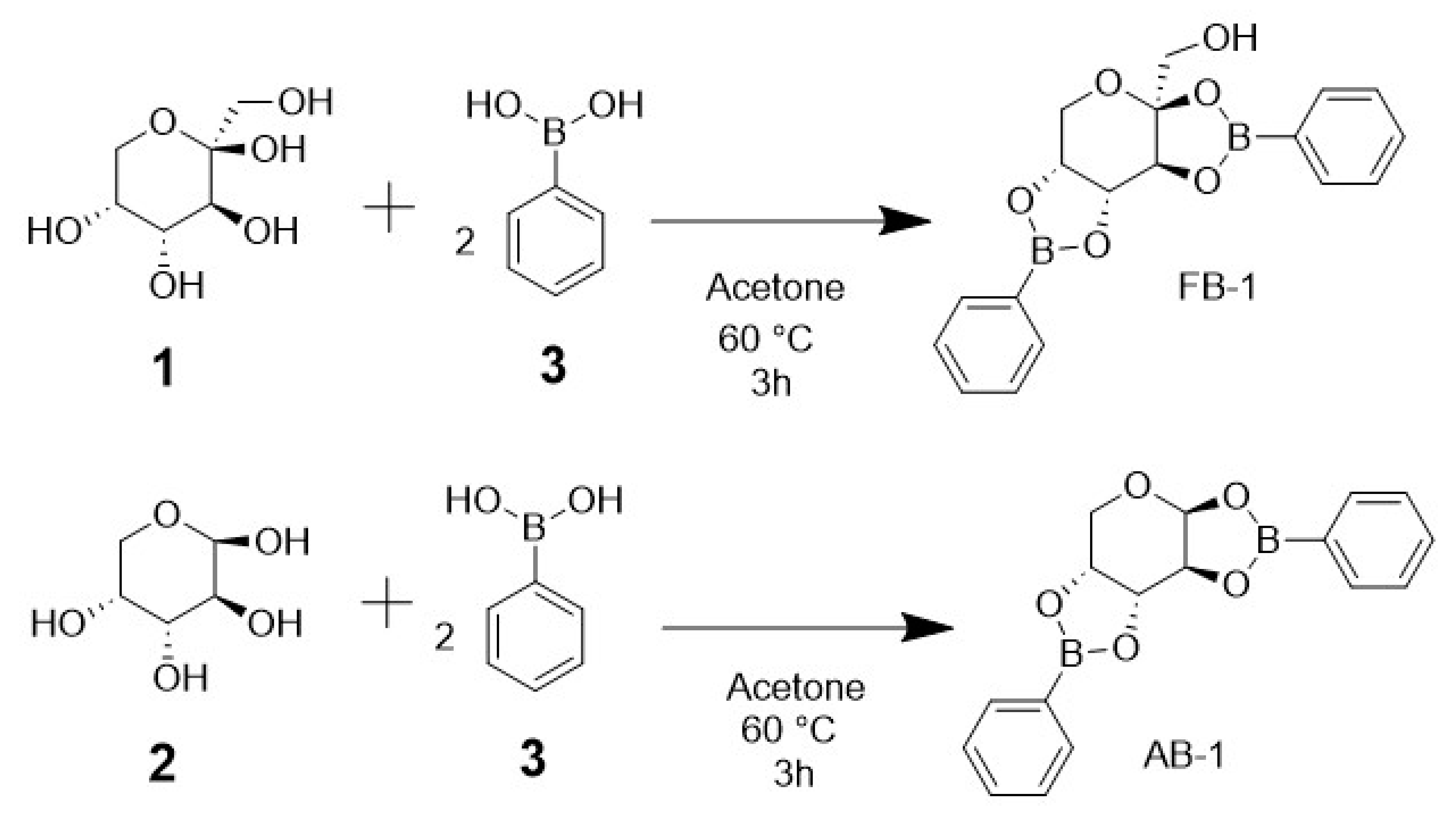
2.1. Chemistry
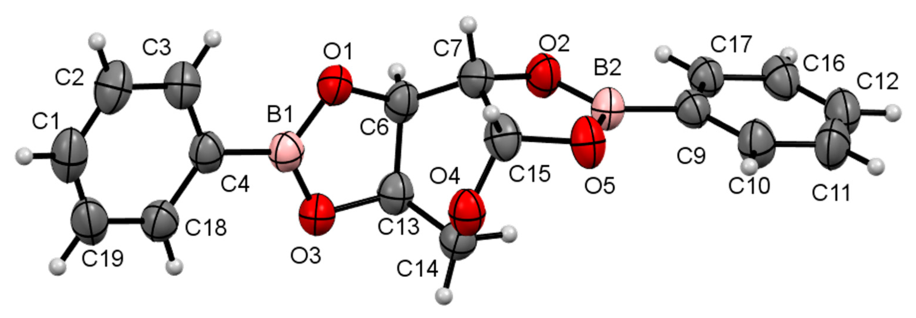
2.2. X-ray Diffraction Crystallography
2.3. In Silico Predictions
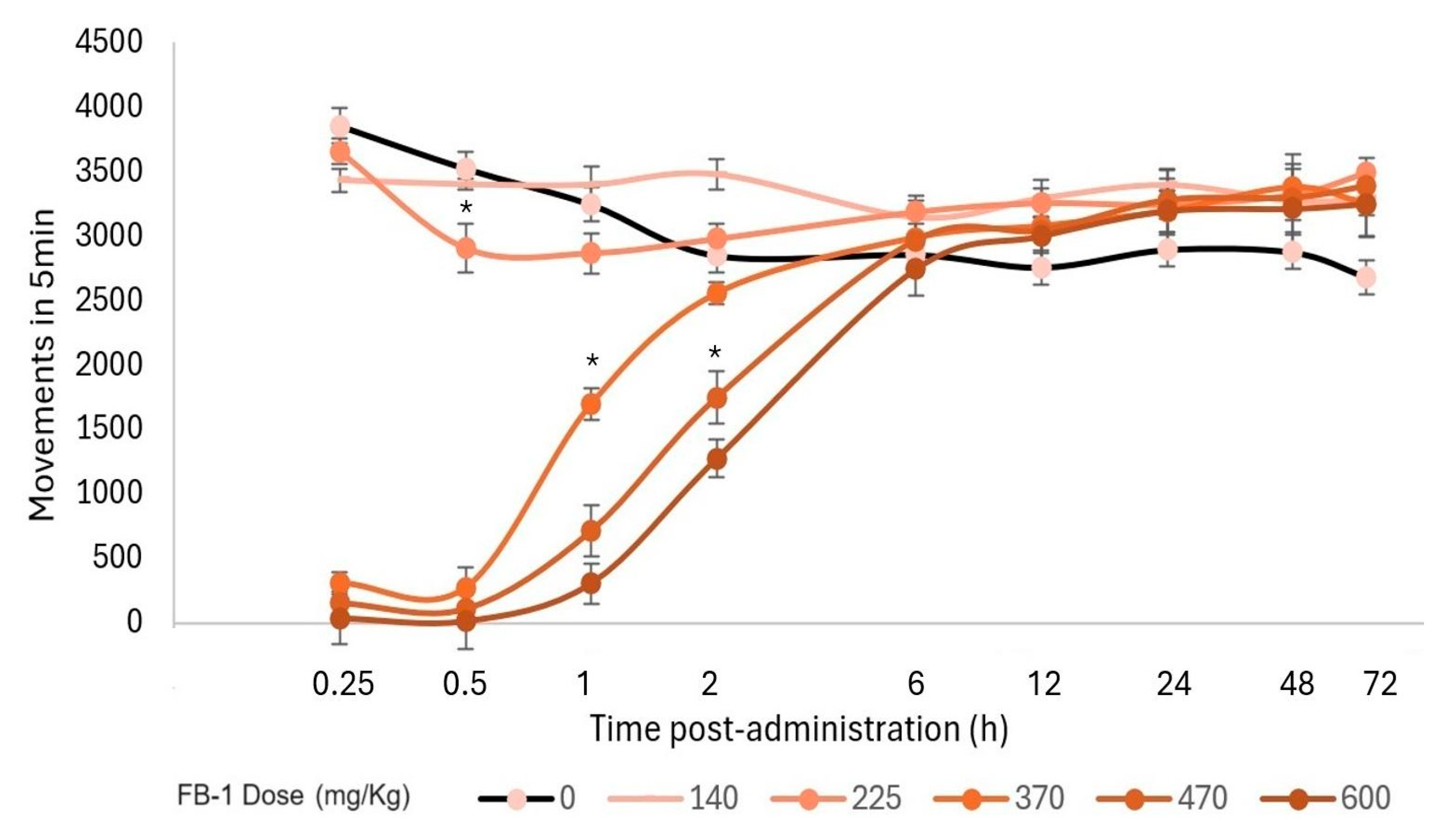
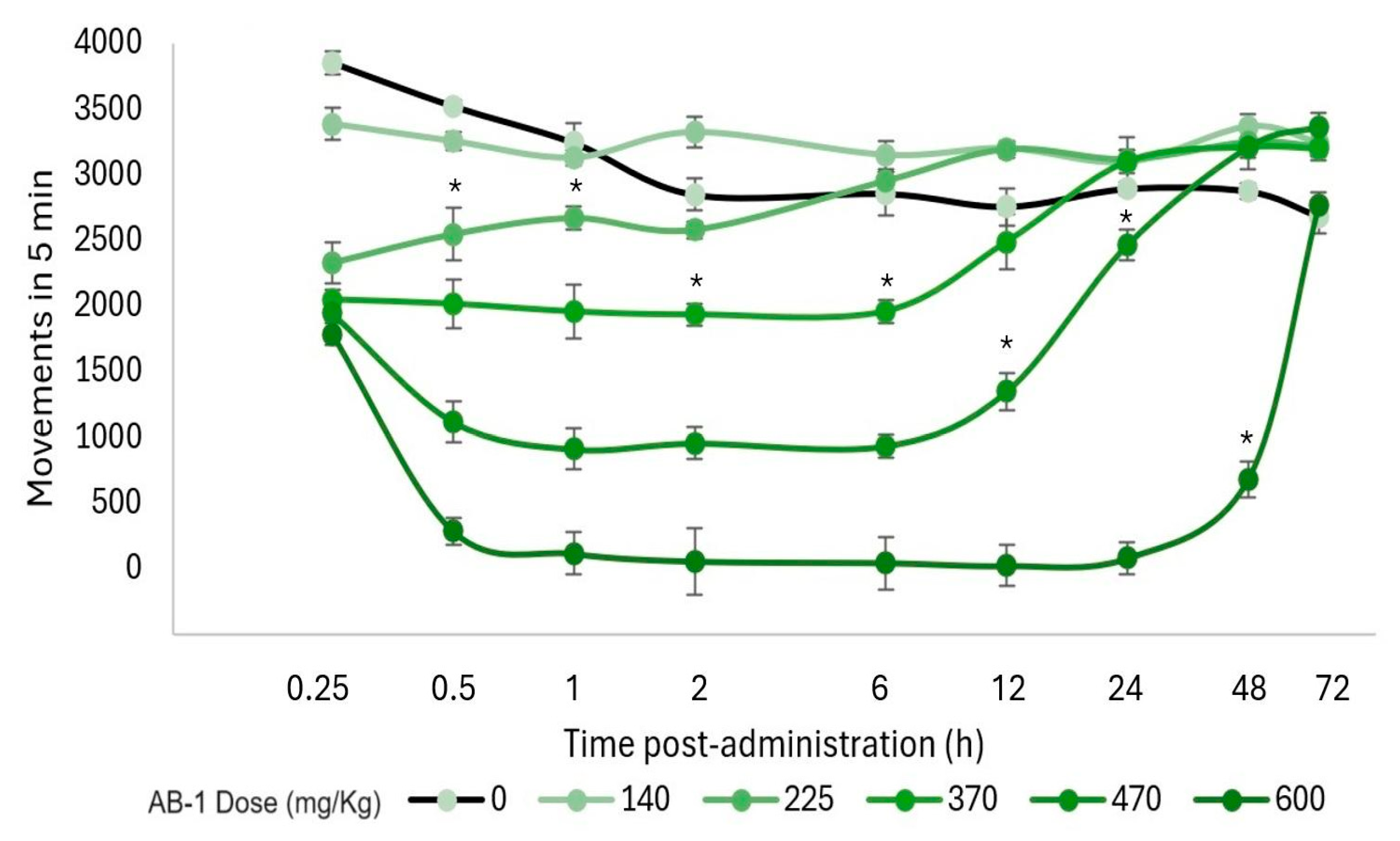
2.4. Acute Toxicity Test
2.5. Determination of the Median Effective Dose (ED50) for Hypnosis and Sedation
3. Discussion
4. Materials and Methods
4.1. Materials
4.1.1. Chemicals
4.1.2. Animals
4.2. Synthesis and Chemical Characterization
4.3. Single-Crystal X-ray Molecular Structure
4.4. In Silico Prediction of Physicochemical Properties
4.5. Biological Evaluation
5. Conclusions
Supplementary Materials
Author Contributions
Funding
Institutional Review Board Statement
Informed Consent Statement
Data Availability Statement
Acknowledgments
Conflicts of Interest
References
- Grams, R.J.; Santos, W.L.; Scorei, I.R.; Abad-García, A.; Rosenblum, C.A.; Bita, A.; Cerecetto, H.; Viñas, C.; Soriano-Ursúa, M.A. The Rise of Boron-Containing Compounds: Advancements in Synthesis, Medicinal Chemistry, and Emerging Pharmacology. Chem. Rev. 2024, 124, 2441–2511. [Google Scholar] [CrossRef] [PubMed]
- Baker, S.J.; Ding, C.Z.; Akama, T.; Zhang, Y.K.; Hernandez, V.; Xia, Y. Therapeutic potential of boron-containing compounds. Future Med. Chem. 2009, 1, 1275–1288. [Google Scholar] [CrossRef] [PubMed]
- Hunter, J.M.; Nemzer, B.V.; Rangavajla, N.; Biţă, A.; Rogoveanu, O.C.; Neamţu, J.; Scorei, I.R.; Bejenaru, L.E.; Rău, G.; Bejenaru, C.; et al. The Fructoborates: Part of a Family of Naturally Occurring Sugar-Borate Complexes-Biochemistry, Physiology, and Impact on Human Health: A Review. Biol. Trace Elem. Res. 2019, 188, 11–25. [Google Scholar] [CrossRef] [PubMed]
- Ban, H.S.; Nakamura, H. Boron-Based Drug Design. Chem. Rec. 2015, 15, 616–635. [Google Scholar] [CrossRef] [PubMed]
- Hunt, C.D. Dietary boron: Progress in establishing essential roles in human physiology. J. Trace Elem. Med. Biol. Organ Soc. Miner. Trace Elem. (GMS) 2012, 26, 157–160. [Google Scholar] [CrossRef]
- Draffin, S.P.; Duggan, P.J.; Fallon, G.D. O2,O3:O4,O5-Bis(phenylboranediyl)-β-d-fructo-pyranose acetone solvate. Acta Crystallogr. 2004, 60, 1520–1522. [Google Scholar] [CrossRef]
- Draffin, S.P.; Duggan, P.J.; Fallon, G.D.; Tyndall, E.M. O1,O2:O3,O5—Bis(phenylboranediyl)-α-d-glucofuranose. Acta Crystallogr. 2005, 61, 1733–1735. [Google Scholar] [CrossRef]
- Reichvilser, M.M.; Heinzl, C.; Klüfers, P. Boronic acid mono- and diesters of the aldopentoses. Carbohydr. Res. 2010, 345, 498–502. [Google Scholar] [CrossRef]
- Wu, X.; Li, Z.; Chen, X.X.; Fossey, J.S.; James, T.D.; Jiang, Y.B. Selective sensing of saccharides using simple boronic acids and their aggregates. Chem. Soc. Rev. 2013, 42, 8032–8048. [Google Scholar] [CrossRef]
- Brooks, W.L.A.; Deng, C.C.; Sumerlin, B.S. Structure-Reactivity Relationships in Boronic Acid-Diol Complexation. ACS Omega 2018, 3, 17863–17870. [Google Scholar] [CrossRef]
- Banach, Ł.; Williams, G.T.; Fossey, J.S. Insulin Delivery Using Dynamic Covalent Boronic Acid/Ester-Controlled Release. Adv. Therap. 2021, 2100118. [Google Scholar] [CrossRef]
- Mohanty, A.R.; Ravikumar, A.; Peppas, N.A. Recent advances in glucose-responsive insulin delivery systems: Novel hydrogels and future applications. Regen. Biomater. 2022, 9, rbac056. [Google Scholar] [CrossRef] [PubMed]
- Itoh, T.; Tamura, K.; Ueda, H.; Tanaka, T.; Sato, K.; Kuroda, R.; Aoki, S. Design and synthesis of boron containing monosaccharides by the hydroboration of d-glucal for use in boron neutron capture therapy (BNCT). Bioorg. Med. Chem. 2018, 26, 5922–5933. [Google Scholar] [CrossRef] [PubMed]
- Soriano-Ursúa, M.A.; Farfán-García, E.D.; Geninatti-Crich, S. Turning Fear of Boron Toxicity into Boron-containing Drug Design. Curr. Med. Chem. 2019, 26, 5005–5018. [Google Scholar] [CrossRef] [PubMed]
- Farfán-García, E.D.; Castillo-Mendieta, N.T.; Ciprés-Flores, F.J.; Padilla-Martínez, I.I.; Trujillo-Ferrara, J.G.; Soriano-Ursúa, M.A. Current data regarding the structure-toxicity relationship of boron-containing compounds. Toxicol. Lett. 2016, 258, 115–125. [Google Scholar] [CrossRef] [PubMed]
- Soriano-Ursúa, M.A.; Farfán-García, E.D.; López-Cabrera, Y.; Querejeta, E.; Trujillo-Ferrara, J.G. Boron-containing acids: Preliminary evaluation of acute toxicity and access to the brain determined by Raman scattering spectroscopy. Neurotoxicology 2014, 40, 8–15. [Google Scholar] [CrossRef] [PubMed]
- Jensen, J.P. The rise and fall of borax as an antiepileptic drug. Arch. Neurol. 2006, 63, 621–622. [Google Scholar] [CrossRef]
- Barrón-González, M.; Montes-Aparicio, A.V.; Cuevas-Galindo, M.E.; Orozco-Suárez, S.; Barrientos, R.; Alatorre, A.; Querejeta, E.; Trujillo-Ferrara, J.G.; Farfán-García, E.D.; Soriano-Ursúa, M.A. Boron-containing compounds on neurons: Actions and potential applications for treating neurodegenerative diseases. J. Inorg. Biochem. 2023, 238, 112027. [Google Scholar] [CrossRef] [PubMed]
- Cordova-Chávez, R.I.; Carrasco-Ruiz, M.F.; Rodríguez-Vera, D.; Pérez-Capistrán, T.; Tamay-Cach, F.; Score, I.R.; Abad-García, A.; Soriano-Ursúa, M.A. Boron-Containing Compounds for Prevention, Diagnosis, and Treatment of Human Metabolic Disorders. Biol. Trace Elem. Res. 2023, 201, 2222–2239. [Google Scholar] [CrossRef]
- Gallop, P.M.; Paz, M.A.; Henson, E. Boradeption: A new procedure for transferring water-insoluble agents across cell membranes. Science 1982, 217, 166–169. [Google Scholar] [CrossRef]
- Altamore, T.M.; Duggan, P.J.; Krippner, G.Y. Improving the membrane permeability of sialic acid derivatives. Bioorganic Med. Chem. 2006, 14, 1126–1133. [Google Scholar] [CrossRef]
- Duggan, P.; Houston, T.; Kiefel-Levonis, S.; Smith-Szydzik, M.L. Enhanced Fructose, Glucose and Lactose Transport Promoted by a 2-(Aminomethyl) phenylboronic Acid. Tetrahedron 2008, 64, 7122–7126. [Google Scholar] [CrossRef]
- Coxon, B. Developments in the Karplus equation as they relate to the NMR coupling constants of carbohydrates. Adv. Carbohydr. Chem. Biochem. 2009, 62, 17–82. [Google Scholar] [CrossRef]
- Williams, D.H.; Bhacca, N.S. Dependency of Vicinal Coupling Constants on the Configuration of Electronegative Substituents. J. Am. Chem. Soc. 1964, 86, 2742–2743. [Google Scholar] [CrossRef]
- Ramsay, W.J.; Bayley, H. Single-Molecule Determination of the Isomers of d-Glucose and d-Fructose that Bind to Boronic Acids. Angew. Chem. 2018, 57, 2841–2845. [Google Scholar] [CrossRef]
- Lorand, J.P.; Edwards, J.O. Polyol Complexes and Structure of the Benzeneboronate Ion. J. Org. Chem. 1959, 24, 769–774. [Google Scholar] [CrossRef]
- Nishiyabu, R.; Kubo, Y.; James, T.D.; Fossey, J.S. Boronic acid building blocks: Tools for sensing and separation. Chem. Commun. 2011, 47, 1106–1123. [Google Scholar] [CrossRef] [PubMed]
- Larkin, J.D.; Fossey, J.S.; James, T.D.; Brooks, B.R.; Bock, C.W. A computational investigation of the nitrogen-boron interaction in o-(N,N-dialkylaminomethyl)arylboronate systems. J. Phys. Chem. A 2010, 114, 12531–12539. [Google Scholar] [CrossRef]
- Bull, S.D.; Davidson, M.G.; van den Elsen, J.M.; Fossey, J.S.; Jenkins, A.T.; Jiang, Y.B.; Kubo, Y.; Marken, F.; Sakurai, K.; Zhao, J.; et al. Exploiting the reversible covalent bonding of boronic acids: Recognition, sensing, and assembly. Acc. Chem. Res. 2013, 46, 312–326. [Google Scholar] [CrossRef]
- Suzuki, Y.; Shimizu, M.; Okamoto, T.; Sugaya, T.; Iwatsuki, S.; Inamo, M.; Takagi, H.D.; Odani, A.; Ishihara, K. Detailed Mechanism of the Reaction of Phenylboronic Acid Derivatives with d-Fructose in Aqueous Solution: A Comprehensive Kinetic Study. ChemistrySelect 2016, 1, 5141–5151. [Google Scholar] [CrossRef]
- Nunez, H.A.; Walker, T.E.; Fuentes, R.; O’Connor, J.; Serianni, A.; Barker, R. Carbon-13 as a tool for the study of carbohydrate structures, conformations and interactions. J. Supramol. Struct. 1977, 6, 535–550. [Google Scholar] [CrossRef] [PubMed]
- Nobre, C.; Teixeira, J.A.; Rodrigues, L.R. New Trends and Technological Challenges in the Industrial Production and Purification of Fructo-oligosaccharides. Crit. Rev. Food Sci. Nutr. 2015, 55, 1444–1455. [Google Scholar] [CrossRef] [PubMed]
- Rodrigues-Borges, M.; de Carvalho-Balaban, R. L-Arabinose (pyranose and furanose rings)-branched poly(vinyl alcohol): Enzymatic synthesis of the sugar esters followed by free radical polymerization. J. Biotechnol. 2014, 192, 42–49. [Google Scholar] [CrossRef] [PubMed]
- Dimmitt, S.; Stampfer, H.; Martin, J.H. When less is more—Efficacy with less toxicity at the ED50. Br. J. Clin. Pharmacol. 2017, 83, 1365–1368. [Google Scholar] [CrossRef] [PubMed]
- Varughese, S.; Azim, Y.; Desiraju, G.R. Molecular complexes of alprazolam with carboxylic acids, boric acid, boronic acids, and phenols. Evaluation of supramolecular heterosynthons mediated by a triazole ring. J. Pharm. Sci. 2010, 99, 3743–3753. [Google Scholar] [CrossRef] [PubMed]
- Goldschen-Ohm, M.P. Benzodiazepine Modulation of GABAA Receptors: A Mechanistic Perspective. Biomolecules 2022, 12, 1784. [Google Scholar] [CrossRef] [PubMed]
- Wisniak, J. Borax, Boric Acid and Boron-From Exotic to Commodity. Indian J. Chem. Technol. 2005, 12, 488–500. [Google Scholar]
- Grand, R.S.; Wegner, E.S. Fatal case of boric acid poisoning. Am. J. Dis. Child. 1948, 6, 910–912. [Google Scholar]
- Bean, E.O. Accidental ingestion of boric acid. Clin. Proc. Child. Hosp. Dist. Columbia 1949, 4, 101–103. [Google Scholar]
- Restuccio, A.; Mortensen, M.E.; Kelley, M.T. Fatal ingestion of boric acid in an adult. Am. J. Emerg. Med. 1992, 10, 545–547. [Google Scholar] [CrossRef]
- Hamilton, R.A.; Wolf, B.C. Accidental boric acid poisoning following the ingestion of household pesticide. J. Forensic Sci. 2007, 52, 706–708. [Google Scholar] [CrossRef] [PubMed]
- Duydu, Y.; Basaran, N.; Hermann, B.M. Human health risk assessment of boric acid and sodium borates. Toxicol. Lett. 2015, 238, S102–S103. [Google Scholar] [CrossRef]
- White, H.S.; Brown, S.D.; Woodhead, J.H.; Skeen, G.A.; Wolf, H.H. Topiramate modulates GABA-evoked currents in murine cortical neurons by a nonbenzodiazepine mechanism. Epilepsia 2000, 41, 17–20. [Google Scholar] [CrossRef] [PubMed]
- Shank, R.P.; Gardocki, J.F.; Streeter, A.J.; Maryanoff, B.E. An overview of the preclinical aspects of topiramate: Pharmacology, pharmacokinetics, and mechanism of action. Epilepsia 2000, 41, 3–9. [Google Scholar] [CrossRef] [PubMed]
- Cavanna, A.E.; Monaco, F.; Mula, M.; Robertson, M.M.; Critchley, H.D. Psychopharmacology of topiramate: From epilepsy to bipolar disorder. Neuropsychiatr. Dis. Treat. 2006, 2, 475–488. [Google Scholar] [CrossRef] [PubMed]
- Zhang, X.; Velumian, A.A.; Jones, O.T.; Carlen, P.L. Modulation of high-voltage-activated calcium channels in dentate granule cells by topiramate. Epilepsia 2000, 41, 52–60. [Google Scholar] [CrossRef] [PubMed]
- Dodgson, S.J.; Shank, R.P.; Maryanoff, B.E. Topiramate as an inhibitor of carbonic anhydrase isoenzymes. Epilepsia 2000, 41, 35–39. [Google Scholar] [CrossRef] [PubMed]
- Gibbs, J.W., 3rd; Sombati, S.; DeLorenzo, R.J.; Coulter, D.A. Cellular actions of topiramate: Blockade of kainate-evoked inward currents in cultured hippocampal neurons. Epilepsia 2000, 41, 10–16. [Google Scholar] [CrossRef] [PubMed]
- Fulmer, G.R.; Miller, A.J.; Sherden, N.H.; Gottlieb, H.E.; Nudelman, A.; Stoltz, B.M.; Goldberg, K.I. NMR chemical shifts of trace impurities: Common laboratory solvents, organics, and gases in deuterated solvents relevant to the organometallic chemist. Organometallics 2010, 29, 2176–2179. [Google Scholar] [CrossRef]
- Bruker. SAINT v8.37A; Bruker AXS Inc.: Madison, WI, USA, 2015. [Google Scholar]
- Blessing, R.H. An empirical correction for absorption anisotropy. Acta Crystallogr. 1995, A51, 33–38. [Google Scholar] [CrossRef]
- Sheldrick, G.M. A short history of SHELX. Acta Crystallogr. 2008, A64, 112–122. [Google Scholar] [CrossRef] [PubMed]
- Farrugia, L.J. WinGX and ORTEP for windows: An update. J. Appl. Crystallogr. 2012, 45, 849–854. [Google Scholar] [CrossRef]
- Spek, A.L. Structure validation in chemical crystallography. Acta Crystallogr. 2009, D65, 148–155. [Google Scholar] [CrossRef]
- Macrae, C.F.; Bruno, I.J.; Chisholm, J.A.; Edgington, P.R.; McCabe, P.; Pidcock, E.; Rodriguez-Monge, L.; Taylor, R.; van de Streek, J.; Wood, P.A. Mercury–CSD, New features for the visualization and investigation of crystal structures. J. Appl. Crystallogr. 2008, 41, 466–470. [Google Scholar] [CrossRef]
- Arulanandam, C.D.; Hwang, J.S.; Rathinam, A.J.; Dahms, H.U. Evaluating different web applications to assess the toxicity of plasticizers. Sci. Rep. 2022, 12, 19684. [Google Scholar] [CrossRef] [PubMed]
- Daina, A.; Michielin, O.; Zoete, V. SwissADME: A free web tool to evaluate pharmacokinetics, drug-likeness and medicinal chemistry friendliness of small molecules. Sci. Rep. 2017, 7, 42717. [Google Scholar] [CrossRef] [PubMed]
- Gfeller, D.; Grosdidier, A.; Wirth, M.; Daina, A.; Michielin, O.; Zoete, V. SwissTargetPrediction: A web server for target prediction of bioactive small molecules. Nucleic Acids Res. 2014, 42, W32–W38. [Google Scholar] [CrossRef] [PubMed]
- Jarrahpour, A.; Motamedifar, M.; Zarei, M.; Youssoufi, M.H.; Mimouni, M.; Chohan, Z.H.; Ben Hadda, T. Petra, Osiris, and molinspiration together as a guide in drug design: Predictions and correlation structure/antibacterial activity relationships of new N-sulfonyl monocyclic β-lactams. Phosphorus Sulfur Silicon Relat. Elem. 2010, 185, 491–497. [Google Scholar] [CrossRef]
- Lin, X.; Li, X.; Lin, X. A Review on Applications of Computational Methods in Drug Screening and Design. Molecules 2020, 25, 1375. [Google Scholar] [CrossRef]
- Sliwoski, G.; Kothiwale, S.; Meiler, J.; Lowe, E.W., Jr. Computational methods in drug discovery. Pharmacol. Rev. 2013, 66, 334–395. [Google Scholar] [CrossRef]
- Lorke, D. A new approach to practical acute toxicity testing. Arch. Toxicol. 1983, 54, 275–287. [Google Scholar] [CrossRef] [PubMed]
- Bin, C.; Xiaohui, W.; Mengrou, S.; Xin, L.; Ting, Z.; Ping, G. Preliminary evaluation of the efficacy and safety of brimonidine for general anesthesia. BMC Anesthesiol. 2021, 21, 305. [Google Scholar] [CrossRef] [PubMed]
- Prut, L.; Belzung, C. The open field as a paradigm to measure the effects of drugs on anxiety-like behaviors: A review. Eur. J. Pharmacol. 2003, 463, 3–33. [Google Scholar] [CrossRef] [PubMed]


Disclaimer/Publisher’s Note: The statements, opinions and data contained in all publications are solely those of the individual author(s) and contributor(s) and not of MDPI and/or the editor(s). MDPI and/or the editor(s) disclaim responsibility for any injury to people or property resulting from any ideas, methods, instructions or products referred to in the content. |
© 2024 by the authors. Licensee MDPI, Basel, Switzerland. This article is an open access article distributed under the terms and conditions of the Creative Commons Attribution (CC BY) license (https://creativecommons.org/licenses/by/4.0/).
Share and Cite
Cordova-Chávez, R.I.; Trujillo-Ferrara, J.G.; Padilla-Martínez, I.I.; González-Espinosa, H.; Abad-García, A.; Farfán-García, E.D.; Ortega-Camarillo, C.; Contreras-Ramos, A.; Soriano-Ursúa, M.A. One-Step Synthesis, Crystallography, and Acute Toxicity of Two Boron–Carbohydrate Adducts That Induce Sedation in Mice. Pharmaceuticals 2024, 17, 781. https://doi.org/10.3390/ph17060781
Cordova-Chávez RI, Trujillo-Ferrara JG, Padilla-Martínez II, González-Espinosa H, Abad-García A, Farfán-García ED, Ortega-Camarillo C, Contreras-Ramos A, Soriano-Ursúa MA. One-Step Synthesis, Crystallography, and Acute Toxicity of Two Boron–Carbohydrate Adducts That Induce Sedation in Mice. Pharmaceuticals. 2024; 17(6):781. https://doi.org/10.3390/ph17060781
Chicago/Turabian StyleCordova-Chávez, Ricardo Ivan, José G. Trujillo-Ferrara, Itzia I. Padilla-Martínez, Héctor González-Espinosa, Antonio Abad-García, Eunice D. Farfán-García, Clara Ortega-Camarillo, Alejandra Contreras-Ramos, and Marvin A. Soriano-Ursúa. 2024. "One-Step Synthesis, Crystallography, and Acute Toxicity of Two Boron–Carbohydrate Adducts That Induce Sedation in Mice" Pharmaceuticals 17, no. 6: 781. https://doi.org/10.3390/ph17060781
APA StyleCordova-Chávez, R. I., Trujillo-Ferrara, J. G., Padilla-Martínez, I. I., González-Espinosa, H., Abad-García, A., Farfán-García, E. D., Ortega-Camarillo, C., Contreras-Ramos, A., & Soriano-Ursúa, M. A. (2024). One-Step Synthesis, Crystallography, and Acute Toxicity of Two Boron–Carbohydrate Adducts That Induce Sedation in Mice. Pharmaceuticals, 17(6), 781. https://doi.org/10.3390/ph17060781