Review

Photoantimicrobial and Photoantiviral Textiles: Underestimated Potential

by
Alexander Efimov
1,* and
Serge Mordon
2
1
Faculty of Engineering and Natural Sciences, Tampere University, 33720 Tampere, Finland
2
Hemerion Therapeutics, 59650 Villeneuve d’Ascq, France
*
Author to whom correspondence should be addressed.
Pharmaceuticals 2024, 17(9), 1164; https://doi.org/10.3390/ph17091164
Submission received: 12 July 2024 / Revised: 23 August 2024 / Accepted: 29 August 2024 / Published: 3 September 2024

Abstract

:
In this review, we summarize the present state of a rapidly developing field of light-activated antimicrobial textiles and their underestimated potential and opportunities.

1. Introduction

Photoantimicrobial textiles (PATXs) are closely related to the fields of photodynamic antimicrobial chemotherapy (PACT) and antimicrobial textiles in general [1]. While the latter two are established fields with over a century’s worth of history, numerous patents, and well-defined ISO standards, PATXs somehow remained in the shadow until very recently, so reviews on the topic are rare [2]. Nevertheless, we see the great potential of PATX to combat the three critical problems of modern protection against pathogens, namely (a) growing antibiotic resistance, (b) reuse of protection barriers, and (c) protection against unknown/unidentified species. In this review, we summarize the state of the art and propose directions of further development. As a special concern, we aim to raise awareness in the research community and to key industrial players of the advantages and potential benefits of this method.
The two essential components of PATXs are the textile fabric itself and the photosensitizer (PS). The PS molecule absorbs a quantum of light and goes into a state of a higher energy, in which it can react with a molecule of oxygen and relay the excess of energy to it. The reactive oxygen species (ROS) generated can be a singlet oxygen molecule 1O2, a radical species (OH, HO2, O3, etc.), or both [1]. Depending on the chemical nature and the photophysical properties of photosensitizer, either pathway (reaction Type I or Type II) can be preferred. The formed ROS molecules (which live from milliseconds to seconds, e.g., 1O2 in the air has a lifetime of ~2.8 s) can diffuse from the PS at a distance within micro- to millimeters and oxidize objects that they collide on their way. This antimicrobial action is based on so-called oxidative stress, where the microbes’ organelles are oxidized simultaneously in many places and through different reactions, making the action efficient and preventing the elaboration of resistance because of the diversity of the reaction pathways. Speaking figuratively, ROS action vs the effect of conventional chemodrugs is like a grenade vs a bullet. Most importantly, the ROS action is a universal process that is generally effective against all kinds of pathogens, including known, unknown, or unidentified bacteria, fungi, or viruses
When combining PACT action with textiles to produce PATXs, a few problems arise. Firstly, the antimicrobial action cannot be accumulated and spared for the future. The ROS are generated directly under light and their lifetime measures from microseconds to seconds, so the antimicrobial action is gone immediately after the light goes off. Secondly, the efficiency of ROS generation is directly related to the light intensity, so thick fabrics can only be efficient at the surface, but not any deeper inside the fiber. Thirdly, the PS molecules and the textile fibers undergo self-oxidation and decomposition under light, thus the photostability of PATXs is an issue. Finally, the bonding of the PS to the fabric might be challenging and requires specific considerations in terms of stability and the ease of the dyeing process.

2. Results

2.1. Textiles

Most of the studies were performed on cotton (cellulose) in woven, non-woven, and yarn/fiber forms. A few studies were made using polypropylene [3,4,5,6], as this is the material from which protection masks are mostly made. In some rare cases, polyamide [7,8,9], silk [10], or wool [11,12,13] have been employed.

2.2. Light Sources

The evaluation of light-driven antibacterial or antiviral textiles requires a well-designed illumination procedure in order to perform reproducible tests and validate the efficiency of the light-driven textile. The analysis of the literature clearly demonstrated that the parameters used for illumination were often missing. Moreover, since there is no standardized procedure, there is a huge diversity of light sources and illumination parameters, such as wavelength, irradiance, fluence, luminance, etc.
This paucity of information and the absence of a standardized illumination procedure is surprising as many organizations have established standards that address lighting design, safety, performance, mounting, and testing, as well as illumination levels. These standards are norms or requirements that establish a basis for common understanding and judgment of materials, products, or processes. Standards are an invaluable tool in industry and business because they streamline business practices and provide a level playing field for businesses to develop products and services. They are also critical to ensuring that products and services are safe for consumers and the environment.
On an international level, a standard has been fixed: EN62471 [14]. In a transversal way, this standard deals with the photobiological risks of lighting sources and especially LED devices and products using specific types of discharge lamps and LED light sources. EN 12464-1 [15] specifies the lighting requirements for humans in indoor workplaces, with all usual visual tasks considered.
First, it seems necessary to give some basic definitions. The illumination of a surface is defined as the luminous flux received by the surface per unit of area. It is represented by the symbol lx and measured in lux (or meter candle, or lumen/m2). However, there is another unit used to characterize the energy falling on a surface, which is irradiance in mW/cm2. It is possible to convert the luminous flux into irradiance. In fact, there is no single conversion factor between lx and mW/m2, as there is a different conversion factor for every wavelength. However, the wavelength of 555 nanometers, which is in the middle of the visible light spectrum, is usually used to perform this conversion.
The question now is whether luminous flux or irradiance should be used in order to activate the antibacterial or antiviral effect of a light-driven textile.
The standard IEC62471 [14] requires 500 lx illuminance for a normal office workplace. The conversion at 555 nm leads to an irradiance of 0.73 mW/cm2. Once again, it is surprising that none of the papers analyzed in this review have clearly defined the minimum amount of light required for efficient inactivation of bacteria or viruses in the textile.
Another factor that must be also considered is photobleaching of the dye impregnated in the textile fabric. Photobleaching (sometimes termed fading) is the photochemical alteration of a dye. This is caused by cleaving of covalent bonds or non-specific reactions between the dye and surrounding molecules. These irreversible modifications of covalent bonds are caused by the transition of the dye from a singlet state to the triplet state. The number of excitation cycles needed to achieve full bleaching varies.
Consequently, if the illuminance is too high the dye will be altered, and the inactivation of bacteria or viruses will be compromised. Only one study has evaluated the photobleaching of phthalocyanine dye 45 [16]. In this study, the light source was an LED white Sundesk 3. The illuminance at the surface of the textile was controlled with the lux meter Model LM 120 from Amprobe and adjusted to 36,000 lux, corresponding to a light power density of 1.6 mW/cm2 at 700 nm (1/3 of the intensity of bright sunlight, or ×60 times the intensity of a typical indoor light). The concentration of the remaining dye in the fabric was calculated. From the slope of the curve, the estimated bleaching time in sunlight (108,000 lux) was found to be 36 h, or 260 days under indoor light. This big difference reflected the light flux difference between natural sunlight and typical indoor light conditions.
Another concern is the control of the illuminance level. Except for four articles, this crucial and determinant parameter was not measured. However, illuminance can be easily determined. An illuminance meter (or a lux meter) measures the amount of visible light energy falling on to a defined area, using lux to express the available light as a measure of perceived brightness.
Inexpensive commercial devices are available. For example, Tang et al. measured light intensity using a light meter (EXTECH, Model # LT300) of around 1000 lux for efficient disinfection of their light-driven textile [17]. As a reference, they measured the light intensity outdoors under the sun (on 22 July 2020, in Davis, CA, USA), outdoors in the shade (on 22 July 2020, in Davis, CA, USA), in an office, and in a supermarket, with measurements of 87,000, 3000, 1000, and 600 lux, respectively.
Kim et al. performed measurements with a digital illuminance meter (Model LX1330B, Union City, CA, USA) to obtain a measurement of 16,000 lux [8]. Another solution consisted of measuring the luminous flux/unit area using a mobile phone. Cuthbert et al. used the Lux Light Meter app on an Apple iPhone [3].
Lastly, Chen et al. and Feese et al. used a NOVA II or an Orion power meter (Orphir Optronics Ltd., Jerusalem, Israel) to determine the fluence rate of light intensity (mW/cm2) [12,18].
Below is a synthesis of the different light sources and parameters (where available) used in the different studies published on light-driven antibacterial or antiviral textiles, where the diversity of light sources and the paucity of reported parameters can be clearly observed.
Sometimes, the light source was a commercial device; for example, the PDT light model LC122 (LumaCare, Salt Lake City, UT, USA) equipped with cold white light from a 150 W halogen light bulb with bundle of optical fibers was used by different teams. This light source delivers visible light (400–700nm). With this light source, Feese et al. used a fluence rate of 60 mW/cm2 for a duration of 15 or 30 min (corresponding to fluences of 54 or 108 J/cm2). Stanley et al. used a similar irradiance (65± 5 mW/cm2) and a duration of illumination varying from 5 to 60 min, corresponding to measurements from 19J/cm2 to 234 J/cm2 [19]. Since the spectrum of this light source can be reduced using a dichroic filter, Chen et al. irradiated their fabrics from 660 nm to 740 nm with an irradiance of 150 mW/cm2 and 30 s: 4.5 J/cm2 [20]. Similar to LumaCare, the Kruss fibre optic light source KL 5125 was put into action by Arenbergerova et al. [21].
The UV light UVA (365 nm) was sometimes proposed. The delivery of an irradiance of 3 mW/cm2 for 60 min (equivalent to a fluence dose of 10.8 J/cm2) has been reported by different authors [22,23,24]. For this wavelength (UVA), a dedicated room or chamber is required to activate the dye.
Kovacova [25] used blue light obtained from a blue LED lamp (LEDart s.r.o., Bratislava, Slovakia) with a wavelength of 470 nm and a power of 50 W.
Several authors decided to use green light, since the absorption of the dye was maximal in this spectrum region. This was the case in studies by Zhu et al. with 520 nm [26], Nie et al. with 532 nm, (85 ± 1 mW/cm2), and Wright et al. with 530 nm light (39 and 8 mW/cm2 at 4 cm, respectively) [27]. Lastly, Morsi et al. resorted to a 100 W mercury lamp (C-SHG1, Nikon corp., Tokio, Japan), equipped with a 540/25 nm filter (43 mW/cm2) [28].
Red light is also an option, as a 10 min irradiation with a light-emitting diode (LED) light 692 nm (12.5 mW/cm2) led to the complete eradication of a virus on medical mask fabric [29].
Most papers claimed that light sources were used to mimic the sunlight. However, the illuminance level was never determined nor provided. Consequently, other teams will be unable to reproduce the experiments. A xenon lamp was often used as solar simulator, for example: (i) xenon lamp, 500 W, 45 min, 15 cm [30]; (ii) xenon lamp, 3500 mW/cm2, 420–780 nm, 30 min illumination [31]; (iii) xenon lamp (500 W) equipped with a long-pass filter (λ ≥ 420 nm), 60 min, 65 ± 5 mW/cm2 , and a (iv) xenon arc lamp (500 W) kept at a lamp-to-sample distance of 12 cm from the bacteria-infected samples [32]. A 100 W tungsten lamp (1250 lm) was also proposed as a visible light source, with an average intensity of ~0.36 mW/cm2 at a distance of 20 cm from the sample [33]. Cardoso et al. placed a daylight lamp system at a distance of 15 cm from the sample; the incident light covered the entire visible spectrum (400–800 nm) obtained with an Osram lamp model (1380 lm, 110–130 V, 400 mA) [34].
One team performed an evaluation with three different light sources; a sunny day (Xenon, λ ≥ 420 nm, 60,000 lux), a cloudy day (lamp, 8000 lux), and a room light (800 lux) [35]. Two authors reported on illumination with a D65 (6500 K) light source. This light-bluish colored light source is used in color matching applications for paints, plastics, textiles, inks, automotive, and other manufactured products. It accentuates blue and subdues green and red. D65 is commonly used as a primary light source in color measurement instrumentation. However, no data were provided on light intensity, and only illumination durations of 10 min, 30 min, and 60 min were mentioned [36]. Li et al. also performed an evaluation of a D65 light source, but they only report the duration of illumination, which was 120 min [4].
In some cases, the irradiance was so high that the temperature of the textile reached 80 °C to 90 °C. Consequently, a photothermal effect (or photothermal therapy, PTT) was obtained, but not a photodynamic therapy (PDT) effect. When using a CEL-S3500/350 simulated daylight lamp (Zhongjiao Jinyuan Co., Beijing, China) to irradiate the samples with a vertical height of 15 cm, a temperature increase up to 90 °C was observed at 200 mW/cm2 [10,28]. This temperature increase was also reached when fabrics were irradiated with 808 nm NIR light. Using different irradiances (0.5, 1.0, and 1.5 W/cm2) for 10 min, Wang et al. monitored the temperature with an infrared thermal imager and obtained a temperature of 80 °C during illumination [13]. Similarly, Yu et al. observed bacterial killing efficiency and high temperatures when irradiating at light power densities below 300 mW/cm2 (808 nm) [37].
The duration of the illumination is not always well defined and can vary drastically from one study to another. A high-intensity white LED light capable of producing ~30,000 lx was used for 10, 100, and 1000 min to perform illumination [3]. Under 6 h of sunlight irradiation, Shivalkar observed that >90% of bacterial growth was inhibited in the presence of sulfur quantum dots [38]. Sometimes illumination is very long; for example, LED light (white light, 10 W) has been used at a vertical distance of 18 cm for 5 days without any other light sources [39].
Lastly, several authors provided very limited or no information. The only information was “white light for 30 min” [40], “limited-to-visible light irradiation” [41,42], or “under light” [11,13,42,43].

2.3. Molecules

Chemical substances used for the preparation of photoantimicrobial textiles were initially derived from the chemicals employed for PACT or PDT. Some modification was required to attach them to the fiber, though this was not always necessary.

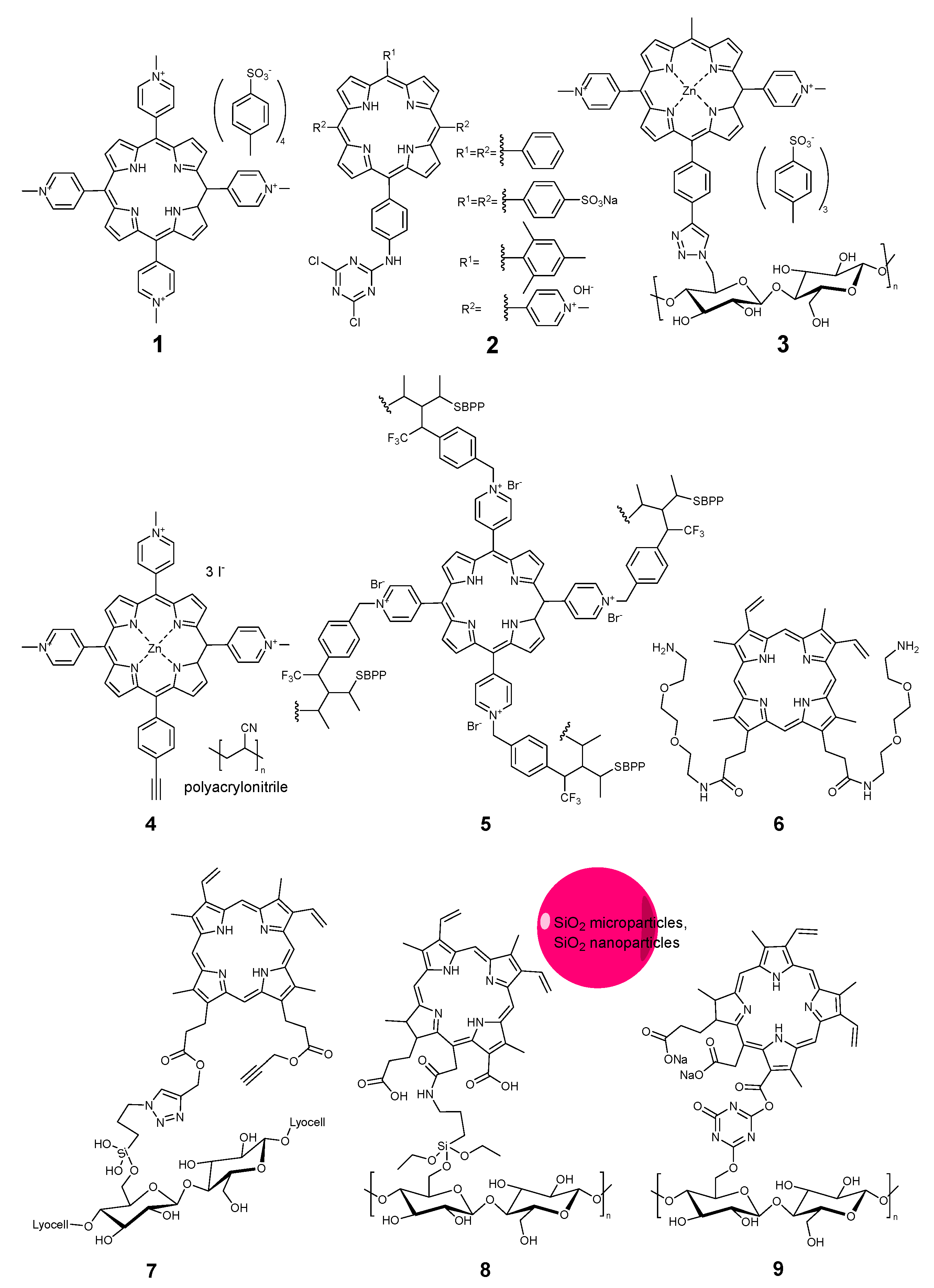
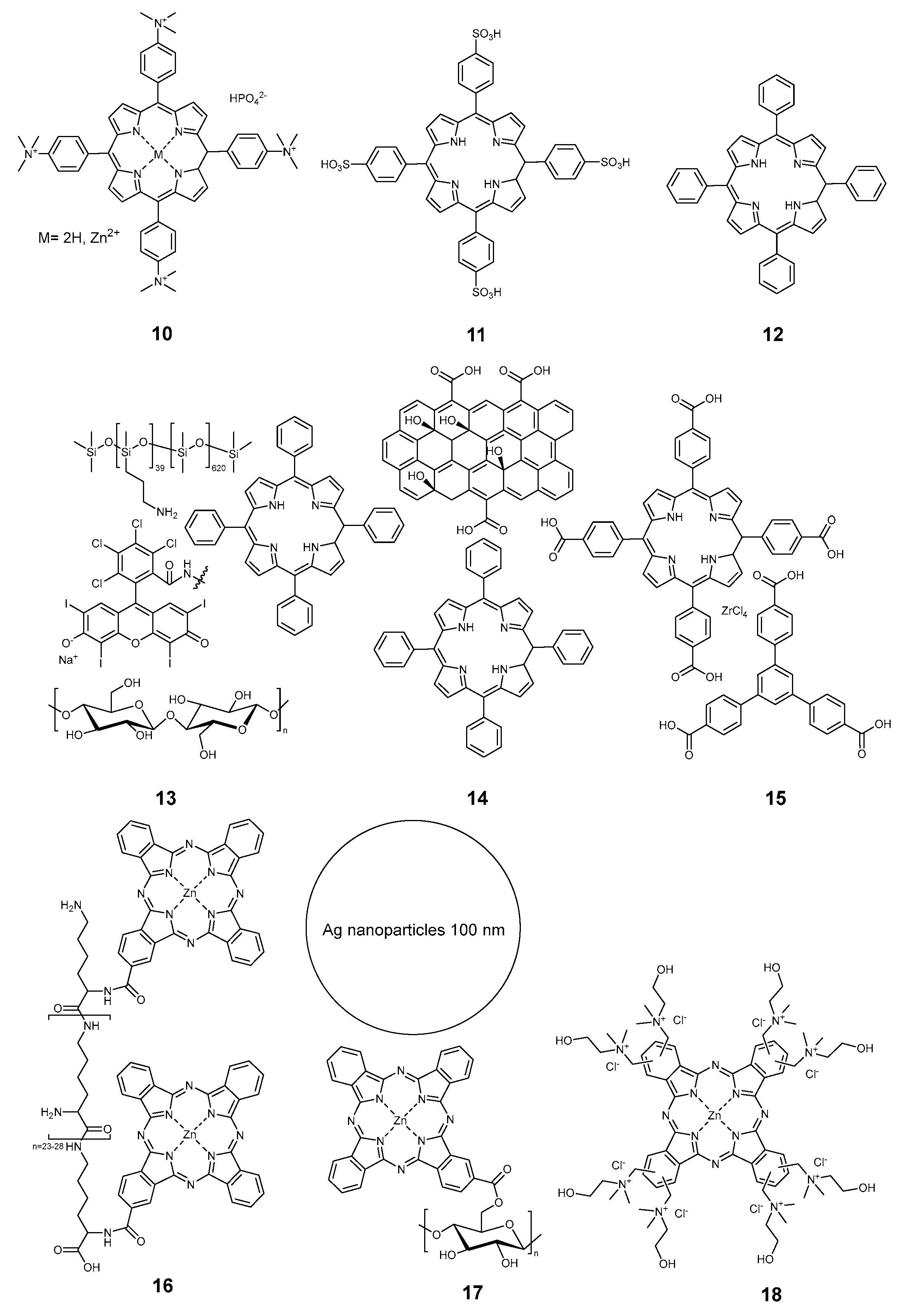
2.3.1. Porphyrinoids

The first mention of a light-activated antimicrobial textile can be found in the conference abstract published by Raymond Bonnett and coworkers in 1997 [44]. They reported on the modification of regenerated cellulose with tetracationic tetrakis(N-methylpyridinium)porphyrin 1 (Figure 1), without specifying which type of textile had been used. This is probably due to the relatively low stability against leaching, as porphyrin 1 was not used alone in other studies. Instead, modified compounds with an anchor group binding to the fiber were prepared and studied. Ringot and coworkers from the group Vincent Sol prepared a set of porphyrins 2, bound to cotton via a 1,3,5-triazine link [45]. Feese and coworkers from the group Resa Ghiladi employed a different click chemistry to bind a tricationic zinc porphyrin 3 to the cellulose surface via a triazole linker [18], although they employed a paper surface, not a textile. The cationic alkylpyridinium motif gave porphyrins higher efficacy against bacteria, so this was also employed by Stanley et al. to bind porphyrin 4 to a polyacrylonitrile non-woven textile [19] and by Cuthbert et al. for the preparation of antiviral non-woven textiles made from polypropylene, to which a porphyrin photosensitizer 5 was covalently bound via diazirine linkers [3].
The development of linkers was proposed by Ringot and coworkers when they introduced protoporphyrin IX with amino-terminated linkers 6 to bind a cellulose fabric modified with polymethacrylic acid [41]. Fadavi [43] and Song [42] proposed their variations of the natural porphyrins 7 and 8 for cotton modification. The first one employed a siloxane linker and a click-triazole lock to immobilize the protoporphyrin IX, and the latter employed chlorin e6 linked via aminopropyl triethoxysilane. In addition, Song et al. embedded silica nanoparticles and coated the cotton fabric with a liquid-repellent siloxane layer. Essentially, the same chlorin structure 9, in the form of trisodium salt (food colorant chlorophyllin), was bound to cotton via a triazine linker by Jiang and coworkers [46].
Unanchored porphyrins were nonetheless used for the modification of textiles too. Compound 10 (Figure 2) had the cationin trialkylammonium groups used by Rahimi et al. [33]. Anionic tetrakis(p-sulfophenyl)porphyrin 11 was proposed by Castriciano and co-workers [47]. Neutral tetraphenyl porphyrin 12 was employed for dyeing textiles [21,40]. The same molecule was also used in more complex systems 13 by Wright [27] and 14 by Morsi [28]. In the work of Wright et al., the porphyrin photosensitizer was used along with polisiloxane-bonded Rose Bengal as a coating for the cotton fabric. Morsi proposed the use of graphene oxide as a co-producer of singlet oxygen over the cellulose acetate membranes. Nie and coworkers employed a rather complex structure, including graphene quantum dots and nanoparticles prepared from a tetracarboxy derivative of TPP and zirconium-based metalorganic frameworks 15 to modify preliminary aminated knitted cotton fabrics [30,48,49,50].
Few phthalocyanines were also used for the preparation of photoantimicrobial textiles. In 2017, Chen et al. covalently linked monosubstituted zinc phthalocyanine to a polylysine chain 16, and a cellulose fabric was impregnated with the obtained polymer [20]. Two years later, same group proposed a combination of covalently-bound phthalocyanine and 100 nm big silver nanoparticles 17 as a combined antibacterial agent on the cotton support [51]. Kurskaya and coworkers proposed polycationic octakis(cholinyl)zinc phthalocyanine 18 for antiviral impregnation of medical masks, and the exact material of the mask was mentioned in the article [29]. A combination of tetrasulfonated copper phthalocyanine and tetracarboxybenzophenone 19 (Figure 3) obtained from commercial sources was employed by Hu et al. to modify cotton fabric [23]. In 2024, Efimov and coworkers proposed an antiviral cotton textile based on tetracationic zinc phthalocyanine 20, which proved to be stable and efficient [16].
All the porphyrinoids mentioned above had antimicrobial functions based on the generation of reactive oxygen species. One work stands out from this trend, as it proposed a red light-induced release of carbon monoxide CO from a sacrificial source of carbonyls sensitized by palladium tetrabenzotetraphenylpoprhyrin 21 [52]. Antimicrobial studies were not performed though, probably due to the low stability of the textile. The authors mentioned that it had to be stored and refrigerated in the dark and used within 10 days to avoid spontaneous decomposition of the dimanganese dodecacarbonyl.

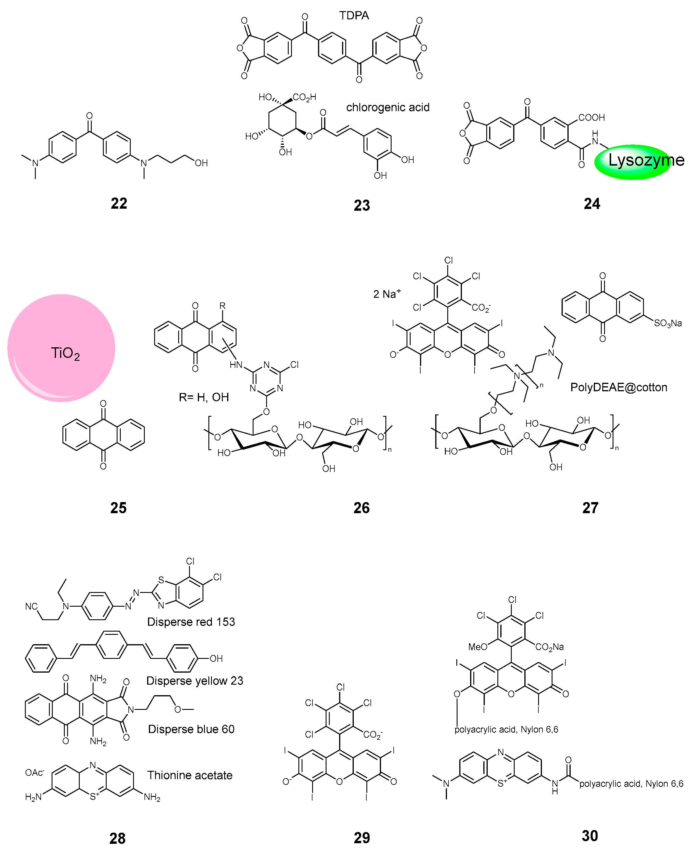
2.3.2. Keto and Quinone Dyes

Benzophenone and anthraquinone dyes were also proposed as photosensitizers for PACT textiles. Oh et al. employed an aminated derivative of benzophenone 22, though the dye had to be activated by UV light [24]. The tetracarboxy benzophenone was also used a co-sensitizer for phthalocyanine, as it was mentioned earlier [23]. Larger benzophenone derivative 23 in combination with chlorogenic acid was used to photosensitize a non-woven polypropylene [4]. The light absorbance occurred chiefly at 400 nm. Very recently, Zhang et al. proposed a phosensitized wool, which was modified by benzophenone tetracarboxylic dianhydride bound to lysozyme 24 [11]. In this case the light absorbance occurred in the 400–500 nm range.
Anthraquinones derivatives might be strongly colored in the visible range, giving red and blue shades. Rahal and coworkers employed unsubstituted anthraquinone in combination with TiO2 25 (Figure 4), which absorbed the violet light at 420 nm [53]. In the work of Cardoso and coworkers, the hydroxy-substituted anthraquinone 26 was covalently bound to an unbleached cotton fabrics via triazines [34]. As a different approach, Tang et al. decorated the cotton cloth with poly(diethylaminoethyl) chains, to which a sulfo-derivative of anthraquinone 27 and Rose Bengal dye was bound electrostatically [17]. A commercial anthraquinone derivative, Disperse blue 60 28 was used by Jiang et al. among other commercial textile dyes [39].

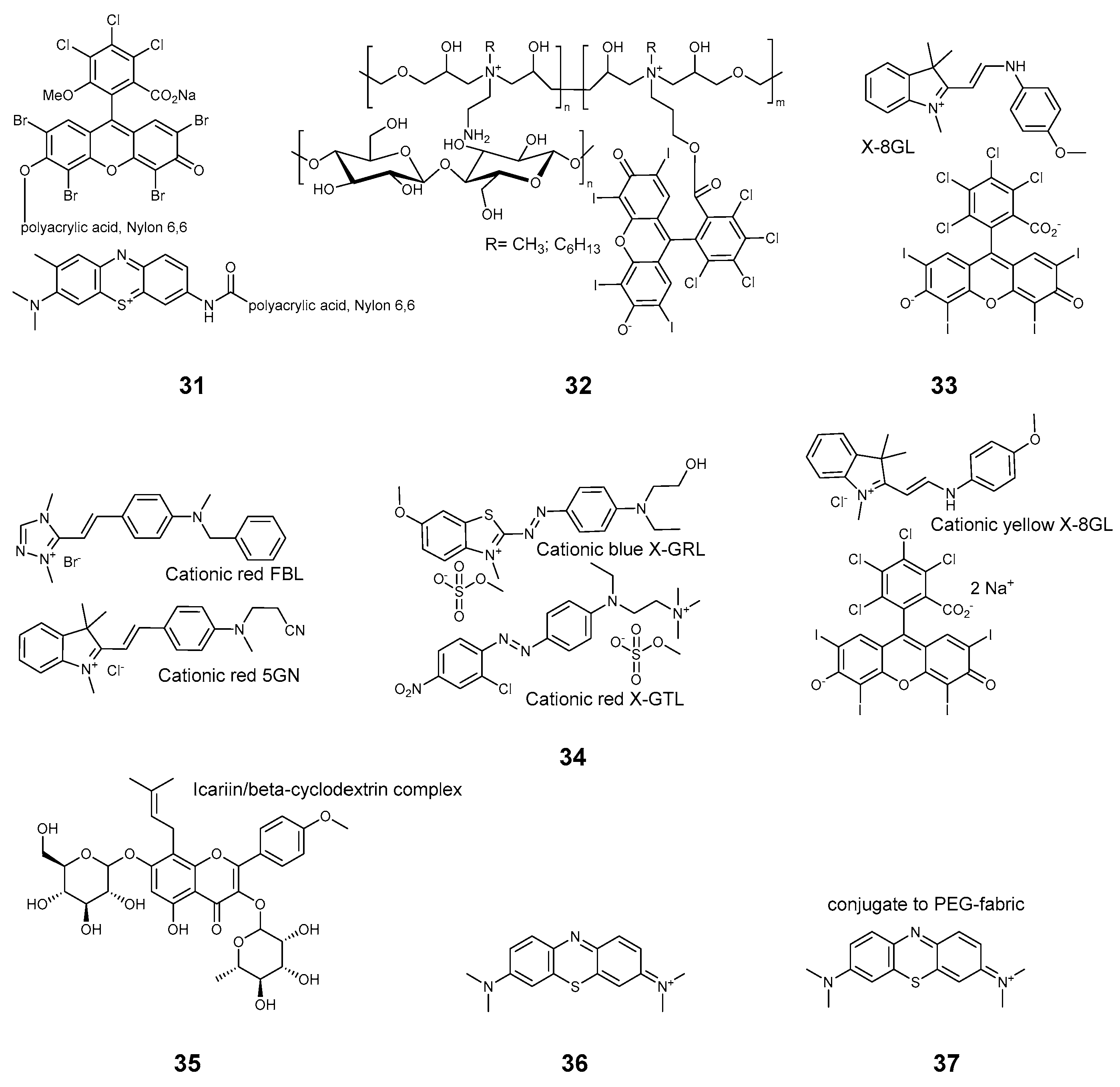
2.3.3. Xanthene Dyes

Rose Bengal (RB) 29 and related compounds are among the most popular dyes used for research on PACT textiles. Available commercially, with a known safety profile, they are beautifully colored and relatively stable. In its unmodified form Rose Bengal was used to color the polyamide fabrics [7]. Jin and coworkers employed it in conjunction with a long phosphorescing SrAl2O4:Eu2+, Dy3+ on cotton fabrics [54]. Lee et al. used Rose Bengal in combination with petal-like nanostructures to prepare self-disinfecting polypropylene cloth [5].
A series of articles was published by Kim and coworkers, in which Rose Bengal and related Phloxine B 30, 31 (Figure 5) were covalently bound to polyacrylic acid [55] and applied to nylon 6,6 [8,9] in combination with a phenothiazine derivative 30. Covalently bonded Rose Bengal was employed by Zhu et al. in a synergistic cotton-based fabric 32, which also included polyammonium motifs [26]. A mixture of RB with commercial dimethylindolinium dye cationic yellow X-8GL 33 was used to prepare antimicrobial wool-acrylic blended fabrics [12]. Later, an expanded set of commercial dyes 34 was tested by this group on a wool-acrylic blend [13]. It should be noted that the set of compounds 34 employed in the study were used as received from commercial sources, and labeled with their commercial names, thus the identification of the substances might be problematic.
The synergistic effects of RB, anthraquinone, and cationic polyamine were studied in the aforementioned works of Tang et al. [17], and the effects of porphyrin were studied by Wright et al. [27]. Not exactly xanthene, but a structurally related flavonoid icariin 35 was used to prepare an antimicrobial cellulose material by Mensah and coworkers [32]. Icariin dye is used in traditional Chinese and Korean medicine as an erectile function enhancer.

2.3.4. Phenothiazines

Methylene blue (MB) and its related compounds are among the oldest and most proven antimicrobial substances used in medicine. Safe for humans, they are usually less light-stable than the other textile dyes. Nonetheless, numerous studies employ various xanthene derivatives. According to Kim et al., MB can be bound covalently to the polyacrilyc acid 8, and this compound can be further employed to modify nylon cloth [8,9]. Unmodified MB 36 can be used to make antimicrobial polyester/cotton blends [31]. The effect of MB was studied on a cotton cloth in the aforementioned test of commercially available dyes [39]. A recent work by Youssef et al. proposed that polyethyleneglycol fabrics be dyed with MB 37 for wound healing [56].

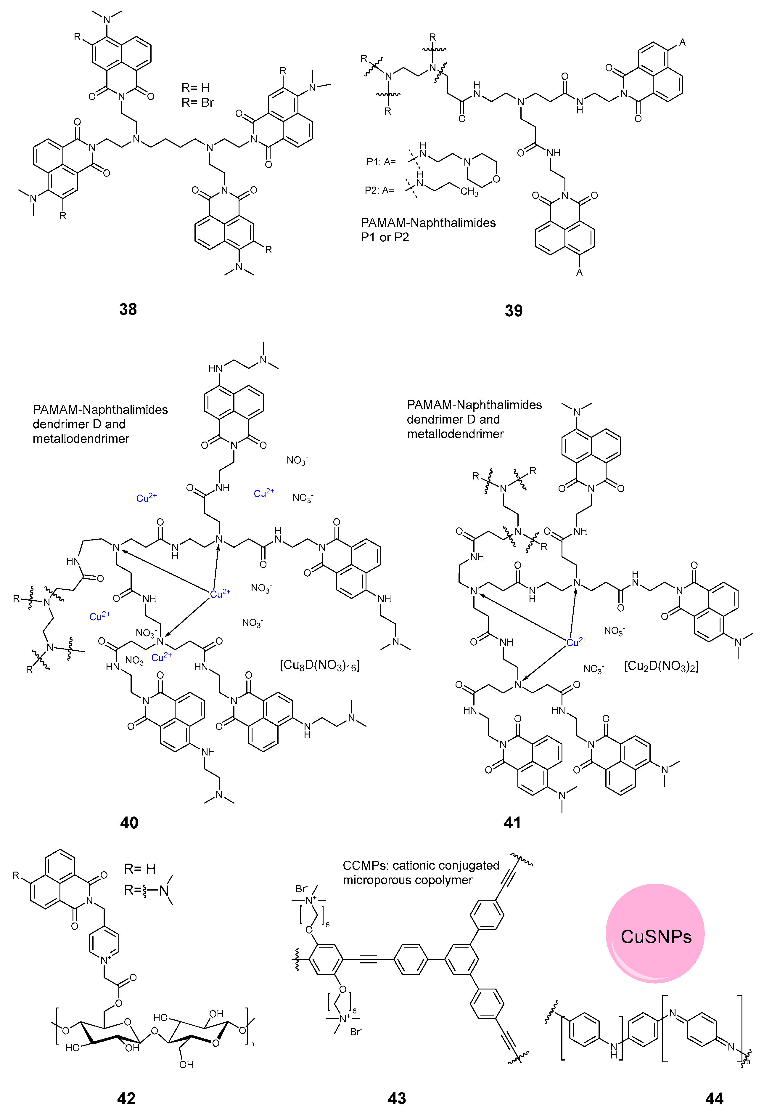
2.3.5. Dendrimers and Polymers

A systematic study of cotton fabrics modified with derivatives of 1,8-naphthalimides was undertaken by the group Grabchev. Starting from a relatively small tetramer 38 (Figure 6) [57], the system was expanded to a PAMAM-napthaalimide dendrimer 39 [58]. The dendrimers were later modified with copper ions 40 [59], and the naphthalimide substitution was varied 41 [60]. Apart from the dendrimers, the naphthalimides were appended directly to cotton cloth 42 using chloroacetyl chloride chemistry [61].
As for the polymeric photoantimicrobial agents, a modification of polypropylene cloth was proposed by Wang et al., employing a cationic conjugated microporous copolymer 43 [6]. The absorbance profile of the fabric was maximal at 400–420 nm. Another amino polymer was proposed by Ren and coworkers [10]. In this work, a conjugated polyaniline 44, in conjunction with copper sulfide nanoparticles and polydimethylsiloxane, was employed to modify silk fabrics and make them highly stable against deterioration.

2.3.6. Nanoparticles and Inorganic Sensitizers

Nanoparticles became a hot topic at the beginning of the century, as their properties significantly vary from the bulk materials. In PATXs, silica nanoparticles 8 were proposed to modify cotton cloth [42] in combination with chlorin e6. Copper sulfide nanoparticles were used in the work of Ren et al. [10] to modify silk. Sulfur quantum dots 45 (Figure 7) were employed to make cotton cloth photoantimicrobial [38], though the difference between light and dark activity was within 1 log. A rather unusual example of nanoparticles was proposed by Liu et al. [62]. In this work, methylene blue was employed to dope melanine nanoparticles on cotton 46 to exert a PACT action. Cotton fabric was used as a textile support, and the bacterial infections were cured on wounds in mice. Novikova et al. employed molybdenum-based nanoclusters 47 for photodynamic inactivation of bacteria and viruses on cotton [63].
Somewhat in relation to the nanoparticles, Li et al. employed the effect of aggregation-induced emission to create a photosensitizer from clusters of a triphenylamine derivative 48, which was then applied to a polypropylene cloth [64].
Inorganic materials have been used for PACT textiles in the form of supramolecular structures. Wang et al. employed rather complex titanium carbide/aluminium/tin sulfide heterojunctions (“MXenes”) 49 with lactate oxidase to render the epsilon-caprolactone fabrics antimicrobial [65]. A combination of similar MXenes with a zinc-methylimidazole metalorganic framework 50 was used to modify cotton [37] and cellulosic non-woven [66] fabrics. Petal nanostructures 51 demonstrated antimicrobial activity on the surface [5]. Hydrophobic quantum dots 52 [25] and silkworm excrements 53 were also proposed to make the antimicrobial coating [35]. In the latter case, however, 70% of the pathogens were eliminated in the dark, with no light irradiation.

2.4. Efficacy against Bacteria, Fungi and Viruses

Antimicrobial efficacy is probably the most complex topic in this review. The results of chemical preparations, photochemical measurements, and stability tests can be obtained via established procedures and reported in a concise manner as a descriptive set of values. Although, the results of the antimicrobial tests are multiparametric by nature and depend on a great number of variables.
When evaluating the antipathogen activity, the following parameters must be controlled.
  • Type and strain of pathogens;
  • Surface-to-volume ratio between the tissue and the pathogen inoculum media;
  • The load of the dye per unit of area of the fabric;
  • The nature and intensity of the activating light;
  • The illumination time;
  • Filtering of heat;
  • Dark activity;
  • Blank/negative control;
  • Numerical assessment of inactivation;
  • Number of parallel experiments.
Ideally, a standardized method should be used in all cases. To date, few standards known for the testing of antibacterial textiles [67]. The quantitative methods are AATCC 100 and JIS L 1902—the absorption method, and the qualitative ones are AATCC 147 and JIS L 1902—the Halo methods. Antiviral textiles can be tested according to ISO 18184:2019 [68,69,70,71]. However, none of these tests were designed for photoactivated textiles, so the protocols should be modified. The modified AATCC 100 method was followed by Cardoso, Castriciano, and Chen et al. [20,34,47,51]. The modified ISO 18184:2019 was used by Efimov et al. [16]. The standard ISO 22196:2011 [72] “Measurement of antibacterial activity on plastics and other non-porous surfaces” was employed in the work of Kovacova et al. [25].
The results of the antimicrobial tests are summarized in Table 1. The numbers are given as rounded up values, taking a logarithm of the eradication of a pathogen; i.e., “6” means 99.9999% inactivation and “0.2” means 63% inactivation. Most of the experiments were performed on bacteria of a so-called ESKAPE set, chiefly E. coli and MRSA. Antiviral experiments were performed on the influenza virus and on variations of SARS-CoV2 (227 and 229E), as well as on non-enveloped types and other types (Table 1). As for fungi, an extensive set was tested by Kim et al. [8,9], including A. fumigates, A. niger, Trichoderma viride, P. funiculosum, Caetonium globosum, P. cinnamoni, M. grisea, and C. albicans. The last one was also employed in the work of Novikova et al. [63].
In most of the tests, the surface activity of textiles was evaluated quantitatively. That is, a small inoculum volume (100 µL) was deposited over a few mm specimen of the textile, and the pathogens were extracted after irradiation, cultivated, counted, and compared to a dark or negative control. In that sense, tests were performed similarly to the AACTT 100 standard, so the results can be directly compared. Also, in most of the works, blank experiments with no photosensitizer were performed.
Few works, however, evaluated the possibility of inactivation in volume, where a centimeter-size textile specimen was placed into a few ml of cultural liquid and the growth reduction measurements or counting of colonies were performed after illumination [7,8,9,31,60]. In one study, the activity was evaluated in a suspension of cellulose nanocrystals with inoculum [18]. Another one employed a fiber placed onto an agar surface, and the inhibition zones were estimated [43]. In these cases, the results are much more difficult to compare and can be used as semi-quantitative or qualitative indications.
For various non-textile antipathogen applications, a borderline 3-log (99.9%) reduction in CFU after treatment is a commonly accepted value to use the word “antimicrobial”. The AATCC 100 test requires a 4-log (99.99%) reduction in CFU over a 24 h contact of the textile and inoculum. In this case the textile can be called “antimicrobial”. On the other hand, the antiviral standard ISO 18184:2019 classifies a 3-log reduction as “excellent” activity.
Since a 24 h irradiation does not seem to be practical, the illumination times in the studies typically vary between 10 min and 4 h, most commonly being 30 and 60 min. Therefore the 3-log reduction in CFU could be taken as a reasonable borderline value. In this case, most of the published works faithfully claimed their textiles to be “antibacterial”. The antiviral studies can be also considered to be mostly successful [3,4,16,39]. Proper antifungal activity is much more difficult to achieve though. Based on the formal criteria, only A. fumigatis was successfully suppressed by photoactive textiles [55].
Photoantimicrobial efficacy depends dramatically on the incident light. The absorption profile of a photosensitizer, the irradiation light power and its density distribution, and the time of irradiation must be counted. These three critical parameters together can be expressed as an absorbed light dose in J/cm2. By neglecting the absorptance profile, the irradiation light can be described by the irradiated light dose in J/cm2.
For the sake of comparing different reports, we propose the use of the irradiated light dose. Indeed, different chromophores absorb at different wavelengths and different light sources have different emission profiles. Antimicrobial textiles, however, are expected to work under available light, be it indoors or outdoors, and the indoor light sources used in living spaces are more or less successfully mimicking sunlight. Also, accurate measurements of light intensity require special equipment, which is not easily available, whereas measurements of illuminance in lux are easy accomplishable with an affordable equipment, and the results can be easily interpreted.
We propose the use of an approximation employed nowadays for estimating the efficiency of photoelectric elements, as presented in Table 2. The power density of direct sunlight can be roughly taken as 100 mW/cm2, which corresponds to 100,000 lux illuminance. A bright daylight outdoor swould produce 10 mW/cm2 and 10,000 lux. In a shadow, the light power would be 1 mW/cm2 and 1000 lux illuminance, and indoors in an office the light would be considered as 0.5 mW/cm2 power and 500 lux illuminance. The data in the Table 1 were recalculated according to this principle. Where possible, the light power density provided in the article was used directly to calculate the light dose as
Light dose (J/cm2) = Light power density (mW/cm2) × Illumination time (s) × 10−3
As in most articles, the light power density was not given, so it was estimated from the illuminance as
Light power density (mW/cm2) = Illuminance (lux) × 10−3
Using these equations one can calculate that 30 min of sunshine (100 Klux) would give a 180 J/cm2 light dose, 30 min on an overcast day (10 Klux) would be 18 J/cm2, 30 min in a shadow (1 Klux) would be 1.8 J/cm2, and 30 min under office lighting (0.5 Klux) would give 0.8 J/cm2. Hence, one could grade all the reported experiments into four categories by the irradiated light dose, so the required illumination conditions for efficient antimicrobial function would be
  • Direct sunlight, 100–1000 J/cm2;
  • Bright day, 10–100 J/cm2;
  • Shadow outdoor, 1–10 J/cm2;
  • Indoor, 0.1–1 J/cm2.
By employing this classification, one can see that only compounds 3 and 20 are suitable for indoor use with acceptable efficiency. The dyes are tetracationic porhyrinoids with alkylpyridinium substituents. On the other hand, a tetracationic porphyrin with alkylammonium substituents 10,showed weaker performance.
Compounds 2, 6, 10, 11, 29, and 48 can be classified as suitable for use in a shadow. These are either porphyrin derivatives or alkylpyridium compounds, which match well with the observations for the most efficient group. Rose Bengal 29 [7] also can be quite an efficient photosensitizer, although other sources reported much higher light doses employed for activation [8,9,12,54,65]. Other compounds, including quantum dots and industrial dyes, require bright or direct sunlight to be sufficiently active.
Photoinactivation of bacteria and viruses can be quite successfully accomplished. Unfortunately, this is not true for fungi. The reported studies [8,9,63] demonstrate that there is still a lot of work to be undertaken.

2.5. Dye Load and Stability Tests

Apart from the light dosage, the amount of the photosensitizer also plays a crucial role in the overall efficiency of the material. The mechanical stability of colored textiles, their light fastness, and washing and rub fastness are also crucially important from a practical point of view. These parameters are summarized in Table 3.
Comparison of the dye loads could give a rough idea about the relative efficacy of different photosensitizers. Unfortunately, the provided data are difficult to compare. In many articles, no dye load values are given at all. In others, they were given either as a mass % of the material, mol per gram of the fabric, or mol or gram per area unit (see Table 3). In many cases, the dye load was estimated as a so-called “owf” value, or as grams of dye taken for dyeing “over the weight of fabric”. Sometimes this corresponds to a dying solution from which all the dye has been expectedly absorbed into the tissue. Indeed, this is not always the case, even though such a calculation can be considered valid as a first approximation. More accurately, the amount of adsorbed dye was calculated by measuring the absorbance of the dyeing solution before and after application to the fabric. Looking at the data collected in the Table 3, most of the dyes were applied in the range of 0.1–3%. Sometimes the amount was as low as 0.02–0.4% [33,52,60,61], or as high as 9–14% [30,49]. The surface concentrations of the dyes ranged from µg/cm2 to mg/cm2. The surface concentrations, however, can only be properly compared when the density of the carrier textile is known. Considering the density of cotton as being between 100 g/m2 (fine shirt) and 300 g/m2 (chino trousers), a 150 g/m2 value can be taken as a good estimate. In this case, 0.02 mg/cm2 of the dye 11 [47] would correspond to 0.13%, 76 µg/cm2 of the dye 48 [64] to 0.5%, and 0.4 mg/cm2 of the dye 2 [45] to 2.6%. Vice versa, the above-mentioned range of 0.1–3% for a 150 g/m2 cotton would mean dye loads of 0.15–4.5 g/m2. For dyes 3 and 20, which were effective under indoor light (see Table 2), the loads were 0.016% and 0.15%, correspondingly [16,18].
Generally speaking, a larger amount of PS yields stronger inactivation. However, the influence has a complex character as the photodynamic action takes place in a porous fibrous material both inside and at the surface. Because of its complexity, this aspect is rarely considered in detail, although some articles studied the influence of the chromophore’s load [7,17,18,19,23,28,31,33,58]. In most of these cases, the efficacy increased with an increased amount of PS, but the dependence was not linear. In the work of Flores et al., the 1% PS concentration was more efficient than the 3%, probably because of the aggregation and screening of the dye molecules [7]. Studies of aggregation were also performed by Efimov et al. [16].
The morphology of the pristine and dyed fabric was most commonly studied by SEM, and in one case by AFM [25]. Typically, the imaging revealed no significant structural difference between the modified and unmodified textile, and the layer of a dye is basically not possible to detect. However, when the dye is combined with a polysiloxane or other polymer matrix, the results can be visible on SEM imaging [42,43,46].
The mechanical properties of the textiles before and after modification were tested in a few articles [12,20,27,39,50,51,54]. The tests revealed that the dyeing process does not affect the mechanical strength of the textile or decreases it slightly. Similarly, the thermal stability of the fabrics was tested in most of the articles with the TGA method and once with DSC [32]. The studies revealed that the dyeing process usually slightly reduces the thermal stability of textiles, but they remain stable up to 300 °C. Rub fastness was assessed for copper phthalocyanine, Rose Bengal, and silkworm excrement extracts 17, 33 [12], 29 [54], and 53 [35].
The light fastness of the PATXs and their ability to maintain their antimicrobial activity upon prolonged exposure to light was studied only by a few groups. Jiang et al. noticed that the activity of tetracationic porphyrin 9 reduced by 10 times after 2 h of irradiation [46]. Wright et al. reported that the combination of tetraphenylpoprhyrin and Rose Bengal (compound 13) bleaches out under disinfection conditions [27]. Tetracaboxylic porphyrin in MOF (compound 15) was stable at least for 30 min [50]. Similar stability was demonstrated for phthalocyanines covalently appended to a polymer support 16 [20]. Zinc phthalocyanine 17 exhibited a 1-log decrease in efficiency after 2 h of irradiation and a 2-log decrease after 4 h of exposure to light [51]. Similar stability was observed for Rose Bengal 29 [54] and its combination with an indolinium dye (compound 33) [51]. Dyes from the set 28 [39] and pigments of the silkworm excrement 53 (most probably chlorophyll derivatives) [35] demonstrated a 1-log decrease in activity after 12 h of exposure to light. Tetracationic zinc phthalocynaine 20 was stable after 6 h of illumination at 36,000 lux without any loss of activity [16]. Triphenilamine derivative 48 remained stable and active after 14 days of illumination under office light [64].
The wash fastness of the dyes was tested in a few different ways. For the compounds 2–7, 9, 10, 15, and 16, the fabric was washed with various solvents after synthesis, but no additional tests of the leaching or activity changes were performed [3,18,19,20,30,33,41,43,45,46,49]. Catsriciano et al. studied the release of porphyrin 11 from the matric fabric [47].
Combined tests of washing and activity were performed by a few groups. Nie et al. revealed that depending on the composition of the fabric, porphyrin 15 may withstand from 1–2 up to 10 washes with a 1-log decrease in activity [48,50]. Similarly, anthraquinone-based dyes, Rose Bengal and indolinium derivatives, and silkworm excrement chromophores 24, 26, 27, 28, 53 demonstrated 1–2-log decreases in activity after 1–10 wash cycles. Phthalocyanine 20 retained its activity after five washing cycles [16]. Wash fastness was thoroughly tested according to AATCC 107-2017 [75] and AATCC 61-1996 [76] for compound 48 by Li et al., and no reduction in activity was observed after 100 wash cycles [64].

2.6. Conclusions

The health system is constantly affected by infectious complications, termed nosocomial infections, which constitute a serious problem for public health worldwide. Bacteria are the main causes of NIs, which result in up to $4.5 billion in additional healthcare expenses annually. Infectious diseases are responsible for 10 million deaths, representing almost 20% of all fatalities worldwide, and it is estimated that approximately 80% of the human infections occur from microbe-contaminated surfaces. The traditional approach to eradicating bacteria is mainly antibiotic drugs, which are not very efficient because of the development of effective resistant mechanisms that bacteria use to survive. In addition, the limited penetration of drugs, from both membrane and biofilm structures, results in reduced susceptibility to this kind of treatment. Additionally, products from the textile industry, especially those made from natural fibers, provide an excellent environment for growth of microorganisms due to their large surface area, ability to retain humidity, and capability to keep oxygen, heat, and nutrients. Most textile materials used in hospitals and hotels are cross-infection vectors or transmitters of diseases caused by microorganisms. Hence, the design of self-sterilizing materials with photoinduced antibacterial and/or antiviral activity is important for different applications, ranging from materials for medical and clinical practices to the disinfection of objects for public use. There is a huge unmet demand for self-disinfecting textiles and filters, which would be equally effective against current and, most importantly, future pandemics.
This review shows that the photodynamic principle is a very efficient technique to eradicate different microorganisms, such as Gram-positive and Gram-negative bacteria, viruses, and fungi. Illumination by visible light with a photosensitizer in the presence of molecular oxygen generates reactive oxygen species, most notably singlet oxygen (1O2), can lead to very efficient antibacterial or antiviral activity.
This review also shows that there is great diversity in the textiles, photosensitizers, and light sources being used. The lack of standardization in (i) the measurement of light doses and (ii) the determination of antimicrobial or antiviral activity is certainly the main factor limiting the spread of these textiles. What is more, very few studies have examined the influence of ageing and leaching on these textiles and their antimicrobial activity. Similarly, the long-term influence of illumination on the efficacy of these textiles has not been addressed almost at all, even though test methods exist in the literature.
It would therefore be desirable for the various researchers involved in the development of photoantimicrobial and photoantiviral textiles to organize themselves, perhaps through an academic society, in order to propose standardized measurement and control protocols for these textiles and to publish articles from which the obtained results could be compared. Such scientific rigor could also be useful to convince the industry and government bodies of the value of these innovative textiles.

Author Contributions

Conceptualization, A.E. and S.M.; writing—original draft preparation, A.E. and S.M.; writing—review and editing, A.E. and S.M.; funding acquisition, A.E. All authors have read and agreed to the published version of the manuscript.

Funding

This research was funded by the Research Council of Finland, Flagship on Photonics Research and Innovation (PREIN), grant number 320164.

Conflicts of Interest

S.M. was employed by Hemerion Therapeutics, 59650 Villeneuve d’Ascq, France. A.E. declare that the research was conducted in the absence of any commercial or financial relationships that could be construed as a potential conflict of interest.

References

  1. Aroso, R.T.; Schaberle, F.A.; Arnaut, L.G.; Pereira, M.M. Photodynamic Disinfection and Its Role in Controlling Infectious Diseases. Photochem. Photobiol. Sci. 2021, 20, 1497–1545. [Google Scholar] [CrossRef]
  2. Wang, Z.; Xu, Y.; Guo, H.; Zhang, K.; Pang, X.; Nie, X.; Zhuge, J.; Wei, Q. Progress in Application of Photodynamic Antibacterial Technology for Textiles. Fangzhi Xuebao/J. Text. Res. 2021, 42, 187–196. [Google Scholar] [CrossRef]
  3. Cuthbert, T.J.; Ennis, S.; Musolino, S.F.; Buckley, H.L.; Niikura, M.; Wulff, J.E.; Menon, C. Covalent Functionalization of Polypropylene Filters with Diazirine–Photosensitizer Conjugates Producing Visible Light Driven Virus Inactivating Materials. Sci. Rep. 2021, 11, 19029. [Google Scholar] [CrossRef] [PubMed]
  4. Li, T.-T.; Shou, B.-B.; Yang, L.; Ren, H.-T.; Hu, X.-J.; Lin, J.-H.; Cai, T.; Lou, C.-W. Modification of Traditional Composite Nonwovens with Stable Storage of Light Absorption Transients and Photodynamic Antibacterial Effect. Photochem. Photobiol. 2024. [CrossRef]
  5. Lee, D.U.; Jeong, S.B.; Lee, B.J.; Park, S.K.; Kim, H.-M.; Shin, J.H.; Lee, S.Y.; Kim, G.; Park, J.; Kim, G.M.; et al. Antimicrobial and Antifouling Effects of Petal-Like Nanostructure by Evaporation-Induced Self-Assembly for Personal Protective Equipment. Small 2024, 20, 2306324. [Google Scholar] [CrossRef] [PubMed]
  6. Wang, Y.; Yuan, Q.; Li, M.; Tang, Y. Cationic Conjugated Microporous Polymers Coating for Dual-Modal Antimicrobial Inactivation with Self-Sterilization and Reusability Functions. Adv. Funct. Mater. 2023, 33, 2213440. [Google Scholar] [CrossRef]
  7. Flores, J.; Blázquez-Moraleja, A.; Bonet-Aracil, M.; Moya, P.; Bosca, F.; Marin, M.L. What Is the Most Effective Percentage of Rose Bengal on Polyamide Fabrics for the Visible-Light Photoinactivation of Gram-Positive Bacteria? J. Environ. Chem. Eng. 2023, 11, 110639. [Google Scholar] [CrossRef]
  8. Kim, J.R.; Michielsen, S. Photodynamic Activity of Nanostructured Fabrics Grafted with Xanthene and Thiazine Dyes against Opportunistic Fungi. J. Photochem. Photobiol. B Biol. 2015, 150, 50–59. [Google Scholar] [CrossRef]
  9. Kim, J.R.; Michielsen, S. Synthesis of Antifungal Agents from Xanthene and Thiazine Dyes and Analysis of Their Effects. Nanomaterials 2016, 6, 243. [Google Scholar] [CrossRef]
  10. Ren, Y.; Yan, B.; Lin, C.; Wang, P.; Zhou, M.; Cui, L.; Yu, Y.; Wang, Q. Multifunctional Textile Constructed via Polyaniline-Mediated Copper Sulfide Nanoparticle Growth for Rapid Photothermal Antibacterial and Antioxidation Applications. ACS Appl. Nano Mater. 2023, 6, 1212–1223. [Google Scholar] [CrossRef]
  11. Zhang, N.; Xu, Y.; Shi, R.; Zhou, M.; Yu, Y.; Wang, P.; Wang, Q. Protein-Based Coating Strategy for Preparing Durable Sunlight-Driven Rechargeable Antibacterial, Super Hydrophilic, and UV-Resistant Textiles. Int. J. Biol. Macromol. 2024, 258, 128761. [Google Scholar] [CrossRef] [PubMed]
  12. Chen, W.; Chen, J.; Li, L.; Wang, X.; Wei, Q.; Ghiladi, R.A.; Wang, Q. Wool/Acrylic Blended Fabrics as Next-Generation Photodynamic Antimicrobial Materials. ACS Appl. Mater. Interfaces 2019, 11, 29557–29568. [Google Scholar] [CrossRef] [PubMed]
  13. Wang, T.; Chen, W.; Dong, T.; Lv, Z.; Zheng, S.; Cao, X.; Wei, Q.; Ghiladi, R.A.; Wang, Q. Color-Variable Photodynamic Antimicrobial Wool/Acrylic Blended Fabrics. Materials 2020, 13, 4141. [Google Scholar] [CrossRef]
  14. European Standard EN 62471:2008; Photobiological Safety of Lamps and Lamp Systems (IEC 62471:2006, modified). Published by European Committee for Electrotechnical Standardization CENELEC: Brussels, Belgium, 2008.
  15. European Standard EN 12464-1:2021; Light and Lighting—Lighting of Work Places—Part 1: Indoor Work Places. Published by European Committee for Standardization CEN-CENELEC: Brussels, Belgium, 2021.
  16. Efimov, A.; Dagallier, C.; Frochot, C.; Myrzakhmetov, B.; Arnoux, P.; Heinonen, T.; Mannerström, M.; Toimela, T.; Ahmed, Z.; Audibert, J.F.; et al. LASU: An Efficient and Stable Phthalocyanine Dye with Tolerable Safety Profile for Self-Disinfecting Anti-COVID Textiles Activated by Ambient Light. Photodiagnosis Photodyn. Ther. 2024, 45, 103978. [Google Scholar] [CrossRef] [PubMed]
  17. Tang, P.; Zhang, Z.; El-Moghazy, A.Y.; Wisuthiphaet, N.; Nitin, N.; Sun, G. Daylight-Induced Antibacterial and Antiviral Cotton Cloth for Offensive Personal Protection. ACS Appl. Mater. Interfaces 2020, 12, 49442–49451. [Google Scholar] [CrossRef]
  18. Feese, E.; Sadeghifar, H.; Gracz, H.S.; Argyropoulos, D.S.; Ghiladi, R.A. Photobactericidal Porphyrin-Cellulose Nanocrystals: Synthesis, Characterization, and Antimicrobial Properties. Biomacromolecules 2011, 12, 3528–3539. [Google Scholar] [CrossRef]
  19. Stanley, S.L.; Scholle, F.; Zhu, J.; Lu, Y.; Zhang, X.; Situ, X.; Ghiladi, R.A. Photosensitizer-Embedded Polyacrylonitrile Nanofibers as Antimicrobial Non-Woven Textile. Nanomaterials 2016, 6, 77. [Google Scholar] [CrossRef]
  20. Chen, J.; Wang, W.; Hu, P.; Wang, D.; Lin, F.; Xue, J.; Chen, Z.; Iqbal, Z.; Huang, M. Dual Antimicrobial Actions on Modified Fabric Leads to Inactivation of Drug-Resistant Bacteria. Dye. Pigment. 2017, 140, 236–243. [Google Scholar] [CrossRef]
  21. Arenbergerova, M.; Arenberger, P.; Bednar, M.; Kubat, P.; Mosinger, J. Light-Activated Nanofibre Textiles Exert Antibacterial Effects in the Setting of Chronic Wound Healing. Exp. Dermatol. 2012, 21, 619–624. [Google Scholar] [CrossRef]
  22. Zhuo, J.; Sun, G. Light-Induced Surface Graft Polymerizations Initiated by an Anthraquinone Dye on Cotton Fibers. Carbohydr. Polym. 2014, 112, 158–164. [Google Scholar] [CrossRef]
  23. Hu, L.; Song, X.; Li, M.; Xie, K.; Hou, A. Scalable Fabrication of Benzophenone/Phthalocyanine-Decorated Photodynamic Cotton Fabrics for Enhanced Dye Degradation and Antibacterial Performance. Cellulose 2023, 30, 4683–4696. [Google Scholar] [CrossRef]
  24. Oh, K.W.; Choi, H.-M.; Kim, J.M.; Park, J.H.; Park, I.S. A Novel Antimicrobial Photosensitizer: Hydroxyethyl Michler’s Ketone. Text. Res. J. 2014, 84, 808–818. [Google Scholar] [CrossRef]
  25. Kováčová, M.; Kleinová, A.; Vajďák, J.; Humpolíček, P.; Kubát, P.; Bodík, M.; Marković, Z.; Špitálský, Z. Photodynamic-Active Smart Biocompatible Material for an Antibacterial Surface Coating. J. Photochem. Photobiol. B Biol. 2020, 211, 112012. [Google Scholar] [CrossRef] [PubMed]
  26. Zhu, Y.; Xu, C.; Zhang, N.; Ding, X.; Yu, B.; Xu, F.-J. Polycationic Synergistic Antibacterial Agents with Multiple Functional Components for Efficient Anti-Infective Therapy. Adv. Funct. Mater. 2018, 28, 1706709. [Google Scholar] [CrossRef]
  27. Wright, T.; Vlok, M.; Shapira, T.; Olmstead, A.D.; Jean, F.; Wolf, M.O. Photodynamic and Contact Killing Polymeric Fabric Coating for Bacteria and SARS-CoV-2. ACS Appl. Mater. Interfaces 2022, 14, 49–56. [Google Scholar] [CrossRef] [PubMed]
  28. Morsi, R.E.; Gentili, D.; Corticelli, F.; Morandi, V.; Figoli, A.; Russo, F.; Galiano, F.; Gentilomi, G.A.; Bonvicini, F.; Manet, I.; et al. Cellulose Acetate Membranes Loaded with Combinations of Tetraphenylporphyrin, Graphene Oxide and Pluronic F-127 as Responsive Materials with Antibacterial Photodynamic Activity. RSC Adv. 2023, 13, 26550–26562. [Google Scholar] [CrossRef]
  29. Kurskaya, O.G.; Sharshov, K.A.; Solomatina, M.V.; Voevoda, M.I.; Shestopalov, A.M.; Meerovich, G.A.; Strakhovskaya, M.G. Photodynamic Inactivation of SARS-CoV-2 on Inanimate Surfaces. Laser Phys. Lett. 2022, 19, 115601. [Google Scholar] [CrossRef]
  30. Nie, X.; Wu, S.; Mensah, A.; Wang, Q.; Huang, F.; Li, D.; Wei, Q. Insight into Light-Driven Antibacterial Cotton Fabrics Decorated by in Situ Growth Strategy. J. Colloid Interface Sci. 2020, 579, 233–242. [Google Scholar] [CrossRef]
  31. Chen, W.; Wang, W.; Ge, X.; Wei, Q.; Ghiladi, R.A.; Wang, Q. Photooxidation Properties of Photosensitizer/Direct Dye Patterned Polyester/Cotton Fabrics. Fibers Polym. 2018, 19, 1687–1693. [Google Scholar] [CrossRef]
  32. Mensah, A.; Yajun, C.; Asinyo, B.K.; Howard, E.K.; Huang, J.; Narh, C.; Wei, Q. Singlet Oxygen (1O2) Induced Photodynamic Inactivation of Bacterials with Bioactive Icariin/Beta-Cyclodextrin/Bacterial Cellulose. Polym. Test. 2022, 112, 107600. [Google Scholar] [CrossRef]
  33. Rahimi, R.; Fayyaz, F.; Rassa, M. The Study of Cellulosic Fabrics Impregnated with Porphyrin Compounds for Use as Photo-Bactericidal Polymers. Mater. Sci. Eng. C 2016, 59, 661–668. [Google Scholar] [CrossRef] [PubMed]
  34. Cardoso, V.; Rittmeyer, T.; Correa, R.J.; Brêda, G.C.; Almeida, R.V.; Simões, G.; de França, B.M.; de Azevedo, P.N.; Bello Forero, J.S. Photoactive Cotton Fabric: Synthesis, Characterization and Antibacterial Evaluation of Anthraquinone-Based Dyes Linked to Cellulose. Dye. Pigment. 2019, 161, 16–23. [Google Scholar] [CrossRef]
  35. Lv, Z.; Wang, Q.; Wang, Y.; Yuan, X.; Xia, X.; Liao, S.; Wei, Q. A Facile and Scalable Strategy for Fabricating Bio-Based Photodynamic Antimicrobial Nonwoven Eco-Textiles. Green Chem. 2024, 26, 2213–2224. [Google Scholar] [CrossRef]
  36. Lin, J.-H.; Yang, L.; Hu, X.; Peng, H.; Ren, H.; Li, T.-T.; Lou, C.-W. Photodynamic Antibacterial Micro/Nanofiber Composite Membrane with High Efficiency and Low Resistance Filtration Performance for Medical Protective Materials. J. Ind. Eng. Chem. 2023, 128, 184–195. [Google Scholar] [CrossRef]
  37. Yu, Z.; Deng, C.; Ding, C.; Zhang, X.; Liu, Y.; Liu, C.; Lou, Z.; Seidi, F.; Han, J.; Yong, Q.; et al. Organic-Inorganic Hybrid ZIF-8/MXene/Cellulose-Based Textiles with Improved Antibacterial and Electromagnetic Interference Shielding Performance. Int. J. Biol. Macromol. 2024, 266, 131080. [Google Scholar] [CrossRef]
  38. Shivalkar, S.; Arshad, F.; Sahoo, A.K.; Sk, M.P. Visible Light-Mediated Photoactivated Sulfur Quantum Dots as Heightened Antibacterial Agents. ACS Omega 2022, 7, 33358–33364. [Google Scholar] [CrossRef] [PubMed]
  39. Jiang, C.; Dejarnette, S.; Chen, W.; Scholle, F.; Wang, Q.; Ghiladi, R.A. Color-Variable Dual-Dyed Photodynamic Antimicrobial Polyethylene Terephthalate (PET)/Cotton Blended Fabrics. Photochem. Photobiol. Sci. 2023, 22, 1573–1590. [Google Scholar] [CrossRef]
  40. Lhotáková, Y.; Plíštil, L.; Morávková, A.; Kubát, P.; Lang, K.; Forstová, J.; Mosinger, J. Virucidal Nanofiber Textiles Based on Photosensitized Production of Singlet Oxygen. PLoS ONE 2012, 7, e49226. [Google Scholar] [CrossRef] [PubMed]
  41. Ringot, C.; Saad, N.; Brégier, F.; Bressollier, P.; Poli, E.; Chaleix, V.; Ouk, T.S.; Sol, V. Antibacterial Activity of a Photosensitive Hybrid Cellulose Fabric. Photochem. Photobiol. Sci. 2018, 17, 1780–1786. [Google Scholar] [CrossRef]
  42. Song, L.; Sun, L.; Zhao, J.; Wang, X.; Yin, J.; Luan, S.; Ming, W. Synergistic Superhydrophobic and Photodynamic Cotton Textiles with Remarkable Antibacterial Activities. ACS Appl. Bio Mater. 2019, 2, 2756–2765. [Google Scholar] [CrossRef]
  43. Fadavi, F.; Abdulkhani, A.; Hamzeh, Y.; Bacher, M.; Gorfer, M.; Bandian, D.; Rosenau, T.; Hettegger, H. Photodynamic Antimicrobial Cellulosic Material Through Covalent Linkage of Protoporphyrin IX onto Lyocell Fibers. J. Wood Chem. Technol. 2019, 39, 57–74. [Google Scholar] [CrossRef]
  44. Bonnett, R.; Evans, R.L.; Galia, A.B.B. Immobilized Photosensitizers: Photosensitizer Films with Microbicidal Effects. In Proceedings of the Photochemotherapy: Photodynamic Therapy and Other Modalities III, San Remo, Italy, 4–6 September 1997; Volume 3191, pp. 79–88. [Google Scholar]
  45. Ringot, C.; Saad, N.; Granet, R.; Bressollier, P.; Sol, V.; Krausz, P. Meso-Functionalized Aminoporphyrins as Efficient Agents for Photo-Antibacterial Surfaces. J. Porphyr. Phthalocyanines 2010, 14, 925–931. [Google Scholar] [CrossRef]
  46. Jiang, C.; Scholle, F.; Jin, F.; Wei, Q.; Wang, Q.; Ghiladi, R.A. Chlorophyllin as a Photosensitizer in Photodynamic Antimicrobial Materials. Cellulose 2024, 31, 2475–2491. [Google Scholar] [CrossRef]
  47. Castriciano, M.A.; Zagami, R.; Casaletto, M.P.; Martel, B.; Trapani, M.; Romeo, A.; Villari, V.; Sciortino, M.T.; Grasso, L.; Guglielmino, S.; et al. Poly(Carboxylic Acid)-Cyclodextrin/Anionic Porphyrin Finished Fabrics as Photosensitizer Releasers for Antimicrobial Photodynamic Therapy. Biomacromolecules 2017, 18, 1134–1144. [Google Scholar] [CrossRef] [PubMed]
  48. Nie, X.; Wu, S.; Liao, S.; Chen, J.; Huang, F.; Li, W.; Wang, Q.; Wei, Q. Light-Driven Self-Disinfecting Textiles Functionalized by PCN-224 and Ag Nanoparticles. J. Hazard. Mater. 2021, 416, 125786. [Google Scholar] [CrossRef]
  49. Nie, X.; Wu, S.; Mensah, A.; Wang, Q.; Huang, F.; Wei, Q. FRET as a Novel Strategy to Enhance the Singlet Oxygen Generation of Porphyrinic MOF Decorated Self-Disinfecting Fabrics. Chem. Eng. J. 2020, 395, 125012. [Google Scholar] [CrossRef]
  50. Nie, X.; Wu, S.; Huang, F.; Wang, Q.; Wei, Q. Smart Textiles with Self-Disinfection and Photothermochromic Effects. ACS Appl. Mater. Interfaces 2021, 13, 2245–2255. [Google Scholar] [CrossRef]
  51. Chen, J.; Yang, L.; Chen, J.; Liu, W.; Zhang, D.; Xu, P.; Dai, T.; Shang, L.; Yang, Y.; Tang, S.; et al. Composite of Silver Nanoparticles and Photosensitizer Leads to Mutual Enhancement of Antimicrobial Efficacy and Promotes Wound Healing. Chem. Eng. J. 2019, 374, 1373–1381. [Google Scholar] [CrossRef]
  52. Askes, S.H.C.; Reddy, G.U.; Wyrwa, R.; Bonnet, S.; Schiller, A. Red Light-Triggered CO Release from Mn2(CO)10 Using Triplet Sensitization in Polymer Nonwoven Fabrics. J. Am. Chem. Soc. 2017, 139, 15292–15295. [Google Scholar] [CrossRef]
  53. Rahal, R.; Le Bechec, M.; Guyoneaud, R.; Pigot, T.; Paolacci, H.; Lacombe, S. Bactericidal Activity under UV and Visible Light of Cotton Fabrics Coated with Anthraquinone-Sensitized TiO2. Catal. Today 2013, 209, 134–139. [Google Scholar] [CrossRef]
  54. Jin, F.; Liao, S.; Wang, Q.; Shen, H.; Jiang, C.; Zhang, J.; Wei, Q.; Ghiladi, R.A. Dual-Functionalized Luminescent/Photodynamic Composite Fabrics: Synergistic Antibacterial Activity for Self-Disinfecting Textiles. Appl. Surf. Sci. 2022, 587, 152737. [Google Scholar] [CrossRef]
  55. Kim, J.R.; Michielsen, S. Photodynamic Antifungal Activities of Nanostructured Fabrics Grafted with Rose Bengal and Phloxine B against Aspergillus fumigatus. J. Appl. Polym. Sci. 2015, 132. [Google Scholar] [CrossRef]
  56. Youssef, D.; Fekry, O.; Badr, A.; Afify, A.; Hamed, E. A New Perspective on Quantitative Assessment of Photodynamic Therapy Mediated Hydrogel Nanocomposite in Wound Healing Using Objective Biospeckle and Morphological Local-Gradient. Comput. Biol. Med. 2023, 163, 107196. [Google Scholar] [CrossRef] [PubMed]
  57. Staneva, D.; Vasileva-Tonkova, E.; Grozdanov, P.; Vilhelmova-Ilieva, N.; Nikolova, I.; Grabchev, I. Synthesis and Photophysical Characterisation of 3-Bromo-4-Dimethylamino-1,8-Naphthalimides and Their Evaluation as Agents for Antibacterial Photodynamic Therapy. J. Photochem. Photobiol. A Chem. 2020, 401, 112730. [Google Scholar] [CrossRef]
  58. Manov, H.; Staneva, D.; Vasileva-Tonkova, E.; Grozdanov, P.; Nikolova, I.; Stoyanov, S.; Grabchev, I. Photosensitive Dendrimers as a Good Alternative to Antimicrobial Photodynamic Therapy of Gram-Negative Bacteria. J. Photochem. Photobiol. A Chem. 2021, 419, 113480. [Google Scholar] [CrossRef]
  59. Staneva, D.; Atanasova, D.; Nenova, A.; Vasileva-Tonkova, E.; Grabchev, I. Cotton Fabric Modified with a Pamam Dendrimer with Encapsulated Copper Nanoparticles: Antimicrobial Activity. Materials 2021, 14, 7832. [Google Scholar] [CrossRef] [PubMed]
  60. Staneva, D.; Manov, H.; Vasileva-Tonkova, E.; Kukeva, R.; Stoyanova, R.; Grabchev, I. Enhancing the Antibacterial Activity of PAMAM Dendrimer Modified with 1,8-Naphthalimides and Its Copper Complex via Light Illumination. Polym. Adv. Technol. 2022, 33, 3163–3172. [Google Scholar] [CrossRef]
  61. Staneva, D.; Said, A.I.; Vasileva-Tonkova, E.; Grabchev, I. Enhanced Photodynamic Efficacy Using 1,8-Naphthalimides: Potential Application in Antibacterial Photodynamic Therapy. Molecules 2022, 27, 5743. [Google Scholar] [CrossRef]
  62. Liu, W.; Yu, Y.; Cheng, W.; Wang, X.; Zhou, M.; Xu, B.; Wang, P.; Wang, Q. D–A Structured High-Performance Photothermal/Photodynamic Thionin-Synthetic Melanin Nanoparticles for Rapid Bactericidal and Wound Healing Effects. Adv. Healthc. Mater. 2023, 12, 2203303. [Google Scholar] [CrossRef]
  63. Novikova, E.D.; Pronina, E.V.; Vorotnikov, Y.A.; Adamenko, L.S.; Alekseev, A.Y.; Shestopalov, A.M.; Tsygankova, A.R.; Gusel’nikova, T.Y.; Kubát, P.; Kirakci, K.; et al. Cotton Fabrics Modified with Molybdenum Nanoclusters for Photodynamic Inactivation of Bacteria and Viruses. J. Environ. Chem. Eng. 2023, 11, 110796. [Google Scholar] [CrossRef]
  64. Li, B.; Wang, D.; Lee, M.M.S.; Wang, W.; Tan, Q.; Zhao, Z.; Tang, B.Z.; Huang, X. Fabrics Attached with Highly Efficient Aggregation-Induced Emission Photosensitizer: Toward Self-Antiviral Personal Protective Equipment. ACS Nano 2021, 15, 13857–13870. [Google Scholar] [CrossRef] [PubMed]
  65. Wang, Z.; Du, B.; Gao, X.; Huang, Y.; Li, M.; Yu, Z.; Li, J.; Shi, X.; Deng, Y.; Liang, K. Endogenous Oxygen-Evolving Bio-Catalytic Fabrics with Fortified Photonic Disinfection for Invasive Bacteria-Caused Refractory Cutaneous Regeneration. Nano Today 2022, 46, 101595. [Google Scholar] [CrossRef]
  66. Yu, Z.; Deng, C.; Sun, J.; Zhang, X.; Liu, Y.; Liu, C.; Seidi, F.; Han, J.; Yong, Q.; Xiao, H. Cellulosic Nonwovens Incorporated with Fully Utilized MXene Precursor as Smart Pressure Sensor and Multi-Protection Materials. Adv. Funct. Mater. 2024, 2402707. [Google Scholar] [CrossRef]
  67. Pinho, E.; Magalhães, L.; Henriques, M.; Oliveira, R. Antimicrobial Activity Assessment of Textiles: Standard Methods Comparison. Ann. Microbiol. 2011, 61, 493–498. [Google Scholar] [CrossRef]
  68. AATCC TM100-2019; Test Method for Antibacterial Finishes on Textile Materials: Assess. American Association of Textile Chemists and Colorists: Durham, NC, USA, 2019.
  69. JIS L 1902:2015; Textiles—Determination of Antibacterial Activity and Efficacy of textile Products. Japanese Standard Association: Tokyo, Japan, 2015.
  70. AATCC TM147-2011 (2016e); Test Method for Antibacterial Activity of Textile Materials: Parallel Streak. American Association of Textile Chemists and Colorists: Durham, NC, USA, 2016.
  71. ISO 18184:2019; Textiles—Determination of Antiviral Activity of Textile Products. The International Organization for Standardization: Geneve, Switzerland, 2019.
  72. ISO 22196:2011; Measurement of Antibacterial Activity on Plastics and Other Non-Porous Surfaces. The International Organization for Standardization: Geneve, Switzerland, 2011.
  73. ASTM E2149-01; Standard Test Method for Determining the Antimicrobial Activity of Immobilized Antimicrobial Agents Under Dynamic Contact Conditions. ASTM International: West Conshohocken, PA, USA, 2001.
  74. AATCC 183-2014; Test Method for Transmittance or Blocking of Erythemally Weighted Ultraviolet Radiation through Fabrics. American Association of Textile Chemists and Colorists: Durham, NC, USA, 2014.
  75. AATCC 107-2017; Colorfastness to Water. American Association of Textile Chemists and Colorists: Durham, NC, USA, 2017.
  76. AATCC 61-1996; Colorfastness to Laundering, Home, and Commercial: Accelerated. American Association of Textile Chemists and Colorists: Durham, NC, USA, 1996.
Figure 1. Compounds 19.
Pharmaceuticals 17 01164 g001
Figure 2. Compounds 1018.
Pharmaceuticals 17 01164 g002
Figure 3. Compounds 1921.
Pharmaceuticals 17 01164 g003
Figure 4. Compounds 2230.
Pharmaceuticals 17 01164 g004
Figure 5. Compounds 3137.
Pharmaceuticals 17 01164 g005
Figure 6. Compounds 3844.
Pharmaceuticals 17 01164 g006
Figure 7. Compounds 4553.
Pharmaceuticals 17 01164 g007
Table 1. Data on the photoinactivation of bacteria, fungi, and viruses. The numbers in the left part of the table indicate the achieved logarithm of inactivation of a pathogen (in the column title) by the compound (in the row name), i.e “6” is 99.9999% inactivation and 0.1 is 40% inactivation.
CompoundRef.Staphylococcus aureusPseudomonas aeruginosaEscherichia coliB. subtilisMRSA Methicillin-Resistant S. aureusS. epidermidisM. smegmatisE. faecalisB. cereusK. pneumoniaeAspergillus fumigatusC. albicansInfluenza SARS-CoV 227Non-Enveloped Feline CalicivirusAutographa Californica Multiple Nuclear Polyhedrosis Virus (AcMNPV),MHV-A59StandardLightTime, minLight Dose, J/cm2Mode
2[45]5 14409.5surface
3[18]4 3 4 0.1suspension
4[19]2 2 surface
5[3] 4 30,000 lux240432surface
6[41]4 0.2 9.5surface
7[43]n/d n/d 1440 lm 15 W LED Agar + fibers
8[42] 1 15,500 lux180 Fabric + liquid
9[46]5 5 6 3 65/80 mW/cm260288surface
10[33]0.50.20.1–0.3 White light 0.36 mW/cm2901.9surface
11[47]40 AATCC10050 W Halogen10/305surface
22[24]2 1.3 365 nm, 4 W or 8 W120 surface
12[40] 1.3 300 W 30 surface
13[27] 1 1 1 530 nm120 surface
14[28]0.5 1 Hg 100 W, 43 mW/cm23077surface
15[49]6666 500 W Xe 15 cm3056surface
15 [30]6666 500 W Xe 15 cm4556surface
15[50]6 6 500 W Xe 15 cm3056surface
15[48]6 6 500W Xe 15 cm, 31.45 W/cm23056surface
16[20]2 2 2 AATCC100150 mW/cm21090surface
17[51]3 3 3 AATCC100660 nm, 75mW/cm21045surface
19[23]1–4 UVA light surface
20[16] 4 ISO 18184LED, 590 lux301surface
21[52] 635 nm, 36 mW 60 surface
22[24] 365 nm, 4 W or 8 W120 surface
23[4]2 2 surface
24[11] 3 3 sunlight-driven surface
25[53] 3 350 or 420 nm Fabric + liquid
26[34] 1.3–6 AATCC1001380 lm 50 W 15cm surface
27[17] 3 500 W Xe, 420 nm60 surface
28[39]4 4 4 500 W Xe 65 mW/cm260234surface
29[7] 5 6.75 mW/cm2208Fabric + liquid
29[55]5 4 Xe 35 mW/cm260126surface
30[56] 6 16,000 lux300288surface
30[8] 0.2–0.3 ASTM E2149-01 [73]15,500 lux180167Fabric + liquid
31[9] 0.2–0.3 ASTM E2149-0115,500 lux180167Fabric + liquid
32[26] 3 3 520 nm3060Fabric + liquid
33[12]4 4 500 W Xe 12 cm60 surface
34[13] 3 Xe 500 W, 12 cm60 surface
35[32]6 6 Xe lamp, 5 mW/cm26018surface
36[31]3 500 W Xe 20 cm30 Fabric + liquid
37[56] 520 nm–25 mW surface
38[57] 1 1 HL 8325, 25 W, 1230 lm60 surface
39[58] 3 sunlight60 surface
40[59] 1 HL 8325, 25 W, 1230 lm60 surface
41[60]1.5 sunlight1080648Fabric + liquid
42[61] sunlight1080648Fabric + liquid
43[6]2 2 Xe 500 W, 12 cm30 surface
44[10]4 4 AATCC 183-2014 [74]Simulated daylight5200surface
45[38] 0.5–10.1–0.2 Sunlight360216surface, Agar
46[62]1 1 808 nm, 2.0 W/cm2 and 660nm6001200surface
47[63]424 1.7 White light 45 mW/cm22054surface
48[64] 5 White light 3 mW/cm281.44surface
49[65]4 Xe 500 W, 12 cm60 surface
50[66]3 3 808 nm, 300 mW/cm230090surface
51[5] 4 White light, 9.5 mW/cm26034surface
52[25]2–3 ISO 22196LED 50 W, 470 nm, 50 cm60 surface
53[35] 4 Xe lamp 60,000/8000/800 lux surface
Table 2. Approximate light doses for different illumination conditions.
Lighting ConditionsluxmW/cm2TimeLight Dose J/cm2Light Dose Range
Direct sunlight100,00010030 min180100–1000
Bright day10,0001030 min1810–100
Shadow outdoors1000130 min1,81–10
Indoors5000,530 min0,90.1–1
Table 3. Summary of the dye loads and stability tests. For the light fastness and wash fastness columns, the logs of loss of antimicrobial activity are given where available.
CompRef.Dye LoadDye Load-ActivityMorphologyMechanical PropertiesRub-FastnessThermal StabilityLight-FastnessWashing Stability
1[44]
2[45]0.4–0.6 mg/cm2 washed
3[18]0.16 umol/mgtested TGA, 320°C washed
4[19]3.9% owftestedSEM TGA, 300 °C washed
5[3] SEM washed
6[41]1.5% SEM TGA, 300 °C washed
7[43]12% SEM washed
8[42]20–30% SEM TGA, 300 °C
9[46]0.4–5 ug/mg SEM TGA, 300 °C1 log loss in 2 hwashed
10[33]0.003–0.3%testedSEM TGA, 300 °C washed
11[47]0.02 mg/cm2 SEM release studied
12[21]
12[40]1% SEM
13[27]1.5% SEMtested bleaches out
14[28]0.1%testedSEM
15[30]9.1% SEM washed
15[48] SEM 1–2 log loss in 5–10 wash cycles
15[49]14% SEM/TEM washed
15[50]0.4 mg/cm2 SEMtested TGA, 300 °C30 min1 log loss in 1–2 washes
16[20]0.2% owf SEMtested TGA, 300 °C30 minwashed
17[51]3% SEMtestedtestedTGA1–2 log loss in 2–4 htested
18[29]
19[23]0.5–8%tested 4 cycles
20[16]0.15% 24 h stable5 washes stable
21[52]0.04%
22[24] SEM TGA2 h UV
23[4]rechargeable SEM TGA
24[11] SEM 0–5–2 log loss 1–5 washes,
25[53]
26[34]0.2–9.6 umol/g SEM TGA, 300 °C 48 whash cycles
27[17]2.5%testedSEM TGA, 300 °C2 log loss for AQ in 7 days3 log for AQ in 3 wash cycles
28[39]0.23–0.33% SEMtested TGA, 270°C1–2 logs loss in 12 h1–2 log loss upon washing
29[7]0.5, 1, 3% owftested
29[54] SEMtestedtestedTGA2 logs loss in 5 h
29[55]30–80 umol/L SEM
30[8] SEM
30[56]
31[9] SEM
32[26]
33[12]1–3% SEMtestedtested 1–2 logs loss in 2–4 htested
34[13]1%
35[32] SEM DSC
36[31]1–3%tested
38[57]0.17–0.19%
39[58]0.02%tested
40[59]0.5–1% SEM
41[60]0.02%
42[61]0.02%
43[6] SEM
44[10] SEM TGA
45[38]24–36 mg/cm2 SEM
46[62] SEM
47[63]0.05–0.3% SEM wash stable
48[64]43–76 ug/cm2 SEM 5–14 days stable100 washes stable
49[65]33% SEM
50[66] SEM
51[5] SEM
52[25]2% AFM
53[35]1% tested 1–2 log loss in 12 h1–2 log loss in 7 washes
Disclaimer/Publisher’s Note: The statements, opinions and data contained in all publications are solely those of the individual author(s) and contributor(s) and not of MDPI and/or the editor(s). MDPI and/or the editor(s) disclaim responsibility for any injury to people or property resulting from any ideas, methods, instructions or products referred to in the content.

