


Unveiling the Neuroprotective Potential of Date Palm (Phoenix dactylifera): A Systematic Review

 , ,
, ,
Abstract

1. Introduction
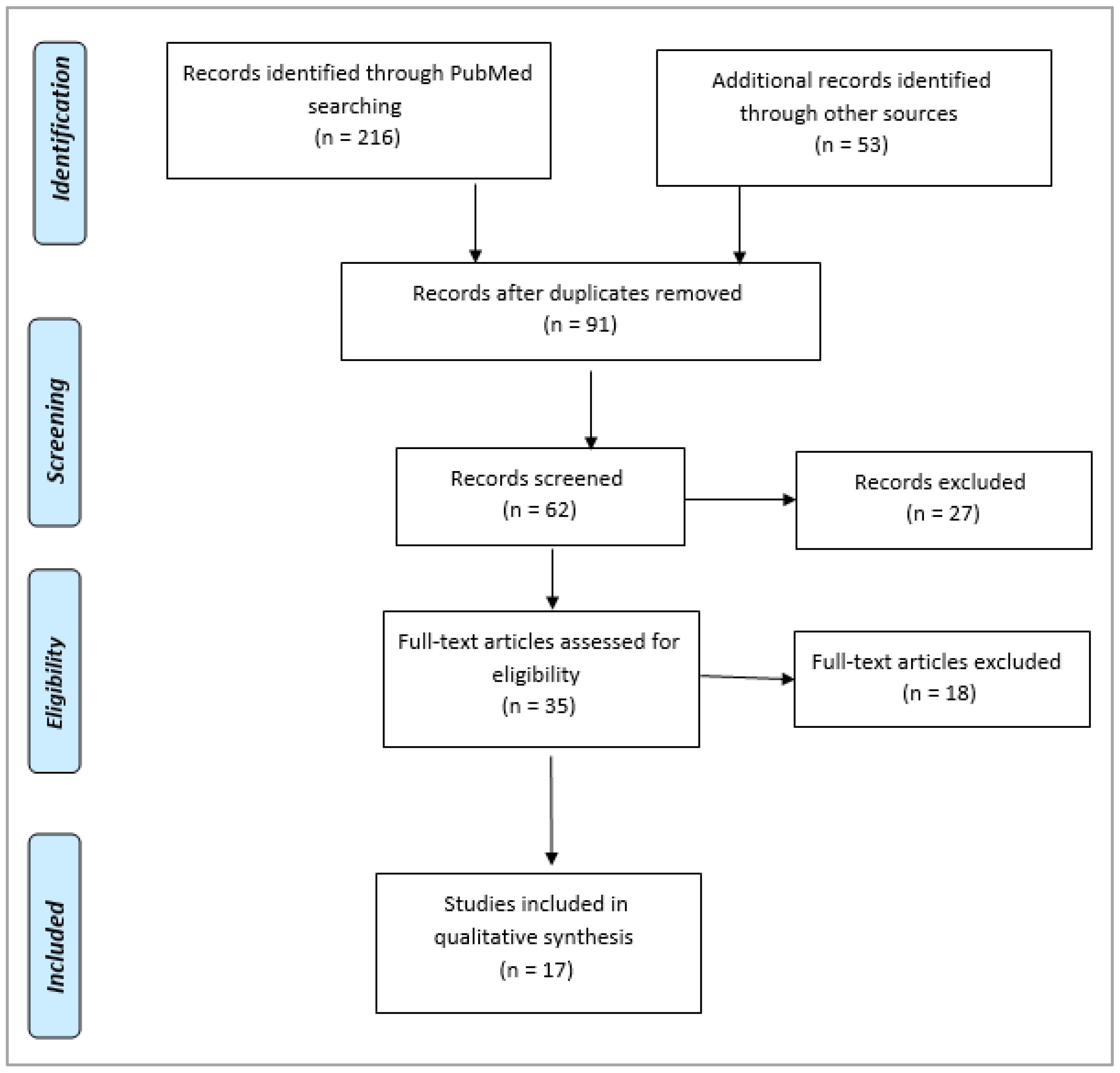
2. Methodology
2.1. Literature Search Strategy
2.2. Eligibility Criteria
- Full-length, English-language articles that include detailed information, particularly details about the P. dactylifera research.
- Cross-sectional studies that were released in the previous two years, 2022–2023.
- Investigations that were conducted to ascertain the impact of P. dactylifera and oxidative stress on neurodegenerative disorders.
- Information about P. dactylifera’s pharmacological properties, including dose, duration, side reactions, and potential mechanisms of action.
- Investigated and evaluated publications from indexed journals that provide comprehensive details on statistics and their significance level.
2.3. Study Selection
2.4. Data Extraction
2.5. Quality Assessment
2.6. Representation of Data
3. Oxidative Stress in Neurodegeneration
4. Major Phytochemicals of Phoenix dactylifera with Antioxidant Potential

| Sl. No. | Name | Chemical Structure | Reference (s) |
|---|---|---|---|
| 1 | Caffeic acid |  | [47,48,49] |
| 2 | Ferulic acid | 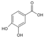 | [46,47,48] |
| 3 | Catechin | 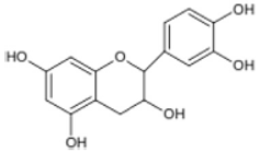 | [54] |
| 4 | Gallic acid | 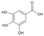 | [53,55] |
| 5 | p-coumaric acid | 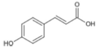 | [49,51] |
| 6 | Resorcinol acid | 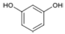 | [59,60,61] |
| 7 | Quercetin | 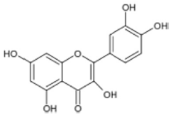 | [52,57] |
| 8 | Protocatechuic acid | 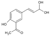 | [49,62] |
| 9 | Rutin | 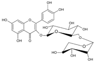 | [56,58] |
| 10 | Apigenin |  | [55,59] |
5. Antioxidant Properties of P. dactylifera
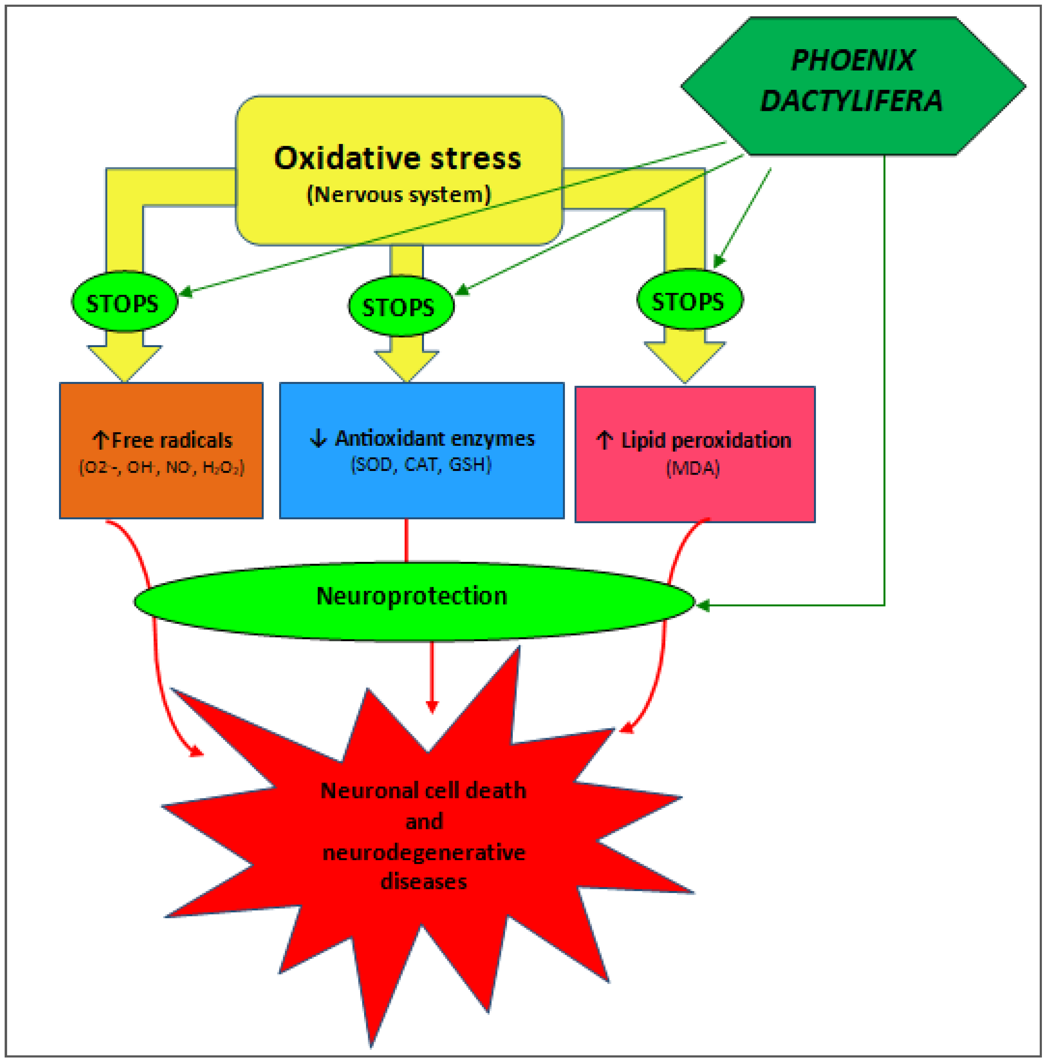
6. Putative Mechanisms of Antioxidant Action of P. dactylifera in Neurodegenerative Diseases
7. Future Implications
8. Conclusions
Supplementary Materials
Author Contributions
Funding
Data Availability Statement
Acknowledgments
Conflicts of Interest
References
- Marín, R.; Abad, C.; Rojas, D.; Chiarello, D.I.; Alejandro, T.G. Biomarkers of oxidative stress and reproductive complications. Adv. Clin. Chem. 2023, 113, 157–233. [Google Scholar] [PubMed]
- Matyas, C.; Haskó, G.; Liaudet, L.; Trojnar, E.; Pacher, P. Interplay of cardiovascular mediators, oxidative stress and inflammation in liver disease and its complications. Nat. Rev. Cardiol. 2021, 18, 117–135. [Google Scholar] [CrossRef] [PubMed]
- Daenen, K.; Andries, A.; Mekahli, D.; Van Schepdael, A.; Jouret, F.; Bammens, B. Oxidative stress in chronic kidney disease. Pediatr. Nephrol. 2019, 34, 975–991. [Google Scholar] [CrossRef] [PubMed]
- Bai, R.; Guo, J.; Ye, X.Y.; Xie, Y.; Xie, T. Oxidative stress: The core pathogenesis and mechanism of Alzheimer’s disease. Ageing Res. Rev. 2022, 77, 101619. [Google Scholar] [CrossRef] [PubMed]
- Hajam, Y.A.; Rani, R.; Ganie, S.Y.; Sheikh, T.A.; Javaid, D.; Qadri, S.S.; Pramodh, S.; Alsulimani, A.; Alkhanani, M.F.; Harakeh, S.; et al. Oxidative Stress in Human Pathology and Aging: Molecular Mechanisms and Perspectives. Cells 2022, 11, 552. [Google Scholar] [CrossRef]
- Salvagno, M.; Sterchele, E.D.; Zaccarelli, M.; Mrakic-Sposta, S.; Welsby, I.J.; Balestra, C.; Taccone, F.S. Oxidative Stress and Cerebral Vascular Tone: The Role of Reactive Oxygen and Nitrogen Species. Int. J. Mol. Sci. 2024, 25, 3007. [Google Scholar] [CrossRef]
- Njoroge, J.N.; Teerlink, J.R. Pathophysiology and Therapeutic Approaches to Acute Decompensated Heart Failure. Circ. Res. 2021, 128, 1468–1486. [Google Scholar] [CrossRef]
- De Franciscis, P.; Colacurci, N.; Riemma, G.; Conte, A.; Pittana, E.; Guida, M.; Schiattarella, A. A Nutraceutical Approach to Menopausal Complaints. Medicina 2019, 55, 544. [Google Scholar] [CrossRef]
- Tasneem, S.; Liu, B.; Li, B.; Choudhary, M.I.; Wang, W. Molecular pharmacology of inflammation: Medicinal plants as anti-inflammatory agents. Pharmacol. Res. 2019, 139, 126–140. [Google Scholar] [CrossRef]
- Zhu, T.; Wang, L.; Wang, L.P.; Wan, Q. Therapeutic targets of neuroprotection and neurorestoration in ischemic stroke: Applications for natural compounds from medicinal herbs. Biomed. Pharmacother. 2022, 148, 112719. [Google Scholar] [CrossRef]
- Chen, S.; Liu, H.; Wang, S.; Jiang, H.; Gao, L.; Wang, L.; Teng, L.; Wang, C.; Wang, D. The Neuroprotection of Verbascoside in Alzheimer’s Disease Mediated through Mitigation of Neuroinflammation via Blocking NF-κB-p65 Signaling. Nutrients 2022, 14, 1417. [Google Scholar] [CrossRef] [PubMed]
- Chen, Y.Y.; Liu, Q.P.; An, P.; Jia, M.; Luan, X.; Tang, J.Y.; Zhang, H. Ginsenoside Rd: A promising natural neuroprotective agent. Phytomedicine 2022, 95, 153883. [Google Scholar] [CrossRef] [PubMed]
- Wu, L.; Song, H.; Zhang, C.; Wang, A.; Zhang, B.; Xiong, C.; Zhuang, X.; Zang, Y.; Li, C.; Fang, Q.; et al. Efficacy and Safety of Panax notoginseng Saponins in the Treatment of Adults with Ischemic Stroke in China: A Randomized Clinical Trial. JAMA Netw. Open 2023, 6, e2317574. [Google Scholar] [CrossRef] [PubMed]
- Singh, S.K.; Srivastav, S.; Castellani, R.J.; Plascencia-Villa, G.; Perry, G. Neuroprotective and Antioxidant Effect of Ginkgo biloba Extract Against AD and Other Neurological Disorders. Neurotherapeutics 2019, 16, 666–674. [Google Scholar] [CrossRef]
- Al-Okbi, S.Y. Date Palm as Source of Nutraceuticals for Health Promotion: A Review. Curr. Nutr. Rep. 2022, 11, 574–591. [Google Scholar] [CrossRef]
- El-Far, A.H.; Oyinloye, B.E.; Sepehrimanesh, M.; Allah, M.A.G.; Abu-Reidah, I.; Shaheen, H.M.; Razeghian-Jahromi, I.; Alsenosy, A.E.A.; Noreldin, A.E.; Al Jaouni, S.K.; et al. Date Palm (Phoenix dactylifera): Novel Findings and Future Directions for Food and Drug Discovery. Curr. Drug Discov. Technol. 2019, 16, 2–10. [Google Scholar] [CrossRef]
- Ahmed, O.S.; Sedraoui, S.; Zhou, B.; Reversat, G.; Rocher, A.; Bultel-Poncé, V.; Guy, A.; Vercauteren, J.; Selim, S.; Galano, J.M.; et al. Phytoprostanes from Date Palm Fruit and Byproducts: Five Different Varieties Grown in Two Different Locations As Potential sources. J. Agric. Food Chem. 2021, 69, 13754–13761. [Google Scholar] [CrossRef]
- Saputri, R.D.; Usman, A.N.; Widaningsih, Y.; Jafar, N.; Ahmad, M.; Ramadhani, S.; Dirpan, A. Date palm (Phoenix dactylifera) consumption as a nutrition source for mild anemia. Gac. Sanit. 2021, 35 (Suppl. 2), S271–S274. [Google Scholar] [CrossRef]
- Alkhoori, M.A.; Kong, A.S.; Aljaafari, M.N.; Abushelaibi, A.; Erin Lim, S.H.; Cheng, W.H.; Chong, C.M.; Lai, K.S. Biochemical Composition and Biological Activities of Date Palm (Phoenix dactylifera L.) Seeds: A Review. Biomolecules 2022, 12, 1626. [Google Scholar] [CrossRef]
- Ibiyeye, R.; Sulaimon, F.; Imam, A.; Adana, M.; Okesina, A.; Ajao, M. Phoenix dactylifera and polyphenols ameliorated monosodium glutamate toxicity in the dentate gyrus of Wistar rats. Niger. J. Physiol. Sci. 2023, 38, 73–78. [Google Scholar] [CrossRef]
- Younas, A.; Naqvi, S.A.; Khan, M.R.; Shabbir, M.A.; Jatoi, M.A.; Anwar, F.; Inam-Ur-Raheem, M.; Saari, N.; Aadil, R.M. Functional food and nutra-pharmaceutical perspectives of date (Phoenix dactylifera L.) fruit. J. Food Biochem. 2020, 44, e13332. [Google Scholar] [CrossRef] [PubMed]
- Al-Dashti, Y.A.; Holt, R.R.; Keen, C.L.; Hackman, R.M. Date Palm Fruit (Phoenix dactylifera): Effects on Vascular Health and Future Research Directions. Int. J. Mol. Sci. 2021, 22, 4665. [Google Scholar] [CrossRef] [PubMed]
- Barakat, H.; Alfheeaid, H.A. Date Palm Fruit (Phoenix dactylifera) and Its Promising Potential in Developing Functional Energy Bars: Review of Chemical, Nutritional, Functional, and Sensory Attributes. Nutrients 2023, 15, 2134. [Google Scholar] [CrossRef] [PubMed]
- Alberti, Á.; Riethmüller, E.; Béni, S. Characterization of diarylheptanoids: An emerging class of bioactive natural products. J. Pharm. Biomed. Anal. 2018, 147, 13–34. [Google Scholar] [CrossRef] [PubMed]
- Hall, E.D.; Wang, J.A.; Miller, D.M.; Cebak, J.E.; Hill, R.L. Newer pharmacological approaches for antioxidant neuroprotection in traumatic brain injury. Neuropharmacology 2019, 145 (Pt B), 247–258. [Google Scholar] [CrossRef]
- Watson, N.; Diamandis, T.; Gonzales-Portillo, C.; Reyes, S.; Borlongan, C.V. Melatonin as an Antioxidant for Stroke Neuroprotection. Cell Transpl. 2016, 25, 883–891. [Google Scholar] [CrossRef]
- Abu-Odeh, A.; Fino, L.; Al-Absi, G.; Alnatour, D.; Al-Darraji, M.; Shehadeh, M.; Suaifan, G. Medicinal plants of Jordan: Scoping review. Heliyon 2023, 9, e17081. [Google Scholar] [CrossRef]
- Głąbska, D.; Guzek, D.; Groele, B.; Gutkowska, K. Fruit and Vegetable Intake and Mental Health in Adults: A Systematic Review. Nutrients 2020, 12, 115. [Google Scholar] [CrossRef]
- Leisegang, K.; Finelli, R.; Sikka, S.C.; Panner Selvam, M.K. Eurycoma longifolia (Jack) Improves Serum Total Testosterone in Men: A Systematic Review and Meta-Analysis of Clinical Trials. Medicina 2022, 58, 1047. [Google Scholar] [CrossRef]
- Awad, A.; Goh, M.S.; Trubiano, J.A. Drug Reaction with Eosinophilia and Systemic Symptoms: A Systematic Review. J. Allergy Clin. Immunol. Pract. 2023, 11, 1856–1868. [Google Scholar] [CrossRef]
- Roy, J.; Galano, J.M.; Durand, T.; Le Guennec, J.Y.; Lee, J.C. Physiological role of reactive oxygen species as promoters of natural defenses. FASEB J. 2017, 31, 3729–3745. [Google Scholar] [CrossRef] [PubMed]
- Yang, D.; Guo, X.; Xie, T.; Luo, X. Reactive oxygen species may play an essential role in driving biological evolution: The Cambrian Explosion as an example. J. Environ. Sci. 2018, 63, 218–226. [Google Scholar] [CrossRef] [PubMed]
- Miwa, S.; Kashyap, S.; Chini, E.; von Zglinicki, T. Mitochondrial dysfunction in cell senescence and aging. J. Clin. Investig. 2022, 132, e158447. [Google Scholar] [CrossRef] [PubMed]
- Roy, Z.; Bansal, R.; Siddiqui, L.; Chaudhary, N. Understanding the Role of Free Radicals and Antioxidant Enzymes in Human Diseases. Curr. Pharm. Biotechnol. 2023, 24, 1265–1276. [Google Scholar] [PubMed]
- Di Meo, S.; Venditti, P. Evolution of the Knowledge of Free Radicals and Other Oxidants. Oxid. Med. Cell Longev. 2020, 2020, 9829176. [Google Scholar] [CrossRef]
- Wang, K.; Liu, H.; Sun, W.; Guo, J.; Jiang, Z.; Xu, S.; Miao, Z. Eucalyptol alleviates avermectin exposure-induced apoptosis and necroptosis of grass carp hepatocytes by regulating ROS/NLRP3 axis. Aquat. Toxicol. 2023, 264, 106739. [Google Scholar] [CrossRef]
- Mortensen, M.S.; Ruiz, J.; Watts, J.L. Polyunsaturated Fatty Acids Drive Lipid Peroxidation during Ferroptosis. Cells 2023, 12, 804. [Google Scholar] [CrossRef]
- Jaganjac, M.; Milkovic, L.; Zarkovic, N.; Zarkovic, K. Oxidative stress and regeneration. Free Radic. Biol. Med. 2022, 181, 154–165. [Google Scholar] [CrossRef]
- Chen, P.P.; Yang, P.; Liu, C.; Deng, Y.L.; Luo, Q.; Miao, Y.; Zhang, M.; Cui, F.P.; Zeng, J.Y.; Shi, T.; et al. Urinary concentrations of phenols, oxidative stress biomarkers and thyroid cancer: Exploring associations and mediation effects. J. Environ. Sci. 2022, 120, 30–40. [Google Scholar] [CrossRef]
- Li, J.; Jia, B.; Cheng, Y.; Song, Y.; Li, Q.; Luo, C. Targeting Molecular Mediators of Ferroptosis and Oxidative Stress for Neurological Disorders. Oxid. Med. Cell Longev. 2022, 2022, 3999083. [Google Scholar] [CrossRef]
- Ma, X.H.; Liu, J.H.; Liu, C.Y.; Sun, W.Y.; Duan, W.J.; Wang, G.; Kurihara, H.; He, R.R.; Li, Y.F.; Chen, Y.; et al. ALOX15-launched PUFA-phospholipids peroxidation increases the susceptibility of ferroptosis in ischemia-induced myocardial damage. Signal Transduct. Target. Ther. 2022, 7, 288. [Google Scholar] [CrossRef] [PubMed]
- Liu, D.; Zhou, L.; Yang, M.; McIntyre, R.S.; Cao, B. Oxidative Stress Mediates the Association Between Dietary Fat Intake and Cognition in US Older Adults. Am. J. Geriatr. Psychiatry 2022, 30, 761–773. [Google Scholar] [CrossRef] [PubMed]
- Videla, L.A.; Hernandez-Rodas, M.C.; Metherel, A.H.; Valenzuela, R. Influence of the nutritional status and oxidative stress in the desaturation and elongation of n-3 and n-6 polyunsaturated fatty acids: Impact on non-alcoholic fatty liver disease. Prostaglandins Leukot. Essent. Fatty Acids 2022, 181, 102441. [Google Scholar] [CrossRef] [PubMed]
- Yasin, B.R.; El-Fawal, H.A.; Mousa, S.A. Date (Phoenix dactylifera) Polyphenolics and Other Bioactive Compounds: A Traditional Islamic Remedy’s Potential in Prevention of Cell Damage, Cancer Therapeutics and Beyond. Int. J. Mol. Sci. 2015, 16, 30075–30090. [Google Scholar] [CrossRef] [PubMed]
- Ahmad Mohd Zain, M.R.; Abdul Kari, Z.; Dawood, M.A.O.; Nik Ahmad Ariff, N.S.; Salmuna, Z.N.; Ismail, N.; Zainal Abidin, S.; Seong Wei, L.; Ahmed Shokri, A. Bioactivity and Pharmacological Potential of Date Palm (Phoenix dactylifera L.) Against Pandemic COVID-19: A Comprehensive Review. Appl. Biochem. Biotechnol. 2022, 194, 4587–4624. [Google Scholar] [CrossRef]
- Gad El-Hak, H.N.; Mahmoud, H.S.; Ahmed, E.A.; Elnegris, H.M.; Aldayel, T.S.; Abdelrazek, H.M.A.; Soliman, M.T.A.; El-Menyawy, M.A.I. Methanolic Phoenix dactylifera L. Extract Ameliorates Cisplatin-Induced Hepatic Injury in Male Rats. Nutrients 2022, 14, 1025. [Google Scholar] [CrossRef]
- Shivanandappa, T.B.; Alotaibi, G.; Chinnadhurai, M.; Dachani, S.R.; Ahmad, M.D.; Aldaajanii, K.A. Phoenix dactylifera (Ajwa Dates) Alleviate LPS-Induced Sickness Behaviour in Rats by Attenuating Proinflammatory Cytokines and Oxidative Stress in the Brain. Int. J. Mol. Sci. 2023, 24, 10413. [Google Scholar] [CrossRef]
- Balasmeh, R.; Jarrar, Y.; Al-Sheikh, I.; Alshaiah, H.; Jarrar, Q.; Alani, R.; Abudahab, S. Effects of Fasting and Phoenix dactylifera on the Expression of Major Drug- Metabolizing Enzymes in the Mouse Livers. Curr. Drug Metab. 2022, 23, 666–676. [Google Scholar] [CrossRef]
- Alkhalidy, H.; Al-Nabulsi, A.A.; Al-Taher, M.; Osaili, T.; Olaimat, A.N.; Liu, D. Date (Phoenix dactylifera L.) seed oil is an agro-industrial waste with biopreservative effects and antimicrobial activity. Sci. Rep. 2023, 13, 17142. [Google Scholar] [CrossRef]
- Osman, K.M.; Kamal, O.E.; Deif, H.N.; Ahmed, M.M. Phoenix dactylifera, mentha piperita and montanide™ ISA-201 as immunological adjuvants in a chicken model. Acta Trop. 2020, 202, 105281. [Google Scholar] [CrossRef]
- Alahyane, A.; ElQarnifa, S.; Ayour, J.; Elateri, I.; Ouamnina, A.; Ait-Oubahou, A.; Benichou, M.; Abderrazik, M. Date seeds (Phoenix dactylifera L.) valorization: Chemical composition of lipid fraction. Braz. J. Biol. 2022, 84, e260771. [Google Scholar] [CrossRef] [PubMed]
- Gantait, S.; El-Dawayati, M.M.; Panigrahi, J.; Labrooy, C.; Verma, S.K. The retrospect and prospect of the applications of biotechnology in Phoenix dactylifera L. Appl. Microbiol. Biotechnol. 2018, 102, 8229–8259. [Google Scholar] [CrossRef] [PubMed]
- Zein, N.; Elewa, Y.H.A.; Alruwaili, M.K.; Dewaard, M.; Alorabi, M.; Albogami, S.M.; Batiha, G.E.; Zahran, M.H. Barhi date (Phoenix dactylifera) extract ameliorates hepatocellular carcinoma in male rats. Biomed. Pharmacother. 2022, 156, 113976. [Google Scholar] [CrossRef] [PubMed]
- Otify, A.M.; Hammam, A.M.; Aly Farag, M. Phoenix dactylifera L. date tree pollen fertility effects on female rats in relation to its UPLC-MS profile via a biochemometric approach. Steroids 2021, 173, 108888. [Google Scholar] [CrossRef] [PubMed]
- Taleb, H.; Maddocks, S.E.; Morris, R.K.; Kanekanian, A.D. Chemical characterisation and the anti-inflammatory, anti-angiogenic and antibacterial properties of date fruit (Phoenix dactylifera L.). J. Ethnopharmacol. 2016, 194, 457–468. [Google Scholar] [CrossRef]
- Ismail, H.; Khalid, D.; Ayub, S.B.; Ijaz, M.U.; Akram, S.; Bhatti, M.Z.; Yousef, F.M.; Waard, M. Effects of Phoenix dactylifera against Streptozotocin-Aluminium Chloride Induced Alzheimer’s Rats and Their In Silico Study. Biomed. Res. Int. 2023, 2023, 1725638. [Google Scholar] [CrossRef]
- Halabi, A.A.; Elwakil, B.H.; Hagar, M.; Olama, Z.A. Date Fruit (Phoenix dactylifera L.) Cultivar Extracts: Nanoparticle Synthesis, Antimicrobial and Antioxidant Activities. Molecules 2022, 27, 5165. [Google Scholar] [CrossRef]
- Bettaieb, I.; Ali Benabderrahim, M.; Rodríguez Arcos, R.; Jose Jiménez Araujo, A.; Elfalleh, W. Date Seeds (Phoenix dactylifera): Antioxidant Potential and Profile of Free and Bound Polyphenols from Different Cultivars. Chem. Biodivers. 2023, 20, e202300179. [Google Scholar] [CrossRef]
- Khan, M.A.; Singh, R.; Siddiqui, S.; Ahmad, I.; Ahmad, R.; Upadhyay, S.; Barkat, M.A.; Ali, A.M.A.; Zia, Q.; Srivastava, A.; et al. Anticancer potential of Phoenix dactylifera L. seed extract in human cancer cells and pro-apoptotic effects mediated through caspase-3 dependent pathway in human breast cancer MDA-MB-231 cells: An in vitro and in silico investigation. BMC Complement. Med. Ther. 2022, 22, 68. [Google Scholar] [CrossRef]
- Djaoudene, O.; López, V.; Cásedas, G.; Les, F.; Schisano, C.; Bachir Bey, M.; Tenore, G.C. Phoenix dactylifera L. seeds: A by-product as a source of bioactive compounds with antioxidant and enzyme inhibitory properties. Food Funct. 2019, 10, 4953–4965. [Google Scholar] [CrossRef]
- Athinarayanan, J.; Periasamy, V.S.; Alshatwi, A.A. Phoenix dactylifera lignocellulosic biomass as precursor for nanostructure fabrication using integrated process. Int. J. Biol. Macromol. 2019, 134, 1179–1186. [Google Scholar] [CrossRef] [PubMed]
- Habib, H.M.; El-Fakharany, E.M.; El-Gendi, H.; El-Ziney, M.G.; El-Yazbi, A.F.; Ibrahim, W.H. Palm Fruit (Phoenix dactylifera L.) Pollen Extract Inhibits Cancer Cell and Enzyme Activities and DNA and Protein Damage. Nutrients 2023, 15, 2614. [Google Scholar] [CrossRef] [PubMed]
- Boroujeni, S.N.; Malamiri, F.A.; Bossaghzadeh, F.; Esmaeili, A.; Moudi, E. The most important medicinal plants affecting sperm and testosterone production: A systematic review. JBRA Assist. Reprod. 2022, 26, 522–530. [Google Scholar] [CrossRef] [PubMed]
- Abbassi, R.; Pontes, M.C.; Dhibi, S.; Duarte Filho, L.A.M.S.; Othmani, S.; Bouzenna, H.; Almeida, J.R.G.S.; Hfaiedh, N. Antioxidant properties of date seeds extract (Phoenix dactylifera L.) in alloxan induced damage in rats. Braz. J. Biol. 2023, 83, e274405. [Google Scholar] [CrossRef] [PubMed]
- Subash, S.; Essa, M.M.; Al-Asmi, A.; Al-Adawi, S.; Vaishnav, R.; Guillemin, G.J. Effect of dietary supplementation of dates in Alzheimer’s disease APPsw/2576 transgenic mice on oxidative stress and antioxidant status. Nutr. Neurosci. 2015, 18, 281–288. [Google Scholar] [CrossRef]
- Al-Qurainy, F.; Khan, S.; Tarroum, M.; Nadeem, M.; Alansi, S.; Alshameri, A. Biochemical and Genetical Responses of Phoenix dactylifera L. to Cadmium Stress. Biomed. Res. Int. 2017, 2017, 9504057. [Google Scholar] [CrossRef]
- Habib, H.M.; El-Fakharany, E.M.; Souka, U.D.; Elsebaee, F.M.; El-Ziney, M.G.; Ibrahim, W.H. Polyphenol-Rich Date Palm Fruit Seed (Phoenix dactylifera L.) Extract Inhibits Labile Iron, Enzyme, and Cancer Cell Activities, and DNA and Protein Damage. Nutrients 2022, 14, 3536. [Google Scholar] [CrossRef]
- Ajiboye, B.O.; Oloyede, H.O.B.; Salawu, M.O. Phoenix dactylifera Linn fruit based-diets palliate hyperglycemia in alloxan-induced diabetic rats. J. Basic. Clin. Physiol. Pharmacol. 2020, 17, 180–185. [Google Scholar] [CrossRef]
- Alqarni, M.M.M.; Osman, M.A.; Al-Tamimi, D.S.; Gassem, M.A.; Al-Khalifa, A.S.; Al-Juhaimi, F.; Mohamed Ahmed, I.A. Antioxidant and antihyperlipidemic effects of Ajwa date (Phoenix dactylifera L.) extracts in rats fed a cholesterol-rich diet. J. Food Biochem. 2019, 43, e12933. [Google Scholar] [CrossRef]
- Shahbaz, K.; Asif, J.A.; Liszen, T.; Nurul, A.A.; Alam, M.K. Cytotoxic and Antioxidant Effects of Phoenix dactylifera L. (Ajwa Date Extract) on Oral Squamous Cell Carcinoma Cell Line. Biomed. Res. Int. 2022, 2022, 5792830. [Google Scholar] [CrossRef]
- Zhang, M.; Wang, L.; Wen, D.; Ren, C.; Chen, S.; Zhang, Z.; Hu, L.; Yu, Z.; Tombran-Tink, J.; Zhang, X.; et al. Neuroprotection of retinal cells by Caffeic Acid Phenylethyl Ester (CAPE) is mediated by mitochondrial uncoupling protein UCP2. Neurochem. Int. 2021, 151, 105214. [Google Scholar] [CrossRef] [PubMed]
- Ayna, A. Caffeic acid prevents hydrogen peroxide-induced oxidative damage in SH-SY5Y cell line through mitigation of oxidative stress and apoptosis. Bratisl. Lek. Listy 2021, 122, 120–124. [Google Scholar] [CrossRef] [PubMed]
- Kulkarni, N.P.; Vaidya, B.; Narula, A.S.; Sharma, S.S. Caffeic Acid Phenethyl Ester (CAPE) Attenuates Paclitaxel-induced Peripheral Neuropathy: A Mechanistic Study. Curr. Neurovasc Res. 2022, 19, 293–302. [Google Scholar]
- Domínguez-Avila, J.A.; Salazar-López, N.J.; Montiel-Herrera, M.; Martínez-Martínez, A.; Villegas-Ochoa, M.A.; González-Aguilar, G.A. Phenolic compounds can induce systemic and central immunomodulation, which result in a neuroprotective effect. J. Food Biochem. 2022, 46, e14260. [Google Scholar] [CrossRef] [PubMed]
- Tavan, M.; Hanachi, P.; de la Luz Cádiz-Gurrea, M.; Segura Carretero, A.; Mirjalili, M.H. Natural Phenolic Compounds with Neuroprotective Effects. Neurochem. Res. 2024, 49, 306–326. [Google Scholar] [CrossRef]
- Rojas-García, A.; Fernández-Ochoa, Á.; Cádiz-Gurrea, M.L.; Arráez-Román, D.; Segura-Carretero, A. Neuroprotective Effects of Agri-Food By-Products Rich in Phenolic Compounds. Nutrients 2023, 15, 449. [Google Scholar] [CrossRef]
- Sahyon, H.A.; Al-Harbi, S.A. Antimicrobial, anticancer and antioxidant activities of nano-heart of Phoenix dactylifera tree extract loaded chitosan nanoparticles: In vitro and in vivo study. Int. J. Biol. Macromol. 2020, 160, 1230–1241. [Google Scholar] [CrossRef]
- Moslemi, E.; Dehghan, P.; Khalafi, M. Effectiveness of supplementation with date seed (Phoenix dactylifera) as a functional food on inflammatory markers, muscle damage, and BDNF following high-intensity interval training: A randomized, double-blind, placebo-controlled trial. Eur. J. Nutr. 2023, 62, 2001–2014. [Google Scholar] [CrossRef]
- Al-Farsi, M.; Alasalvar, C.; Morris, A.; Baron, M.; Shahidi, F. Compositional and sensory characteristics of three native sun-dried date (Phoenix dactylifera L.) varieties grown in Oman. J. Agric. Food Chem. 2005, 53, 7586–7591. [Google Scholar] [CrossRef]
- Kiełczykowska, M.; Kocot, J.; Paździor, M.; Musik, I. Selenium—A fascinating antioxidant of protective properties. Adv. Clin. Exp. Med. 2018, 27, 245–255. [Google Scholar] [CrossRef]
- Bjørklund, G.; Shanaida, M.; Lysiuk, R.; Antonyak, H.; Klishch, I.; Shanaida, V.; Peana, M. Selenium: An Antioxidant with a Critical Role in Anti-Aging. Molecules 2022, 27, 6613. [Google Scholar] [CrossRef] [PubMed]
- Huang, J.; Xie, L.; Song, A.; Zhang, C. Selenium Status and Its Antioxidant Role in Metabolic Diseases. Oxid. Med. Cell Longev. 2022, 2022, 7009863. [Google Scholar] [CrossRef] [PubMed]
- Groth, S.; Budke, C.; Weber, T.; Neugart, S.; Brockmann, S.; Holz, M.; Sawadski, B.C.; Daum, D.; Rohn, S. Relationship between Phenolic Compounds, Antioxidant Properties, and the Allergenic Protein Mal d 1 in Different Selenium-Biofortified Apple Cultivars (Malus domestica). Molecules 2021, 26, 2647. [Google Scholar] [CrossRef]
- Pujari, R.R.; Vyawahare, N.S.; Kagathara, V.G. Evaluation of antioxidant and neuroprotective effect of date palm (Phoenix dactylifera L.) against bilateral common carotid artery occlusion in rats. Indian. J. Exp. Biol. 2011, 49, 627–633. [Google Scholar] [PubMed]
- Salem, G.A.; Shaban, A.; Diab, H.A.; Elsaghayer, W.A.; Mjedib, M.D.; Hnesh, A.M.; Sahu, R.P. Phoenix dactylifera protects against oxidative stress and hepatic injury induced by paracetamol intoxication in rats. Biomed. Pharmacother. 2018, 104, 366–374. [Google Scholar] [CrossRef] [PubMed]
- Mani, V.; Arfeen, M.; Dhaked, D.K.; Mohammed, H.A.; Amirthalingam, P.; Elsisi, H.A. Neuroprotective Effect of Methanolic Ajwa Seed Extract on Lipopolysaccharide-Induced Memory Dysfunction and Neuroinflammation: In Vivo, Molecular Docking and Dynamics Studies. Plants 2023, 12, 934. [Google Scholar] [CrossRef]
- Xiao, Q.; Liu, H.; Yang, C.; Chen, Y.; Huang, Y.; Xiao, X.; Pan, Y.; He, J.; Du, Q.; Wang, Q.; et al. Bushen-Yizhi formula exerts neuroprotective effect via inhibiting excessive mitophagy in rats with chronic cerebral hypoperfusion. J. Ethnopharmacol. 2023, 310, 116326. [Google Scholar] [CrossRef]
- Mallhi, T.H.; Qadir, M.I.; Ali, M.; Ahmad, B.; Khan, Y.H.; Rehman, A. Review: Ajwa date (Phoenix dactylifera)—An emerging plant in pharmacological research. Pak. J. Pharm. Sci. 2014, 27, 607–616. [Google Scholar]
- Behl, C.; Moosmann, B. Antioxidant neuroprotection in Alzheimer’s disease as preventive and therapeutic approach. Free Radic. Biol. Med. 2002, 33, 182–191. [Google Scholar] [CrossRef]
- Moslemi, E.; Dehghan, P.; Khani, M.; Sarbakhsh, P.; Sarmadi, B. The effects of date seed (Phoenix dactylifera) supplementation on exercise-induced oxidative stress and aerobic and anaerobic performance following high-intensity interval training sessions: A randomised, double-blind, placebo-controlled trial. Br. J. Nutr. 2022, 14, 1151–1162. [Google Scholar] [CrossRef]
- Al-Alawi, R.A.; Al-Mashiqri, J.H.; Al-Nadabi, J.S.M.; Al-Shihi, B.I.; Baqi, Y. Date Palm Tree (Phoenix dactylifera L.): Natural Products and Therapeutic Options. Front. Plant Sci. 2017, 8, 845. [Google Scholar] [CrossRef] [PubMed]
- Zidan, N.S.; Omran, A.M.E.; Rezk, S.M.; Atteia, H.H.; Sakran, M.I. Anti-Alzheimer’s disease potential of Arabian coffee versus Date palm seed extract in male rats. J. Food Biochem. 2022, 46, e14017. [Google Scholar] [CrossRef] [PubMed]
- Khan, A.; Park, J.S.; Kang, M.H.; Lee, H.J.; Ali, J.; Tahir, M.; Choe, K.; Kim, M.O. Caffeic Acid, a Polyphenolic Micronutrient Rescues Mice Brains against Aβ-Induced Neurodegeneration and Memory Impairment. Antioxidants 2023, 12, 1284. [Google Scholar] [CrossRef] [PubMed]
- Razak, A.M.; Tan, J.K.; Mohd Said, M.; Makpol, S. Modulating Effects of Zingiberaceae Phenolic Compounds on Neurotrophic Factors and Their Potential as Neuroprotectants in Brain Disorders and Age-Associated Neurodegenerative Disorders: A Review. Nutrients 2023, 15, 2564. [Google Scholar] [CrossRef] [PubMed]
- Carrera, I.; Martínez, O.; Cacabelos, R. Neuroprotection with Natural Antioxidants and Nutraceuticals in the Context of Brain Cell Degeneration: The Epigenetic Connection. Curr. Top. Med. Chem. 2019, 19, 2999–3011. [Google Scholar] [CrossRef]
- Uddin, M.S.; Al Mamun, A.; Kabir, M.T.; Ahmad, J.; Jeandet, P.; Sarwar, M.S.; Ashraf, G.M.; Aleya, L. Neuroprotective role of polyphenols against oxidative stress-mediated neurodegeneration. Eur. J. Pharmacol. 2020, 886, 173412. [Google Scholar] [CrossRef]
- Hsueh, Y.J.; Chen, Y.N.; Tsao, Y.T.; Cheng, C.M.; Wu, W.C.; Chen, H.C. The Pathomechanism, Antioxidant Biomarkers, and Treatment of Oxidative Stress-Related Eye Diseases. Int. J. Mol. Sci. 2022, 23, 1255. [Google Scholar] [CrossRef]
- AlFaris, N.A.; AlTamimi, J.Z.; AlGhamdi, F.A.; Albaridi, N.A.; Alzaheb, R.A.; Aljabryn, D.H.; Aljahani, A.H.; AlMousa, L.A. Total phenolic content in ripe date fruits (Phoenix dactylifera L.): A systematic review and meta-analysis. Saudi J. Biol. Sci. 2021, 28, 3566–3577. [Google Scholar] [CrossRef]
- Singh, A.; Dhaneshwar, S.; Mazumder, A. Investigating Neuroprotective Potential of Berberine, Levetiracetam and their Combination in the Management of Alzheimer’s Disease Utilizing Drug Repurposing Strategy. Curr. Rev. Clin. Exp. Pharmacol. 2023, 18, 182–190. [Google Scholar]
Disclaimer/Publisher’s Note: The statements, opinions and data contained in all publications are solely those of the individual author(s) and contributor(s) and not of MDPI and/or the editor(s). MDPI and/or the editor(s) disclaim responsibility for any injury to people or property resulting from any ideas, methods, instructions or products referred to in the content. |
© 2024 by the authors. Licensee MDPI, Basel, Switzerland. This article is an open access article distributed under the terms and conditions of the Creative Commons Attribution (CC BY) license (https://creativecommons.org/licenses/by/4.0/).
Share and Cite
Asdaq, S.M.B.; Almutiri, A.A.; Alenzi, A.; Shaikh, M.; Shaik, M.A.; Alshehri, S.; Rabbani, S.I. Unveiling the Neuroprotective Potential of Date Palm (Phoenix dactylifera): A Systematic Review. Pharmaceuticals 2024, 17, 1221. https://doi.org/10.3390/ph17091221
Asdaq SMB, Almutiri AA, Alenzi A, Shaikh M, Shaik MA, Alshehri S, Rabbani SI. Unveiling the Neuroprotective Potential of Date Palm (Phoenix dactylifera): A Systematic Review. Pharmaceuticals. 2024; 17(9):1221. https://doi.org/10.3390/ph17091221
Chicago/Turabian StyleAsdaq, Syed Mohammed Basheeruddin, Abdulaziz Ali Almutiri, Abdullah Alenzi, Maheen Shaikh, Mujeeb Ahmed Shaik, Sultan Alshehri, and Syed Imam Rabbani. 2024. "Unveiling the Neuroprotective Potential of Date Palm (Phoenix dactylifera): A Systematic Review" Pharmaceuticals 17, no. 9: 1221. https://doi.org/10.3390/ph17091221
APA StyleAsdaq, S. M. B., Almutiri, A. A., Alenzi, A., Shaikh, M., Shaik, M. A., Alshehri, S., & Rabbani, S. I. (2024). Unveiling the Neuroprotective Potential of Date Palm (Phoenix dactylifera): A Systematic Review. Pharmaceuticals, 17(9), 1221. https://doi.org/10.3390/ph17091221