Article

FLT3 and IRAK4 Inhibitor Emavusertib in Combination with BH3-Mimetics in the Treatment of Acute Myeloid Leukemia

1
Department for Biomedical Research, University of Bern, 3008 Bern, Switzerland
2
Department of Hematology, University Hospital Bern, 3010 Bern, Switzerland
3
Department of Medical Oncology, University Hospital Bern, 3010 Bern, Switzerland
*
Authors to whom correspondence should be addressed.
Curr. Issues Mol. Biol. 2024, 46(4), 2946-2960; https://doi.org/10.3390/cimb46040184
Submission received: 27 February 2024 / Revised: 22 March 2024 / Accepted: 22 March 2024 / Published: 29 March 2024
(This article belongs to the Special Issue Molecular Research and Pathological Mechanism of Leukemia)

Abstract

:
Targeting the FLT3 receptor and the IL-1R associated kinase 4 as well as the anti-apoptotic proteins MCL1 and BCL2 may be a promising novel approach in the treatment of acute myeloid leukemia (AML). The FLT3 and IRAK4 inhibitor emavusertib (CA4948), the MCL1 inhibitor S63845, the BCL2 inhibitor venetoclax, and the HSP90 inhibitor PU-H71 were assessed as single agents and in combination for their ability to induce apoptosis and cell death in leukemic cells in vitro. AML cells represented all major morphologic and molecular subtypes, including FLT3-ITD and NPM1 mutant AML cell lines and a variety of patient-derived AML cells. Emavusertib in combination with MCL1 inhibitor S63845 or BCL2 inhibitor venetoclax induced cell cycle arrest and apoptosis in MOLM-13 cells. In primary AML cells, the response to emavusertib was associated with the presence of the FLT3 gene mutation with an allelic ratio >0.5 and the presence of NPM1 gene mutations. S63845 was effective in all tested AML cell lines and primary AML samples. Blast cell percentage was positively associated with the response to CA4948, S63845, and venetoclax, with elevated susceptibility of primary AML with blast cell fraction >80%. Biomarkers of the response to venetoclax included the blast cell percentage and bone marrow infiltration rate, as well as the expression levels of CD11b, CD64, and CD117. Elevated susceptibility to CA4948 combination treatments with S63845 or PU-H71 was associated with FLT3-mutated AML and CD34 < 30%. The combination of CA4948 and BH3-mimetics may be effective in the treatment in FLT3-mutated AML with differential target specificity for MCL1 and BCL2 inhibitors. Moreover, the combination of CA4948 and PU-H71 may be a candidate combination treatment in FLT3-mutated AML.

Graphical Abstract

1. Introduction

The fms-like tyrosine kinase receptor 3 (FLT3) gene is overexpressed in up to 93% and mutated in over 30% of primary acute myeloid leukemia (AML) [1]. FLT3 signaling pathways are highly active in AML cells, with consequential induction of protein translation and cell proliferation as well as reduced apoptosis. Several FLT3 kinase inhibitors have been approved for the treatment of FLT3-mutated AML; however, the therapeutic effects may be short-lived due to the emergence of adaptive resistance [2]. Therapy resistance occurs through target-dependent mechanisms resulting from point mutations in the FLT3 kinase domain or through target-independent mechanisms, such as an alternate activation of survival and proliferation pathways [3], including the elevated expression of anti-apoptotic proteins of the B-cell lymphoma 2 (BCL2) family. MCL1 and BCL2 proteins are both frequently overexpressed in AML and critical for the survival of AML cells and leukemic stem cells [4]. While the BCL2 inhibitor venetoclax, in combination with azacitidine, decitabine, or low-dose cytarabine (LDAC), has been approved for the treatment of adults with newly diagnosed AML [5], the MCL1 inhibitor S63845 has been evaluated as a candidate treatment in AML in combination with the MEK inhibitor trametinib, the BMI1 inhibitor PTC596, or the HSP90 inhibitor PU-H71 in preclinical studies [6,7,8].
Interleukin-1 receptor-associated kinases are emerging therapeutic targets in hematologic malignancies [9]. IRAK4 is a key mediator in interleukin- and Toll-like receptor (TLR) signaling in the innate immune response [10]. Recruitment and subsequent activation of IRAK4 upon interleukin 1 (IL-1) binding to IL-1R or TLR4 stimulation by bacterial lipopolysaccharides (LPS) can be mediated by the myeloid differentiation primary response 88 (MYD88) adaptor protein [11]. In AML cells, LPS binding to TLR4 promotes cell proliferation, inhibits apoptosis, and increases resistance to chemotherapy [12]. Like FLT3, TLRs and their associated signal transducers are frequently overexpressed and/or constitutively activated in hematological malignancies [13]. IRAK4 inhibitors were originally developed for the treatment of chronic inflammatory auto-immune diseases such as rheumatoid arthritis [14]. Multikinase FLT3-IRAK1/4 inhibitors have been developed to overcome the adaptive resistance of myeloid cells to specific FLT3 inhibitors [15]. Pacritinib, a multi-kinase inhibitor of IRAK1, JAK2, and FLT3, demonstrated anti-leukemic activity in combination with chemotherapy in patients with FLT3 mutations [16,17]. Emavusertib (CA4948), a multi-kinase inhibitor of IRAK4 and FLT3, has been proposed for the treatment of B-cell lymphoma and myeloid malignancies [10,18]. Emavusertib is currently being evaluated in a phase 1/2 clinical trial in R/R AML as a monotherapy and in combination with azacitidine or venetoclax (NCT04278768) [19]. New insights into the biology of IRAK4, the development of IRAK4 inhibitors, and synergies with established treatments such as chemotherapy and targeted inhibitors were discussed at the first symposium on IRAK4 in cancer [20].
HSP90 inhibitors may destabilize oncoproteins associated with the cell cycle, angiogenesis, RAS-MAPK activity, histone modification, kinases, and growth factors [21,22]. However, due to toxicity and limited efficacy, no HSP90 inhibitor has been approved for clinical use as single agent [23]. Combining HSP90 inhibitors with other anticancer therapies might be a more advisable strategy to reduce toxicity and increase efficacy [24]. The combination of the HSP90 inhibitor PU-H71 and the MCL1 inhibitor S63845 may be a candidate treatment for FLT3-mutated AML with moderate CD34 positivity, while the combination of HSP90 inhibitor PU-H71 and BCL2 inhibitor venetoclax may be more effective in the treatment of primitive AML with high CD117 and low CD11b positivity [8].
In this study, we assessed the multikinase inhibitor emavusertib (CA4948), the MCL1 inhibitor S63845, the BCL2 inhibitor venetoclax (ABT-199), and the HSP90 inhibitor PU-H71 as single agents and in combination for their ability to induce apoptosis and cell death in leukemic cells in vitro. AML cells represented all major morphologic and molecular subtypes, including FLT3-ITD and FLT3 wild type, NPM1 mutant and wild type, as well as TP53 mutant and wild type AML cell lines and primary AML cells.

2. Materials and Methods

2.1. Patient Samples

Mononuclear cells of AML patients diagnosed and treated at the University Hospital, Bern, Switzerland, between 2015 and 2022 were included in this study. Informed consent was obtained according to the Declaration of Helsinki, and the studies were approved by decisions of the Ethics Committee of the Canton of Bern, Switzerland. Peripheral blood mononuclear cells (PBMCs) and bone marrow mononuclear cells (BMMCs) were collected at the time of diagnosis before the initiation of treatment. AML cells were analyzed at the central hematology laboratory of the University Hospital Bern according to current practice and techniques [25], including mutational screening of FLT3, NPM1, and TP53 genes and conventional karyotype analysis of at least 20 metaphases. All samples were analyzed by NGS sequencing of the myeloid panel genes, encoding spliceosome components (U2AF1, SF3B1, SRSF2, ZRSR2), epigenetic modifiers (TET2, DNMT3A, BCOR, ASXL1, IDH1, IDH2), cohesins (STAG2, RAD21, SMC3), transcription factors (TP53, RUNX1, WT1, ETV6), signaling molecules (FLT3, NF1, NRAS, CBL, PTPN11, JAK2), and chromatin modifiers (ASXL1, EZH2, SUZ12). Flow-cytometry immuno-phenotyping was performed according to the international consensus with CD markers in the initial evaluation of myeloid leukemia, including CD7, CD11b, CD13, CD14, CD15, CD16, CD33, CD34, CD45, CD56, CD117, and HLA-DR.

2.2. Cell Lines and Cell Culture

Human AML cell lines OCI-AML3 (AML-M4, FLT3wt, DNMT3A R882C, NPM1mut, TP53wt), MOLM-13 (AML-M5, t (9;11), FLT3-ITD, TP53wt), SKM-1 (AML-M5, FLT3wt, TP53mut), and ML-2 (AML-M4, t (6;11), FLT3wt, TP53wt) cells were supplied by the Leibniz Institute DSMZ, German Collection of Microorganisms and Cell Cultures. AML cells were grown in RPMI 1640 media (SIGMA-ALDRICH, St. Louis, MO, USA) supplemented with 20% fetal bovine serum (FBS, Biochrom GmbH, Berlin, Germany) in a standard cell culture incubator at 37 C with 5% CO2. The human bone marrow stroma cell line HS-5 was supplied by the American Type Culture Collection (ATCC) (ATCC® CRL-11882™). HS-5 cells were grown in DMEM media (SIGMA-ALDRICH, St. Louis, MO, USA) supplemented with 10% fetal bovine serum (FBS, Biochrom GmbH, Berlin, Germany). The HS-5 cells secreted granulocyte colony-stimulating factor (G-CSF), granulocyte-macrophage-CSF (GM-CSF), macrophage-CSF (M-CSF), Kit ligand (KL), macrophage-inhibitory protein-1 alpha, interleukin-1 alpha (IL-1alpha), IL-1beta, IL-1RA, IL-6, IL-8, IL-11, and leukemia inhibitory factor (LIF). For the co-culture assays, HS-5 cells were plated on six-well plates on day 1. On day 2, Nunc 0.4 uM cell culture inserts (ThermoFisher, Roskilde, Denmark) were placed over the HS-5 feeder layer and AML cells were filled into the cell culture inserts. On day 3, AML cells were collected from the six-well inserts and replated on 96-well plates before the addition of compounds. Cytotoxicity assays were performed on day 4.

2.3. Cytotoxicity Assays

The IRAK4 inhibitor Emavusertib (CA4948), the MCL1 inhibitor S63845, the HSP90 inhibitor PU-H71, and lipopolysaccharides (LPS) were purchased from MedChem-Express (Monmouth Junction, NJ, USA). A stock solution of Venetoclax (ABT-199) was prepared by dissolving a tablet in DMSO (Venclexta®, Abbvie Inc., North Chicago, IL, USA). Cryopreserved patient-derived mononuclear cells were thawed and cultured overnight prior to the start of treatment. AML cells were plated at a density of 5 × 10⁵/mL. Cell viability was determined after 20 h of treatment using the MTT-based cell proliferation kit I (Roche Diagnostics GmbH, Mannheim, Germany). This time point was selected because the cellular responses were effectual for the calculation of combination indexes after 20 h of treatment with two compounds in leukemic cells. For AML cell lines, four independent assays (biological replicates) with four measurements (technical replicates) per dosage were performed. For hematological patient samples, two independent assays with three technical replicates per dosage were performed. For the calculation of combination indexes, three dosages of CA4948 and two dosages of the other compounds were applied alone and in combination. Combination indexes were calculated on Compusyn software version 1.0 (Informer technologies, Inc., multinational (Los Angeles, CA, USA)) according to Chou Talalay [26]. Data are depicted as XY graphs, column plots, or scatter plots with mean values and SD. Statistical analysis was carried out using GraphPad Prism version 10 (GraphPad, San Diego, CA, USA) in grouped analysis, and the significance was calculated using the t-test for column graphs or the Mann–Whitney test for scatter plots.

2.4. Imaging Cytometry

Imaging cytometry was carried out on an NC-3000 cell analyzer (ChemoMetec, Allerod, Denmark), with reagents supplied by ChemoMetec. To determine the induction of apoptosis, cells were stained with AnnexinV-CF488A conjugate (Biotium, Fremont, CA, USA) in AnnexinV buffer and Hoechst 33,342 (10 μg/mL) for 15 min at 37 °C, followed by several washes. Propidium iodide was added shortly before imaging. According to Annexin V and PI staining intensity, cells were classified as vital (Ann lo, PI lo), early apoptotic (Ann hi, PI lo), late apoptotic (Ann hi, PI hi) or necrotic (Ann lo, PI hi). For cell cycle analysis, cells were incubated in lysis buffer with DAPI (10 μg/mL) for 5 min at 37 C before imaging on the NC-3000 cell analyzer. According to DAPI staining intensity, cells were classified as subG1 (<2 N), G0/G1 (2 N), S phase (2–4 N), or G2 phase (4 N). Statistical analysis was performed using the unpaired t-test on GraphPad Prism version 10. Data are depicted as stacked column bar graphs.

3. Results

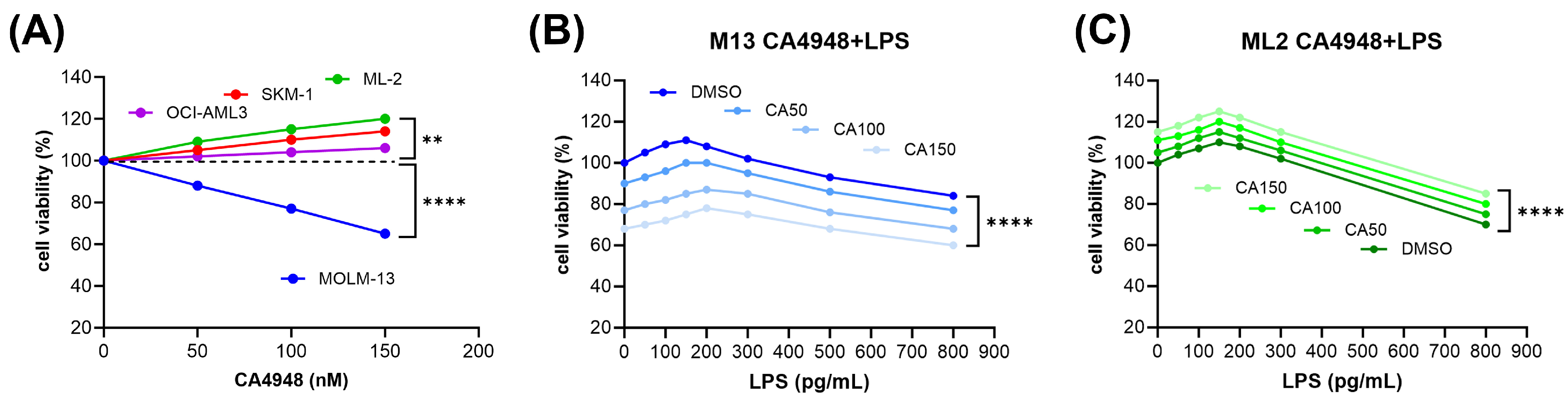
3.1. Inverse Response of AML Cell Lines to Emavusertib (CA4948) Monotherapy

To determine the susceptibility to emavusertib (CA4948), a variety of AML cell lines were subjected to in vitro cytotoxicity assays. AML cells represented the major morphologic and molecular subtypes, including FLT3-ITD and FLT3 wild type, NPM1 mutant and wild type, as well as TP53 mutant and wild type AML cell lines. AML cells were treated for 20 h in dose escalation experiments before the cell viability assessment.
A dose-dependent decrease in cell viability in the presence of CA4948 was detected in the FLT3-ITD-positive MOLM-13 cells, with IC50 at 150 nM. In contrast, there was a dose-dependent increase in cell viability in the presence of CA4948 in the FLT3 wild-type cell lines ML-2, OCI-AML3, and SKM-1 (Figure 1A).

3.2. Susceptibility of AML Cells to Emavusertib (CA4948) Treatment under Exposure to Lipopolysaccharide

Lipopolysaccharide (LPS) is an endotoxin derived from the outer membrane of Gram-negative bacteria, a pathogen-associated molecular pattern (PAMP) recognized by Toll-like receptors (TLRs) and other pattern recognition receptors (PRRs) [27]. In monocytes, LPS binding promotes the secretion of pro-inflammatory cytokines, including IL-1β and IL-6 [28]. In AML cells, LPS binding to TLR4 promotes cell proliferation, inhibits apoptosis, and increases resistance to chemotherapy [12]. Plasma LPS concentrations have been reported to be in the range of 10 to 50 pg/mL in healthy humans, 50 to 250 pg/mL in the peripheral blood of AML patients [23], and 500 pg/mL in patients with sepsis [24]. AML cell lines were exposed to LPS within the physiological concentration ranges and subjected to in vitro cytotoxicity assays. Exposure to LPS in the range of 50 to 250 pg/mL induced cell viability of AML cells in a dose-dependent manner in both FLT3-ITD-positive MOLM-13 (Figure 1B) and FLT3 wild-type ML-2 cells (Figure 1C). Exposure to LPS over 500 pg/mL reduced the cell viability of AML cells in a dose-dependent manner. CA4948 treatment of AML cells exposed to LPS led to reduced cell viability in MOLM-13 cells (Figure 1B) and enhanced cell viability in ML-2 cells (Figure 1C). The combined effects appeared to be additive, not synergistic, in nature, indicating an absence of cooperative interactions on the molecular level. LPS is one of several ligands binding to the TLR4/MD2 receptor complex (Figure 2).

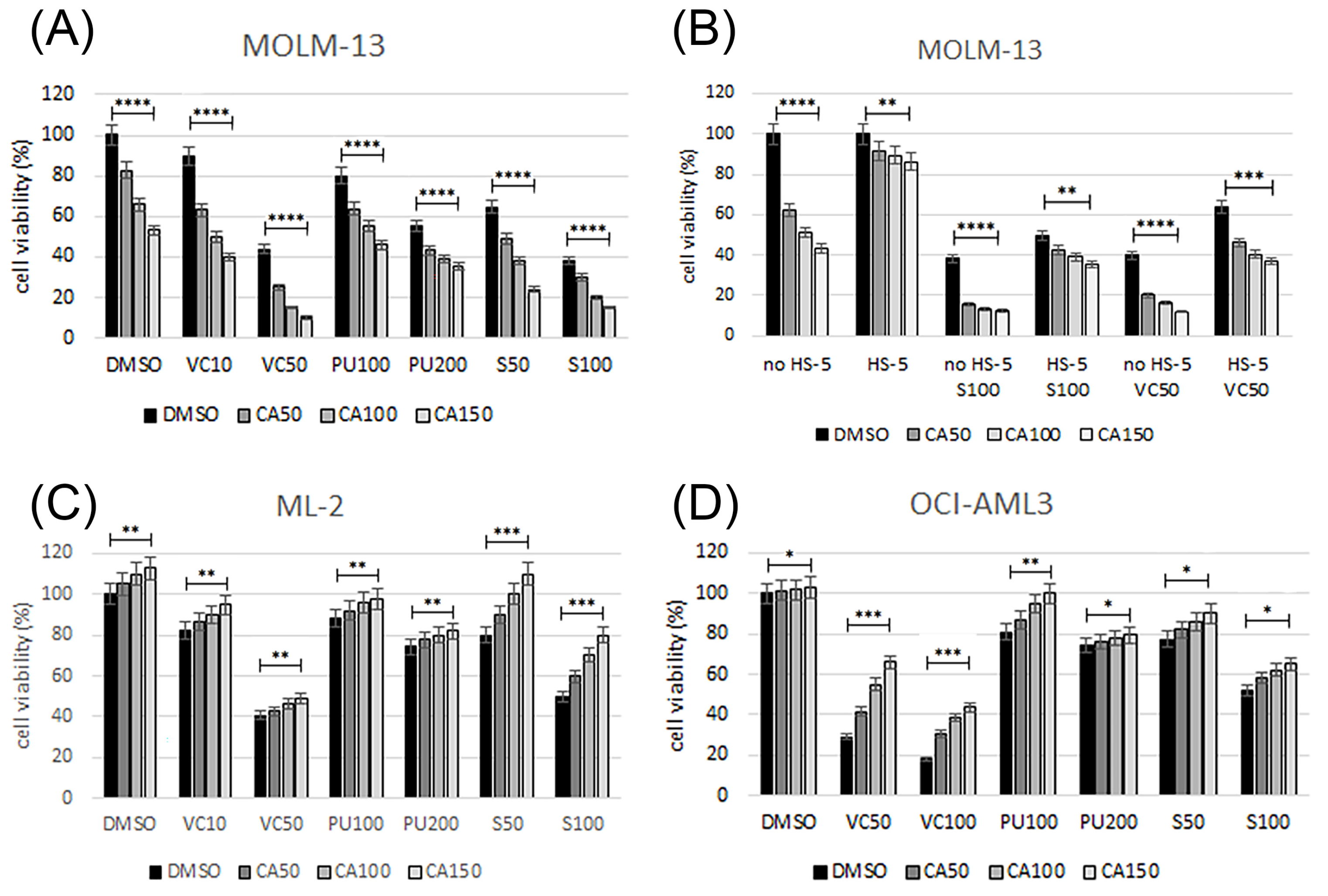
3.3. Emavusertib Combination Treatments in AML Cell Lines

In order to define effective treatment combinations, we focused on inhibitors expected to elicit synergistic effects in combination with the IRAK4/FLT3 inhibitor CA4948 based on previous studies with BCL2, MCL1, and HSP90 inhibitors (Figure 2). Cell viability was determined in AML cell lines treated with increasing dosages of single compounds, as well as in combination treatments using the IRAK4 inhibitor CA4948 and a variety of targeted therapies, including the BCL2 inhibitor venetoclax, the MCL1 inhibitor S63845, and the HSP90 inhibitor PU-H71. FLT3-ITD-positive MOLM-13 cells were susceptible to CA4948, S63845, venetoclax, and PU-H71, with enhanced effects on cell viability in the combination treatments (Figure 3A). Combination index (CI) values were calculated according to Chou-Talalay [26]. In MOLM-13 cells, the combination indexes indicated synergistic effects (CI 0.5–0.7) for the combination CA4948 and S63845, or venetoclax and additive effects (CI 0.9–1.1) for the combination CA4948 and PU-H71. Cell viability was determined in MOLM-13 cells grown in the absence or presence of bone marrow stroma cells secreting granulocyte and macrophage colony-stimulating factors, as well as other cytokines, including IL-1 and stem cell factor SCF (Figure 2). In the presence of bone marrow stroma, MOLM-13 cells were protected from the cytotoxic effects of CA4948, S63845, and venetoclax, and combination treatments were less effective (Figure 3B). FLT3 wild-type OCI-AML3 and ML-2 cells were susceptible to S63845, venetoclax, and PU-H71, but, with a dose-dependent increase in cell viability in the presence of CA4948 in the combination treatments (Figure 3C,D).

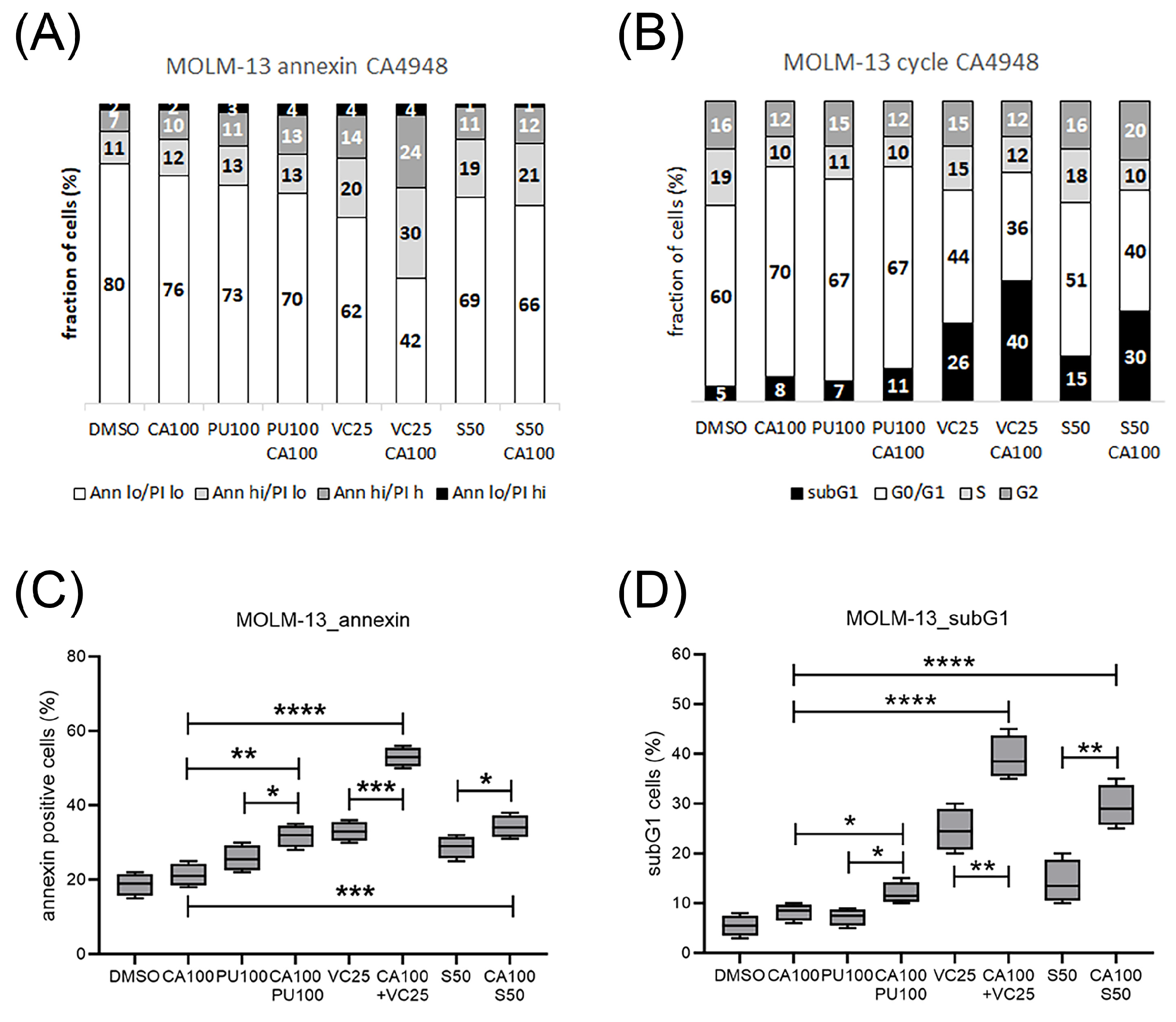
3.4. Emavusertib Treatment Induced Cell Cycle Arrest, Apoptosis and Cell Death

The effects of treatment with CA4948 and S63845, venetoclax, or PU-H71, alone and in combination, on the induction of apoptosis, cell cycle arrest, and cell death were determined in MOLM-13 cells by cytometric analysis. Apoptosis was induced in MOLM-13 cells treated with CA4948, PU-H71, S63845, or venetoclax, with enhanced effects in the combination treatments (Figure 4A,C). G1 cell cycle arrest was induced in CA4948- and PU-H71-treated MOLM-13 cells (Figure 4B). Cell death was induced in MOLM-13 cells treated with CA4948, PU-H71, S63845, or venetoclax, with enhanced effects in the combination treatments (Figure 4B,D).

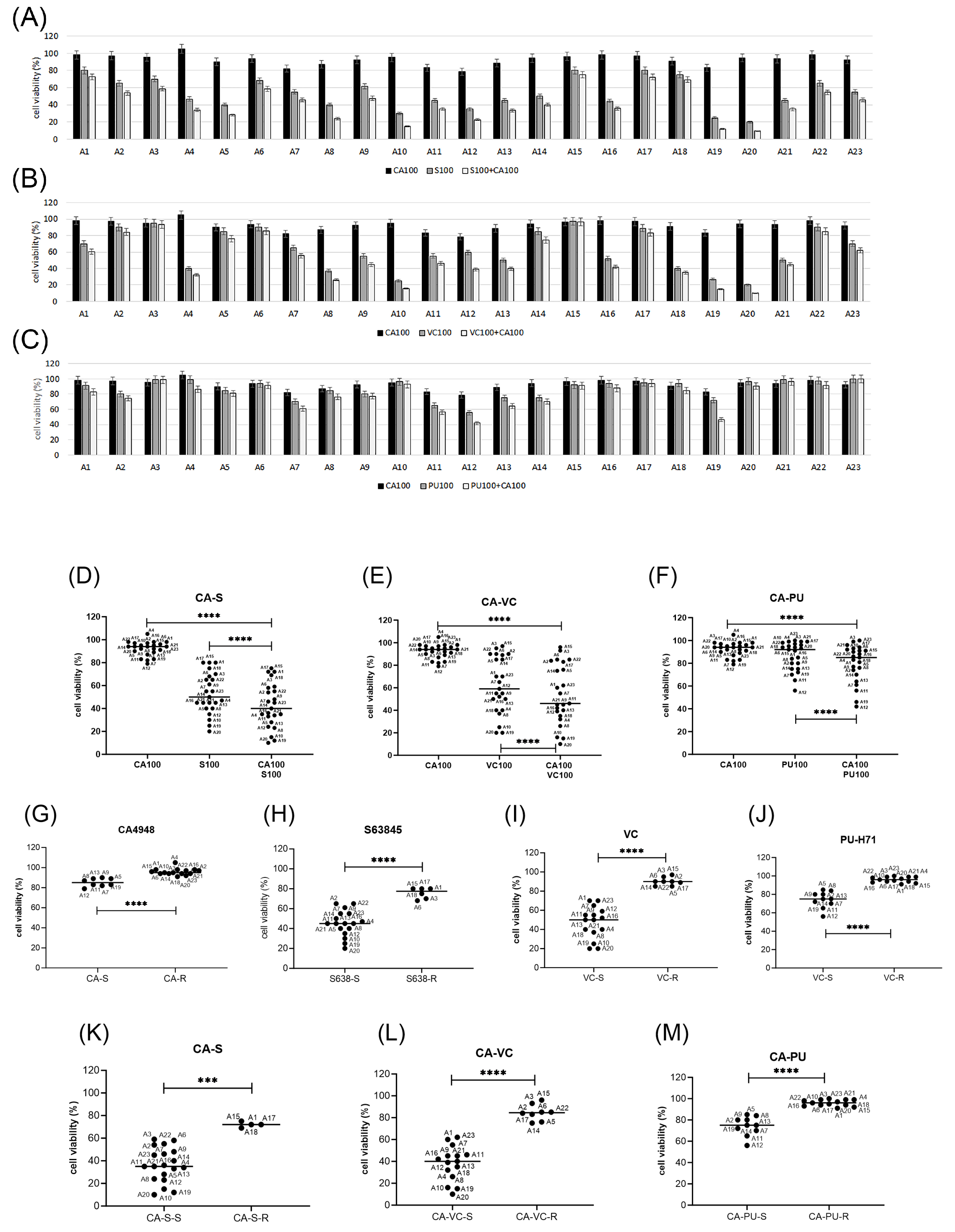
3.5. Emavusertib Combination Treatments in Primary AML Cells In Vitro

After initial studies in AML cell lines, the treatment combinations of emavusertib (CA4948) with S63845, venetoclax, or PU-H71 were applied to patient-derived mononuclear cells isolated from peripheral blood (PB) or bone marrow (BM). A total of twenty-three primary AML cells were subjected to single compound and combination treatments, with eleven FLT3 mutated and twelve FLT3 wild-type primary AML. In vitro cytotoxic effects were mild to moderate in CA4948 and PU-H71 monotherapy and moderate to strong in S63845 or venetoclax monotherapy, with enhanced effects in the combination treatments (Figure 5A–C).
CA4948 monotherapy was effective in 35% of primary AML (Figure 5D). S63845 monotherapy was effective in all tested AML samples, with enhanced toxicity in 75% of primary AML (Figure 5E). Venetoclax monotherapy was effective in 65% of primary AML (Figure 5F), and PU-H71 monotherapy in 44% of primary AML (Figure 5G). The patient samples were sorted into response groups, susceptible (S) and resistant (R), treated with 100 nM CA4948 alone (Figure 6D) and in combination with 100 nM S63845 (Figure 5E), 100 nM venetoclax (Figure 6F), or 100 nM PU-H71 (Figure 6G). The cut-off values of cell viability were set arbitrarily, in CA4948 and PU-H71 monotherapy at 90%, venetoclax at 80%, and S63845 at 70%. In combination treatments, they were set to 85% (CA-PU), 70% (CA-VC), or 60% (CA-S). CA4948 and S63845 combination treatments were effective in all tested AML samples, with enhanced toxicity in 83% of primary AML (Figure 5H). CA4948 and venetoclax combination treatments were effective in 65% of primary AML (Figure 5I), and CA4948 and PU-H71 in 44% of primary AML (Figure 5J).

3.6. Biomarkers of Response to Emavusertib and Combination Treatments in Primary AML Cells

Potential response markers were deduced from the correlation analysis of cell viabilities grouped according to diagnostic parameters, including gene mutation status; peripheral blood and bone marrow blast cell percentage; and expression levels of CD markers, including CD11b, CD34, CD64, and CD117 (c-KIT). The clinical characteristics of the hematological samples are listed in Table 1. The presence of a FLT3 gene mutation was the major indicator of a response to CA4948, S63845, and PU-H71 treatment, with elevated susceptibility of AML cells with the FLT3 gene mutation at an allelic ratio >0.5 (Figure 6A).
The presence of a NPM1 gene mutation was positively associated with the response to CA4948 (Figure 6B). NPM1 gene mutations were present in most of the FLT3-mutated AML (8/11) and a few of the FLT3 wild-type primary AML (3/12). The one FLT3 wild-type and NPM1-mutated sample with substantial response to CA4948 and S63845 (AML5) also carried a specific PTPN11 gene mutation (E69K). The blast cell percentage was positively associated with response to CA4948, S63845, and venetoclax, along with elevated susceptibility of primary AML with a blast cell count >80% (Figure 6C). Bone marrow (BM) infiltration was positively associated with the venetoclax response, with elevated susceptibility of primary AML with BM infiltration of >80% (Figure 6D). CD11b was negatively associated with the venetoclax response, with elevated susceptibility of primary AML with CD11b < 20% (Figure 6E). CD34 was negatively associated with the response to CA4948, S63845, and PU-H71, with elevated susceptibility of primary AML samples with CD34 < 30% (Figure 6F). CD64 was negatively associated with the venetoclax response, with elevated susceptibility of primary AML samples with CD64 < 10% (Figure 6G). CD117 was positively associated with the venetoclax response, with elevated susceptibility of primary AML samples with CD117 > 80% (Figure 6H). Biomarkers of the responses to individual treatments and statistical significances are listed in Figure 6I. Two primary AMLs carried TP53 gene mutations, one in a FLT3 wild-type AML (A1) and the other in a FLT3-TKD AML (A2). Both TP53-mutant AMLs were resistant to CA4948 monotherapy, but susceptible to S63845 and venetoclax, with enhanced effects in the combination treatments. Mutations in spliceosome genes U2AF1 and SF3B1 have been linked to the expression of oncogenic IRAK4 isoforms in myeloid malignancies [29,30]. U2AF1 gene mutations were present in one FLT3 wild-type AML (A3) and in one FLT3-ITD AML (A7), while a SF3B1 gene mutation was present in one FLT3 wild-type AML (A22). Of the three primary AMLs with U2AF1 mutations, only the FLT3-ITD-positive AML was susceptible to CA4948. Mutations in the spliceosome gene SRSF2 were present in five primary AMLs (A2, A10, A15, A18, A21), none of them susceptible to CA4948. The majority of primary AML (17/23) carried mutations in epigenetic modifiers (TET2, DNMT3A, BCOR, ASXL1, IDH1, IDH2). The majority of primary AMLs with wild-type status of epigenetic modifiers and TP53 (4/5), and a minority of AMLs with mutations in epigenetic modifiers (4/17), were susceptible to CA4948.
In summary, emavusertib (CA4948) monotherapy appeared to be most effective in primary AML with FLT3 and NPM1 gene mutations and the absence of gene mutations in TP53 and epigenetic modifiers, as well as moderate CD34 positivity. Combination treatments with CA4948 and venetoclax were effective in primary AMLs with elevated blast counts and bone marrow infiltration, as well as high CD117 expression and low levels of CD11b and CD64. Combination treatments with CA4948 and PU-H71 or S63845 were effective in FLT3-mutated AML with moderate CD34 positivity (<30%). A possible scenario of intracellular signaling in AML cells affected by targeted inhibition of IRAK4, FLT3, HSP90, BCL2, and MCL1 is presented in Figure 2.

4. Discussion

In order to determine the susceptibility of AML cell lines to the IRAK4 inhibitor emavusertib (CA4948), an initial dose escalation screening was performed in AML cell lines. Cytotoxic effects were present in the FLT3-ITD-positive cell line MOLM-13, with a dose-dependent decrease in cell viability. In contrast, there was a complete absence of cytotoxic effects in three FLT3 wild-type cell lines, OCI-AML3, ML-2, and SKM-1, treated with 50–250 nM CA4948 with a dose-dependent increase in cell viability. A possible molecular mechanism underlying the accelerated cell growth may be CA4948-induced IRAK1 activation in FLT3 wild-type AML cells, as IRAK4 inhibition can lead to functional compensation of IRAK1 in THP1 cells [31] indicating a requirement for cotargeting IRAK1 and IRAK4 in the treatment of FLT3 wild-type AML.
In AML cells, LPS binding to TLR4 may promote cell proliferation, inhibit apoptosis, and increase resistance to chemotherapy [10]. Indeed, cell viability was induced in MOLM-13 and ML-2 AML cells exposed to bacterial LPS, a TLR4 agonist. Plasma LPS concentrations have been reported to be in the range of 10 to 50 pg/mL in healthy humans, 50 to 250 pg/mL in the peripheral blood of AML patients [23], and 500 pg/mL in patients with sepsis [24]. In AML patients, the intestinal barrier function may be compromised, leading to an increased translocation of bacteria and endotoxins into the blood. LPS may exacerbate leukemia progression in vitro and in vivo [32]. While cell viability was induced in AML cells exposed to intermediate LPS concentrations (50–250 pg/mL), cytotoxic effects were elicited in AML cells under LPS exposure over 500 pg/mL, equivalent to physiological conditions of bacterial sepsis. Sepsis has profound impacts on myeloid cell response. In the bone marrow, a progressive shift toward the release of immature myeloid cells (including myeloid-derived suppressor cells), at the expense of mature neutrophils, takes place [33,34]. Emavusertib (CA4948) treatment of AML cells exposed to LPS lead to reduced cell viability in FLT3-ITD-positive MOLM-13 cells and enhanced cell viability in FLT3 wild-type ML-2 cells, indicating that a CA4948-induced compensatory IRAK1 activation occurs in FLT3 wild-type cells.
Emavusertib as a monotherapy and in combination with azacitidine or venetoclax is currently being evaluated in a phase 1/2 clinical trial in R/R AML (NCT04278768) [19]. Combining emavusertib with other anticancer therapies may be a more effective treatment strategy. In order to define effective treatment combinations, we focused on inhibitors expected to elicit synergistic cytotoxic effects in combination with CA4948 based on previous studies with BCL2, MCL1, and HSP90 inhibitors [7,8,35]. FLT3-ITD-positive MOLM-13 cells were susceptible to CA4948, S63845, venetoclax, or PU-H71, with enhanced effects on cell viability in the combination treatments. After initial studies in AML cell lines, the treatment combinations of CA4948 with S63845, venetoclax, or PU-H71 were applied to primary AML cells. CA4948 and S63845 combination treatments were effective in all tested AML samples, with enhanced toxicity in 83% of primary AML; CA4948 and venetoclax combination treatments in 65% of primary AML; and CA4948 and PU-H71 combination treatments in 44% of primary AML. The in vitro concentrations of the tested inhibitors were in the range of physiologically relevant concentrations for all compounds. Emavusertib plasma concentrations of 1–5 μg/mL (2–10 μM) were detected in patients receiving 50 to 400 mg BID [18]. Venetoclax plasma concentrations of 1–3 μg/mL (1.2–3.5 μM) were observed in patients receiving 400 mg/day [36]. PU-H71 plasma concentrations of 1–5 μM were measured in patients infused with 100–400 mg/m2 [37]. S63845 displayed impressive potency at low nanomolar concentrations in preclinical in vitro and in vivo models of hematological malignancies, including MM, AML, CML, and c-MYC-driven Burkitt lymphoma [38].
Potential response markers were deduced from the correlation analysis of cell viabilities and grouped according to diagnostic parameters, including gene mutation status; peripheral blood and bone marrow blast cell percentage; and presence of CD markers, including CD11b, CD34, CD64, and CD117 (c-KIT). The FLT3 gene mutation was the major biomarker of response to CA4948, S63845, and PU-H71 treatment, with elevated susceptibility of AML cells with the FLT3 gene mutation at an allelic ratio of >0.5. Both FLT3 and NPM1 genes were mutated in the susceptible primary AML samples. The FLT3 gene was overexpressed in up to 93% and mutated in over 30% of primary AML [1]. FLT3-ITD and FLT3-TKD are constitutively active growth factor receptors linked to the upregulation of BCL2 and MCL-1 expression in myeloid cells. BH3-mimetics are small compounds that antagonize anti-apoptotic BCL2 family proteins, and are specifically effective in apoptosis induction in FLT3-mutated AML cells [39]. NPM1 gene mutations represent the most common genetic lesion in AML and cause aberrant localization of the mutant NPM1c protein [40]. NPM1c is exported to the cytoplasm, where it inhibits the tumor suppressor protein p53 by cytoplasmic retention. In a previous study, we found that the presence of the NPM1 gene mutation was negatively associated with the susceptibility of FLT3-ITD AML cells to the FLT3 inhibitor midostaurin and the MDM2 inhibitor NVP-HDM201 [41]. In this preclinical study, however, the presence of the NPM1 gene mutation was positively associated with the susceptibility of FLT3-ITD AML cells to the IRAK4 inhibitor CA4948. The presence of NPM1 gene mutations has previously been correlated with the general response to TLR stimulation in AML cells [42]. Moreover, TLR4 may be a receptor for extracellular NPM1 protein [43]. Extracellular NPM1 protein acts as a potent inflammatory stimulator by promoting cytokine production (e.g., tumor necrosis factor-α (TNF-α)), which suggests that NPM1 acts as a damage-associated molecular pattern (DAMP). The wild-type NPM1 and mutant NPM1c proteins may differ in their abilities to activate TLR4 receptor signaling.
The surface marker CD34 was negatively associated with the response to CA4948, S63845, and PU-H71 treatment, with elevated susceptibility of primary AML samples with CD34 <30%. CD34 is a hematopoietic stem cell marker and may mediate the attachment to the bone marrow extracellular matrix and stromal cells [44]. Leukemic stem cells are a subpopulation of leukemia cells characterized by the CD34 + CD38- phenotype, and are considered to be resistant to standard treatment [45]. To address the adverse risk of CD34+ AMLs, the combination of the BMI1 inhibitor PTC596 with the MCL1 inhibitor S63845 may be a more effective treatment option [7]. We observed that three markers were associated with the venetoclax response: CD11b, CD64, and CD117. The markers CD11b and CD64 were negatively associated with the venetoclax response. Both CD11b (Integrin alpha M) and CD64 (FcγRI) are monocytic differentiation markers. CD11b+ CD64+ cells may exhibit resistance to venetoclax-based therapies due to a loss of BCL2 expression [46]. CD117 was positively associated with the venetoclax response. CD-117 is a tyrosine kinase receptor expressed on the majority of AML cells, where it induces signaling pathways similar to FLT3-ITD that are linked to the upregulation of BCL2 expression.
Mutations in the spliceosome genes U2AF1 and SF3B1 have been linked to the expression of oncogenic IRAK4 isoforms in myeloid malignancies [18,29,30], and may impose sensitivity to IRAK4 inhibition [31]. Mutations in epigenetic modifiers may also be linked to treatment responses to targeted therapies, e.g., enhanced sensitivity to venetoclax and azacitidine [47,48], venetoclax and bimiralisib [35], or AC-4-130 and S63845 [49]. In our preclinical study, the majority of primary AMLs with wild-type status of epigenetic modifiers and a minority of AMLs with mutations in epigenetic modifiers were susceptible to CA4948. In order to investigate a potential association of the emavusertib treatment response with spliceosome or epigenetic modifiers, preclinical studies in larger cohorts are required.

5. Conclusions

In this preclinical study, we assessed the IRAK4 inhibitor emavusertib (CA4948) in combination with the MCL1 inhibitor S63845, the BCL2 inhibitor venetoclax, and the HSP90 inhibitor PU-H71 in the treatment of acute myeloid leukemia and investigated the associated biomarkers of response. Our data suggest that treatment combinations with CA4948 and BH3-mimetics can effectively induce cell cycle arrest, apoptosis, and cell death in FLT3-mutated AML cells. In primary AMLs, the response to emavusertib was associated with the presence of the FLT3 and NPM1 gene mutations, blast cell percentage, and CD34 positivity. Biomarkers of the response to CA4948 combination treatment with S63845 included the blast cell percentage and CD34. Biomarkers of the response to CA4948 combination treatment with venetoclax included FLT3 gene mutations as well as expression levels of CD11b, CD64, and CD117. The combination of emavusertib and BH3-mimetics may be effective in the treatment of AML with differential target specificities for MCL1 and BCL2 inhibitors. The combination of emavusertib and venetoclax may be an effective treatment in FLT3-mutated AML, where CD11b < 20%, CD64 < 10%, and CD117 > 80%. The combination of emavusertib and S63845 may be more effective in FLT3-mutated AMLs with blast cell counts >80% and CD34 < 30%.

Author Contributions

Conceptualization, K.S. and H.M.; investigation, K.S.; writing—original draft preparation, K.S.; writing—review and editing, K.S., H.M., U.B. and T.P.; visualization, K.S.; resources: H.M., U.B. and T.P. All authors have read and agreed to the published version of the manuscript.

Funding

This research received no external funding.

Institutional Review Board Statement

The study was conducted according to the guidelines of the Declaration of Helsinki and approved by the Ethics Committee of the Canton of Bern, Switzerland (decision number: 2022-00203 and date of approval: 4 May 2022).

Informed Consent Statement

Informed consent was obtained from all subjects involved in the study according to the Declaration of Helsinki. The general consent form is available.

Data Availability Statement

Data available upon request due to restrictions, privacy, and ethics.

Acknowledgments

The authors wish to thank the data management, the apheresis, the flow cytometry, and the stem cell laboratory teams of the ASCT program at the University hospital of Bern and its associated partner hospitals and collaborators for the documentation of data relevant to this study.

Conflicts of Interest

The authors declare no conflicts of interest.

References

  1. Kennedy, V.E.; Smith, C.C. FLT3 Mutations in Acute Myeloid Leukemia: Key Concepts and Emerging Controversies. Front. Oncol. 2020, 10, 612880. [Google Scholar] [CrossRef]
  2. Zhao, J.C.; Agarwal, S.; Ahmad, H.; Amin, K.; Bewersdorf, J.P.; Zeidan, A.M. A Review of FLT3 Inhibitors in Acute Myeloid Leukemia. Blood Rev. 2022, 52, 100905. [Google Scholar] [CrossRef]
  3. Larrosa-Garcia, M.; Baer, M.R. FLT3 Inhibitors in Acute Myeloid Leukemia: Current Status and Future Directions. Mol. Cancer Ther. 2017, 16, 991–1001. [Google Scholar] [CrossRef]
  4. Carter, B.Z.; Mak, P.Y.; Tao, W.; Warmoes, M.; Lorenzi, P.L.; Mak, D.; Ruvolo, V.; Tan, L.; Cidado, J.; Drew, L.; et al. Targeting MCL-1 Dysregulates Cell Metabolism and Leukemia-Stroma Interactions and Resensitizes Acute Myeloid Leukemia to BCL-2 Inhibition. Haematologica 2022, 107, 58–76. [Google Scholar] [CrossRef]
  5. DiNardo, C.D.; Pratz, K.; Pullarkat, V.; Jonas, B.A.; Arellano, M.; Becker, P.S.; Frankfurt, O.; Konopleva, M.; Wei, A.H.; Kantarjian, H.M.; et al. Venetoclax Combined with Decitabine or Azacitidine in Treatment-Naive, Elderly Patients with Acute Myeloid Leukemia. Blood 2019, 133, 7–17. [Google Scholar] [CrossRef]
  6. Seipel, K.; Schmitter, K.; Bacher, U.; Pabst, T. Rationale for a Combination Therapy Consisting of MCL1- and MEK-Inhibitors in Acute Myeloid Leukemia. Cancers 2019, 11, 1779. [Google Scholar] [CrossRef]
  7. Seipel, K.; Kopp, B.; Bacher, U.; Pabst, T. BMI1-Inhibitor PTC596 in Combination with MCL1 Inhibitor S63845 or MEK Inhibitor Trametinib in the Treatment of Acute Leukemia. Cancers 2021, 13, 581. [Google Scholar] [CrossRef]
  8. Seipel, K.; Kohler, S.; Bacher, U.; Pabst, T. HSP90 Inhibitor PU-H71 in Combination with BH3-Mimetics in the Treatment of Acute Myeloid Leukemia. Curr. Issues Mol. Biol. 2023, 45, 7011–7026. [Google Scholar] [CrossRef]
  9. Bennett, J.; Starczynowski, D.T. IRAK1 and IRAK4 as Emerging Therapeutic Targets in Hematologic Malignancies. Curr. Opin. Hematol. 2022, 29, 8–19. [Google Scholar] [CrossRef]
  10. Pereira, M.; Gazzinelli, R.T. Regulation of Innate Immune Signaling by IRAK Proteins. Front. Immunol. 2023, 14, 1133354. [Google Scholar] [CrossRef]
  11. Suzuki, N.; Suzuki, S.; Duncan, G.S.; Millar, D.G.; Wada, T.; Mirtsos, C.; Takada, H.; Wakeham, A.; Itie, A.; Li, S.; et al. Severe Impairment of Interleukin-1 and Toll-like Receptor Signalling in Mice Lacking IRAK-4. Nature 2002, 416, 750–756. [Google Scholar] [CrossRef] [PubMed]
  12. Zhong, C.; Wang, R.; Hua, M.; Zhang, C.; Han, F.; Xu, M.; Yang, X.; Li, G.; Hu, X.; Sun, T.; et al. NLRP3 Inflammasome Promotes the Progression of Acute Myeloid Leukemia via IL-1β Pathway. Front. Immunol. 2021, 12, 661939. [Google Scholar] [CrossRef] [PubMed]
  13. Rhyasen, G.W.; Starczynowski, D.T. IRAK Signalling in Cancer. Br. J. Cancer 2015, 112, 232–237. [Google Scholar] [CrossRef] [PubMed]
  14. Wiese, M.D.; Manning-Bennett, A.T.; Abuhelwa, A.Y. Investigational IRAK-4 Inhibitors for the Treatment of Rheumatoid Arthritis. Expert. Opin. Investig. Drugs 2020, 29, 475–482. [Google Scholar] [CrossRef]
  15. Melgar, K.; Walker, M.M.; Jones, L.M.; Bolanos, L.C.; Hueneman, K.; Wunderlich, M.; Jiang, J.-K.; Wilson, K.M.; Zhang, X.; Sutter, P.; et al. Overcoming Adaptive Therapy Resistance in AML by Targeting Immune Response Pathways. Sci. Transl. Med. 2019, 11, eaaw8828. [Google Scholar] [CrossRef]
  16. Hosseini, M.M.; Kurtz, S.E.; Abdelhamed, S.; Mahmood, S.; Davare, M.A.; Kaempf, A.; Elferich, J.; McDermott, J.E.; Liu, T.; Payne, S.H.; et al. Inhibition of Interleukin-1 Receptor-Associated Kinase-1 Is a Therapeutic Strategy for Acute Myeloid Leukemia Subtypes. Leukemia 2018, 32, 2374–2387. [Google Scholar] [CrossRef] [PubMed]
  17. Jeon, J.Y.; Zhao, Q.; Buelow, D.R.; Phelps, M.; Walker, A.R.; Mims, A.S.; Vasu, S.; Behbehani, G.; Blachly, J.; Blum, W.; et al. Preclinical Activity and a Pilot Phase I Study of Pacritinib, an Oral JAK2/FLT3 Inhibitor, and Chemotherapy in FLT3-ITD-Positive AML. Investig. New Drugs 2020, 38, 340–349. [Google Scholar] [CrossRef]
  18. Parrondo, R.D.; Iqbal, M.; Von Roemeling, R.; Von Roemeling, C.; Tun, H.W. IRAK-4 Inhibition: Emavusertib for the Treatment of Lymphoid and Myeloid Malignancies. Front. Immunol. 2023, 14, 1239082. [Google Scholar] [CrossRef]
  19. Garcia-Manero, G.; Winer, E.S.; DeAngelo, D.J.; Tarantolo, S.; Sallman, D.A.; Dugan, J.; Groepper, S.; Giagounidis, A.; Götze, K.; Metzeler, K.H.; et al. S129: Takeaim Leukemia—A Phase 1/2A Study of the IRAK4 Inhibitor Emavusertib (CA-4948) AS Monotherapy or in Combination with Azacitidine or Venetoclax in Relapsed/Refractory AML or MDS. HemaSphere 2022, 6, 30. [Google Scholar] [CrossRef]
  20. Garcia-Manero, G.; Platzbecker, U.; Lim, K.-H.; Nowakowski, G.; Abdel-Wahab, O.; Kantarjian, H.; Verma, A.; Starczynowski, D.T. Research and Clinical Updates on IRAK4 and Its Roles in Inflammation and Malignancy: Themes and Highlights from the 1st Symposium on IRAK4 in Cancer. Front. Hematol. 2024, 3, 1339870. [Google Scholar] [CrossRef]
  21. Albakova, Z.; Mangasarova, Y.; Albakov, A.; Gorenkova, L. HSP70 and HSP90 in Cancer: Cytosolic, Endoplasmic Reticulum and Mitochondrial Chaperones of Tumorigenesis. Front. Oncol. 2022, 12, 829520. [Google Scholar] [CrossRef] [PubMed]
  22. Cabaud-Gibouin, V.; Durand, M.; Quéré, R.; Girodon, F.; Garrido, C.; Jego, G. Heat-Shock Proteins in Leukemia and Lymphoma: Multitargets for Innovative Therapeutic Approaches. Cancers 2023, 15, 984. [Google Scholar] [CrossRef]
  23. Yu, J.; Zhang, C.; Song, C. Pan- and Isoform-Specific Inhibition of Hsp90: Design Strategy and Recent Advances. Eur. J. Med. Chem. 2022, 238, 114516. [Google Scholar] [CrossRef]
  24. Ren, X.; Li, T.; Zhang, W.; Yang, X. Targeting Heat-Shock Protein 90 in Cancer: An Update on Combination Therapy. Cells 2022, 11, 2556. [Google Scholar] [CrossRef] [PubMed]
  25. Shumilov, E.; Flach, J.; Kohlmann, A.; Banz, Y.; Bonadies, N.; Fiedler, M.; Pabst, T.; Bacher, U. Current Status and Trends in the Diagnostics of AML and MDS. Blood Rev. 2018, 32, 508–519. [Google Scholar] [CrossRef]
  26. Chou, T.-C. Drug Combination Studies and Their Synergy Quantification Using the Chou-Talalay Method. Cancer Res. 2010, 70, 440–446. [Google Scholar] [CrossRef] [PubMed]
  27. Tang, D.; Kang, R.; Coyne, C.B.; Zeh, H.J.; Lotze, M.T. PAMPs and DAMPs: Signal 0s That Spur Autophagy and Immunity. Immunol. Rev. 2012, 249, 158–175. [Google Scholar] [CrossRef]
  28. Chaiwut, R.; Kasinrerk, W. Very Low Concentration of Lipopolysaccharide Can Induce the Production of Various Cytokines and Chemokines in Human Primary Monocytes. BMC Res. Notes 2022, 15, 42. [Google Scholar] [CrossRef]
  29. Smith, M.A.; Choudhary, G.S.; Pellagatti, A.; Choi, K.; Bolanos, L.C.; Bhagat, T.D.; Gordon-Mitchell, S.; Von Ahrens, D.; Pradhan, K.; Steeples, V.; et al. U2AF1 Mutations Induce Oncogenic IRAK4 Isoforms and Activate Innate Immune Pathways in Myeloid Malignancies. Nat. Cell Biol. 2019, 21, 640–650. [Google Scholar] [CrossRef]
  30. Choudhary, G.S.; Pellagatti, A.; Agianian, B.; Smith, M.A.; Bhagat, T.D.; Gordon-Mitchell, S.; Sahu, S.; Pandey, S.; Shah, N.; Aluri, S.; et al. Activation of Targetable Inflammatory Immune Signaling Is Seen in Myelodysplastic Syndromes with SF3B1 Mutations. Elife 2022, 11, e78136. [Google Scholar] [CrossRef]
  31. Bennett, J.; Ishikawa, C.; Agarwal, P.; Yeung, J.; Sampson, A.; Uible, E.; Vick, E.; Bolanos, L.C.; Hueneman, K.; Wunderlich, M.; et al. Paralog-Specific Signaling by IRAK1/4 Maintains MyD88-Independent Functions in MDS/AML. Blood 2023, 142, 989–1007. [Google Scholar] [CrossRef]
  32. Wang, R.; Yang, X.; Liu, J.; Zhong, F.; Zhang, C.; Chen, Y.; Sun, T.; Ji, C.; Ma, D. Gut Microbiota Regulates Acute Myeloid Leukaemia via Alteration of Intestinal Barrier Function Mediated by Butyrate. Nat. Commun. 2022, 13, 2522. [Google Scholar] [CrossRef]
  33. Venet, F.; Demaret, J.; Gossez, M.; Monneret, G. Myeloid Cells in Sepsis-Acquired Immunodeficiency. Ann. N. Y. Acad. Sci. 2021, 1499, 3–17. [Google Scholar] [CrossRef]
  34. Schrijver, I.T.; Théroude, C.; Roger, T. Myeloid-Derived Suppressor Cells in Sepsis. Front. Immunol. 2019, 10, 447308. [Google Scholar] [CrossRef]
  35. Seipel, K.; Brügger, Y.; Mandhair, H.; Bacher, U.; Pabst, T. Rationale for Combining the BCL2 Inhibitor Venetoclax with the PI3K Inhibitor Bimiralisib in the Treatment of IDH2- and FLT3-Mutated Acute Myeloid Leukemia. Int. J. Mol. Sci. 2022, 23, 12587. [Google Scholar] [CrossRef]
  36. Roberts, A.W.; Davids, M.S.; Pagel, J.M.; Kahl, B.S.; Puvvada, S.D.; Gerecitano, J.F.; Kipps, T.J.; Anderson, M.A.; Brown, J.R.; Gressick, L.; et al. Targeting BCL2 with Venetoclax in Relapsed Chronic Lymphocytic Leukemia. N. Engl. J. Med. 2016, 374, 311–322. [Google Scholar] [CrossRef]
  37. Speranza, G.; Anderson, L.; Chen, A.P.; Do, K.; Eugeni, M.; Weil, M.; Rubinstein, L.; Majerova, E.; Collins, J.; Horneffer, Y.; et al. First-in-Human Study of the Epichaperome Inhibitor PU-H71: Clinical Results and Metabolic Profile. Investig. New Drugs 2018, 36, 230–239. [Google Scholar] [CrossRef]
  38. Kotschy, A.; Szlavik, Z.; Murray, J.; Davidson, J.; Maragno, A.L.; Le Toumelin-Braizat, G.; Chanrion, M.; Kelly, G.L.; Gong, J.-N.; Moujalled, D.M.; et al. The MCL1 Inhibitor S63845 Is Tolerable and Effective in Diverse Cancer Models. Nature 2016, 538, 477–482. [Google Scholar] [CrossRef]
  39. Parry, N.; Wheadon, H.; Copland, M. The Application of BH3 Mimetics in Myeloid Leukemias. Cell Death Dis. 2021, 12, 222. [Google Scholar] [CrossRef]
  40. Falini, B.; Brunetti, L.; Sportoletti, P.; Martelli, M.P. NPM1-Mutated Acute Myeloid Leukemia: From Bench to Bedside. Blood 2020, 136, 1707–1721. [Google Scholar] [CrossRef]
  41. Seipel, K.; Marques, M.A.T.; Sidler, C.; Mueller, B.U.; Pabst, T. MDM2- and FLT3-Inhibitors in the Treatment of FLT3-ITD Acute Myeloid Leukemia, Specificity and Efficacy of NVP-HDM201 and Midostaurin. Haematologica 2018, 103, 1862–1872. [Google Scholar] [CrossRef]
  42. Brenner, A.K.; Bruserud, Ø. Functional Toll-Like Receptors (TLRs) Are Expressed by a Majority of Primary Human Acute Myeloid Leukemia Cells and Inducibility of the TLR Signaling Pathway Is Associated with a More Favorable Phenotype. Cancers 2019, 11, 973. [Google Scholar] [CrossRef]
  43. Nakatomi, K.; Ueno, H.; Ishikawa, Y.; Salim, R.C.; Mori, Y.; Kanemoto, I.; Tancharoen, S.; Kikuchi, K.; Miura, N.; Omori, T.; et al. TLR4/MD-2 Is a Receptor for Extracellular Nucleophosmin 1. Biomed. Rep. 2021, 14, 21. [Google Scholar] [CrossRef]
  44. Radu, P.; Zurzu, M.; Paic, V.; Bratucu, M.; Garofil, D.; Tigora, A.; Georgescu, V.; Prunoiu, V.; Pasnicu, C.; Popa, F.; et al. CD34—Structure, Functions and Relationship with Cancer Stem Cells. Medicina 2023, 59, 938. [Google Scholar] [CrossRef]
  45. Vergez, F.; Green, A.S.; Tamburini, J.; Sarry, J.-E.; Gaillard, B.; Cornillet-Lefebvre, P.; Pannetier, M.; Neyret, A.; Chapuis, N.; Ifrah, N.; et al. High Levels of CD34+CD38low/−CD123+ Blasts Are Predictive of an Adverse Outcome in Acute Myeloid Leukemia: A Groupe Ouest-Est Des Leucémies Aiguës et Maladies Du Sang (GOELAMS) Study. Haematologica 2011, 96, 1792–1798. [Google Scholar] [CrossRef]
  46. Pei, S.; Pollyea, D.A.; Gustafson, A.; Stevens, B.M.; Minhajuddin, M.; Fu, R.; Riemondy, K.A.; Gillen, A.E.; Sheridan, R.M.; Kim, J.; et al. Monocytic Subclones Confer Resistance to Venetoclax-Based Therapy in Acute Myeloid Leukemia Patients. Cancer Discov. 2020, 10, 536–551. [Google Scholar] [CrossRef]
  47. Rahmani, N.E.; Ramachandra, N.; Sahu, S.; Gitego, N.; Lopez, A.; Pradhan, K.; Bhagat, T.D.; Gordon-Mitchell, S.; Pena, B.R.; Kazemi, M.; et al. ASXL1 Mutations Are Associated with Distinct Epigenomic Alterations That Lead to Sensitivity to Venetoclax and Azacytidine. Blood Cancer J. 2021, 11, 157. [Google Scholar] [CrossRef]
  48. Feld, J.; Tremblay, D.; Dougherty, M.; Czaplinska, T.; Sanchez, G.; Brady, C.; Kremyanskaya, M.; Bar-Natan, M.; Keyzner, A.; Marcellino, B.K.; et al. Safety and Efficacy: Clinical Experience of Venetoclax in Combination With Hypomethylating Agents in Both Newly Diagnosed and Relapsed/Refractory Advanced Myeloid Malignancies. Hemasphere 2021, 5, e549. [Google Scholar] [CrossRef]
  49. Seipel, K.; Graber, C.; Flückiger, L.; Bacher, U.; Pabst, T. Rationale for a Combination Therapy with the STAT5 Inhibitor AC-4-130 and the MCL1 Inhibitor S63845 in the Treatment of FLT3-Mutated or TET2-Mutated Acute Myeloid Leukemia. Int. J. Mol. Sci. 2021, 22, 8092. [Google Scholar] [CrossRef]
Figure 1. In vitro dose response to emavusertib (CA4948) treatment in AML cell lines. AML cells were treated with the dual IRAK4 and FLT3 inhibitor CA4948 at the indicated dosages for 20 h (A). Susceptibility of AML cells to CA4948 under exposure to lipopolysaccharides (B,C). Cell viability was determined in AML cells after exposure to 50–250 pg/mL LPS and 50 to 150 nM CA4948 in FLT3-ITD positive MOLM-13 (B) and FLT3 wild-type ML-2 (C) cells. Cell viability data are average values of multiple repeat measurements per dosage. The standard deviation was 3–6%. Significance of differences denoted for p < 0.01 (**); and p < 0.0001 (****).
Cimb 46 00184 g001
Figure 2. Schematic presentation of the FLT3 and SCFR (CD117, c-KIT) as well as IL1R and TLR4 signaling pathways and downstream effects. FLT3-ITD and FLT3-TKD are constitutively active growth factor receptors. SCFR (c-KIT) is an inducible growth factor receptor activated by stem cell factor (SCF). Both tyrosine receptor kinases activate PI3K-AKT, RAS-MEK-ERK, and STAT5, leading to cell growth and proliferation via inhibition of the tumor suppressor TP53 and induction of the apoptosis regulators MCL1 and BCL2. IL1R and TLR4 are inducible receptors signaling via MyD88 and IRAK4. HSP90 protein can bind and stabilize client proteins, including AKT, BCL2, FLT3, JAK, MDM2, STAT5, SHP2, and BRAF. Hsp90 proteins are indicated in yellow, oncogenic protein functions in red, tumor suppressor functions in green ovals, and targeted inhibitors in blue rectangles. Sharp arrows and blunt arrows indicate target induction and inhibition, respectively.
Cimb 46 00184 g002
Figure 3. Susceptibility of AML cell lines to various treatment combinations. Cell viability was determined in AML cell lines MOLM-13, ML-2, and OCI-AML3 after 20 h of treatment with single compounds and in combination with 50–150 nM CA4948 (CA) and 10–100 nM venetoclax (VC100), 100–200 nM PU-H71 (PU), or 50–100 nM S63845 (S100). Susceptibility of MOLM-13 cells to various treatment combinations in the absence or presence of HS-5 bone marrow stroma (B). Cell viability data are average values of multiple repeat measurements per dosage. Synergistic effects of combination treatments are depicted in MOLM-13 (A). Oppositional effects in ML-2 (C) and OCI-AML3 (D) cells. Cell viability data are average values of multiple repeat measurements per dosage. Significance of differences denoted for p < 0.05 (*); p < 0.01 (**); p < 0.001 (***); and p < 0.0001 (****).
Cimb 46 00184 g003
Figure 4. Induction of cell cycle arrest, apoptosis, and cell death in AML cells treated with CA4948 in combination with PU-H71, S63845, or venetoclax. Cytometric analysis of MOLM-13 cells treated with 100 nM PU-H71 (PU), 50 nM S63845 (S), or 25 nM venetoclax (VC) and stained with Annexin-V and PI (A,C) or DAPI (B,D). According to Annexin V and PI staining intensity, cells were classified as vital (Ann lo, PI lo), early apoptotic (Ann hi, PI lo), late apoptotic (Ann hi, PI hi), or necrotic (Ann lo, PI hi). According to DAPI staining intensity, cells were classified as subG1 (<2 N), G0/G1 (2 N), S phase (2–4 N), or G2 phase (4 N). Significance of differences denoted for p < 0.05 (*); p < 0.01 (**); p < 0.001 (***); and p < 0.0001 (****).
Cimb 46 00184 g004
Figure 5. Primary AML cells’ in vitro response to Emavusertib and combination treatments. Cell viability was determined in mononuclear cells isolated from the peripheral blood or bone marrow of 23 AML patients (A1 to A23) after 20 h of treatment. (A) Primary AML cells treated with 100 nM CA4948 (CA100) and 100 nM S63845 (S100), alone and in combination. (B) Primary AML cells treated with 100 nM CA4948 (CA100) and 100 nM venetoclax (VC100), alone and in combination. (C) Primary AML cells treated with 100 nM CA4948 (CA100) and 100 nM PU-H71 (PU100), alone and in combination. Treatment for 20 h with 100 nM CA4948 and 100 nM S63845 (D), 100 nM CA4948 and 100 nM venetoclax (E), and 100 nM CA4948 and 100 nM PU-H71 (F). Significance was calculated by paired t-test. The patient samples were sorted into two groups, susceptible (S) and resistant (R), in a single compound treatment with CA4948 (G), S63845 (H), venetoclax (I), and PU-H71 (J), as well as in different combination treatments with CA4948 and S63845 (K), CA4948 and venetoclax (L), and CA4948 and PU-H71 (M). Significance of differences in median values was calculated using the Mann–Whitney test. Significance is denoted for p < 0.001 (***) and p < 0.0001 (****).
Cimb 46 00184 g005
Figure 6. Biomarkers of response to emavusertib and combination treatments. Cell viability was determined in mononuclear cells isolated from AML patients’ peripheral blood or bone marrow after 20 h of treatment with 100 nM CA4948 (CA100), 100 nM S63845 (S100), 100 nM venetoclax (VC100), and 100 nM PU-H71 (PU100), alone and in combination. Response was correlated to FLT3 gene mutation status (A), NPM1 gene mutation status (B), blast cell percentage (C), bone marrow infiltration (D), CD11b (E), CD34 (F), CD64 (G), and CD117 (H). Best fit lines are according to simple linear regression analysis. Association of response and biomarkers was analyzed by unpaired t-test (I). Significance denoted for p < 0.05 (*); p < 0.01 (**); p < 0.001 (***). Legend for treatment combinations (J).
Cimb 46 00184 g006
Table 1. Clinical characteristics of primary AML cells.
IDDiseaseMutation Profile (VAF)KaryotypeSourceCD34
%
CD117
%
Blast
(%)
BM Inf (%)CD11b
%
A1AML-M0TP53 G245S (92%)complexBM979374805
A2AML-M4FLT3-TKD (0.565), NPM1 (19%),
SRSF2 (47%), TET2 Q1357fs (42%),
TET2 L1816fs (35%), TP53 G245D (5%)
triple 8PB11465096
A3AML-M5ASXL1 G646fs (41%), KRAS Q61R (39%), SH2B3 L14P (17%), TET2 Y1294H (47%), U2AF1 Q157R (41%)mono 7BM1865380<1
A4AML-M1/2CEBPA K313del (49%), GATA2 A318G (14%), TET2 H436fs (47%)normalBM68680901
A5AML-M4NPM1 (42%), PTPN11 E69K (40%)naBM616656586
A6AML-M4ASXL1 G646fs (42%), KRAS G12D (64%), TET2 R1214W (32%)normalPB11na80100
A7AML-M1FLT3-ITD (0,783), BCOR G1382fs (79%), TET2 L1420* (39%), U2AF1 S34F (40%)triple 8BM1830nana12
A8AML-M5FLT3-ITD (0.5), DNMT3A R882H (48%), NPM1 (49%), IDH2 R140Q (49%) normalBM1868790<1
A9AML-M1/2FLT3-ITD (1.1), NPM1 (43%)normalBM48650908
A10AML-NOSFLT3-ITD (0.45), TET2 R1261H (47%),
TET2 H1904R (48%), SRSF2 (54%)
normalPB979961803
A11AML-M1FLT3-ITD (1.2), NPM1 (35%),
WT1 R462P (47%)
normalPB1990869010
A12AML-M1/2FLT3-ITD (1.0), DNMT3A R882C (50%),
NPM1 (39%), RUNX1 P263S (51%)
normalPB108492901
A13AML_M1FLT3-ITD (0.56)naPB<18092na5
A14AML-M4FLT3-ITD (0.86), DNMT3A (45%),
NPM1 (36%), SUZ12 (51%)
normalPB<18421207
A15MDS-AMLTET2 Q278* (42%), TET2 M1701fs (35%),
NPM1 (31%), ASXL1 (38%), SRSF2 (42%)
triple 8PB5150698035
A16AML-M1NRAS (45%), DNMT3A (46%),
NPM1 (22%), RAD21 (44%)
+ 21PB<1978990<1
A17AML-M4NRAS (32%), PTPN11 F285I (46%), DNMT3A S243fs (45%),
DNMT3A M880V (48%)
der (7;14)PB872082512
A18AML-M0ASXL1 (48 %), IDH2 (45 %), RUNX1 (43%), SRSF2 (34 %), STAG2 (9 %)+13PB989464801
A19AML-M1FLT3-ITD (1.0), IDH2 (47%),
NPM1 (48%)
normalPB2095949040
A20AML-M1normalnormalPB16208990<1
A21PV-AMLFLT3-TKD (0.16), PTPN11 Y62D (18%),
IDH1 (42%), NPM1 (38%), SRSF2 (40%)
t(8;21), -YPB7837839032
A22AML-M2SF3B1 (50%), TET2 S689fs*4 (50%),
CBL (87%)
normalBM7664698025
A23AML-M4DNMT3A V895M (46%), NPM1 (33%),
IDH2 R140Q (46%)
normalPB<130868557
Abbreviations: Acute myeloid leukemia (AML), bone marrow (BM), bone marrow infiltration (BM Inf), peripheral blood (PB), polycytemia vera (PV), myelodysplatic syndrome (MDS). FLT3 gene mutant allele ratio indicated in parentheses. Variant allele frequency (VAF) indicated in parenthesis. * (asterisk) = translation termination (stop) codon.
Disclaimer/Publisher’s Note: The statements, opinions and data contained in all publications are solely those of the individual author(s) and contributor(s) and not of MDPI and/or the editor(s). MDPI and/or the editor(s) disclaim responsibility for any injury to people or property resulting from any ideas, methods, instructions or products referred to in the content.

