Article

Autism Spectrum Disorder- and/or Intellectual Disability-Associated Semaphorin-5A Exploits the Mechanism by Which Dock5 Signalosome Molecules Control Cell Shape

1
Laboratory of Molecular Neurology, Tokyo University of Pharmacy and Life Sciences, Tokyo 192-0392, Japan
2
Laboratory for Protein Functional and Structural Biology, Center for Biosystems Dynamics Research, RIKEN, Yokohama 230-0045, Japan
3
Laboratory of Molecular Pharmacology, National Research Institute for Child Health and Development, Tokyo 157-8535, Japan
4
Diabetic Neuropathy Project, Tokyo Metropolitan Institute of Medical Science, Tokyo 156-8506, Japan
*
Author to whom correspondence should be addressed.
These authors contributed equally to this study.
Curr. Issues Mol. Biol. 2024, 46(4), 3092-3107; https://doi.org/10.3390/cimb46040194
Submission received: 12 February 2024 / Revised: 27 March 2024 / Accepted: 27 March 2024 / Published: 2 April 2024
(This article belongs to the Section Biochemistry, Molecular and Cellular Biology)

Abstract

:
Autism spectrum disorder (ASD) is a neurodevelopmental disorder that includes autism, Asperger’s syndrome, and pervasive developmental disorder. Individuals with ASD may exhibit difficulties in social interactions, communication challenges, repetitive behaviors, and restricted interests. While genetic mutations in individuals with ASD can either activate or inactivate the activities of the gene product, impacting neuronal morphogenesis and causing symptoms, the underlying mechanism remains to be fully established. Herein, for the first time, we report that genetically conserved Rac1 guanine-nucleotide exchange factor (GEF) Dock5 signalosome molecules control process elongation in the N1E-115 cell line, a model line capable of achieving neuronal morphological changes. The increased elongation phenotypes observed in ASD and intellectual disability (ID)-associated Semaphorin-5A (Sema5A) Arg676-to-Cys [p.R676C] were also mediated by Dock5 signalosome molecules. Indeed, knockdown of Dock5 using clustered regularly interspaced short palindromic repeat (CRISPR)/CasRx-based guide(g)RNA specifically recovered the mutated Sema5A-induced increase in process elongation in cells. Knockdown of Elmo2, an adaptor molecule of Dock5, also exhibited similar recovery. Comparable results were obtained when transfecting the interaction region of Dock5 with Elmo2. The activation of c-Jun N-terminal kinase (JNK), one of the primary signal transduction molecules underlying process elongation, was ameliorated by either their knockdown or transfection. These results suggest that the Dock5 signalosome comprises abnormal signaling involved in the process elongation induced by ASD- and ID-associated Sema5A. These molecules could be added to the list of potential therapeutic target molecules for abnormal neuronal morphogenesis in ASD at the molecular and cellular levels.

1. Introduction

During the development of the central nervous system (CNS), both neuronal and glial cells undergo continuous morphological changes [1,2]. In neuronal cells, morphogenesis comprises multiple stages [3,4], including the outgrowth of primitive neurites, elongation of neurites, navigation of neuronal processes, and the subsequent formation of synapses to form neuronal networks [3,4,5,6]. These processes are generated together with interactions with glial cells [5,6]. Neuronal plasticity, a key property, relies on the formation of flexible networks [5,6]. Although it is generally believed that the outgrowth of primitive neurites and their elongation represent the initial stages in the establishment of neuronal networks [3,4,5,6], the molecular mechanisms that govern changes in neuronal cell shapes, controlling neurite outgrowth and elongation, are not completely understood. Conversely, in neurological and neurodevelopmental diseases, cell morphogenesis can be particularly affected, especially at the early stage [5,6,7,8]. Aberrant cell morphogenesis in neuronal cells appears to be related to the onset or specific phenotype of neurological and neurodevelopmental diseases [3,4,5,6,7,8].
Individuals with autism spectrum disorder (ASD) encounter challenges in interpreting others’ thoughts and expressing their own thoughts through non-verbal means such as words, facial expressions, gaze, and gestures [9,10,11,12]. They can also exhibit a distinct inclination towards restricted interests and repetitive behaviors [9,10,11,12]. The collective term for conditions such as autism, pervasive developmental disorder, and Asperger’s syndrome is ASD [9,10,11,12]. ASD can be characterized as a neuronal developmental disorder that affects the integral sensory input and output system [9,10,11,12]. ASD is believed to be caused by genetic and/or environmental factors [13,14,15,16]. The majority of ASD cases (50–90%) are suggested to result from familial or sporadic (de novo) mutations [13,14,15,16]. The activities of the mutated gene products can be either activated or inactivated, resulting in gain-of-function or loss-of-function [13,14,15,16]. In either case, the gene mutations of ASD have multiple effects on neuronal and possibly glial cell morphogenesis during neurogenesis and neuritegenesis, particularly at developmental stages [13,14,15,16].
Semaphorin family molecules, which are mainly composed of the Sema domain, the plexin, semaphorin, and integrin (PSI) domain, and the thrombospondin (TSP) domain, are conserved transmembrane proteins found in both invertebrates and vertebrates. They function as axon guidance cues that respond to neuronal process elongation and navigation [17,18,19,20]. Increasing evidence illustrates the role of semaphorin family molecules in morphogenetic processes, including immunological responses, angiogenesis, cardiac and skeletal muscle morphological changes, and renal morphogenesis [17,18]. Semaphorins have different effects on different development periods, depending on their expression in different tissue and cell types [17,18]. Plexin family proteins have the function of binding to semaphorin family molecules and transmitting them into cells, especially for short-distance signaling [19,20]. Signals can also be transmitted in the opposite direction [17,18,19,20]. Each class of plexin (classes A to D) has a different specificity for the respective semaphorins (semaphorins-3 to -7 in humans and mammals), meaning that it can specifically bind to one or more semaphorin protein(s) [19,20].
Semaphorin-5A (Sema5A) is thought to be one such axon guidance cue to bind to plexin-B3, acting as both a ligand and, occasionally, a receptor [21,22,23,24]. In the CNS, Sema5A is expressed in specific neuronal cells and, to a lesser extent, primitive cells of the oligodendrocyte lineage [22,23]. The gene is strongly associated with ASD [25,26,27,28] and intellectual disability (ID) primarily during the developmental period [25,26,27,28]. Certain mutations of the gene are associated with ASD and/or epilepsy [29,30]. Analyses using genetically modified mice have demonstrated that Sema5A is involved in connections between different neurons in certain brain regions [31,32]. Herein, we report that neurite-like process elongation induced by the ASD- and ID-associated Arg676-to-Cys (R676C) mutation [30] within the TSP domain of Sema5A is mediated by a signalosome, including Rac1 guanine-nucleotide exchange factor (GEF) Dock5 as the Rac1 activator, in the N1E-115 cell line [33,34,35,36]. This provides evidence for a unique group of molecules involved in potential therapeutic target pathways in Sema5A-related ASD at the molecular and cellular experimental levels.

2. Materials and Methods

2.1. Key Antibodies and Plasmids

The key antibodies used in this study are listed in Table 1, and the key plasmids are also detailed in the table.

2.2. Generation of Plasmids

Sense and antisense Dock5 and Elmo2 guide(g)RNAs (Fasmac, Atsugi, Japan) were annealed and inserted into the mammalian small RNA transcription vector pSINsi-mU6 (Takara Bio, Kyoto, Japan, Takara Bio Gene No. X06980) in accordance with the manufacturer’s instructions. The mammalian expression plasmid encoding CasRx, integrated into the clustered regularly interspaced short palindromic repeats (CRISPR)/CasRx system, was purchased from Addgene (Watertown, MA, USA; Addgene Gene No. 109049). The isolated N-terminal region (amino acids 1 to 71) of human Dock5 was chemically synthesized and fused with the mammalian expression vector pcDNA3.1-N-eGFP (GenScript, Piscataway, NJ, USA).
The R676C mutations of Sema5A were generated from the plasmids encoding mouse Sema5A (Addgene Gene No. 72161) using the PrimeStar Mutagenesis Basal kit (Takara Bio) in accordance with the manufacturer’s instructions [36].

2.3. RNA Isolation and Reverse Transcription–Polymerase Chain Reaction (RT-PCR)

Total RNA was isolated with the Isogen kit (Nippon Gene, Tokyo, Japan). RNA-derived cDNAs were generated using the PrimeScript RT Master Mix kit (Takara Bio) in accordance with the manufacturer’s instructions.
RT products were used for PCR amplification with Gflex DNA polymerase (Takara Bio) in accordance with the manufacturer’s instructions. Their PCR products were subjected into agarose gels.

2.4. Culture of the Cell Line, Generation of a Stable Clone, and Induction of Neurite-Like Process Elongation

The mouse N1E-115 cell line was kindly donated by Japan Health Science Foundation (Tokyo, Japan). The cells were cultured on cell culture dishes (Nunc brand of ThermoFisher Scientific, Waltham, MA, USA) in high-glucose Dulbecco’s modified Eagle medium (DMEM; Nacalai Tesque, Kyoto, Japan; Fujifilm, Tokyo, Japan), 10% heat-inactivated fetal bovine serum (FBS) (Gibco brand of ThermoFisher Scientific), and penicillin–streptomycin mixed solution (Nacalai Tesque) in accordance with the manufacturer’s instructions.
Stable clones harboring the wild-type (indicated as WT in the figures) Sema5A or the gene with the R676C mutation (indicated as R676C in figures) were selected in the presence of the antibiotic G418 (Nacalai Tesque) in accordance with the manufacturer’s instructions. Their cells were cultured without cryopreservation.
To induce neurite-like process elongation, the cells were cultured in DMEM and 1% FBS containing penicillin–streptomycin mixed solution in 5% CO2 at 37 °C for 2 days, unless otherwise indicated. Cells with processes exceeding two cell bodies in length were considered to be neurite-like process-bearing cells [36].
Under these conditions, the estimated percentage of attached cells incorporating trypan blue was less than 5% in each experiment.

2.5. Plasmid Transfection

The ScreenFect A transfection kit (Fujifilm) was used for plasmid transfection in accordance with the manufacturer’s instructions. The medium was replaced 4 h after transfection. Unless otherwise indicated, transfected cells were generally used for cell biological and biochemical experiments at 48 h or more after transfection.
Under these conditions, the estimated percentage of attached cells incorporating trypan blue was less than 7.5% in each experiment.

2.6. Polyacrylamide Gel Electrophoresis and Immunoblotting

Collected cells were lysed in lysis buffer [36,37,38]. Their centrifugally collected supernatants (20 mg per sample) were denatured in sample buffers (Fujifilm). They were separated on a sodium dodecylsulfate (SDS) polyacrylamide gel. The cell lysate-derived proteins or the resultant precipitates were separated using polyacrylamide gel and transferred to a polyvinylidene fluoride (PVDF) membrane (Fujifilm). They were blocked with Blocking One (Nacalai Tesque) and immunoblotted using the respective primary antibodies and peroxidase enzyme–conjugated secondary antibodies.
Enzymatically reactive bands were detected using CanoScan LIDE400 (Canon, Tokyo, Japan) and scanned using CanoScan software (ver. 2023, Canon). The blots shown in the figures are representative of 3 blots. We performed multiple sets of experiments and quantified other bands with one control’s band as 100% using Image J software (ver. Java 8, https://imagej.nih.gov/ accessed on 5 December 2023).

2.7. Affinity Precipitation Assay to Detect GTP-Bound, Active Rac1

Recombinant glutathione-s-transferase (GST)-tagged conserved Cdc42 and Rac interactive binding (CRIB) domain, which specifically binds to GTP-bound, active Rac1 as well as Cdc42, was generated by E. coli. GST-tagged CRIB (20 µg per sample) was mixed with glutathione-resin (ThermoFisher Scientific). The resin was washed with lysis buffer [36]. The supernatants (1 mg per sample) of the lysed cells were mixed with GST-CRIB-absorbed resin, collected via centrifugation, and washed with lysis buffer.
The washed resin was denatured in sample buffers and applied to polyacrylamide gel via immunoblotting to detect GTP-bound, active Rac1.

2.8. Statistical Analysis

Values are shown as the means ± standard deviation (SD) of separate experiments. For intergroup comparisons, the unpaired Student’s t-test in Excel (ver. 2021, Microsoft, Redmond, WA, USA) was used.
If multiple comparisons were required, a one-way analysis of variance (ANOVA) followed by Tukey’s multiple comparisons test in Graph Pad Prism (ver. 5, GraphPad Software, San Diego, CA, USA) was used. Differences were considered statistically significant when p < 0.05.

2.9. Ethics Statement

The protocol approved by the Tokyo University of Pharmacy and Life Sciences Gene and Animal Care Committee (Approval Nos. LS28-20 and LSR3-011) was utilized for genetically modified cells and related techniques.

3. Results

3.1. Dock5 and Partner Elmo2 Are Involved in the Regulation of Process Elongation during Normal Morphogenesis

The increased process elongation induced by the ASD-associated Sema5A mutant protein was recovered through co-expression of the isolated RhoG-binding domain [36]. Therefore, we sought to identify which Dock-A subfamily molecule of the typical Dock family and the Elmo family molecule as the tight partner [39,40,41,42,43,44,45], acting downstream of RhoG, are present in N1E-115 cells. RT-PCR analyses revealed the specific detection of a Dock5 transcript of Dock-A members and an Elmo2 transcript of Elmo members (Figure S1). Next, we examined whether the Dock5 and Elmo2 signaling unit is actually involved in the regulation of process elongation under normal conditions. Knockdown of Dock5 using the CRISPR/CasRx-based specific gRNA (panel A of Figure S2) greatly decreased process elongation. Its knockdown also resulted in decreased expression levels of neuronal differentiation marker proteins growth-/growth cone-associated protein 43 (GAP43) and beta3 tubulin whereas control actin proteins remained comparable in control and Dock5-knocked down cells (Figure S3). Similarly, the knockdown of Elmo2 using the CRISPR/CasRx-based specific gRNA (panel B of Figure S2) decreased process elongation and the expression levels of neuronal differentiation marker proteins (Figures S4 and S5), demonstrating that the Dock5 and Elmo2 signaling unit specifically participates in neuronal morphological changes.

3.2. Dock5 and Elmo2 Are Required for Mutated Sema5A-Induced Process Elongation

We asked whether Dock5 contributes to the increased process elongation induced by Sema5A harboring the ASD-associated R676C mutation in N1E-115 cells (Figure S5). Knockdown of Dock5 using CRISPR/CasRx-based gRNA resulted in the recovery of increased process elongation by mutated Sema5A (Figure 1A,B). These results were accompanied by the recovered expression levels of neuronal differentiation marker proteins GAP43 and Tau (Figure 1C,D), indicating that mutated Sema5A-induced phenotypes are formed by Dock5.
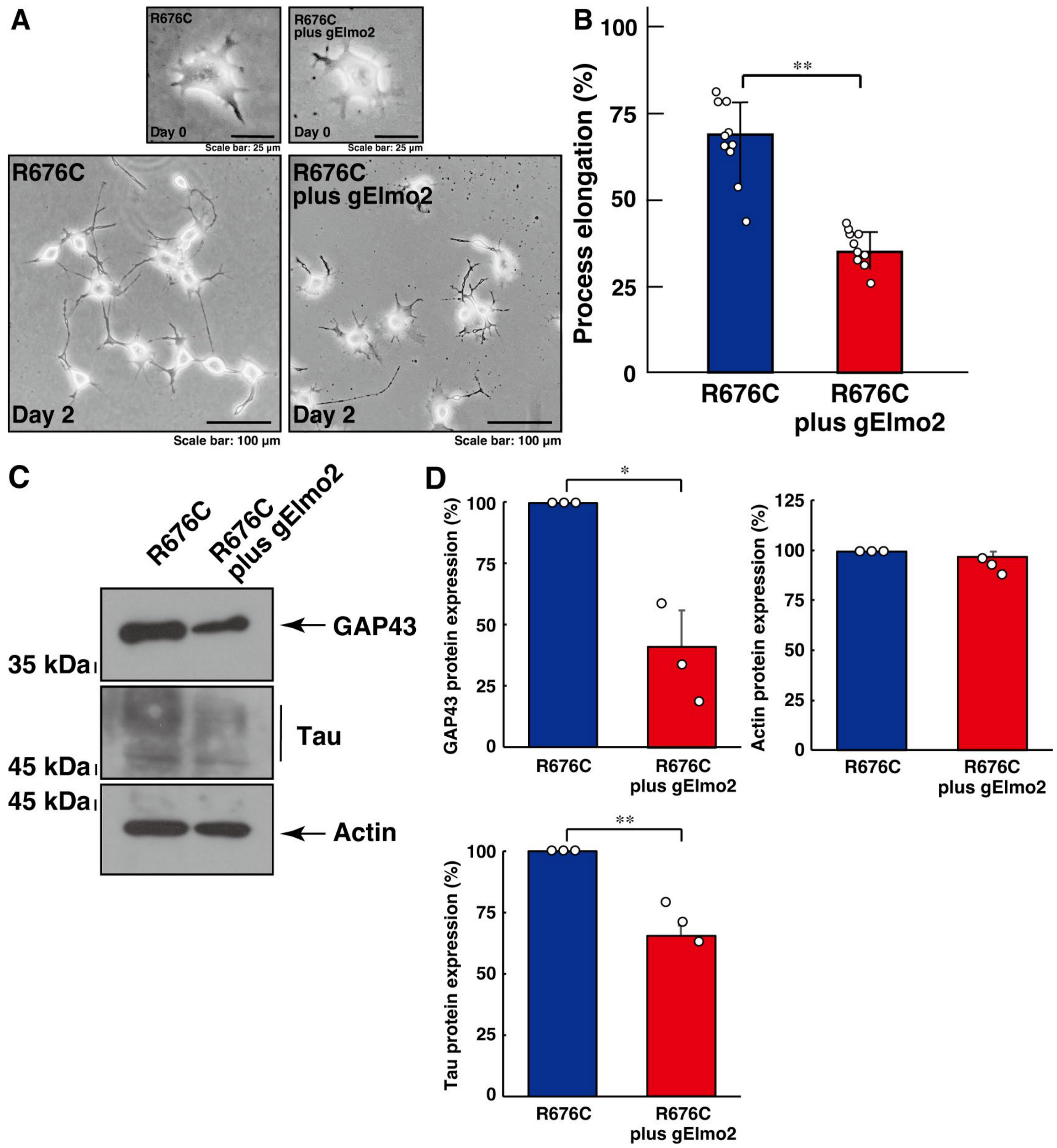
Next, we examined the involvement of Elmo2 in the increased process elongation induced by Sema5A harboring the R676C mutation. Knockdown of Elmo2 led to the recovery of increased process elongation (Figure 2A,B) and increased expression levels of neuronal differentiation marker proteins (Figure 2C,D), indicating that the Dock5 and Elmo2 signaling unit mediates mutated Sema5A-induced phenotypes.
We further verified whether the interaction of Dock5 and Elmo2 is needed for the formation of increased process elongation phenotypes by Sema5A harboring the R676C mutation. We co-transfected the plasmid encoding the N-terminal region (amino acids 1 to 71) of Dock5, which provides a binding region with Elmo members [41,42,43,44,45], into cells. Dock5 (1-71) resulted in the recovery of increased process elongation (Figure 3A,B) and increased expression levels of neuronal differentiation marker proteins (Figure 3C,D), indicating that mutated Sema5A-induced phenotypes are formed by the interaction between Dock5 and Elmo2.

3.3. Rac1 Acts Downstream of Dock5 and Elmo2 in Mutated Sema5A-Induced Process Elongation

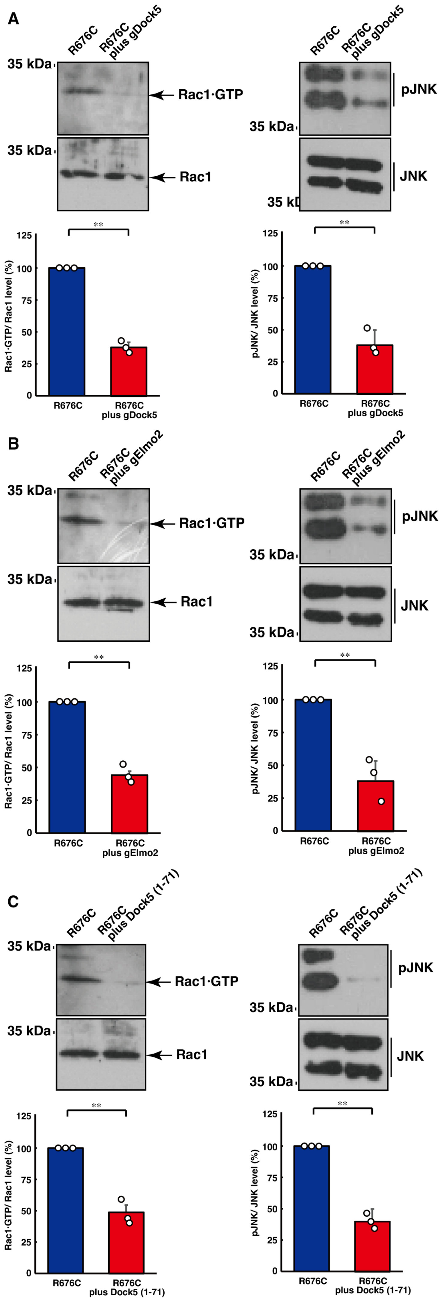
Finally, we examined whether Rac1 actually acts downstream of Dock5 during the elongation of processes induced by Sema5A harboring the ASD-associated R676C mutation in N1E-115 cells. Knockdown of Dock5 decreased the levels of active GTP-bound Rac1, as determined by a pull-down assay using the isolated Rac1·GTP-binding domain. Similar results were obtained in the case of either knockdown of Elmo2 or co-expression of the N-terminal region of Dock5 (Figure 4A,B). Consequently, we explored whether the JNK cascade, a major Rac1 effector [41,42,43,44], actually acts downstream of Dock5 during the elongation of processes. Phosphorylated JNK, an active form, was decreased by knockdown of Dock5, Elmo2, or co-expression of the N-terminal region of Dock5 (Figure 4C). Taken together, these findings suggest that Rac1 and the effector cascade act downstream of Dock5 and Elmo2 in this potential pathological signaling.

4. Discussion

Genetic mutations in individuals with ASD can affect the cell morphology and function of neurons as well as the relationships between excitatory and inhibitory neurons, neurons and glial cells, and neurons and immune cells in the brain. The formation of ASD as a neurodevelopmental disorder is likely to involve the abnormal morphogenesis and developmental patterns of neurons [27,28]. The structure and function of Sema5A may be altered by amino acid substitutions in autism, affecting neuronal cell morphogenesis and morphologies [27,28]. Mutated Sema5A has been observed to exhibit increased elongation of processes in a neuronal cell line in an autocrine manner [36]. However, despite this relation of ASD-associated amino acid mutations with cell morphologies [36], it remains unclear what signaling pathways these mutations utilize to cause aberrant neuromorphological changes.
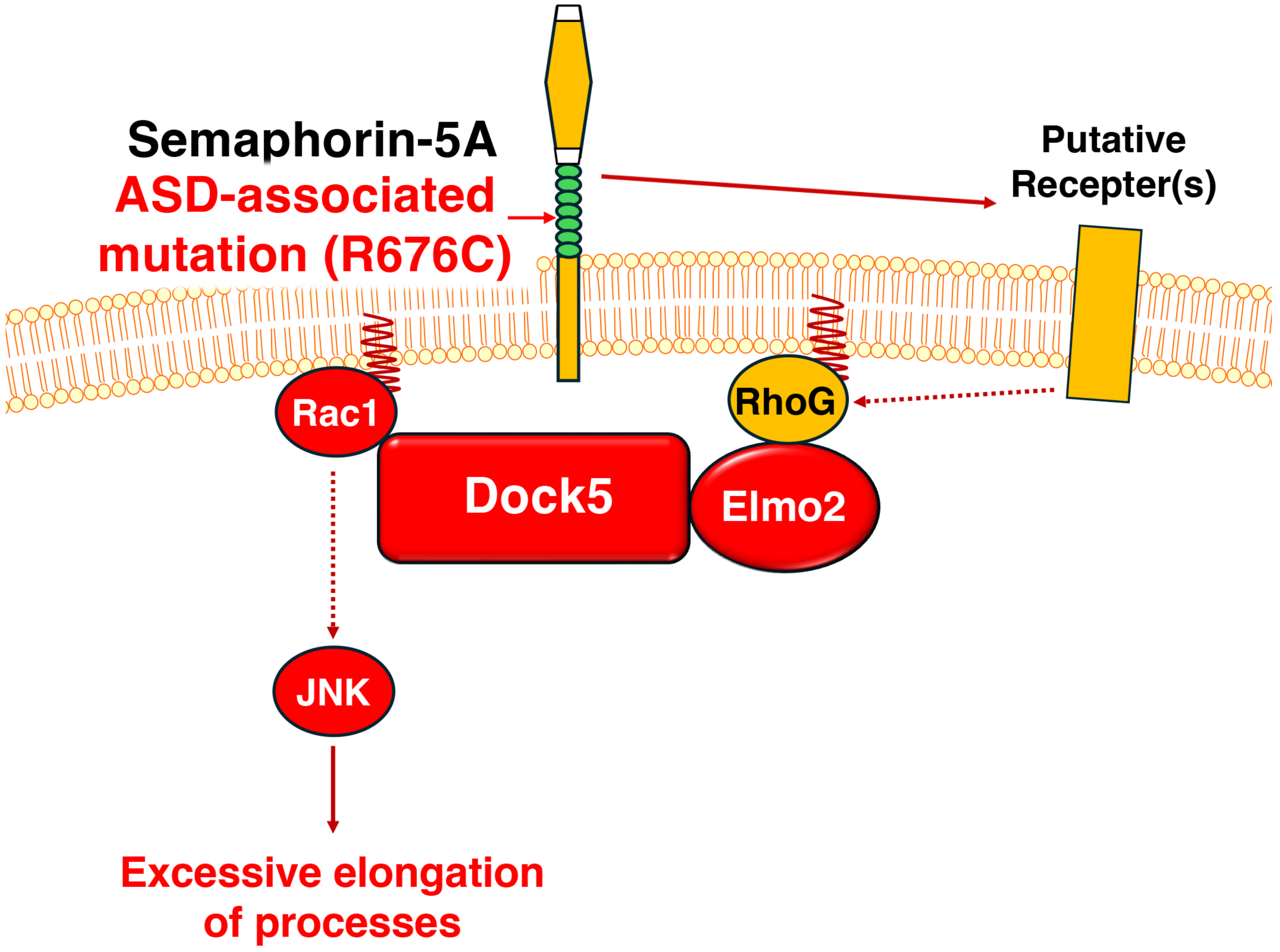
Using the N1E-115 cell line as a model, we have identified that mutated Sema5A-induced neuromorphological changes, particularly process elongation, are mediated by signaling complexes containing Dock5. The promoting phenotypes are supported by the expression levels of neuronal differentiation markers, implying that the process elongation is related to changes in biochemical properties. In addition, the activities of Rac1, a downstream effector of Dock5, and the phosphorylation levels of JNK, recognized as neuronal differentiation markers, contribute to the mutated Sema5A-induced morphological changes. These results suggest that mutated Sema5A controls morphological changes by taking advantage of neuromorphogenesis-related intracellular signaling through Dock5 (see Figure 5).
In studies on human genetics, deletion of the short arm region of chromosome 5 is associated with phenotypic features such as microcephaly, ID, and Cri du chat syndrome in infancy [26]. Classical transcriptome analysis indicates that some transcripts, including the gene, are downregulated in some individuals with autism [25]. This deleted region includes the gene and some other genes, with studies associating it with causing infantile epilepsy [29,30]. Importantly, the respective mutations of Arg-676 and Ser-951 of Sema5A are critically associated with ASD and/or ID, probably depending on individual patients [27,28], suggesting that Sema5A is actually the responsible gene product of ASD and/or ID. These mutations occur between the extracellular and intracellular regions of Sema5a, allowing for potential disulfide bond formation in the extracellular region and a reducing group in the intracellular region within a predicted topological domain structure. The activities of ASD-associated mutant proteins generally involve activation or inactivation compared to the wild type [13,14,15,16]. In this case, Sema5A is activated by mutations at Arg-676 or Ser-951, and its mutation at Arg-676 is particularly active through effective cell surface transportation and/or secretion of Sema5A [36]. Sema5A acts autocrinally by interacting with an unknown receptor on the cell surface, suggesting a role as a ligand. In addition, since Sema5A is a bifunctional transmembrane protein, acting not only as a ligand but also as a receptor [21,22,23,24], this case may also involve effective cell surface transportation [36]. In either scenario, it is hypothesized that signaling through Dock5 underlies the effects of mutated Sema5A.
Dock family GEFs and Rac1 compose a genetically conserved molecular family of signaling pathways related to cell morphogenesis in developing stages, spanning from Nematoda to mammals but not to abnormal cell morphogenesis in disease states [39,40,41]. These molecules also trigger apoptosis during development [39,40,41]. In humans and rodents, Dock family molecules are divided into four subfamilies: Dock-A, -B. -C, and -D [42]. Dock5 is a member of the Rac1-GEF Dock-A subfamily within the typical Dock family [42]. Since Dock1 and Dock5 have multiple functions in various cell types, it is likely that the function of Dock5 is compensated by Dock1 [41,42,43,44,45,46].
Studies involving the mapping of naturally mutated mice with the rupture of lens cataract have identified the deletion of a continuous 27 nucleotides at the end of the 15th exon, which encodes the dock5 gene in mouse chromosome 14. This deletion is identified as a candidate autosomal recessive disease gene [47]. The deletion results in decreased protein levels of Dock5, indicating that the loss-of-function of Dock5 is linked to abnormal morphogenesis in the formation of the eye lens, resulting in eye disease [47]. Thus, we asked whether and how Dock5 is involved in the regulation of morphogenesis in neuronal cells. The findings suggest that Dock5 is a specific signal transducer involved in both normal process elongation and mutated Sema5A-induced process elongation.
Elmo family molecules are well-established essential partners of all Dock-A family molecules [39,40,41,42,43,44,45]. Elmo1 exhibits wide expression profiles in the brain [48], and a similar expression pattern is observed for Elmo2. However, the expression levels of Elmo2 and Elmo1 differ depending on the respective brain and spinal cord regions [48]. It is unlikely that Elmo2 is completely compensated by Elmo1. In contrast to Dock5, it is well-known that Elmo2 positively regulates the elongation of neuronal dendrites in hippocampal neurons [49,50]. In addition, Elmo2 and Elmo1 play an essential role in the developmental stage when Sonic Hedgehog (Shh) guides commissural axons toward the floor plate [51]. It is suggested that Elmo2 responds to the Shh signal to control cytoskeletal remodeling, which underlies growth cone rotation [51]. Elmo2 and Elmo1 appear to act either specifically or cooperatively in controlling cytoskeletal proteins in neurons, depending on the respective extracellular signals; however, their partners are Dock3 and/or Dock4 [49,50,51]. Dock5 is unlikely to be involved in dendrite or commissural axon elongation. The formation of dendrites or axons by the signaling complex of Elmo2 or Elmo1 and Dock5 may depend on the type of neuron. It is possible that Elmo2 and Dock5 also exhibit specificity in certain types of neurological diseases.
On the other hand, JNK is a genetically conserved molecule of the signaling pathway related to cell morphogenesis during developmental stages from flies to mammals but not abnormal cell morphogenesis in disease states [52,53]. Like other members of the mitogen-activated protein kinase (MAPK) family, JNK is part of the signal complexes involved in the morphogenesis of almost all tissues and organs and is implicated in many diseases [52,53]. It is well-known that JNK kinase (JNKKK), an upstream kinase of JNK, is activated by Rac1 [52,53]. JNK itself phosphorylates cytoskeletal molecules and controls their activities [52,53]. The signaling coupling of Sema5A to Elmo2/Dock5 in both health and disease conditions may have specificity for process elongation in certain types of neuronal cells.
Plexin-B3 has been biochemically identified as the binding partner of Sema5A in neurons [54]. Plexin-B3, acting together with the interaction involving the Met proto-oncogene product (c-Met)’s receptor tyrosine kinase, links non-receptor tyrosine kinases c-Src and focal adhesion kinase (FAK) to small GTPases. This linkage controls cytoskeletal proteins, leading to cell morphological changes in cell types other than neuronal cells [55]. Given that Plexin-B3 is a multifunctional protein [17,18,19,20], Met could be one of the major signal transducers for Plexin-B3. Despite an unknown association of Plexin-B3 with ASD, it is of note that Met is recognized as a risk gene product for ASD [56,57]. It is also possible that signaling through Dock5 acts downstream of Plexin-B3 and c-Met.
The relationship between semaphorin family molecules and Alzheimer’s disease is increasingly recognized [58,59]. It is known that Sema3A is a component of extracellular matrix structures surrounding certain types of neuronal cells in the adult CNS. It plays a positive role in ending a critical period of plasticity [59]. Conversely, the failure of the system is believed to be one of the important factors contributing to Alzheimer’s disease-like phenotypes. Additionally, Sema5A and/or Sema7A are involved in the regulation of neurogenesis in adults, and dysfunction is also associated with Alzheimer’s disease-like phenotypes [58,59]. Genome-wide association studies (GWASs) suggest an association between single nucleotide polymorphisms (SNPs) of the gene and Alzheimer’s disease [60,61]. Further studies may reveal the association of Sema5A with many neurological diseases in addition to ASD and ID. Herein, we demonstrate that the increased process elongation induced by the Sema5A protein harboring the ASD-associated R676C mutation is mediated by signaling through Dock5. Knockdown of the respective molecules using CRISPR/CasRx-based gRNAs results in the recovery of increased process elongation. Additional studies may promote our understanding of not only the detailed mechanism by which mutated Sema5A intracellularly leads to the phosphorylation and activation of JNK via signaling through Dock5 but also the extent to which mutated Sema5A utilizes signaling through Dock5 and other signal transduction pathway molecules in increased process elongation, in both primary neuronal cells and genetically modified mice. These studies can allow us to clarify the relationship between the possible cellular phenotypes observed in this study and those of cells in patients. This line of investigation may facilitate a direct link between experimental results and potential therapeutic methods for Sema5A-associated ASD and ID and axon guidance molecule-related ASD.

Supplementary Materials

The following supporting information can be downloaded at: https://www.mdpi.com/article/10.3390/cimb46040194/s1, Figure S1. Transcripts of typical Dock family members and their binding partner proteins. Figure S2. Effects of Dock5 or Elmo2 CRISPR/CasRx-based gRNA on knockdown in N1E-115 cells. Figure S3. Effects of Dock5 knockdown on process elongation in N1E-115 cells. Figure S4. Effects of Elmo2 knockdown on process elongation in N1E-115 cells. Figure S5. Effects of mutated Sema5A in process elongation in N1E-115 cells.

Author Contributions

Conceptualization, J.Y.; Methodology, M.O. (Miyu Okabe), T.S., M.T., A.H., M.O. (Maho Okawa), M.I., M.K.-N., M.S. and Y.M.; Software, M.O. (Miyu Okabe), T.S., M.T., A.H., M.O. (Maho Okawa), M.I., M.K.-N., M.S. and Y.M.; Validation, M.O. (Miyu Okabe), T.S., M.T., A.H., M.O. (Maho Okawa), M.I., M.K-N., M.S. and Y.M.; Formal analysis, M.O. (Miyu Okabe), T.S., M.T., A.H., M.O. (Maho Okawa), M.I., M.K-N., M.S. and Y.M.; Investigation, M.O. (Miyu Okabe), T.S., M.T., A.H., M.O. (Maho Okawa), M.I., M.K-N. and M.S.; Resources, M.O. (Miyu Okabe), T.S., M.T., A.H., M.O. (Maho Okawa), M.I., M.K-N., M.S. and Y.M.; Data curation, M.O. (Miyu Okabe), T.S., M.T. and Y.M.; Writing—original draft, Y.M. and J.Y.; Writing—review & editing, Y.M. and J.Y.; Visualization, M.O. (Miyu Okabe), T.S., M.T., Y.M. and J.Y.; Supervision, J.Y.; Project administration, Y.M. and J.Y.; Funding acquisition, Y.M. and J.Y. All authors have read and agreed to the published version of the manuscript.

Funding

This work was supported by Core Research for Evolutional Science and Technology (CREST) of the Japan Science and Technology Agency (JST). This work was also supported by grants-in-aid for scientific research from the Japanese Ministry of Education, Culture, Sports, Science and Technology (MEXT), grants-in-aid for medical scientific research from the Japanese Ministry of Health, Labour and Welfare (MHLW), and grants from the Daiichi Sankyo Science Foundation, Japan Foundation for Pediatric Research, Mishima Kaiun Memorial Foundation, Mitsubishi Tanabe Science Foundation, Otsuka Science Foundation, and Takeda Science Foundation.

Institutional Review Board Statement

Techniques using genetically modified cells and related techniques were performed in accordance with a protocol approved by the Tokyo University of Pharmacy and Life Sciences Gene and Animal Care Committee (Approval Nos. LS28-20 and LSR3-011 on 1 April 2023).

Informed Consent Statement

Not applicable.

Data Availability Statement

The datasets used and/or analyzed in the current study are available from the corresponding author upon reasonable request.

Acknowledgments

We thank Takako Morimoto, Yoichi Seki, and Remina Shirai for their insightful comments throughout this study.

Conflicts of Interest

The authors declare no conflicts of interest.

References

  1. Craig, A.M.; Banker, G. Neuronal polarity. Annu. Rev. Neurosci. 1994, 17, 267–310. [Google Scholar] [CrossRef] [PubMed]
  2. Da Silva, J.S.; Dotti, C.G. Breaking the neuronal sphere: Regulation of the actin cytoskeleton in neuritogenesis. Nat. Rev. Neurosci. 2002, 3, 694–704. [Google Scholar] [CrossRef] [PubMed]
  3. Arimura, N.; Kaibuchi, K. Neuronal polarity: From extracellular signals to intracellular mechanisms. Nat. Rev. Neurosci. 2007, 8, 194–205. [Google Scholar] [CrossRef] [PubMed]
  4. Park, H.; Poo, M.-M. Neurotrophin regulation of neural circuit development and function. Nat. Rev. Neurosci. 2013, 14, 7–23. [Google Scholar] [CrossRef] [PubMed]
  5. Bray, D. Surface movements during the growth of single explanted neurons. Proc. Natl. Acad. Sci. USA 1970, 65, 905–910. [Google Scholar] [CrossRef] [PubMed]
  6. Markus, A.; Patel, T.D.; Snider, W.D. Neurotrophic factors and axonal growth. Curr. Opin. Neurobiol. 2002, 12, 523–531. [Google Scholar] [CrossRef] [PubMed]
  7. Rigby, M.J.; Gomez, T.M.; Puglielli, L. Glial cell-axonal growth cone interactions in neurodevelopment and regeneration. Front. Neurosci. 2020, 14, 203. [Google Scholar] [CrossRef]
  8. Taylor, K.R.; Monje, M. Neuron–oligodendroglial interactions in health and malignant disease. Nat. Rev. Neurosci. 2023, 24, 733–746. [Google Scholar] [CrossRef] [PubMed]
  9. Bernier, R.; Mao, A.; Yen, J. Psychopathology, families, and culture: Autism. Child Adolesc. Psychiatr. Clin. N. Am. 2010, 19, 855–867. [Google Scholar] [CrossRef]
  10. Mazza, M.; Pino, M.C.; Mariano, M.; Tempesta, D.; Ferrara, M.; De Berardis, D.; Masedu, F.; Valenti, M. Affective and cognitive empathy in adolescents with autism spectrum disorder. Front. Hum. Neurosci. 2014, 8, 791. [Google Scholar] [CrossRef]
  11. Arnett, A.B.; Trinh, S.; Bernier, R.A. The state of research on the genetics of autism spectrum disorder: Methodological, clinical and conceptual progress. Curr. Opin. Psychol. 2019, 27, 1–5. [Google Scholar] [CrossRef] [PubMed]
  12. Peterson, J.L.; Earl, R.K.; Fox, E.A.; Ma, R.; Haidar, G.; Pepper, M.; Berliner, L.; Wallace, A.S.; Bernier, R.A. Trauma and autism spectrum disorder: Review, proposed treatment adaptations and future directions. J. Child Adolesc. Trauma 2019, 12, 529–547. [Google Scholar] [CrossRef] [PubMed]
  13. Modabbernia, A.; Velthorst, E.; Reichenberg, A. Environmental risk factors for autism: An evidence-based review of systematic reviews and meta-analyses. Mol. Autism 2017, 8, 13. [Google Scholar] [CrossRef] [PubMed]
  14. Varghese, M.; Keshav, N.; Jacot-Descombes, S.; Warda, T.; Wicinski, B.; Dickstein, D.L.; Harony-Nicolas, H.; De Rubeis, S.; Drapeau, E.; Buxbaum, J.D.; et al. Autism spectrum disorder: Neuropathology and animal models. Acta Neuropathol. 2017, 134, 537–566. [Google Scholar] [CrossRef] [PubMed]
  15. Genovese, A.; Butler, M.G. Clinical assessment, genetics, and treatment approaches in autism spectrum disorder (ASD). Int. J. Mol. Sci. 2020, 21, 4726. [Google Scholar] [CrossRef] [PubMed]
  16. Wang, L.; Wang, B.; Wu, C.; Wang, J.; Sun, M. Autism spectrum disorder: Neurodevelopmental risk factors, biological mechanism, and precision therapy. Int. J. Mol. Sci. 2023, 24, 1819. [Google Scholar] [CrossRef] [PubMed]
  17. Lin, L.; Lesnick, T.G.; Maraganore, D.M.; Isacson, O. Axon guidance and synaptic maintenance: Preclinical markers for neurodegenerative disease and therapeutics. Trends Neurosci. 2009, 32, 142–149. [Google Scholar] [CrossRef]
  18. Worzfeld, T.; Offermanns, S. Semaphorins and plexins as therapeutic targets. Nat. Rev. Drug Discov. 2014, 13, 603–621. [Google Scholar] [CrossRef]
  19. Limoni, G.; Niquille, M. Semaphorins and Plexins in central nervous system patterning: The key to it all? Curr. Opin. Neurobiol. 2021, 66, 224–232. [Google Scholar] [CrossRef]
  20. Zang, Y.; Chaudhari, K.; Bashaw, G.J. New insights into the molecular mechanisms of axon guidance receptor regulation and signaling. Curr. Top. Dev. Biol. 2021, 142, 147–196. [Google Scholar]
  21. Adams, R.H.; Betz, H.; Püschel, A.W. A novel class of murine semaphorins with homology to thrombospondin is differentially expressed during early embryogenesis. Mech. Dev. 1996, 57, 33–45. [Google Scholar] [CrossRef] [PubMed]
  22. Oster, S.F.; Bodeker, M.O.; He, F.; Sretavan, D.W. Invariant Sema5A inhibition serves an ensheathing function during optic nerve development. Development 2003, 130, 775–784. [Google Scholar] [CrossRef]
  23. Goldberg, J.L.; Vargas, M.E.; Wang, J.T.; Mandemakers, W.; Oster, S.F.; Sretavan, D.W.; Barres, B.A. An oligodendrocyte lineage-specific semaphorin, Sema5A, inhibits axon growth by retinal ganglion cells. J. Neurosci. 2004, 24, 4989–4999. [Google Scholar] [CrossRef] [PubMed]
  24. Matsuoka, R.L.; Chivatakarn, O.; Badea, T.C.; Samuels, I.S.; Cahill, H.; Katayama, K.-I.; Kumar, S.R.; Suto, F.; Chédotal, A.; Peachey, N.S.; et al. Class 5 transmembrane semaphorins control selective mammalian retinal lamination and function. Neuron 2011, 71, 460–473. [Google Scholar] [CrossRef] [PubMed]
  25. Melin, M.; Carlsson, B.; Anckarsater, H.; Rastam, M.; Betancur, C.; Isaksson, A.; Gillberg, C.; Dahl, N. Constitutional Downregulation of SEMA5A Expression in Autism. Neuropsychobiology 2006, 54, 64–69. [Google Scholar] [CrossRef] [PubMed]
  26. Zhang, B.; Willing, M.; Grange, D.K.; Shinawi, M.; Manwaring, L.; Vineyard, M.; Kulkarni, S.; Cottrell, C.E. Multigenerational autosomal dominant inheritance of 5p chromosomal deletions. Am. J. Med. Genet. Part A 2016, 170, 583–593. [Google Scholar] [CrossRef] [PubMed]
  27. Ito, H.; Morishita, R.; Nagata, K.-I. Autism spectrum disorder-associated genes and the development of dentate granule cells. Med. Mol. Morphol. 2017, 50, 123–129. [Google Scholar] [CrossRef]
  28. Mosca-Boidron, A.-L.; Gueneau, L.; Huguet, G.; Goldenberg, A.; Henry, C.; Gigot, N.; Pallesi-Pocachard, E.; Falace, A.; Duplomb, L.; Thevenon, J.; et al. A de novo microdeletion of SEMA5A in a boy with autism spectrum disorder and intellectual disability. Eur. J. Hum. Genet. 2016, 24, 838–843. [Google Scholar] [CrossRef]
  29. Depienne, C.; Magnin, E.; Bouteiller, D.; Stevanin, G.; Saint-Martin, C.; Vidailhet, M.; Apartis, E.; Hirsch, E.; LeGuern, E.; Labauge, P.; et al. Familial cortical myoclonic tremor with epilepsy: The third locus (FCMTE3) maps to 5p. Neurology 2010, 74, 2000–2003. [Google Scholar] [CrossRef]
  30. Wang, Q.; Liu, Z.; Lin, Z.; Zhang, R.; Lu, Y.; Su, W.; Li, F.; Xu, X.; Tu, M.; Lou, Y.; et al. De Novo Germline Mutations in SEMA5A Associated With Infantile Spasms. Front. Genet. 2019, 10, 605. [Google Scholar] [CrossRef]
  31. Gunn, R.K.; Huentelman, M.J.; Brown, R.E. Are Sema5a mutant mice a good model of autism? A behavioral analysis of sensory systems, emotionality and cognition. Behav. Brain Res. 2011, 225, 142–150. [Google Scholar] [CrossRef]
  32. Duan, Y.; Wang, S.H.; Song, J.; Mironova, Y.; Ming, G.L.; Kolodkin, A.L.; Giger, R.J. Semaphorin 5A inhibits synaptogenesis in early postnatal- and adult-born hippocampal dentate granule cells. eLife 2014, 3, e04390. [Google Scholar] [CrossRef]
  33. Hirose, M.; Ishizaki, T.; Watanabe, N.; Uehata, M.; Kranenburg, O.; Moolenaar, W.H.; Matsumura, F.; Maekawa, M.; Bito, H.; Narumiya, S. Molecular Dissection of the Rho-associated Protein Kinase (p160ROCK)-regulated Neurite Remodeling in Neuroblastoma N1E-115 Cells. J. Cell Biol. 1998, 141, 1625–1636. [Google Scholar] [CrossRef] [PubMed]
  34. Miyamoto, Y.; Yamauchi, J. Cellular signaling of Dock family proteins in neural function. Cell Signal. 2010, 22, 175–182. [Google Scholar] [CrossRef] [PubMed]
  35. Miyamoto, Y.; Torii, T.; Yamamori, N.; Ogata, T.; Tanoue, A.; Yamauchi, J. Akt and PP2A reciprocally regulate the guanine nucleotide exchange factor Dock6 to control axon growth of sensory neurons. Sci. Signal. 2013, 6, ra15. [Google Scholar] [CrossRef]
  36. Okabe, M.; Miyamoto, Y.; Ikoma, Y.; Takahashi, M.; Shirai, R.; Kukimoto-Niino, M.; Shirouzu, M.; Yamauchi, J. RhoG-binding domain of Elmo1 ameliorates excessive process elongation induced by autism spectrum disorder-associated Sema5A. Pathophysiology 2023, 30, 548–566. [Google Scholar] [CrossRef] [PubMed]
  37. Numakawa, T.; Yokomaku, D.; Kiyosue, K.; Adachi, N.; Matsumoto, T.; Numakawa, Y.; Taguchi, T.; Hatanaka, H.; Yamada, M. Basic fibroblast growth factor evokes a rapid glutamate release through activation of the MAPK pathway in cultured cortical neurons. J. Biol. Chem. 2002, 277, 28861–28869. [Google Scholar] [CrossRef] [PubMed]
  38. Hwang, S.; Lee, S.-E.; Ahn, S.-G.; Lee, G.H. Psoralidin stimulates expression of immediate-early genes and synapse development in primary cortical neurons. Neurochem. Res. 2018, 43, 2460–2472. [Google Scholar] [CrossRef] [PubMed]
  39. Matsuda, M.; Kurata, T. Emerging components of the Crk oncogene product: The first identified adaptor protein. Cell Signal. 1996, 8, 335–340. [Google Scholar] [CrossRef]
  40. Reddien, P.W.; Horvitz, H.R. The engulfment process of programmed cell death in Caenorhabditis elegans. Annu. Rev. Cell Dev. Biol. 2004, 20, 193–221. [Google Scholar] [CrossRef]
  41. Ravichandran, K.S.; Lorenz, U. Engulfment of apoptotic cells: Signals for a good meal. Nat. Rev. Immunol. 2007, 7, 964–974. [Google Scholar] [CrossRef] [PubMed]
  42. Laurin, M.; Côté, J.-F. Insights into the biological functions of Dock family guanine nucleotide exchange factors. Minerva Anestesiol. 2014, 28, 533–547. [Google Scholar] [CrossRef] [PubMed]
  43. Gadea, G.; Blangy, A. Dock-family exchange factors in cell migration and disease. Eur. J. Cell Biol. 2014, 93, 466–477. [Google Scholar] [CrossRef] [PubMed]
  44. Samani, A.; English, K.G.; Lopez, M.A.; Birch, C.L.; Brown, D.M.; Kaur, G.; Worthey, E.A.; Alexander, M.S. DOCKopathies: A systematic review of the clinical pathologies associated with human DOCK pathogenic variants. Hum. Mutat. 2022, 43, 1149–1161. [Google Scholar] [CrossRef] [PubMed]
  45. Kukimoto-Niino, M.; Ihara, K.; Murayama, K.; Shirouzu, M. Structural insights into the small GTPase specificity of the DOCK guanine nucleotide exchange factors. Curr. Opin. Struct. Biol. 2021, 71, 249–258. [Google Scholar] [CrossRef] [PubMed]
  46. Moore, C.A.; Parkin, C.A.; Bidet, Y.; Ingham, P.W. A role for the Myoblast city homologues Dock1 and Dock5 and the adaptor proteins Crk and Crk-like in zebrafish myoblast fusion. Development 2007, 134, 3145–3153. [Google Scholar] [CrossRef] [PubMed]
  47. Omi, N.; Kiyokawa, E.; Matsuda, M.; Kinoshita, K.; Yamada, S.; Yamada, K.; Matsushima, Y.; Wang, Y.; Kawai, J.; Suzuki, M.; et al. Mutation of Dock5, a member of the guanine exchange factor Dock180 superfamily, in the rupture of lens cataract mouse. Exp. Eye Res. 2008, 86, 828–834. [Google Scholar] [CrossRef] [PubMed]
  48. Katoh, H.; Fujimoto, S.; Ishida, C.; Ishikawa, Y.; Negishi, M. Differential distribution of ELMO1 and ELMO2 mRNAs in the developing mouse brain. Brain Res. 2006, 1073–1074, 103–108. [Google Scholar] [CrossRef] [PubMed]
  49. Ueda, S.; Fujimoto, S.; Hiramoto, K.; Negishi, M.; Katoh, H. Dock4 regulates dendritic development in hippocampal neurons. J. Neurosci. Res. 2008, 86, 3052–3061. [Google Scholar] [CrossRef]
  50. Ueda, S.; Negishi, M.; Katoh, H.; Jaudon, F.; Raynaud, F.; Wehrlé, R.; Bellanger, J.-M.; Doulazmi, M.; Vodjdani, G.; Gasman, S.; et al. Rac GEF Dock4 interacts with cortactin to regulate dendritic spine formation. Mol. Biol. Cell 2013, 24, 1602–1613. [Google Scholar] [CrossRef]
  51. Makihara, S.; Morin, S.; Ferent, J.; Côté, J.-F.; Yam, P.T.; Charron, F. Polarized Dock activity drives Shh-mediated axon guidance. Dev. Cell 2018, 46, 410–425.e7. [Google Scholar] [CrossRef]
  52. Cano, E.; Mahadevan, L.C. Parallel signal processing among mammalian MAPKs. Trends Biochem. Sci. 1995, 20, 117–122. [Google Scholar] [CrossRef]
  53. Kim, E.K.; Choi, E.-J. Pathological roles of MAPK signaling pathways in human diseases. Biochim. Biophys. Acta (BBA) Mol. Basis Dis. 2010, 1802, 396–405. [Google Scholar] [CrossRef]
  54. Artigiani, S.; Conrotto, P.; Fazzari, P.; Gilestro, G.F.; Barberis, D.; Giordano, S.; Comoglio, P.M.; Tamagnone, L. Plexin-B3 is a functional receptor for semaphorin 5A. Embo Rep. 2004, 5, 710–714. [Google Scholar] [CrossRef]
  55. Zuo, Q.; Yang, Y.; Lyu, Y.; Yang, C.; Chen, C.; Salman, S.; Huang, T.Y.; Wicks, E.E.; Jackson, W., 3rd; Datan, E.; et al. Plexin-B3 expression stimulates MET signaling, breast cancer stem cell specification, and lung metastasis. Cell Rep. 2023, 42, 112164. [Google Scholar] [CrossRef]
  56. Heuer, L.; Braunschweig, D.; Ashwood, P.; Van de Water, J.; Campbell, D.B. Association of a MET genetic variant with autism-associated maternal autoantibodies to fetal brain proteins and cytokine expression. Transl. Psychiatry 2011, 1, e48. [Google Scholar] [CrossRef]
  57. Desole, C.; Gallo, S.; Vitacolonna, A.; Montarolo, F.; Bertolotto, A.; Vivien, D.; Comoglio, P.; Crepaldi, T. HGF and MET: From Brain Development to Neurological Disorders. Front. Cell Dev. Biol. 2021, 9, 683609. [Google Scholar] [CrossRef]
  58. Carulli, D.; de Winter, F.; Verhaagen, J. Semaphorins in adult nervous system plasticity and disease. Front. Synaptic Neurosci. 2021, 13, 672891. [Google Scholar] [CrossRef]
  59. Zhang, L.; Qi, Z.; Li, J.; Li, M.; Du, X.; Wang, S.; Zhou, G.; Xu, B.; Liu, W.; Xi, S.; et al. Roles and Mechanisms of Axon-Guidance Molecules in Alzheimer’s Disease. Mol. Neurobiol. 2021, 58, 3290–3307. [Google Scholar] [CrossRef] [PubMed]
  60. Ueki, M.; Kawasaki, Y.; Tamiya, G.; Alzheimer’s Disease Neuroimaging Initiative. Detecting genetic association through shortest paths in a bidirected graph. Genet. Epidemiol. 2017, 41, 481–497. [Google Scholar] [CrossRef] [PubMed]
  61. Zhang, C.; Tan, G.; Zhang, Y.; Zhong, X.; Zhao, Z.; Peng, Y.; Cheng, Q.; Xue, K.; Xu, Y.; Li, X.; et al. Comprehensive analyses of brain cell communications based on multiple scRNA-seq and snRNA-seq datasets for revealing novel mechanism in neurodegenerative diseases. CNS Neurosci. Ther. 2023, 29, 2775–2786. [Google Scholar] [CrossRef]
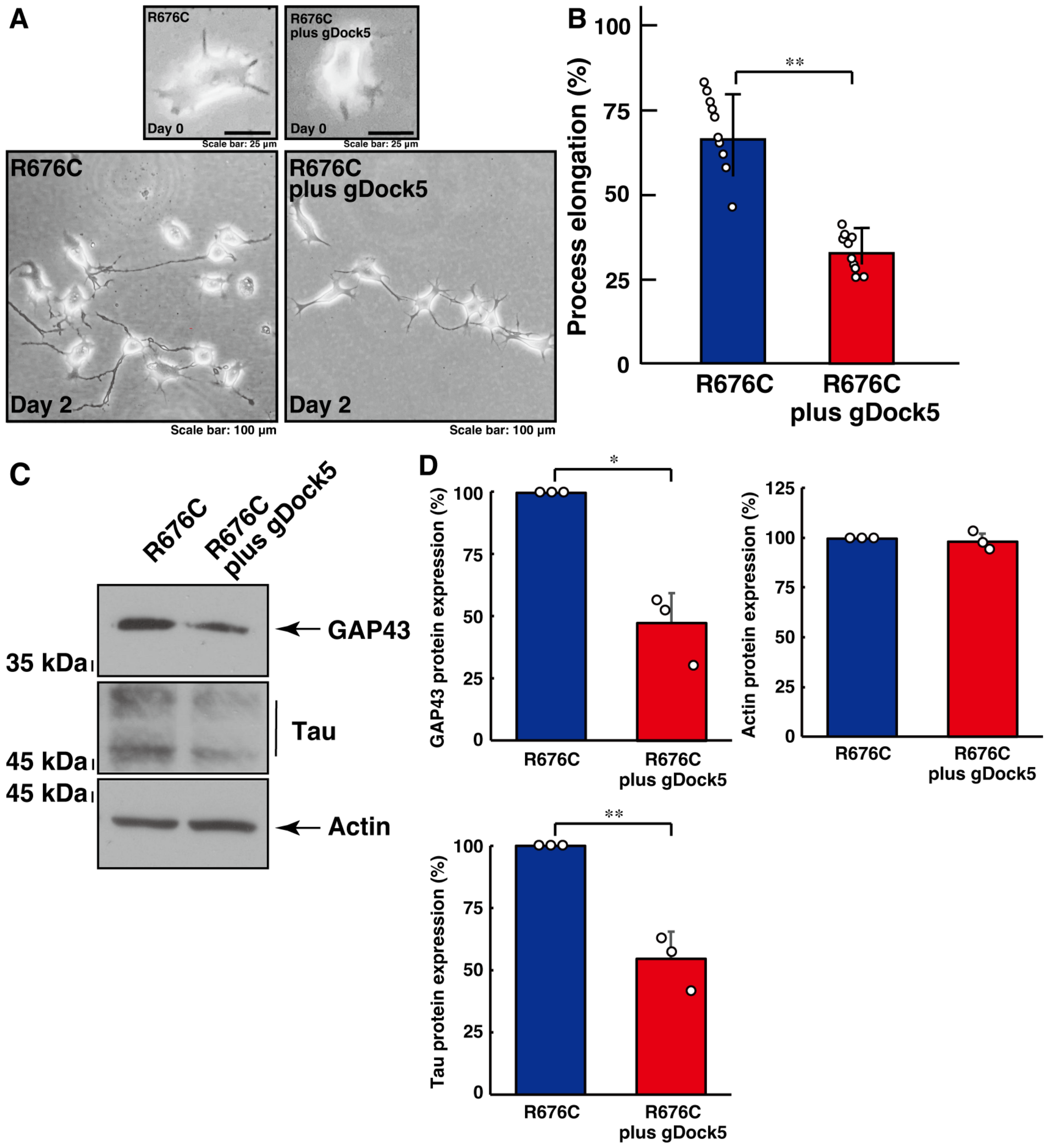
Figure 1. Knockdown of Dock5 using CRISPR/CasRx-based gRNA recovers mutated Sema5A-induced excessive process elongation. (A,B) N1E-115 cells harboring Sema5A with the ASD-associated R676C mutation were transfected with or without the plasmid encoding CasRx and gRNA specific for Dock5 and were allowed to differentiate for 0 or 2 days. Cells with processes exceeding a body length of two cells were counted as cells with neurite-like process elongation and are statistically depicted in the graph (** p < 0.01; n = 10 fields). (C,D) The lysates of cells at day 2 were immunoblotted with an antibody against a neuronal differentiation marker protein (GAP43 or Tau) or an internal control protein actin. Their immunoreactive band intensities are statistically depicted (** p < 0.01 and * p < 0.05; n = 3 blots).
Cimb 46 00194 g001
Figure 2. Knockdown of Elmo1 using CRISPR/CasRx-based gRNA recovers mutated Sema5A-induced excessive process elongation. (A,B) N1E-115 cells harboring Sema5A with the ASD-associated R676C mutation were transfected with or without the plasmid encoding CasRx and gRNA specific for Elmo1 and were allowed to differentiate for 0 or 2 days. Cells with processes exceeding a body length of two cells were counted as cells with neurite-like process elongation and are statistically depicted in the graph (** p < 0.01; n = 10 fields). (C,D) The lysates of cells at day 2 were immunoblotted with an antibody against a neuronal differentiation marker protein (GAP43 or Tau) or an internal control protein actin. Their immunoreactive band intensities are statistically depicted (** p < 0.01 and * p < 0.05; n = 3 blots).
Cimb 46 00194 g002
Figure 3. Transfection of an isolated binding region with Elmo members recovers mutated Sema5A-induced excessive process elongation. (A,B) N1E-115 cells harboring Sema5A with the ASD-associated R676C mutation were transfected with or without the plasmid encoding the Dock5 N-terminal region (amino acids 1 to 71) and were allowed to differentiate for 0 or 2 days. Cells with processes exceeding a body length of two cells were counted as cells with neurite-like process elongation and are statistically depicted in the graph (** p < 0.01; n = 10 fields). (C,D) The lysates of cells at day 2 were immunoblotted with an antibody against a neuronal differentiation marker protein (GAP43 or Tau) or an internal control protein actin. Their immunoreactive band intensities are statistically depicted (** p < 0.01 and * p < 0.05; n = 3 blots).
Cimb 46 00194 g003
Figure 4. Transfection of Dock5 or Elmo2 gRNA or the Dock5 N-terminal region recovers Rac1 and JNK activated states induced by mutated Sema5A. (A,B) The transfected cell lysates in N1E-115 cells harboring Sema5A with the ASD-associated R676C mutation were used for the affinity precipitation assay to monitor GTP-bound Rac1 protein. The lysates were also subjected to immunoblotting with an antibody against Rac1. (C) The lysates were immunoblotted with an antibody against phosphorylated JNK or JNK. Their immunoreactive band intensities are statistically depicted (** p < 0.01; n = 3 blots).
Cimb 46 00194 g004
Figure 5. Schematic diagram of Dock5 signaling evoked by the ASD- and/or ID-associated Sema5A mutant protein underlying excessive elongation. Mutated Sema5A autocrinally regulates elongation of neurite-like processes through an unidentified putative receptor(s), which possibly transduces the signal to Elmo2 adaptor protein-mediated Dock5 GEF. The complex couples RhoG to Rac1 and the downstream JNK, enhancing excessive elongation.
Cimb 46 00194 g005
Table 1. Key materials used in this study.
Reagents or MaterialsCompanies or SourcesCat. Nos.Lot. Nos.Concentrations Used
Key antibodies
Anti-growth-associated protein 43 (GAP43)Santa Cruz Biotechnology (Santa Cruz, CA, USA)sc-17790J0920Immunoblotting (IB), 1:5000
Anti-beta III tubulinSanta Cruz sc-80005H2721IB, 1:5000
Anti-TauSanta Cruz Biotechnologysc-21796007IB, 1:500
Anti-actin (also called pan-beta type actin)MBL (Tokyo, Japan)M177-3J2521IB, 1:5000
Anti-c-Jun N-terminal kinase (JNK, pan-JNK)Santa Cruz Biotechnologysc-7345C1722IB, 1:250
Anti-phospho-c-Jun N-terminal kinase (pJNK, pan-pJNK) Santa Cruz Biotechnologysc-6254sc-6254IB, 1:500
anti-Rac1Proteintech (Rosemont, IL, USA)66122-1-Ig10011346IB, 1:500
Anti-IgG (H + L chain) (rabbit) pAb-HRPMBL458353IF, 1:5000
Anti-IgG (H + L chain) (mouse) pAb-HRPMBL330365IF, 1:5000
Recombinant DNAs
pCMV-SV40ori-mouse Sema5A-HisAddgene (Watertown, MA, USA)72035n.d.1.25 microgram of DNA per 6 cm dish
pCMV-SV40ori-mouse Sema5A (R676C)-HisA construct generated using Cat. No. 72035
(Addgene)
derived from
72035
n.d.1.25 microgram of DNA per 6 cm dish
CasRx (NLS-RfxCas13d-NLS)Addgene109049n.d.1.25 microgram of DNA per 6 cm dish
pSINsi-mU6-gRNA for mouse Dock5
(nucleotide numbers 1266 and 1410)
Generated in this studyn.d.n.d.1.25 microgram of DNA per 6 cm dish
pSINsi-mU6-gRNA for mouse Elmo2
(nucleotide numbers 909 and 1239)
Generated in this studyn.d.n.d.1.25 microgram of DNA per 6 cm dish
pcDNA3.1-N-eGFP-human Dock5
(amino acids 1 to 71 of the Elmo family molecule-binding region)
GenScript (Piscataway, NJ, USA)n.d.n.d.1.25 microgram of DNA per 6 cm dish
pET42a-conserved Cdc42 and Rac interactive binding (CRIB) domain of human Pak1see Pathophysiology 2023 30:548-566n.d.n.d.5 micrograms of DNA per 1 litter size bacteria culture bottle
Disclaimer/Publisher’s Note: The statements, opinions and data contained in all publications are solely those of the individual author(s) and contributor(s) and not of MDPI and/or the editor(s). MDPI and/or the editor(s) disclaim responsibility for any injury to people or property resulting from any ideas, methods, instructions or products referred to in the content.