Article

Efficacy of SGPP2 Modulation-Mediated Materials in Ameliorating Facial Wrinkles and Pore Sagging

LG Household & Health Care (LG H&H) R&D Center, Seoul 07795, Republic of Korea
*
Authors to whom correspondence should be addressed.
Curr. Issues Mol. Biol. 2024, 46(8), 9122-9135; https://doi.org/10.3390/cimb46080539
Submission received: 31 July 2024 / Revised: 12 August 2024 / Accepted: 19 August 2024 / Published: 20 August 2024
(This article belongs to the Section Biochemistry, Molecular and Cellular Biology)

Abstract

:
Skin aging is a complex process with internal and external factors. Recent studies have suggested that enlargement and elongation of skin pores may be early signs of aging in addition to wrinkles and loss of elasticity. This study explores the potential of targeting the SGPP2 gene in keratinocytes to address these emerging concerns. Using siRNA knockdown, we demonstrated that SGPP2 modulates the production of inflammatory cytokines (interleukin (IL)-1β and IL-8). Furthermore, conditioned media experiments revealed that keratinocytes with high SGPP2 expression indirectly influence fibroblast extracellular matrix remodeling, potentially contributing to enlarged pores and wrinkle formation. Based on these findings, we explored a complex formulation containing four SGPP2-modulating compounds. In vitro and in vivo experiments demonstrated the efficacy of the formulation in mitigating fine wrinkles and pore enlargement. This study highlights the significant implications of developing a more effective antiaging cosmetic formulation by targeting underlying inflammatory processes that drive skin aging.

1. Introduction

Skin aging is a multi-factorial process driven by the intricate interplay of intrinsic (time, genetic factors, and hormonal changes) and extrinsic (ultraviolet (UV) exposure and pollution) factors. Among these factors, inflammation is pivotal in intrinsic aging, characterized by the progressive increase in inflammatory cytokines such as prostaglandin E2 (PGE-2), IL-1β, and tumor necrosis factor (TNF)-α [1,2]. A key regulator of this chronic inflammatory state, also known as senescent-associated secretory phenotype (SASP), is the nuclear factor kappa-light-chain-enhancer of activated B cells (NF-κB) [3]. NF-κB upregulates various inflammatory cytokines and chemokines, including IL-6 and IL-8, as well as proteases such as matrix metalloproteinases (MMPs). This inflammatory milieu paracrinely induces senescence in neighboring cells, rendering them hypersensitive to external stimuli and primed for exaggerated inflammatory responses mediated by the p38 mitogen-activated protein kinase (MAPK) and NF-κB pathways [4]. Consequently, SASP factors like MMPs degrade surrounding extracellular matrix components, such as collagen and elastin, contributing to skin aging and wrinkle formation [5,6,7,8,9].
The clinical hallmarks of skin aging extend beyond wrinkle formation and encompass a range of manifestations, including reduced skin elasticity, epidermal thinning, and enlarged pores. Notably, enlarged pores are a cosmetic concern that involves not merely an uneven skin surface but is increasingly recognized as a precursor to wrinkle development [10]. With age, pores lengthen, expand, and coalesce, forming pre-wrinkles and exacerbating wrinkle severity [11,12,13].
Pore enlargement is associated with a complex interplay of factors, including skin elasticity, sebum level, and hormonal balance [14]. Elevated sebum production and reduced skin elasticity have been linked to wider pores. Beyond the enlargement, pore elongation is also a significant hallmark of skin aging. This elongation is attributed to a combination of factors, including the breakdown of dermal collagen and elastin fibers, inflammation, and alterations in the pore microenvironment [15,16,17]. While PGE-2 has been extensively studied in relation to pore enlargement [18], the roles of other inflammatory mediators and cytokines in this process remain largely unexplored. Consequently, clinical reports of pore enlargement or elongation are common; however, the underlying causes and effective treatment strategies remain poorly understood.
Recent studies have used genome-wide association studies (GWAS) to identify genetic markers associated with facial wrinkles [19,20], and some of these markers have been subjected to functional studies and gene regulation to determine whether they improve wrinkles [21,22]. Among the GWAS-identified genes implicated in wrinkle formation, we aimed to investigate the relationship between sphingosine 1-phosphate (S1P) phosphatase 2 (SGPP2), the gene encoding SGPP2 involved in inflammatory signaling [23], and the enlargement of pores, including elongation and wrinkle formation. SGPP2, an enzyme involved in the sphingolipid metabolic pathway, exhibits differential functions compared with its closely related paralog SGPP1 [24]. SGPP1 is involved in keratinocyte differentiation and skin homeostasis [25], whereas the role of SGPP2 in the skin is relatively not well understood. Given that S1P is downregulated in skin inflammatory conditions, such as atopic dermatitis, SGPP2 is upregulated in psoriasis, and SGPP2 is involved in inflammatory signaling in immune and endothelial cells [26,27], we hypothesized that SGPP2 contributes to inflammatory pathways in the skin. Notably, as an NF-κB-dependent gene that regulates pro-inflammatory cytokines IL-1β and IL-8, SGPP2 may play a role in skin cell aging and wrinkle formation related to SASP.
To elucidate the role of SGPP2 in skin aging and wrinkle formation, we examined its expression and function in keratinocytes under inflammatory conditions. Using siRNA-knockdown, the study demonstrated that SGPP2 modulates the production of inflammatory cytokines IL-1β and IL-8 in keratinocytes. Furthermore, conditioned media experiments revealed that keratinocytes with elevated SGPP2 expression paracrinely modulate fibroblast extracellular matrix (ECM) remodeling, contributing to pore sagging and wrinkle formation.
Building upon these findings, the study explored the efficacy of four SGPP2-modulating compounds (fucoidan, feruloyl serotonin, hyaluronic acid, and Polygonum cuspidatum root extract) in a complex formulation. In vitro and in vivo experiments demonstrated the ability of this formulation to mitigate fine wrinkles and pore enlargement.
This study highlights the potential of SGPP2 gene modulation as a therapeutic strategy to combat skin aging, particularly facial wrinkles, by targeting the underlying inflammatory processes. By regulating SGPP2 expression and activity, effective control of inflammatory mediators is expected, alleviating skin aging and promoting skin health.

2. Materials and Methods

2.1. Cell Culture and Preparation

Human keratinocyte HaCaT cells (AddexBio, San Diego, CA, USA) were cultured in Dulbecco’s modified eagle medium (DMEM; Gibco, Grand Island, NY, USA) supplemented with 10% fetal bovine serum (FBS; Gibco), penicillin-streptomycin (Gibco), 1 mM sodium pyruvate (Gibco), 2 mM L-glutamine (Gibco), and 0.01 mM CaCl2 (Sigma-Aldrich, St. Louis, MO, USA) at 37 °C in a humidified atmosphere containing 5% CO2. Normal human dermal fibroblasts (NHDFs; ATCC, Manassas, VA, USA) were cultured in DMEM supplemented with 10% FBS and penicillin-streptomycin at 37 °C.
Hyaluronic acid, retinol, L-hydroxyproline, ginsenoside Rh1, cedrol, asiaticoside, luteolin, and D-panthenol were purchased from Sigma-Aldrich. Ronacare Balmance® containing feruloyl serotonin and Fuligo® containing fucoidan were purchased from Merck KGaA (Darmstadt, Germany) and Biospectrum, Inc. (Yongin, Republic of Korea), respectively. Myristica fragrans extract and Polygonum cuspidatum root extract were purchased from Bilco (Gunpo, Republic of Korea) and Alphacryptec (Cheongwon, Republic of Korea), respectively. Polygonum cuspidatum root extract, extracted with a water-butylene glycol mixture using a modified experimental method of Jeong et al. (2010) [28], is standardized to contain 4–6% polydatin as its active substance. Myristica frangrans extract, an ethanol-based extract, is standardized to contain a macelignan content of 2% or more. All extracts are managed in accordance with the manufacturer’s standards.

2.2. Transfection of Keratinocytes with siRNA for RNAi Experiments

Three types of SGPP2 siRNAs and one negative control siRNA (siNC) were purchased from Bioneer (Daejeon, Republic of Korea), including AccuTarget™ genome-wide predesigned siRNA (SDH-1001) No. 130367-1, 130367-2, and 130367-3 for SGPP2 and AccuTarget™ negative control siRNA (SN-1003). The siRNAs were dissolved in diethyl pyrocarbonate (DEPC)-treated distilled water to yield a final concentration of 100 μM. HaCaT cells were seeded at 2 × 10⁵ cells per well in a six-well plate and transfected with 100 nM of the siRNAs using Lipofectamine® 2000 (Thermo Fisher Scientific, Waltham, MA, USA) following the manufacturer’s instructions. After transfection for 6 h, the cells were washed with phosphate-buffered saline. Subsequently, fresh DMEM supplemented with 10% FBS was added to each well. After incubation for 16 h, the cells were harvested for further analysis. The effectiveness of SGPP2 knockdown was assessed using real-time quantitative polymerase chain reaction (RT-qPCR) and compared with that of cells treated with the negative control siRNA (siNC).

2.3. RNA Extraction and RT-qPCR

HaCaT and NHDFs were seeded in six-well plates at a density of 2 × 10⁵ and 3 × 10⁵ cells/well, respectively. After incubation for 24 h at 37 °C with 5% CO2, the cells were treated with different concentrations of the active ingredients for another 24 h in a serum-free medium. Following incubation, RNA was extracted from HaCaT and NHDFs using the RNeasy mini kit (Qiagen, Hilden, Germany). The concentration and purity of the extracted RNA were evaluated using a Nanodrop (Thermofisher, Wilmington, DE, USA). For further analysis, 1 μg of total RNA was used to generate cDNA through reverse transcription with a cDNA synthesis kit (Philekorea, Seoul, Republic of Korea), following the manufacturer’s protocol, and RT-qPCR was performed using the StepOnePlus® Real-Time PCR System (Applied Biosystems, Waltham, MA, USA). The following TaqMan probes were used for RT-qPCR: SGPP2 (Hs00544786_m1), collagen type 1 alpha 1 chain (Col1a1; Hs00164004_m1), Col4a1 (Hs00266237_m1), elastin (ELN; Hs00899658_m1), microfibril-associated glycoprotein-1 (MAGP-1, Hs01027737_m1), IL-1β (Hs01555410_m1), IL-6 (Hs00174131_m1), IL-8 (Hs00174103_m1), 11β-hydroxysteroid dehydrogenase type (11β-HSD1; (Hs00194153_m1), and human glyceraldehyde 3-phosphate dehydrogenase (GAPDH; Hs02786624_g1) endogenous control (Thermo Fisher Scientific).

2.4. Conditioned-Media Experiment

HaCaT cells were seeded at 2 × 10⁵ cells/well in six-well plates and cultured for one day. Subsequently, the medium was replaced with serum-free DMEM supplemented with 1 μg/mL lipopolysaccharide (LPS) and active materials for 24 h. The cell suspension was centrifuged at 1300× g for 5 min, and the supernatant was collected for further analysis on NHDFs. The NHDFs seeded at 3 × 10⁵ cells/well before one day were treated with the conditioned media (CM) for 24 h and harvested. This method was adapted from the experimental protocols described by Oh, S. et al. (2023) and Seok, J.K. et al. (2015) [29,30].

2.5. Reconstructed Three-Dimensional (3D) Skin Experiment

The premade reconstructed 3D human skin model Neoderm®-ED was purchased from Tego Science (Seoul, Republic of Korea). The 3D skin was maintained and cultured following the manufacturer’s instructions. For the experiment, the test formulation containing Fuligo® (2%), Ronacare Balmance® (0.3%), hyaluronic acid (0.05%), and Polygonum cuspidatum root extract (PCE; 0.25%) was topically applied. For the control group, the same formulation without the active ingredients mentioned above was applied. The 3D skin was cultured for two days and stained with Masson’s trichrome. The stained sections of the 3D human skin model were imaged using the EVOS™ FL Auto2 Imaging System (Thermo Fisher Scientific). Dermal collagen area and epidermal thickness were qualified using the Image J Software version 1.54 (NIH, Bethesda, MD, USA).

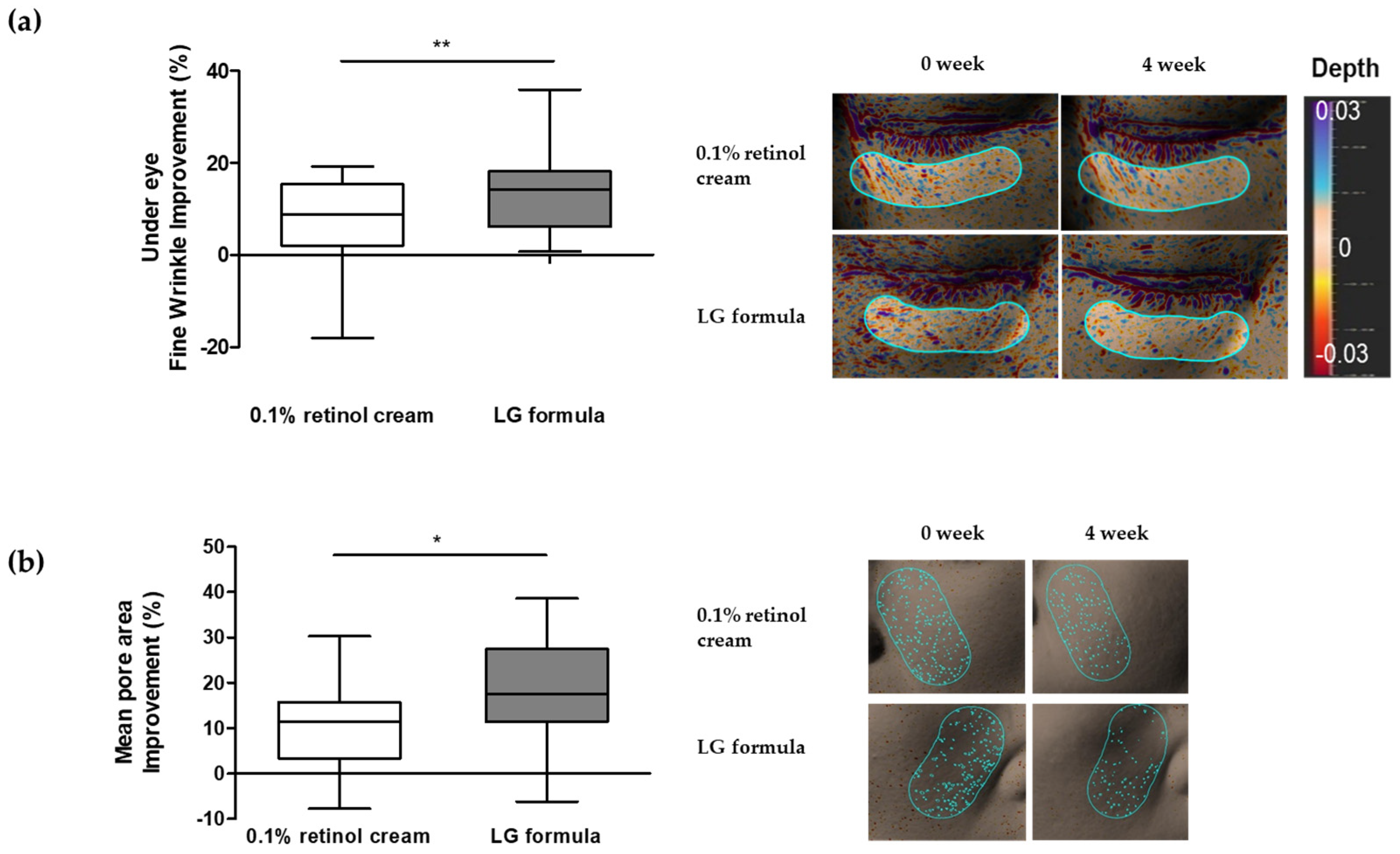
2.6. Human Clinical Trial

This study was ethically approved by the LG H&H Institutional Review Board (LGHH20221110-AA-01-01; 10 November 2022). Nineteen healthy Korean women between the ages of 40 and 62 (average age: 49.2 years) were recruited. Firstly, we excluded volunteers who did not have enough fine wrinkles or enlarged pores according to our standards. The possible side effects of the study were thoroughly explained to each participant, and written informed consent was obtained before they participated in the clinical trial. Pregnant women or individuals undergoing dermatological procedures were excluded from the test. A double-blind, half-face design was used for the experiment. The test formulation containing specific ingredients, including Fuligo® (2%), Ronacare Balmance® (0.3%), hyaluronic acid (0.05%), and PCE (0.25%), was topically applied to the right side of the participants’ faces twice daily for four weeks. For the control, 0.1% retinol formulation without the active ingredients mentioned above was applied on the left side of the face. Before facial wrinkle measurements, all participants were required to rest for a minimum of 20 min in a controlled environment with a specific humidity (45 ± 5%) and temperature (22 ± 2 °C) after their faces were cleansed. Wrinkles on the participants’ faces were measured using an Antera 3D camera (Miravex, Dublin, Ireland). To analyze the effectiveness of the formulation, texture Ra and pore mean area parameters were used to assess changes in fine wrinkles under the eyes and the average pore area of the cheek, respectively.

2.7. Statistical Analysis

The data are presented as the mean value and standard deviation derived from at least three independent experiments. Statistical analysis of data was performed using the Student’s t-test. A p-value less than 0.05 (* p < 0.05, ** p < 0.01, *** p < 0.001) indicated a significant difference.

3. Results and Discussion

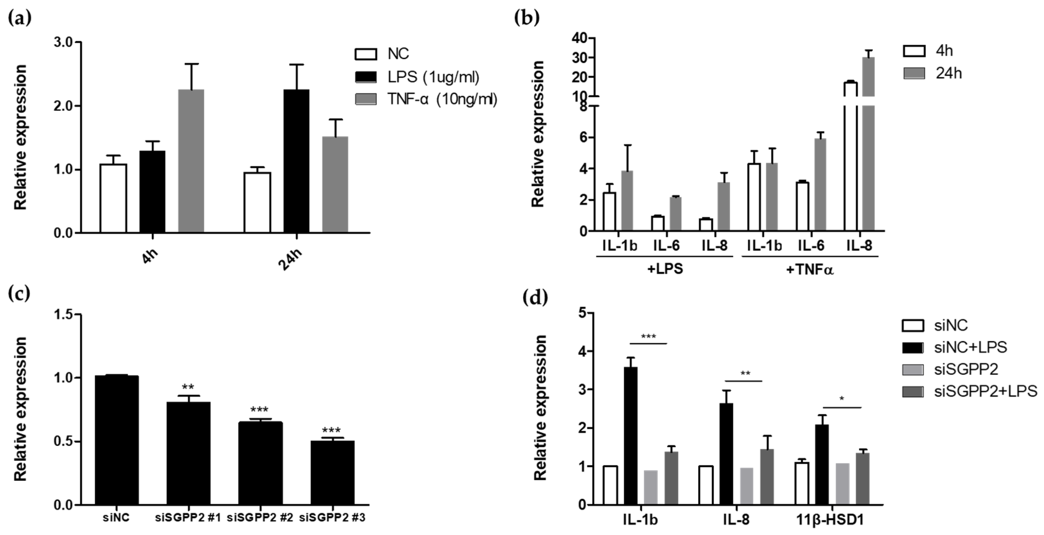
3.1. Functional Study of Wrinkle-Related Gene SGPP2 in HaCaT Cells

SGPP2 is a gene primarily expressed under inflammatory conditions [27]. While SGPP1 has been implicated in skin keratinocyte differentiation, the role of SGPP2 in this context remains unclear. However, SGPP2 has been shown to contribute to the production of IL-1β and IL-8, inflammatory cytokines induced by the TNF-α inflammatory cytokine in endothelial cells, and plays a major role in pro-inflammatory signaling. SGPP2 is reportedly expressed within 24 h, particularly within a short duration, in response to inflammatory stimuli such as LPS or TNF-α, and siRNA-mediated inhibition of SGPP2 expression also suppresses the IL-1β and IL-8 inflammatory response. Since these inflammatory responses can be involved in various processes of wrinkle formation and pore size regulation, it is necessary to confirm whether SGPP2 is also involved in the inflammatory response in skin cells to determine its association with skin wrinkle formation.
Therefore, to investigate whether this phenomenon also occurs in keratinocytes, we examined the transcriptional level of SGPP2 in keratinocytes treated with LPS and TNF-α at 4 and 24 h post-treatment (Figure 1a). Similar to the observations in endothelial cells [27], we found that SGPP2 upregulation was more rapid upon TNF-α treatment. The transcriptional level of SGPP2 was 2.25-fold higher 4 h after TNF-α treatment, while a similar transcriptional level was observed 24 h after LPS treatment. We also examined the transcriptional levels of IL-1β, IL-6, and IL-8, which are the cytokines that share downstream signaling pathways at the same time points. This analysis revealed a pattern of increased expression of inflammatory cytokines along with SGPP2 upregulation (Figure 1b). The differences in the response patterns of SGPP2 upregulation and inflammatory cytokine expression to LPS and TNF-α, respectively, are believed to be owing to differences in the response rates of NF-κB signaling induced by their respective receptors [31]. Based on the findings presented in Figure 1a,b, we confirmed that SGPP2 upregulation and the expression of other inflammatory cytokines occur in skin keratinocytes upon LPS or TNF-α treatment.
To further investigate whether SGPP2 downregulation directly regulates the inflammatory cytokines IL-1β and IL-8, we performed additional experiments using siSGPP2-transfected keratinocytes to assess their response to LPS. As shown in Figure 1c, siSGPP2 transfection significantly reduced SGPP2 mRNA expression compared with that in the control cells. Notably, SGPP2 knockdown significantly attenuated LPS-induced expression of IL-1β and IL-8 and inhibited 11β-HSD1 upregulation (Figure 1d), which is involved in the inflammatory response, interacting with NF-kB, regulating IL-6 production upstream, and is associated with skin barrier stress formation [32,33,34]. While the extent to which the enzymatic activity of SGPP2 influences S1P or sphingosine levels remains unclear, its minimal impact on skin and blood sphingosine/ceramide concentrations in knockout mice, along with previous studies highlighting the primary role of SGPP1 in S1P regulation, suggests SGPP2 functions more directly in downstream inflammatory signaling pathways [27]. These findings provide evidence that SGPP2 contributes to LPS-mediated inflammatory cytokine production and skin barrier stress in keratinocytes.

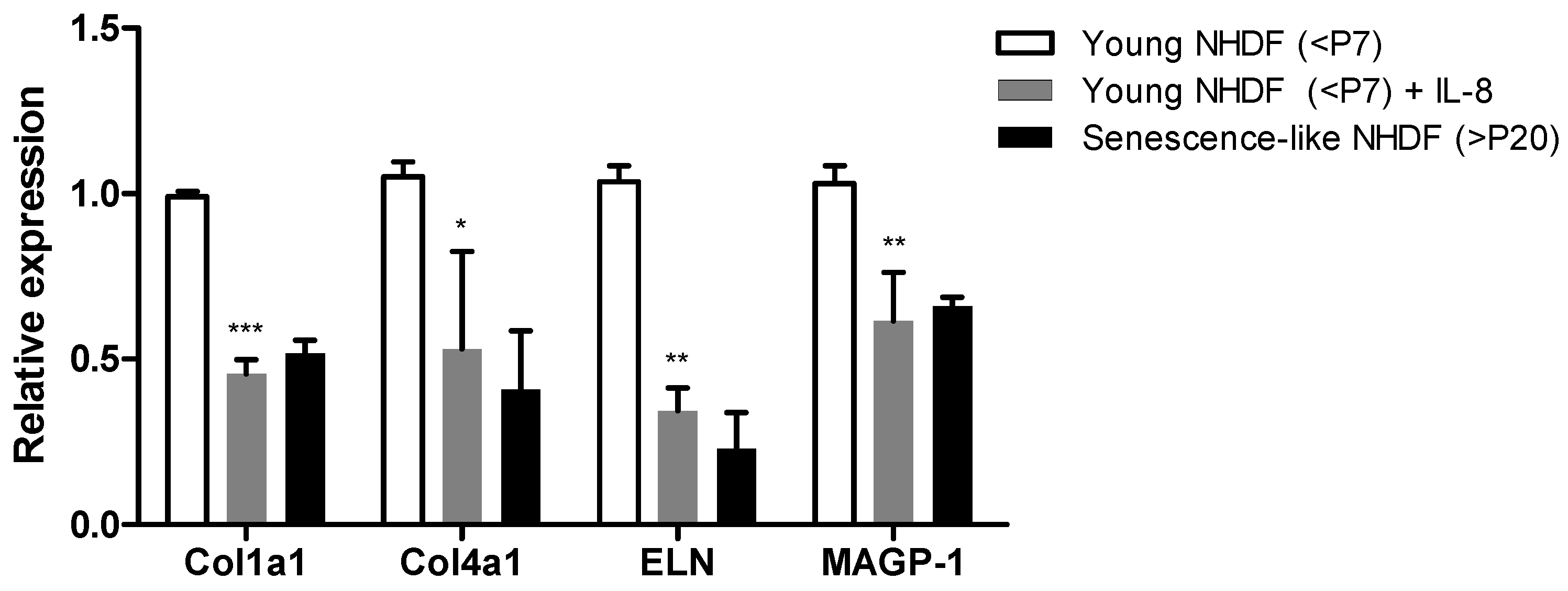
3.2. Inflammation Exacerbates Skin Fibroblast-Mediated Wrinkle Formation

Skin wrinkles, a hallmark of aging, are attributed to the loss of dermal ECM, such as collagen and elastin [35]. Fibroblasts in the dermis are the primary producers of these fibers, and their function can be affected by various factors, including epidermal inflammation [36,37].
Skin fibroblasts exhibiting an “aged” phenotype, characterized by altered morphology, reduced proliferation activity, and high beta-galactosidase (β-gal) activity, have been studied using various methods, including continuous UV exposure, prolonged culture or isolation from elderly donors [38,39,40,41,42]. In vitro, prolonged culture induces a senescence-like state in skin fibroblasts, characterized by increased expression of calponin 1 (CNN1) and alpha-smooth muscle actin (α-SMA), associated with a reticular fibroblast phenotype and decreased expression of podoplanin (PDPN), associated with a papillary fibroblast phenotype. This suggests that in vitro prolonged culture-induced senescence-like skin fibroblasts may also contribute to aging-associated structural changes [41]. In this study, we used NHDFs that had undergone extensive passaging more than 20 times, resulting in a senescence-like state characterized by larger morphology, high expression of CNN1 and α-SMA, and low expression of PDPN (Figure S1).
To elucidate how epidermal inflammation contributes to wrinkle formation through fibroblast dysfunction, we investigated how IL-8, an inflammatory mediator produced under the control of SGPP2 in the epidermis, affects the expression of ECM-related genes (Col1a1, Col4a1, and ELN) and MAGP-1, a crucial component in elastic fiber involved in pore size regulation, in fibroblasts [14,43]. We compared the expression of ECM-related genes in IL-8-treated fibroblasts with early passage (negative control) and senescence-like NHDFs (>P20). In senescence-like NHDFs, Col1a1, Col4a1, ELN, and MAGP-1 were all downregulated, similar to that in early passage fibroblasts treated with IL-8 (Figure 2). While Col1a1, Col4a1, and ELN are known to be regulated by inflammation, the regulation of MAGP-1, an ECM-related gene that organizes elastic fibers and is essential for supporting pore structures [43] by IL-8 treatment, is novel. This experiment confirms that Col1a1, Col4a1, and ELN in skin fibroblasts, which are essential for wrinkle formation, and MAGP-1, related to sagging pores, are upregulated by the SGPP2-regulated inflammatory substance IL-8, thereby promoting an elderly-like phenotype in skin fibroblasts.

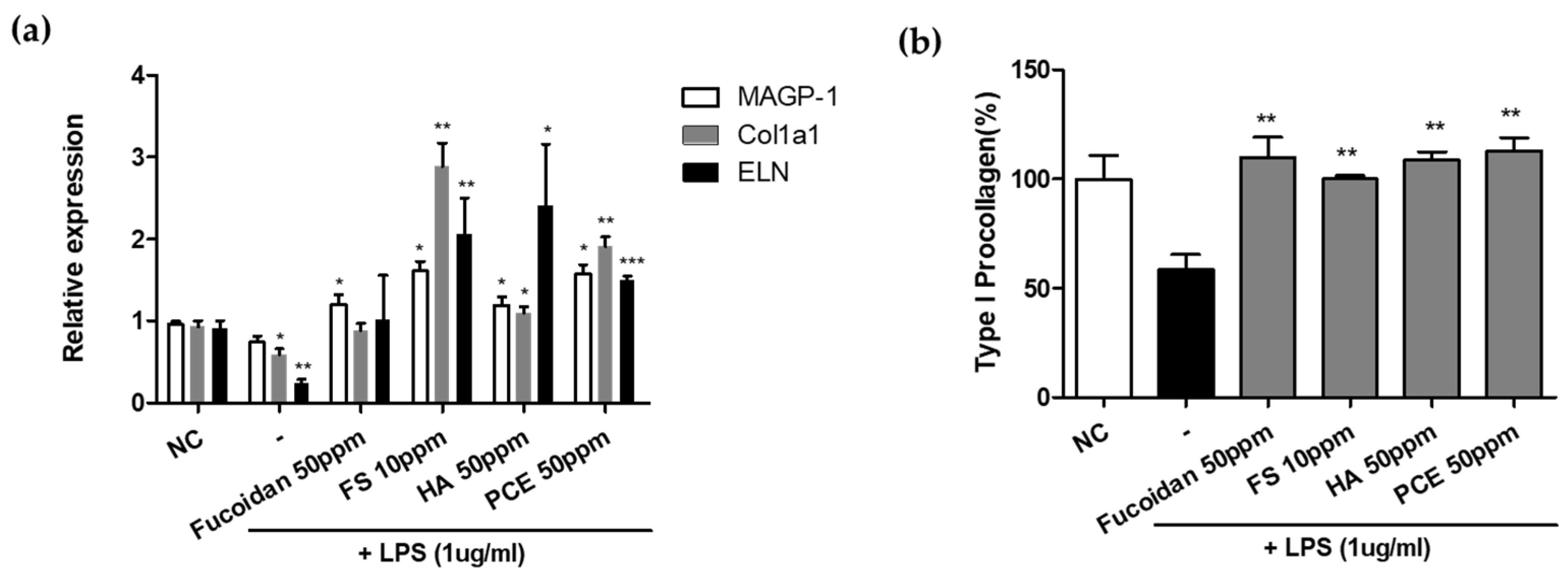
3.3. Screening Active Materials That Regulate SGPP2 Expression

We observed an upregulation of SGPP2 and pro-inflammatory cytokine expression in keratinocytes under an LPS-induced inflammatory condition. This phenomenon downregulates ECM factors in fibroblasts, potentially contributing to skin wrinkle formation.
To investigate the modulation of SGPP2 expression and its impact on skin wrinkle formation and pore sagging, we evaluated the effects of various anti-inflammatory substances on SGPP2 regulation in LPS-induced inflammation. We assessed the ability of fucoidan, feruloyl serotonin, hyaluronic acid, and PCE to downregulate SGPP2 expression in LPS-treated keratinocytes. These substances are known for their anti-inflammatory properties, including reducing LPS-induced pro-inflammatory cytokine expression (fucoidan and feruloyl serotonin) and inhibiting LPS-toll-like receptor 4 binding (hyaluronic acid) [44,45,46]. Our findings revealed that fucoidan (50 and 100 μg/mL) significantly decreased SGPP2 expression by 35% and 61%, respectively, compared with LPS alone (Figure 3a). Similarly, feruloyl serotonin (10 ppm) downregulated SGPP2 expression by 69%. Hyaluronic acid (50 and 100 ppm) also effectively reduced SGPP2 levels by 58% and 67%, respectively. Additionally, PCE (≥10 ppm) exhibited a significant decrease in SGPP2 expression by 65%. We also found that these materials also inhibited LPS-induced IL-8 expression in keratinocytes (Figure 3b). Further analysis of other anti-inflammatory and wrinkle-improving agents demonstrated that L-hydroxyproline, retinol, cedrol, asiaticoside, and ginsenoside Rh1 also significantly downregulated SGPP2 expression (Figure S2).

3.4. Antiaging Effect of SGPP2 Expression-Regulating Materials In Vitro

3.4.1. Recovery of Inflammation-Induced Decrease in ECM-Related Gene in NHDFs

Paracrine signaling, a form of cell-to-cell communication, is crucial in various biological processes. In skin aging and wrinkle formation, the paracrine effects of keratinocytes on fibroblasts are particularly significant. CM experiments are used in vitro to investigate paracrine signaling between two cell populations [29]. Skin fibroblasts play a central role in regulating elastic fibers and are key components associated with wrinkle formation [47]. Our previous findings in Figure 2 demonstrated that IL-8, an inflammatory cytokine potentially induced by inflammation in keratinocytes, downregulates elastic fibers in fibroblasts.
To exclude the direct effects of these substances on collagen regulation in fibroblasts and investigate the paracrine regulation mediated by the inflammation-modulating effect of keratinocytes, we examined whether these substances could mitigate the LPS-induced downregulation of elastic fibers in fibroblasts through paracrine signaling.
CM from LPS-treated keratinocytes significantly downregulated MAGP-1, Col1a1, and ELN expression in fibroblasts by 23%, 37%, and 75%, respectively, compared with the control CM (Figure 4a). Fucoidan-containing CM markedly reversed the LPS-induced decrease in MAGP-1 expression. Additionally, CM containing feruloyl serotonin, hyaluronic acid, or PCE substantially significantly increased the expression of MAGP-1, Col1a1, and ELN, which were downregulated by LPS treatment.
These findings suggest that modulating substances that regulate SGPP2 in keratinocytes can paracrinely influence the regulation of wrinkle-related elastic fibers in dermal fibroblasts. This restoration of elastic fiber expression by modulating substances in CM is possibly attributed to the reduced secretion of inflammatory cytokines such as IL-8 and IL-1b due to SGPP2 inhibition and downregulation of inflammatory signaling in LPS-treated keratinocytes.

3.4.2. Effect of Enhanced Type I Procollagen Synthesis

We discovered that SGPP2-regulating substances applied to keratinocytes in the epidermis can paracrinely affect the regulation of wrinkle-related elastic fibers in dermal fibroblasts.
To validate whether the observed modulation of elastic fiber expression in CM also translates to protein synthesis at the protein level, we assessed type I collagen protein levels, the primary subtype in human skin, using an enzyme-linked immunosorbent assay.
Keratinocytes were treated with LPS alone or LPS in combination with modulating substances (fucoidan, feruloyl serotonin, hyaluronic acid, or PCE) to generate the CM. This CM was applied to dermal skin fibroblasts. Treatment with 50 μg/mL fucoidan, 50 μg/mL feruloyl serotonin, 50 μg/mL hyaluronic acid, and 50 μg/mL PCE resulted in 1.23-fold, 1-fold, 1.21-fold, and 1.31-fold increases in type I collagen restoration compared with that of LPS-only CM (Figure 4b). These results demonstrate that the paracrine effects of keratinocytes treated with these four modulating substances regulate collagen transcription and impact protein expression levels in fibroblasts.

3.5. Dermal Collagen Enhances the Efficacy of the Selected Materials within 3D Skin Model

While 2D monolayer cell culture systems offer convenience for studying molecular mechanisms, they may not accurately reflect in vivo conditions. Therefore, to assess the efficacy of our material in a more realistic environment, we used a reconstructed 3D human skin model (Neoderm®-ED) [48,49].
We applied creams containing our four active materials (LG formula) to the 3D skin model and compared the results to those obtained with a vehicle cream. Through a comprehensive evaluation of in vitro efficacy concentrations, stability, and solubility, an optimal combination of the four active materials was determined and formulated. After treatment for 48 h, the relative area of dermal collagen significantly increased by +68.1% in the LG formula-treated samples (Figure 5a). This enhancement in the dermal collagen area was further confirmed through histological analysis (Figure 5b).
These results demonstrate that the four active materials, when incorporated into a cosmetic formulation, can promote collagen production and potentially contribute to wrinkle improvement in a 2D cell culture and also in a more human-like 3D culture environment.

3.6. Improvement in Skin Wrinkles and Pores Using LG Formula-Containing Materials That Modulated SGPP2 Expression

Subsequently, we conducted an experiment to determine if the formulation containing four types of active materials improved the appearance of facial wrinkles and pores. Based on the 3D human skin model test, we formulated LG formulas containing each of the four active materials and tested them to determine if they improved the appearance of wrinkles on the under eye and pores. To accurately measure the efficacy of the LG formula, a half-test method experiment was conducted, where 0.1% retinol cream of the same base formula was applied to the left side of the face, and the LG formula cream containing four active substances was applied to the right side of the face. To assess the efficacy of four active material complexes, we selected 0.1% retinol cream as a control, given its established reputation as a highly effective anti-wrinkle ingredient. The changes in the area of facial wrinkles and pores were measured with Antera 3D, among which the Ra value of texture, which is a common parameter for measuring fine wrinkles [50], and the average pore area, which is a suitable parameter for showing stretched pores or pore elongation, were measured [15,51].
After applying both creams once daily for four weeks, the results showed a 14.3% improvement in fine wrinkles in the under-eye area (Figure 6a) and a 17.22% improvement in the mean pore area (Figure 6b). In contrast, the 0.1% retinol cream alone showed a relatively low improvement rate of 6.52% and 10.13% in the control group, showing significant differences in under-eye fine wrinkle and mean pore area improvements, respectively.
Although retinol alone can improve under-eye fine wrinkles and stretched pores, individual differences in retinol reactivity exist, and the perception of wrinkle improvement may be lower than expected owing to irritation (such as dryness, redness, and hyperkeratinization) caused by retinol. However, this study shows that LG formula with various ingredient combinations can provide similar or better results than retinol, a well-known anti-wrinkle ingredient, in reducing under-eye fine and pore wrinkles without irritation. Although a significant level of improvement was confirmed after four weeks of use, the limited sample size and experimental duration of this study preclude definitive conclusions about the long-term efficacy of the product. While the observed improvements are encouraging, further research with a larger sample size and extended treatment periods is necessary to fully assess the potential benefits of the formula [52,53].
In this study, based on the hypothesis that SGPP2 is involved in the development of facial wrinkles, we showed how four materials that modulate SGPP2 can ameliorate facial wrinkles in vitro and in vivo. However, the effect of SGPP2 as a phosphatase enzyme and the limited conditions, such as sample size and the duration of clinical study, are the limitations of this study. An in-depth investigation of SGPP2 signaling and S1P after inflammatory stimuli is required to further clarify the role of SGPP2 in wrinkle formation.

4. Conclusions

In this study, we highlighted the potential of regulating SGPP2 in keratinocytes, which influences LPS-induced pro-inflammatory signaling and inflammatory cytokines such as IL-8, commonly known as SASP. This pro-inflammatory cytokine acts on dermal fibroblasts, prompting the expression of ECM-related genes associated with an aged phenotype.
We identified four active substances that effectively reduce SGPP2 expression under inflammatory conditions. Using CM experiments, we confirmed their potential to alleviate the inhibition of dermal collagen production caused by epidermal inflammation. To assess the applicability beyond 2D cultures, the application of a cosmetic formulation (LG formula) containing the four selected substances demonstrated a significant improvement in collagen synthesis in a 3D skin model. This indicates their effectiveness in an environment mimicking human skin conditions.
Finally, a clinical evaluation was conducted using the LG formula. Compared with 0.1% retinol, a well-established anti-wrinkle treatment, the LG formula demonstrated superior efficacy in improving fine wrinkles under the eyes and enlarged/elongated pores.
This study elucidates the potential of SGPP2 modulation as a novel anti-wrinkle strategy that mitigates inflammation. The identification of these four active ingredients is promising for the development of effective antiaging products that address a broader spectrum of facial wrinkles, including pore enlargement and elongation.

Supplementary Materials

The following supporting information can be downloaded at https://www.mdpi.com/article/10.3390/cimb46080539/s1, Figure S1. Changes in morphology and gene expression (CNN1, αSMA, PDPN) of NHDFs under prolonged culture conditions (more than 20 passages). Figure S2: The effect of anti-inflammatory and wrinkle-improving materials on SGPP2 expression.

Author Contributions

Conceptualization, J.K., S.Y., S.-H.J. and N.-G.K.; data curation J.K.; formal analysis J.K.; investigation, J.K. and S.Y.; methodology J.K. and S.Y.; project administration, S.-H.J. and N.-G.K.; resources, J.K.; software, J.K.; supervision, S.-H.J. and N.-G.K.; validation, J.K., S.Y., S.-H.J. and N.-G.K.; writing of the original draft, J.K.; writing of the review and editing, J.K. and S.-H.J. All authors have read and agreed to the published version of the manuscript.

Funding

This research was funded by LG H&H.

Institutional Review Board Statement

This study was conducted according to the guidelines of the Declaration of Helsinki and approved by the Ethics Committee of LG H&H Institutional Review Board (LGHH20221110-AA-01-01, 10 November 2022).

Informed Consent Statement

Informed consent was obtained from all subjects involved in the study.

Data Availability Statement

The data that support the findings of this study will be made available by the corresponding author upon request.

Conflicts of Interest

The authors Juhyun Kim, Sanghyun Ye, Seung-Hyun Jun, and Nae-Gyu Kang were employed by the company LG Household and Healthcare Ltd. The company LG Household and Health Care had no role in the design of the study; in the collection, analyses, or interpretation of the data; in the writing of the manuscript; or in the decision to publish the results.

References

  1. Wang, A.S.; Dreesen, O. Biomarkers of Cellular Senescence and Skin Aging. Front. Genet. 2018, 9, 247. [Google Scholar] [CrossRef]
  2. Fuller, B. Role of PGE-2 and Other Inflammatory Mediators in Skin Aging and Their Inhibition by Topical Natural Anti-Inflammatories. Cosmetics 2019, 6, 6. [Google Scholar] [CrossRef]
  3. Salminen, A.; Kauppinen, A.; Kaarniranta, K. Emerging role of NF-kappaB signaling in the induction of senescence-associated secretory phenotype (SASP). Cell Signal. 2012, 24, 835–845. [Google Scholar] [CrossRef]
  4. Budamagunta, V.; Manohar-Sindhu, S.; Yang, Y.; He, Y.; Traktuev, D.O.; Foster, T.C.; Zhou, D. Senescence-associated hyper-activation to inflammatory stimuli in vitro. Aging 2021, 13, 19088–19107. [Google Scholar] [CrossRef] [PubMed]
  5. Bielak-Zmijewska, A.; Grabowska, W.; Ciolko, A.; Bojko, A.; Mosieniak, G.; Bijoch, L.; Sikora, E. The Role of Curcumin in the Modulation of Ageing. Int. J. Mol. Sci. 2019, 20, 1239. [Google Scholar] [CrossRef] [PubMed]
  6. Shim, J.H. Prostaglandin E2 Induces Skin Aging via E-Prostanoid 1 in Normal Human Dermal Fibroblasts. Int. J. Mol. Sci. 2019, 20, 5555. [Google Scholar] [CrossRef]
  7. Baechle, J.J.; Chen, N.; Makhijani, P.; Winer, S.; Furman, D.; Winer, D.A. Chronic inflammation and the hallmarks of aging. Mol. Metab. 2023, 74, 101755. [Google Scholar] [CrossRef]
  8. Chin, T.; Lee, X.E.; Ng, P.Y.; Lee, Y.; Dreesen, O. The role of cellular senescence in skin aging and age-related skin pathologies. Front. Physiol. 2023, 14, 1297637. [Google Scholar] [CrossRef]
  9. Zhang, J.; Yu, H.; Man, M.Q.; Hu, L. Aging in the dermis: Fibroblast senescence and its significance. Aging Cell 2024, 23, e14054. [Google Scholar] [CrossRef]
  10. Jang, S.I.; Kim, E.J.; Lee, H.K. A method of evaluating facial pores using optical 2D images and analysis of age-dependent changes in facial pores in Koreans. Skin Res. Technol. 2018, 24, 304–308. [Google Scholar] [CrossRef]
  11. Jung, H.J.; Ahn, J.Y.; Lee, J.I.; Bae, J.Y.; Kim, H.L.; Suh, H.Y.; Youn, J.I.; Park, M.Y. Analysis of the number of enlarged pores according to site, age, and sex. Skin Res. Technol. 2018, 24, 367–370. [Google Scholar] [CrossRef] [PubMed]
  12. Dissanayake, B.; Miyamoto, K.; Purwar, A.; Chye, R.; Matsubara, A. New image analysis tool for facial pore characterization and assessment. Skin Res. Technol. 2019, 25, 631–638. [Google Scholar] [CrossRef]
  13. Lee, S.; Cherel, M.; Gougeon, S.; Jeong, E.; Lim, J.M.; Park, S.G. Identifying patterns behind the changes in skin pores using 3-dimensional measurements and K-means clustering. Skin Res. Technol. 2022, 28, 3–9. [Google Scholar] [CrossRef] [PubMed]
  14. Dong, J.; Lanoue, J.; Goldenberg, G. Enlarged facial pores: An update on treatments. Cutis 2016, 98, 33–36. [Google Scholar]
  15. Messaraa, C.; Metois, A.; Walsh, M.; Flynn, J.; Doyle, L.; Robertson, N.; Mansfield, A.; O’Connor, C.; Mavon, A. Antera 3D capabilities for pore measurements. Skin Res. Technol. 2018, 24, 606–613. [Google Scholar] [CrossRef]
  16. Nkengne, A.; Pellacani, G.; Ciardo, S.; De Carvalho, N.; Vie, K. Visible characteristics and structural modifications relating to enlarged facial pores. Skin Res. Technol. 2021, 27, 560–568. [Google Scholar] [CrossRef] [PubMed]
  17. Qiu, W.; Chen, F.; Feng, X.; Shang, J.; Luo, X.; Chen, Y. Potential role of inflammaging mediated by the complement system in enlarged facial pores. J. Cosmet. Dermatol. 2024, 23, 27–32. [Google Scholar] [CrossRef]
  18. Inui, S.; Mori, A.; Ito, M.; Hyodo, S.; Itami, S. Reduction of conspicuous facial pores by topical fullerene: Possible role in the suppression of PGE2 production in the skin. J. Nanobiotechnol. 2014, 12, 6. [Google Scholar] [CrossRef]
  19. Lee, S.G.; Shin, J.G.; Kim, Y.; Leem, S.; Park, S.G.; Won, H.H.; Kang, N.G. Identification of Genetic Loci Associated with Facial Wrinkles in a Large Korean Population. J. Investig. Dermatol. 2022, 142, 2824–2827. [Google Scholar] [CrossRef]
  20. Cha, M.-Y.; Choi, J.-E.; Lee, D.-S.; Lee, S.-R.; Lee, S.-I.; Park, J.-H.; Shin, J.-H.; Suh, I.S.; Kim, B.H.; Hong, K.-W. Novel Genetic Associations for Skin Aging Phenotypes and Validation of Previously Reported Skin GWAS Results. Appl. Sci. 2022, 12, 11422. [Google Scholar] [CrossRef]
  21. Lee, S.; Ye, S.; Kim, M.; Lee, H.; Jun, S.H.; Kang, N.G. Fine Wrinkle Improvement through Bioactive Materials That Modulate EDAR and BNC2 Gene Expression. Biomolecules 2024, 14, 279. [Google Scholar] [CrossRef]
  22. Lee, H.; Ye, S.; Kim, J.; Jun, S.H.; Kang, N.G. Improvement in Facial Wrinkles Using Materials Enhancing PPARGC1B Expression Related to Mitochondrial Function. Curr. Issues Mol. Biol. 2024, 46, 5037–5051. [Google Scholar] [CrossRef]
  23. Kleuser, B.; Baumer, W. Sphingosine 1-Phosphate as Essential Signaling Molecule in Inflammatory Skin Diseases. Int. J. Mol. Sci. 2023, 24, 1456. [Google Scholar] [CrossRef]
  24. Noujarede, J.; Carrie, L.; Garcia, V.; Grimont, M.; Eberhardt, A.; Mucher, E.; Genais, M.; Schreuder, A.; Carpentier, S.; Segui, B.; et al. Sphingolipid paracrine signaling impairs keratinocyte adhesion to promote melanoma invasion. Cell Rep. 2023, 42, 113586. [Google Scholar] [CrossRef]
  25. Allende, M.L.; Sipe, L.M.; Tuymetova, G.; Wilson-Henjum, K.L.; Chen, W.; Proia, R.L. Sphingosine-1-phosphate phosphatase 1 regulates keratinocyte differentiation and epidermal homeostasis. J. Biol. Chem. 2013, 288, 18381–18391. [Google Scholar] [CrossRef]
  26. Baumer, W.; Rossbach, K.; Mischke, R.; Reines, I.; Langbein-Detsch, I.; Luth, A.; Kleuser, B. Decreased concentration and enhanced metabolism of sphingosine-1-phosphate in lesional skin of dogs with atopic dermatitis: Disturbed sphingosine-1-phosphate homeostasis in atopic dermatitis. J. Investig. Dermatol. 2011, 131, 266–268. [Google Scholar] [CrossRef]
  27. Mechtcheriakova, D.; Wlachos, A.; Sobanov, J.; Kopp, T.; Reuschel, R.; Bornancin, F.; Cai, R.; Zemann, B.; Urtz, N.; Stingl, G.; et al. Sphingosine 1-phosphate phosphatase 2 is induced during inflammatory responses. Cell Signal. 2007, 19, 748–760. [Google Scholar] [CrossRef]
  28. Jeong, E.T.; Jin, M.H.; Kim, M.S.; Chang, Y.H.; Park, S.G. Inhibition of melanogenesis by piceid isolated from Polygonum cuspidatum. Arch. Pharm. Res. 2010, 33, 1331–1338. [Google Scholar] [CrossRef]
  29. Seok, J.K.; Boo, Y.C. p-Coumaric Acid Attenuates UVB-Induced Release of Stratifin from Keratinocytes and Indirectly Regulates Matrix Metalloproteinase 1 Release from Fibroblasts. Korean J. Physiol. Pharmacol. 2015, 19, 241–247. [Google Scholar] [CrossRef]
  30. Oh, S.R.; Park, S.K.; Lee, P.; Kim, Y.M. The ginsenoside Rg2 downregulates MMP-1 expression in keratinocyte (HaCaT)-conditioned medium-treated human fibroblasts (Hs68). Appl. Biol. Chem. 2023, 66, 85. [Google Scholar] [CrossRef]
  31. Cheong, R.; Hoffmann, A.; Levchenko, A. Understanding NF-kappaB signaling via mathematical modeling. Mol. Syst. Biol. 2008, 4, 192. [Google Scholar] [CrossRef]
  32. Choe, S.J.; Kim, D.; Kim, E.J.; Ahn, J.S.; Choi, E.J.; Son, E.D.; Lee, T.R.; Choi, E.H. Psychological Stress Deteriorates Skin Barrier Function by Activating 11beta-Hydroxysteroid Dehydrogenase 1 and the HPA Axis. Sci. Rep. 2018, 8, 6334. [Google Scholar] [CrossRef]
  33. Ishii-Yonemoto, T.; Masuzaki, H.; Yasue, S.; Okada, S.; Kozuka, C.; Tanaka, T.; Noguchi, M.; Tomita, T.; Fujikura, J.; Yuji Yamamoto, Y.; et al. Glucocorticoid reamplification within cells intensifies NF-κB and MAPK signaling and reinforces inflammation in activated preadipocytes. Am. J. Physiol. Endocrinol. Metab. 2010, 298, E930–E940. [Google Scholar]
  34. Itoi, S.; Terao, M.; Murota, H.; Katayama, I. 11beta-Hydroxysteroid dehydrogenase 1 contributes to the pro-inflammatory response of keratinocytes. Biochem. Biophys. Res. Commun. 2013, 440, 265–270. [Google Scholar] [CrossRef]
  35. Cannarozzo, G.; Fazia, G.; Bennardo, L.; Tamburi, F.; Amoruso, G.F.; Del Duca, E.; Nistico, S.P. A New 675 nm Laser Device in the Treatment of Facial Aging: A Prospective Observational Study. Photobiomodul Photomed. Laser Surg. 2021, 39, 118–122. [Google Scholar] [CrossRef]
  36. Varani, J.; Dame, M.K.; Rittie, L.; Fligiel, S.E.; Kang, S.; Fisher, G.J.; Voorhees, J.J. Decreased collagen production in chronologically aged skin: Roles of age-dependent alteration in fibroblast function and defective mechanical stimulation. Am. J. Pathol. 2006, 168, 1861–1868. [Google Scholar] [CrossRef]
  37. Lee, D.H.; Oh, J.H.; Chung, J.H. Glycosaminoglycan and proteoglycan in skin aging. J. Dermatol. Sci. 2016, 83, 174–181. [Google Scholar] [CrossRef]
  38. Weinmullner, R.; Zbiral, B.; Becirovic, A.; Stelzer, E.M.; Nagelreiter, F.; Schosserer, M.; Lammermann, I.; Liendl, L.; Lang, M.; Terlecki-Zaniewicz, L.; et al. Organotypic human skin culture models constructed with senescent fibroblasts show hallmarks of skin aging. npj Aging Mech. Dis. 2020, 6, 4. [Google Scholar] [CrossRef]
  39. Lago, J.C.; Puzzi, M.B. The effect of aging in primary human dermal fibroblasts. PLoS ONE 2019, 14, e0219165. [Google Scholar] [CrossRef]
  40. Lammermann, I.; Terlecki-Zaniewicz, L.; Weinmullner, R.; Schosserer, M.; Dellago, H.; de Matos Branco, A.D.; Autheried, D.; Sevcnikar, B.; Kleissl, L.; Berlin, I.; et al. Blocking negative effects of senescence in human skin fibroblasts with a plant extract. npj Aging Mech. Dis. 2018, 4, 4. [Google Scholar] [CrossRef]
  41. Kamiya, Y.; Odama, M.; Mizuguti, A.; Murakami, S.; Ito, T. Puerarin blocks the aging phenotype in human dermal fibroblasts. PLoS ONE 2021, 16, e0249367. [Google Scholar] [CrossRef]
  42. Yang, J.; Dungrawala, H.; Hua, H.; Manukyan, A.; Abraham, L.; Lane, W.; Mead, H.; Wright, J.; Schneider, B.L. Cell size and growth rate are major determinants of replicative lifespan. Cell Cycle 2011, 10, 144–155. [Google Scholar] [CrossRef]
  43. Zheng, Q.; Chen, S.; Chen, Y.; Lyga, J.; Wyborski, R.; Santhanam, U. Investigation of age-related decline of microfibril-associated glycoprotein-1 in human skin through immunohistochemistry study. Clin. Cosmet. Investig. Dermatol. 2013, 6, 317–323. [Google Scholar] [CrossRef] [PubMed]
  44. Han, W.; Lv, Y.; Sun, Y.; Wang, Y.; Zhao, Z.; Shi, C.; Chen, X.; Wang, L.; Zhang, M.; Wei, B.; et al. The anti-inflammatory activity of specific-sized hyaluronic acid oligosaccharides. Carbohydr. Polym. 2022, 276, 118699. [Google Scholar] [CrossRef]
  45. Fernando, I.P.S.; Dias, M.; Madusanka, D.M.D.; Han, E.J.; Kim, M.J.; Heo, S.J.; Lee, K.; Cheong, S.H.; Ahn, G. Low molecular weight fucoidan fraction ameliorates inflammation and deterioration of skin barrier in fine-dust stimulated keratinocytes. Int. J. Biol. Macromol. 2021, 168, 620–630. [Google Scholar] [CrossRef]
  46. Shanura Fernando, I.P.; Asanka Sanjeewa, K.K.; Samarakoon, K.W.; Kim, H.-S.; Gunasekara, U.K.D.S.S.; Park, Y.-J.; Abeytunga, D.T.U.; Lee, W.W.; Jeon, Y.-J. The potential of fucoidans from Chnoospora minima and Sargassum polycystum in cosmetics: Antioxidant, anti-inflammatory, skin-whitening, and antiwrinkle activities. J. Appl. Phycol. 2018, 30, 3223–3232. [Google Scholar] [CrossRef]
  47. Zorina, A.; Zorin, V.; Isaev, A.; Kudlay, D.; Vasileva, M.; Kopnin, P. Dermal Fibroblasts as the Main Target for Skin Anti-Age Correction Using a Combination of Regenerative Medicine Methods. Curr. Issues Mol. Biol. 2023, 45, 3829–3847. [Google Scholar] [CrossRef] [PubMed]
  48. Langhans, S.A. Three-Dimensional in Vitro Cell Culture Models in Drug Discovery and Drug Repositioning. Front. Pharmacol. 2018, 9, 6. [Google Scholar] [CrossRef]
  49. Zhang, Z.; Michniak-Kohn, B.B. Tissue engineered human skin equivalents. Pharmaceutics 2012, 4, 26–41. [Google Scholar] [CrossRef] [PubMed]
  50. Messaraa, C.; Metois, A.; Walsh, M.; Hurley, S.; Doyle, L.; Mansfield, A.; O’Connor, C.; Mavon, A. Wrinkle and roughness measurement by the Antera 3D and its application for evaluation of cosmetic products. Skin Res. Technol. 2018, 24, 359–366. [Google Scholar] [CrossRef] [PubMed]
  51. Lu, S.; Zhang, S.; Wang, Y.; Ni, J.; Zhao, T.; Xiao, G. Anti-skin aging effects and bioavailability of collagen tripeptide and elastin peptide formulations in young and middle-aged women. J. Dermatol. Sci. Cosmet. Technol. 2024, 1, 100019. [Google Scholar]
  52. Kong, R.; Cui, Y.; Fisher, G.J.; Wang, X.; Chen, Y.; Schneider, L.M.; Majmudar, G. A comparative study of the effects of retinol and retinoic acid on histological, molecular, and clinical properties of human skin. J. Cosmet. Dermatol. 2016, 15, 49–57. [Google Scholar] [CrossRef] [PubMed]
  53. Tucker-Samaras, S.; Zedayko, T.; Cole, C.; Miller, D.; Wallo, W.; Leyden, J.J. A stabilized 0.1% retinol facial moisturizer improves the appearance of photodamaged skin in an eight-week, double-blind, vehicle-controlled study. J. Drugs Dermatol. 2009, 8, 932–936. [Google Scholar] [PubMed]
Figure 1. SGPP2 expression and SGPP2 knockdown effects in HaCaTs under inflammatory conditions. (a) Changes in sphingosine-1-phosphate phosphatase 2 (SGPP2) gene expression over time in lipopolysaccharide (LPS)-treated (1 ug/mL) and tumor necrosis factor (TNF)-α treated (10 ng/mL) HaCaT cells (b) Expression of other inflammatory cytokines in LPS-treated and TNF-α treated HaCaT cells (c) Downregulation of SGPP2 expression using three siRNAs (siSGPP2) compared to negative control siRNA (siNC) (d) Expression of genes associated with skin inflammation and barrier stress-related genes in SGPP2-knockdown cells. Bars indicate standard deviation (n = 3). *** p < 0.001, ** p < 0.01, * p < 0.05; Student’s t-test.
Cimb 46 00539 g001
Figure 2. Expression of genes associated with human skin ECM under inflammatory conditions compared to senescence-like normal human dermal fibroblasts (NHDFs). Relative expression of Col1a1, Col4a1, ELN, and MAGP-1 in young NHDFs with or without IL-8 (10 ng/mL) for 24 h and in senescence-like NHDFs, which were cultured for more than 20 passages. Bars indicate standard deviation (n = 3). Data were compared between young NHDFs treated with or without IL-8. *** p < 0.001, ** p < 0.01, * p < 0.05; Student’s t-test.
Cimb 46 00539 g002
Figure 3. Screening materials with the ability to downregulate SGPP2 expression and IL-8 inflammatory cytokine. (a) Relative expression of SGPP2 in keratinocytes (HaCaT) treated with Fucoidan, Feruloyl Serotonin (FS), Hyaluronic acid (HA), and Polygonum cuspidatum root extract (PCE) at the mentioned concentrations. All samples except Negative control (NC) are treated with LPS (1 ug/mL). (b) Anti-inflammatory effect of SGPP2-downregulating materials that decreases IL-8 expression in LPS-treated keratinocytes. Bars indicate standard deviation (n = 3). *** p < 0.001, ** p < 0.01, * p < 0.05; Student’s t-test.
Cimb 46 00539 g003
Figure 4. Analysis of ECM-enhancing effects in dermal fibroblast of SGPP2 expression-regulating materials through in vitro experiments. (a) Relative expression of MAGP-1, Col1a1, and ELN genes in normal human dermal fibroblast treated with conditioned medium (CM) for 24 h produced by LPS-treated HaCaT cells cultured in Fucoidan, Feruloyl Serotonin (FS), Hyaluronic acid (HA) and Polygonum cuspidatum Root extract (PCE) for 24 h at the mentioned concentrations. (b) Analysis of changes in type I collagen synthesis in human dermal fibroblasts treated with CM. Bars indicate standard deviation (n = 3). Data were compared between LPS-treated groups treated with and without each substance. *** p < 0.001, ** p < 0.01, * p < 0.05; Student’s t-test.
Cimb 46 00539 g004
Figure 5. Dermal collagen upregulation in 3D skin model by treatment with formulations containing SGPP2 modulating materials. (a) Effect of two cram formulations on increasing dermal collagen area in 3D skin equivalent; (b) Representative images cross sections of 3D skin equivalent; scale bar = 275 um. Black arrows indicate the dermis as visualized with Masson Trichrome stain. Bars indicate standard deviation (n = 3). ** p < 0.01; Student’s t-test.
Cimb 46 00539 g005
Figure 6. Facial wrinkle and pore improvement by the LG formula containing SGPP2 expression-regulating materials. (a) Comparison of the undereye fine wrinkle improvement rate; (b) Comparison of the mean pore area improvement rate between LG formula- and 0.1% retinol-treated groups after four weeks. Representative images were acquired using an Antera 3D camera both pre- and post-treatment (4 weeks). Bars indicate standard deviation (n = 19). ** p < 0.01, * p < 0.05; Student’s t-test.
Cimb 46 00539 g006
Disclaimer/Publisher’s Note: The statements, opinions and data contained in all publications are solely those of the individual author(s) and contributor(s) and not of MDPI and/or the editor(s). MDPI and/or the editor(s) disclaim responsibility for any injury to people or property resulting from any ideas, methods, instructions or products referred to in the content.

