Article

Gibberellins Play an Essential Role in the Bud Growth of Petunia hybrida

College of Horticulture, Anhui Agricultural University, Hefei 230036, China
*
Author to whom correspondence should be addressed.
These authors contributed equally to this work.
Curr. Issues Mol. Biol. 2024, 46(9), 9906-9915; https://doi.org/10.3390/cimb46090590
Submission received: 1 August 2024 / Revised: 23 August 2024 / Accepted: 3 September 2024 / Published: 5 September 2024
(This article belongs to the Section Molecular Plant Sciences)

Abstract

:
This study delves into the role of gibberellin (GA) in governing plant branch development, a process that remains incompletely understood. Through a combination of exogenous hormone treatment, gene expression analysis, and transgenic phenotype investigations, the impact of GA on petunia’s branch development was explored. The results showed that GA3 alone did not directly induce axillary bud germination. However, paclobutrazol (PAC), an inhibitor of GA synthesis, effectively inhibited bud growth. Interestingly, the simultaneous application of GA3 and 6-BA significantly promoted bud growth in both intact and decapitated plants compared to using 6-BA alone. Moreover, this study observed a significant downregulation of GA synthesis genes, including GA20ox1, GA20ox2, GA20ox3, GA3ox1, and CPS1, alongside an upregulation of GA degradation genes such as GA2ox2, GA2ox4, and GA2ox8. The expression of GA signal transduction gene GID1 and GA response factor RGA was found to be upregulated. Notably, the PhGID1 gene, spanning 1029 bp and encoding 342 amino acids, exhibited higher expression in buds and the lowest expression in leaves. The overexpression of PhGID1 in Arabidopsis resulted in a noteworthy rise in the number of branches. This study highlights the crucial role of GA in bud germination and growth and the positive regulatory function of GA signaling in shoot branching processes.

1. Introduction

Plants are inevitably exposed to complex environmental stresses throughout their lifecycle. Consequently, they have evolved developmental plasticity to cope with adverse conditions. A notable example is the regulation of branching in plants. During this process, axillary meristem forms in the leaf axils and gradually develops into axillary buds, which can remain dormant or activate to form lateral branches [1].
Research conducted over the last few decades has demonstrated that a variety of phytohormones influence branching. In addition to auxin (IAA), cytokinin (CK), and strigolactones (SLs), GA is also involved in regulating branch development, though its mechanism of action is complex. GA plays important roles in many developmental processes, including seed germination, stem elongation, flowering transition, and reproductive development [2]. At present, more than 136 kinds of GAs have been found, but only a very small part of them are biologically active, such as GA1, GA3, GA4, and GA7 [3].
The synthesis and metabolic pathways of GA have been comprehensively investigated. The GA biosynthesis process commences with geranylgeranyl diphosphate (GGDP) localized in plastids. This compound is enzymatically transformed by ent-copalyl diphosphate synthase (CPS) and ent-kaurene synthase (KS) to yield ent-kaurene, a crucial precursor in the production of GA. Subsequently, ent-kaurene is converted to GA12-aldehyde through the catalytic actions of ent-kaurene oxidase (KO) and ent-kaurenoic acid oxidase (KAO). Ultimately, GA12-aldehyde is metabolized into various GA species by the enzymes GA20ox, GA3ox, and GA2ox [4]. In the signaling pathway, the GA receptor GIBBERELLIN INSENSITIVE DWARF1 (GID1) binds to active GA molecules, transmitting the GA signal to the DELLA protein to form a GID1-GA-DELLA protein complex [5].
GA is frequently considered a branch inhibitor due to observations that GA biosynthesis and GA perception mutants in Arabidopsis, along with GA-deficient transgenic plants in diverse species, typically display branching phenotypes. For instance, the Arabidopsis GA-insensitive mutant, known as ga, exhibits characteristics such as reduced apical dominance and increased branching [6]. The GA-deficient mutant ga1-3 displays more axillary buds [7]. The overexpression of GA2-oxidase (GA2ox) has resulted in an increased number of tillers or branches in Oryza sativa [8], Solanum lycopersicum [9], Paspalum notatum [10], and Populus tremula [11]. Contrary to these findings, several studies have shown that GA stimulates branching. In strawberries, GA biosynthesis mutants have reduced branching, and the application of GA3 rescues the phenotype [12]. The application of exogenous GA3 to axillary buds can break dormancy in many plants, including woody plants such as Jatropha curcas [13], Prunus avium [14], and S. tuberosum [15], with GA synthesis gene expression levels in buds being positively correlated with bud growth.
GA also regulates branching by interacting with other hormones. Studies have shown that auxin can promote GA synthesis. In pea (Pisum sativum), auxin maintains apical dominance by increasing the expression of GA3-oxidase 1 (GA3ox) and inhibiting the expression of GA2ox1 [16]. When corn tillers are active, endogenous ZR content increases, while GA content decreases, indicating that GA and CK may have antagonistic effects [17]. Numerous studies have shown that GA and SL interact in the growth of lateral buds in various plants. For example, researchers found that D14 and the DELLA protein SLR1 interacted in an SL-dependent manner in O. sativa, suggesting an interaction between the SL and GA signaling pathways [18]. In Malus pumila, GA3 treatment significantly affected the expression of MdMAX1, MdD14, and MdMAX2 [19]. These studies indicate that GA regulates branch development through interactions with auxin, CK, and SL.
Although the role of GA in regulating branch development has been studied in multiple species and significant progress has been made, the mechanism of action of GA is still unclear, which requires us to search for more experimental evidence in more species. P. hybrida, originally from South America, has now spread all over the world and is known as the king of herbaceous flowers. The number of branches determines the quantity of petunia flowers, thereby affecting their ornamental characteristics. Therefore, the regulation of branch number is an important research direction in P. hybrida. In this study, the role of GA in regulating branching was preliminarily investigated through hormone application, gene expression analysis, and a functional analysis of GA pathway-related genes. This research lays a foundation for understanding the mechanism of branch development regulation in petunia and provides an experimental basis for the future application of hormones in agriculture.

2. Materials and Methods

2.1. Plant Growth Conditions

The petunia cultivar employed in our research was Petunia × hybrida cv ‘Mitchell Diploid’. Both petunia and Arabidopsis Columbia (Col-0) plants, as well as transgenic plants, were cultivated in tissue culture rooms under the following growth conditions: temperature 23 ± 2 °C, photoperiod of 16 h/8 h (light/dark), and light intensity ranging from 3500 to 4000 LX.

2.2. Hormonal Application

Forty-five-day-old petunia plants were selected, and 10 µL of hormone solution was applied to the axillary bud using a pipette. The treatments included GA3, 6-BA+GA3, PAC, and 6-BA. Fifteen plants were used for each treatment. Bud length was measured and photographed every three days. This experiment was repeated three times to ensure accuracy. The hormone concentrations used in this experiment are shown in Supplemental Table S1.

2.3. Isolation of Genes

According to the published petunia genome sequence, the full-length cDNA of PhGID1, PhBRC1, and PhDRM1 was obtained using primers specified in Table 1. The amplified products, produced with MegaFiTM Fidelity 2 × PCR MasterMix (Abm, Cologne, Germany), were sequenced by General Biosystems (Anhui) Company in Chuzhou, China.
The GID1 sequences were all downloaded from NCBI (https://www.ncbi.nlm.nih.gov/, accessed on 2 January 2023). In the process of constructing the phylogenetic tree, MEGA11 software was utilized. The neighbor-joining (NJ) method was applied, accompanied by 1000 bootstrap replicates for enhanced reliability [20]. The alignment of multiple sequences was carried out using DNAMAN 7.0 software. In addition, the physicochemical characteristic analysis of PhGID1 was carried out using ProtParamtool (https://web.expasy.org/protparam/, accessed on 2 January 2023) [21].

2.4. Analysis of Gene Expression

For the tissue expression experiment, flowers, buds, leaves, stems, and roots of 90-day-old petunia plants were collected. For the hormone treatment experiment, buds subjected to different treatments were collected. RNA extraction was carried out using the PastPure® Universal Plant Total RNA Isolation Kit (Vazyme, Nanjing, China). In RT-PCRs, two micrograms of total RNA was utilized alongside MonScriptTM RTIII Super Mix, which incorporates dsDNase (Two-Step) (Monad, Suzhou, China). qRT-PCR was performed following the protocol of the SYBER premix ExTaq kit (TaKaRa, Dalian, China) on an Applied Biosystems Plus Real-Time PCR System. PhGAPDH expression served as the normalization reference. Primers used for the qRT-PCR were designed utilizing Primer Premier 5 software (PREMIER Biosoft Int., Palo Alto, CA, USA) (Table 1). Three technical replicates and three biological replicates were performed for every sample. The target genes’ relative expression levels were verified based on the 2−ΔΔCt method [22].

2.5. Arabidopsis Transformation and Phenotype Analysis

Primers PhGID1-1300-F and PhGID1-1300-R were utilized to amplify PhGID1. The resulting PCR products and pSuper1300 vector were both digested with HindIII and SalI. The recombinant reaction was carried out following the guidelines of the NovoRec® plus One-step PCR Cloning Kit (Novoprotein, Shanghai, China). Subsequently, the recombinant product was transformed into competent cells of Escherichia coli DH5α. Upon screening for positive clones, the plasmid of pSuper1300-PhGID1 was isolated and transformed into an Agrobacterium tumefaciens GV3101 strain using the freeze/thaw method.
The constructed 35S::PhGID1 was delivered into Arabidopsis plants (Col-0) utilizing the floral-dipping technique [23] with the Agrobacterium strain. Seeds harvested from the injected plants were cultured on Murashige and Skoog (MS) medium supplemented with 50 mg·L−1 kanamycin. After 15 days of germination, seedlings resistant to kanamycin were transplanted to soil and placed in a growth chamber. To conduct the phenotypic analysis, the height of the main stems and the count of branches (bud length ≥10 mm) were evaluated.

2.6. Data Analysis

The bar chart and line chart data in this experiment were calculated using Excel 2016 software and are expressed as the mean ± standard deviation. Meanwhile, significance analysis was performed on the data. A p-value less than 0.05 (* p < 0.05, ** p < 0.01, *** p < 0.001) indicated a significant difference.

3. Results

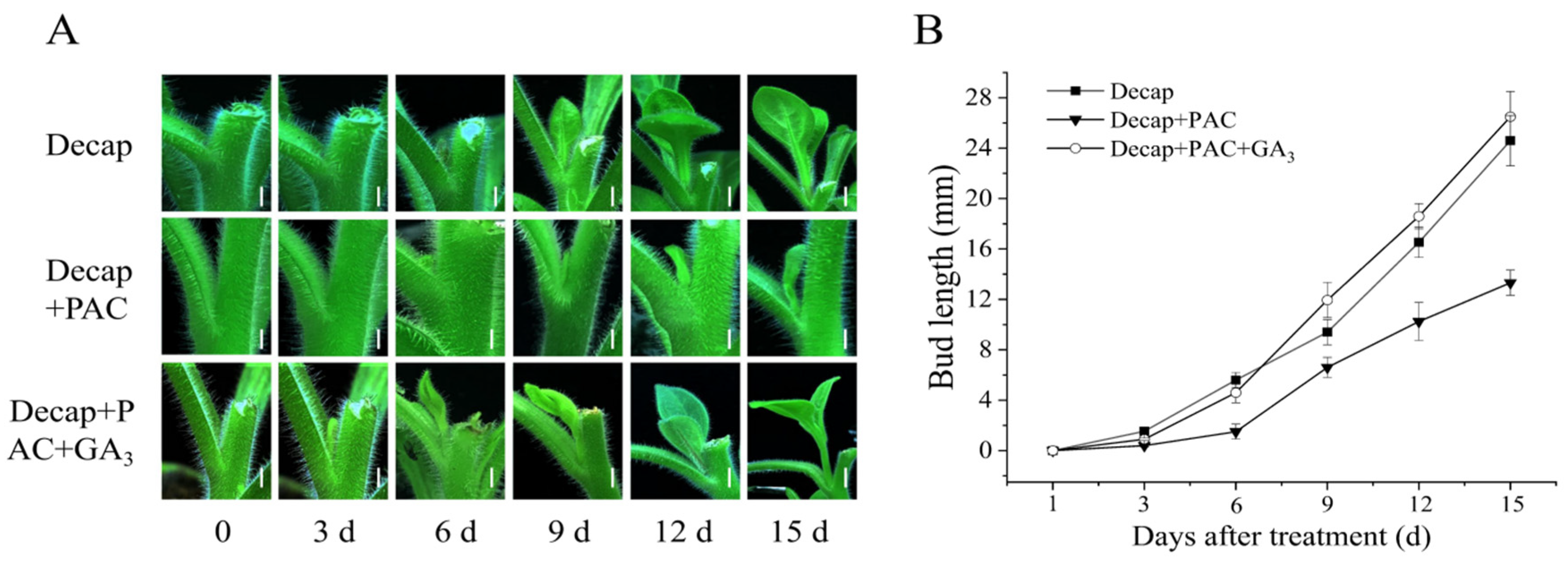
3.1. Exogenous GA3 Could Not Promote Axillary Bud Germination in Petunia

To investigate the effect of GA on bud germination in petunia, exogenous GA3 was applied to the fourth axillary bud from top to bottom, and the growth of the axillary bud was recorded over 15 days. As shown in Figure 1, the application of GA3 to buds did not promote bud germination.
Furthermore, we sampled the leaf axils treated with GA3 and measured the expression levels of PhBRC1 and PhDRM1, which are key genes in bud dormancy [24,25]. The results showed that after six hours of 6-BA treatment, PhBRC1 and PhDRM1 were downregulated to 0.42 and 0.09 times the control levels, respectively. However, GA3 treatment did not cause meaningful changes in the expression levels of PhDRM1 and PhBRC1 (Figure 1). These findings indicated that exogenous GA3 could not activate bud germination in petunia.

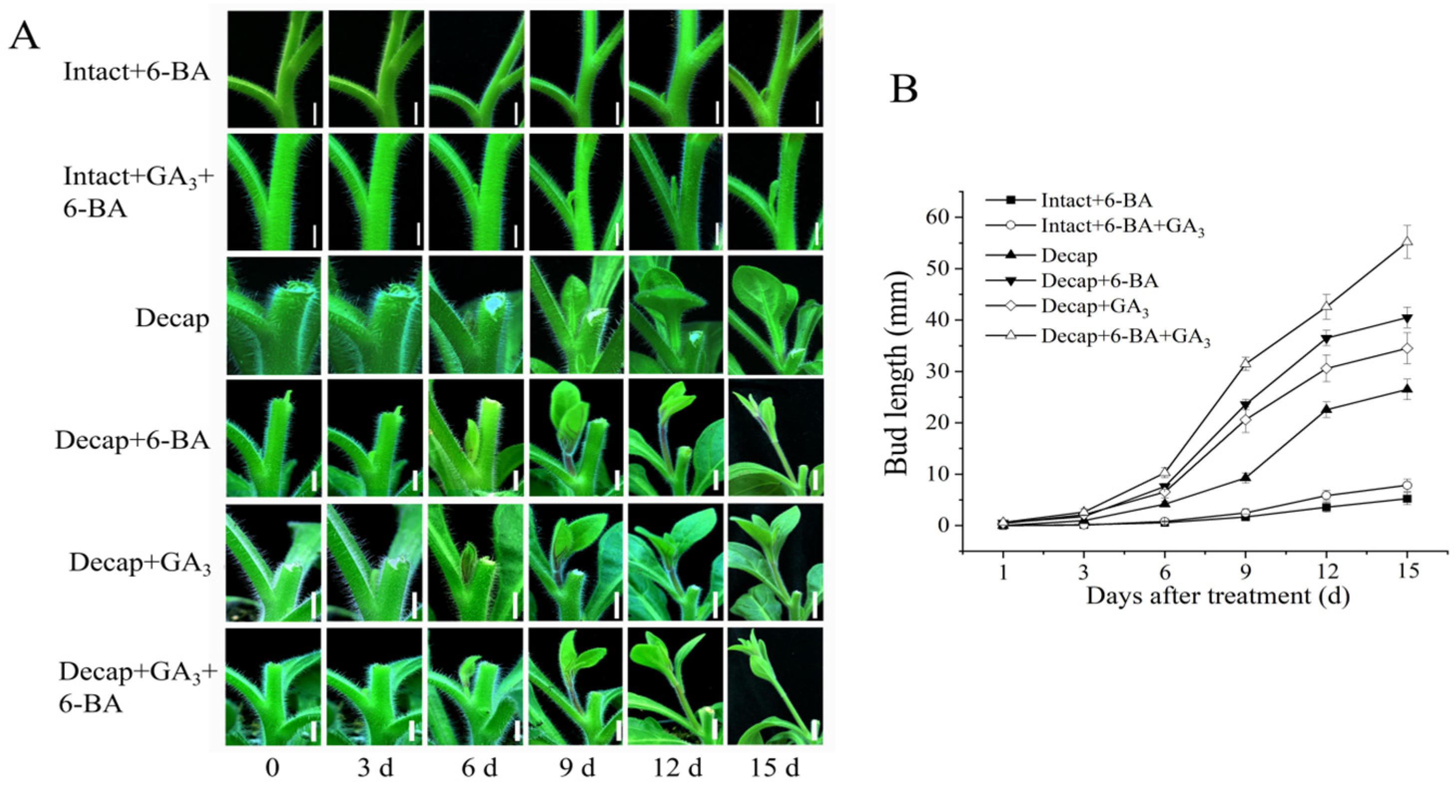
3.2. GA Promotes Bud Elongation

Since GA cannot promote the germination of axillary buds, we aimed to determine whether GA is involved in the growth of axillary buds after germination. PAC and PAC+GA3 were applied to the buds after decapitation. As shown in Figure 2, on day 15, the axillary buds of the decapitation (Decap) group reached 24.6 mm, the bud length in the Decap+PAC group was 13.32 mm, and the bud length in the Decap+PAC+GA3 group was 26.5 mm. These results indicated that PAC significantly inhibited bud elongation after decapitation but did not affect bud germination. The application of GA3 counteracted the inhibitory effect of PAC on bud elongation, implying that the inhibition of axillary bud growth is indeed caused by the deficiency of endogenous GA3.

3.3. GA and CK Synergistically Promote Bud Elongation

To study the interaction between GA and CK in regulating bud elongation, we treated the buds with various solutions. As shown in Figure 3, on day 15, the bud length for the Intact+6-BA, Intact+GA3+6-BA, Decap, Decap+6-BA, Decap+GA3, and Decap+GA3+6-BA groups were 5.23 mm, 7.84 mm, 28.5 mm, 40.5 mm, 34.5 mm, and 55.2 mm, respectively. These results indicated that the simultaneous application of GA3 and 6-BA could significantly promote bud elongation in both intact plants and decapitated plants, compared to the single application of GA3 and 6-BA. This suggests that GA and CK have a synergistic effect in regulating the bud growth of petunia.

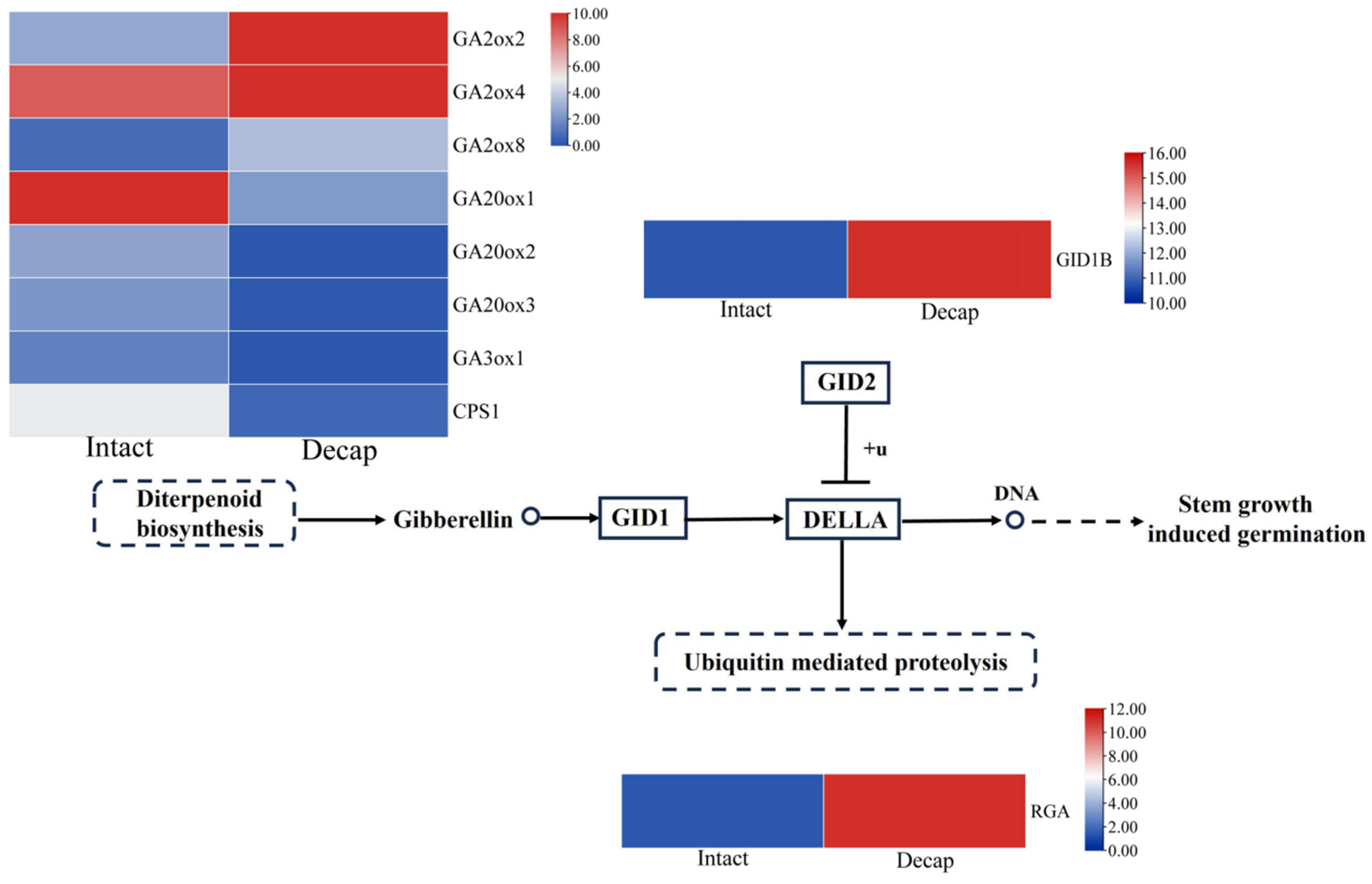
3.4. GA Pathway Genes Respond to Decapitation

Given that exogenous GA cannot directly promote the germination of axillary buds in petunia, we investigated whether endogenous GA plays a crucial role in bud germination. To explore this issue, we measured the expression levels of GA pathway genes after decapitation. As depicted in Figure 4, 1 h after decapitation, the expression levels of bioactive GA-degrading enzyme genes GA2ox2, GA2ox4, and GA2ox8 were upregulated by 5.44, 3.86, and 4.06 times compared to the control, respectively. The expression levels of GA synthetic genes GA20ox1, GA20ox2, GA20ox3, and GA3ox1 and the first key enzyme of the GA biosynthetic pathway CPS1 decreased to 0.05, 0.05, 0.07, 0.09, and 0.13 times the control levels, respectively. Moreover, the expression of the GA signal transduction gene GID1B was upregulated from 10.8 to 15.6, and the REPRESSOR OF ga1-3 (RGA), a repressor of GA signaling, was upregulated by 7.86 times compared to the control. These results suggest that these GA pathway genes may be involved in bud activation.

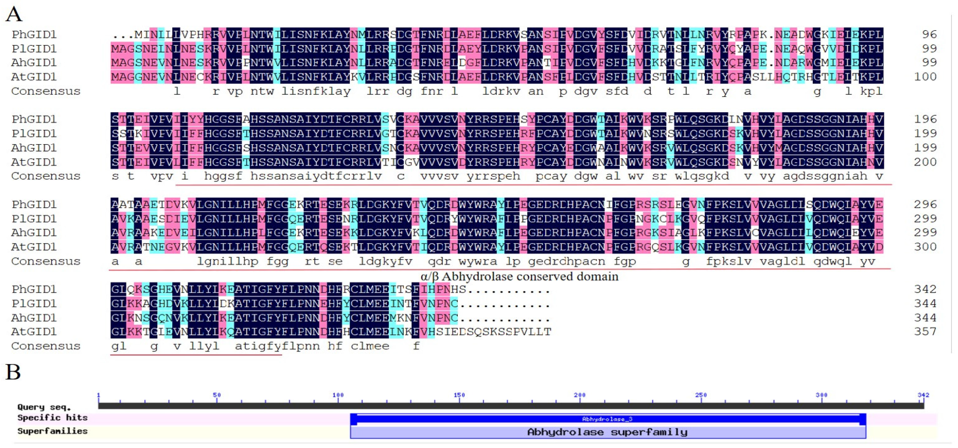
3.5. Cloning of PhGID1 and Sequence Analysis

Considering the well-documented branching phenotype of GA synthesis genes in transgenic plants and the relatively limited research on the involvement of the GA signal transduction gene GID1 in branch development, we selected this gene for further study. The PhGID1 sequence is 1029 bp long and encodes 342 amino acids. The molecular formula of the PhGID1 protein was estimated to be C1756H2693N483O501S9. The amino acid composition of the protein includes 9.9% Leu, 9.1% Val, 7.6% Ser, 6.4% Ala, and 6.4% Arg. The total number of negatively charged residues (Asp+Glu) is 39, while the total number of positively charged residues (Arg+Lys) is 38. Comparative sequence analysis revealed that the protein sequences of PhGID1 share similarities of 78.26% with Paeonia lactiflora, 77.10% with Arachis hypogaea, and 72.91% with A. thaliana, (Figure 5A). Additionally, sequence analysis showed that PhGID1 contains an α/β hydrolase-conserved domain (Figure 5B).

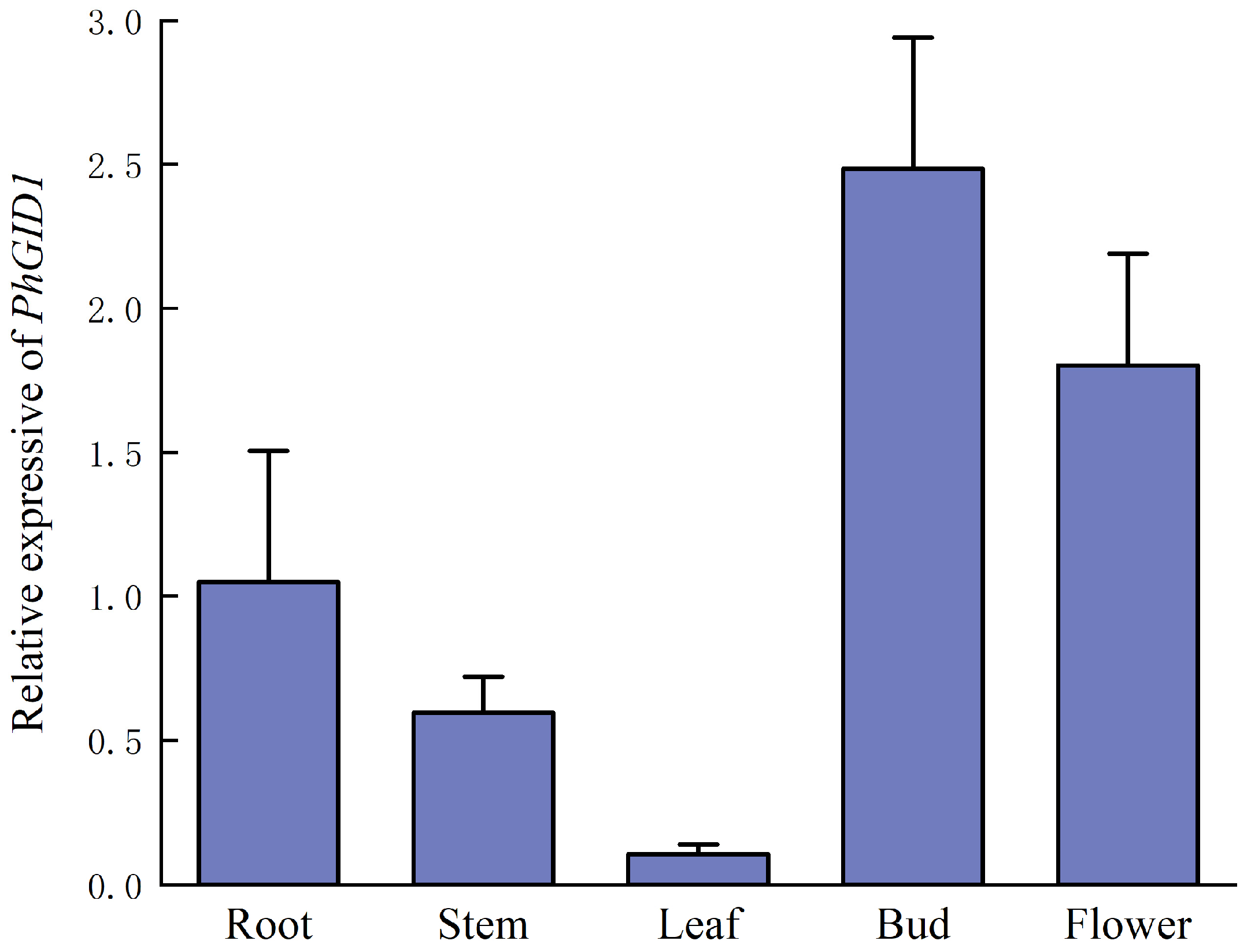
3.6. Analysis of Tissue-Specific Expression of PhGID1

To investigate the expression characteristics of PhGID1, we measured its expression levels in various tissues, including the roots, stems, leaves, buds, and flowers of petunia (Figure 6). The results revealed that PhGID1 is expressed in all examined tissues, with the highest expression levels observed in buds and the lowest in leaves. The expression levels of PhGID1 in the roots, stems, leaves, and flowers were 0.42, 0.24, 0.04, and 0.72 times that in buds, respectively.

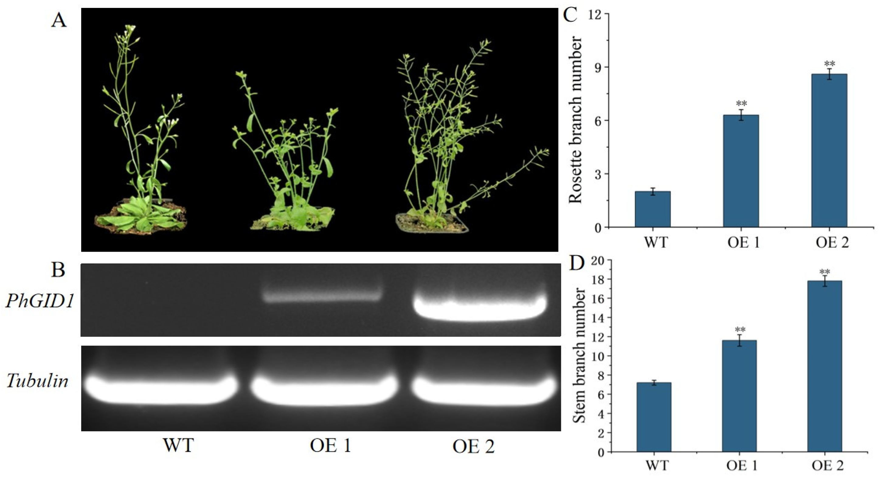
3.7. Overexpression of PhGID1 Increases Branch Number

To elucidate the function of PhGID1, we constructed the 35S::PhGID1 vector and transformed it into Arabidopsis, resulting in nine transgenic lines. Two transgenic lines (OE 1 and OE 2) were selected for phenotypic analysis. As shown in Figure 7, the overexpression of PhGID1 led to a significant increase in the number of branches. Compared with control plants, the average number of rosette branches in OE 1 and OE 2 was 5.2 and 6.5, respectively, while the average number of stem branches was 11.4 and 16.8, respectively.

4. Discussion

The branching stage of plants includes the establishment of axillary meristem (AM), the formation and germination of axillary buds, and the elongation of buds. Each stage is regulated by a variety of genes and hormones [26]. Although many studies have reported on the regulation of branch development by GA, their findings are inconsistent, making the role of GA in this process unclear.
In our study, we revealed partial functions of GA through hormone application and transgenic experiments. Directly applying GA3 to the axillary buds of petunia did not promote their germination, nor did it alter the expression levels of PhBRC1 and PhDRM1. This finding is consistent with studies in pea and M. pumila [19,27], but it is contrary to research on J. curcas and sweet cherry [13,14]. These differences may be related to the state of axillary buds treated, rather than species-specific responses to GA. Both our experiment and Cao et al.’s study found that GA can promote the growth of axillary buds when they are in an activated state.
Even though GA cannot directly stimulate bud germination like CK can, the application of GA3 following decapitation can promote bud elongation, which aligns with the finding that GA promotes the growth of buds released by decapitation in peas [27]. Applying PAC to axillary buds after decapitation significantly inhibited their growth but did not affect their germination, indicating that GA’s role in bud germination is not necessary. The combination of PAC+GA3 restored PAC’s inhibition of bud growth, indicating that GA is crucial in regulating bud elongation.
Simultaneously applying GA3 and 6-BA to axillary buds significantly promoted bud elongation, both after decapitation and in intact plants. This phenomenon was observed in both pea and M. pumila, suggesting that the synergistic effect of GA and CK in promoting axillary bud elongation is widespread. Studies on J. curcas have shown that this regulation by GA and CK is due to CK promoting the biosynthesis of GA in J. curcas buds and inhibiting GA degradation [13]. Further research is needed to determine whether other mechanisms underlie this synergistic effect.
To examine whether GA is involved in the activation of axillary buds, we performed a heatmap analysis on the expression levels of GA pathway genes 1 h after decapitation. The results showed that the synthesis genes of GA (GA20ox1, GA20ox2, GA20ox3, GA3ox1, and CPS1) were significantly downregulated, while the degradation genes (GA2ox2, GA2ox4, and GA2ox8) were significantly upregulated. Earlier studies have revealed that rising GA levels in leaf axils through the ectopic expression of the GA biosynthesis enzyme GA20ox2 markedly impaired axillary meristem initiation [7]. These investigations indicate that low GA content plays a vital role in the initiation of the axillary meristem and the activation of axillary buds.
In the transcriptome data, we observed an upregulation of PhGID1B expression levels, though not significantly, and did not detect other members of GID1. Despite the modest upregulation, PhGID1B expression levels were higher than most other genes both before and after decapitation. Combined with tissue-specific expression experiments, PhGID1B showed the highest expression level in axillary buds, suggesting its potential involvement in regulating branch development in petunia. However, there is limited research on the role of GID1B in branch development. Therefore, we cloned this gene and analyzed its expression in a variety of tissues. We found that this gene was highly expressed in buds and had the lowest expression in leaves. This contrasts with results in P. tomentosa, where higher expression was observed in leaves compared to roots [28]. To clarify the function of this gene, we constructed the overexpression vector 35S::PhGID1B and transformed it into Arabidopsis. The overexpression of PhGID1B resulted in changes in both rosette and stem branches in Arabidopsis, implying that PhGID1B is involved in regulating branch development.
Based on the above research, we propose that GA plays a critical role in the activation and elongation of axillary buds. Low levels of GA are beneficial for the activation of axillary buds, while high levels of GA contribute to the growth of buds. Therefore, the synthesis of GA may be dynamic throughout the entire process from the formation of the AM to the development of lateral branches, exhibiting different levels at various stages. For the GA signal transduction gene, although there was no significant change in expression level during the activation of buds after decapitation, it still played a role in bud germination, as evidenced by the increased number of branches in transgenic plants overexpressing PhGID1B. This research provides a theoretical reference for enriching the regulatory mechanism of GA in branch development.

Supplementary Materials

The following supporting information can be downloaded at https://www.mdpi.com/article/10.3390/cimb46090590/s1. Table S1: Hormone concentrations.

Author Contributions

J.D., H.Y. and S.J. performed experiments and analyzed data; J.D. and X.D. wrote the manuscript; and L.D. led the experiment and commented on the manuscript. All authors have read and agreed to the published version of the manuscript.

Funding

This work was financially supported by the National Natural Science Foundation of China (grant number: 31902042).

Institutional Review Board Statement

Not applicable.

Informed Consent Statement

Not applicable.

Data Availability Statement

Enquiries about data availability should be directed to the authors.

Conflicts of Interest

The authors declare that they have no conflicts of interest.

References

  1. Nahas, Z.; Ticchiarelli, F.; van Rongen, M.; Dillon, J.; Leyser, O. The activation of Arabidopsis axillary buds involves a switch from slow to rapid committed outgrowth regulated by auxin and strigolactone. New Phytol. 2024, 242, 1084–1097. [Google Scholar] [CrossRef]
  2. Hedden, P. The current status of research on gibberellin biosynthesis. Plant Cell Physiol. 2020, 61, 1832–1849. [Google Scholar] [CrossRef]
  3. Xie, Y.; Chen, L. Epigenetic regulation of gibberellin metabolism and signaling. Plant Cell Physiol. 2020, 61, 1912–1918. [Google Scholar] [CrossRef]
  4. He, Y.; Zhang, T.; Sun, H.; Zhan, H.; Zhao, Y. A reporter for noninvasively monitoring gene expression and plant transformation. Hortic. Res. 2020, 7, 152. [Google Scholar] [CrossRef]
  5. Hirano, K.; Ueguchi-Tanaka, M.; Matsuoka, M. GID1-mediated gibberellin signaling in plants. Trends Plant Sci. 2008, 13, 192–199. [Google Scholar] [CrossRef] [PubMed]
  6. Koorneef, M.; Elgersma, A.; Hanhart, C.J.; van Loenen-Martinet, E.P.; van Rijn, L.; Zeevaart, J.A.D. A gibberellin insensitive mutant of Arabidopsis thaliana. Physiol. Plant. 1985, 65, 33–39. [Google Scholar] [CrossRef]
  7. Zhang, Q.; Wang, J.; Wang, L.; Wang, J.; Wang, Q.; Yu, P.; Bai, M.; Fan, M. Gibberellin repression of axillary bud formation in Arabidopsis by modulation of DELLA-SPL9 complex activity. J. Integr. Plant Biol. 2020, 62, 421–432. [Google Scholar] [CrossRef] [PubMed]
  8. Lo, S.F.; Yang, S.Y.; Chen, K.T.; Hsing, Y.I.; Zeevaart, J.A.; Chen, L.J.; Yu, S.M. A Novel Class of Gibberellin 2-Oxidases Control Semidwarfism, Tillering, and Root Development in Rice. Plant Cell 2008, 20, 2603–2618. [Google Scholar] [CrossRef] [PubMed]
  9. Martínez-Bello, L.; Moritz, T.; López-Díaz, I. Silencing C19-GA2-oxidases induces parthenocarpic development and inhibits lateral branching in tomato plants. J. Exp. Bot. 2015, 66, 5897–5910. [Google Scholar] [CrossRef]
  10. Agharkar, M.; Lomba, P.; Altpeter, F.; Zhang, H.; Kenworthy, K.; Lange, T. Stable expression of AtGA2ox1 in a low-input turfgrass (Paspalum notatum Flugge) reduces bioactive gibberellin levels and improves turf quality under field conditions. Plant Biotechnol. J. 2007, 5, 791–801. [Google Scholar] [CrossRef]
  11. Mauriat, M.; Sandberg, L.G.; Moritz, T. Proper gibberellin localization in vascular tissue is required to control auxin-dependent leaf development and bud outgrowth in hybrid aspen. Plant J. 2011, 67, 805–816. [Google Scholar] [CrossRef] [PubMed]
  12. Tenreira, T.; Lange, M.J.P.; Lange, T.; Bres, C.; Labadie, M.; Monfort, A.; Hernould, M.; Rothan, C.; Denoyes, B. A Specific Gibberellin 20-Oxidase Dictates the Flowering-Runnering Decision in Diploid Strawberry. Plant Cell 2017, 29, 2168–2182. [Google Scholar] [CrossRef]
  13. Ni, J.; Gao, C.; Chen, M.S.; Pan, B.Z.; Ye, K.; Xu, Z.F. Gibberellin Promotes Shoot Branching in the Perennial Woody Plant Jatropha curcas. Plant Cell Physiol. 2015, 56, 1655–1666. [Google Scholar] [CrossRef] [PubMed]
  14. Elfving, D.C.; Visser, D.B.; Henry, J.L. Gibberellins Stimulate Lateral Branch Development in Young Sweet Cherry Trees in the Orchard. Int. J. Fruit Sci. 2011, 11, 41–54. [Google Scholar] [CrossRef]
  15. Hartmann, A.; Senning, M.; Hedden, P.; Sonnewald, U.; Sonnewald, S. Reactivation of Meristem Activity and Sprout Growth in Potato Tubers Require Both Cytokinin and Gibberellin. Plant Physiol. 2010, 155, 776–796. [Google Scholar] [CrossRef]
  16. O’Neill, D.P.; Ross, J.J. Auxin Regulation of the Gibberellin Pathway in Pea. Plant Physiol. 2002, 130, 1974–1982. [Google Scholar] [CrossRef]
  17. Wang, R.F.; Zhang, J.Y.; Peng, L.; Dong, S.T.; Liu, P.; Zhao, B. Effects of endogenous hormones on tiller development process of different maize varieties. Sci. Agric. Sin. 2012, 45, 840–847. [Google Scholar] [CrossRef]
  18. Nakamura, H.; Xue, Y.-L.; Miyakawa, T.; Hou, F.; Qin, H.-M.; Fukui, K.; Shi, X.; Ito, E.; Ito, S.; Park, S.-H.; et al. Molecular mechanism of strigolactone perception by DWARF14. Nat. Commun. 2013, 4, 2613. [Google Scholar] [CrossRef]
  19. Tan, M.; Li, G.; Liu, X.; Cheng, F.; Ma, J.; Zhao, C.; Zhang, D.; Han, M. Exogenous application of GA3 inactively regulates axillary bud outgrowth by influencing of branching-inhibitors and bud-regulating hormones in apple (Malus domestica Borkh.). Mol. Genet. Genom. 2018, 293, 1547–1563. [Google Scholar] [CrossRef]
  20. Tamura, K.; Stecher, G.; Kumar, S. MEGA11: Molecular Evolutionary Genetics Analysis Version 11. Mol. Biol. Evol. 2021, 38, 3022–3027. [Google Scholar] [CrossRef]
  21. Pramanik, K.; Pal, P.; Soren, T.; Mitra, S.; Ghosh, P.K.; Sarkar, A.; Maiti, T.K. In silico structural, functional and phylogenetic analysis of Klebsiella phytases. J. Plant Biochem. Biotechnol. 2018, 27, 362–372. [Google Scholar] [CrossRef]
  22. Livak, K.J.; Schmittgen, T.D. Analysis of relative gene expression data using real-time quantitative PCR and the 2−ΔΔCT Method. Methods 2001, 25, 402–408. [Google Scholar] [CrossRef] [PubMed]
  23. Clough, S.J.; Bent, A.F. Floral dip: A simplified method for Agrobacterium-mediated transformation of Arabidopsis thaliana. Plant J. 1998, 16, 735–743. [Google Scholar] [CrossRef]
  24. González-Grandío, E.; Poza-Carrión, C.; Sorzano, C.O.S.; Cubas, P. BRANCHED1Promotes Axillary Bud Dormancy in Response to Shade in Arabidopsis. Plant Cell 2013, 25, 834–850. [Google Scholar] [CrossRef] [PubMed]
  25. Rae, G.M.; David, K.; Wood, M. The Dormancy Marker DRM1/ARP Associated with Dormancy but a Broader Role In Planta. Dev. Biol. J. 2013, 2013, 632524. [Google Scholar] [CrossRef]
  26. Barbier, F.F.; Dun, E.A.; Kerr, S.C.; Chabikwa, T.G.; Beveridge, C.A. An Update on the Signals Controlling Shoot Branching. Trends Plant Sci. 2019, 24, 220–236. [Google Scholar] [CrossRef]
  27. Cao, D.; Chabikwa, T.; Barbier, F.; Dun, E.A.; Fichtner, F.; Dong, L.; Kerr, S.C.; Beveridge, C.A. Auxin-independent effects of apical dominance induce changes in phytohormones correlated with bud outgrowth. Plant Physiol. 2023, 192, 1420–1434. [Google Scholar] [CrossRef]
  28. Yang, H.; Yang, X.; Wang, L.; Gong, C.; Chen, B.; Du, Q.; Zhang, D. Single nucleotide polymorphisms in two GID1 orthologs associate with growth and wood property traits in Populus tomentosa. Tree Genet. Genomes 2016, 12, 109. [Google Scholar] [CrossRef]
Figure 1. Exogenous GA3 cannot promote bud germination in petunia. (A) Axillary bud growth of petunia plants after GA3 treatment, bars = 1 cm. (B) Statistical analysis of bud length of petunia. (C,D) Regulation of GA3 and 6-BA on PhBRC1 and PhDRM1 expression. (** p < 0.01).
Cimb 46 00590 g001
Figure 2. GA promoted bud elongation in petunia. (A) Bud growth of petunia after different treatments, bars = 1 cm. (B) Statistical analysis of bud length of petunia.
Cimb 46 00590 g002
Figure 3. GA and CK synergistically promoted bud growth in petunia. (A) Bud growth of petunia with different treatments, bars = 1 cm. (B) Statistical analysis of bud length of petunia.
Cimb 46 00590 g003
Figure 4. Expression analysis of GA pathway genes after decapitation for 1 h.
Cimb 46 00590 g004
Figure 5. Sequence analysis of PhGID1. (A) Comparison of GID1 amino acid sequences of various plants. Note: Pl: P. lactiflora; Ah: A. hypogaea; At: A. thaliana. (B) Conserved domain analysis of PhGID1.
Cimb 46 00590 g005
Figure 6. The relative expression of PhGID1 in various tissues of petunia.
Cimb 46 00590 g006
Figure 7. Phenotypic analysis of transgenic plants overexpressing PhGID1. (A) Phenotypic comparisons of WT and transgenic plants overexpressing PhGID1. Different lines overexpressing PhGID1 are represented by OE 1 and OE 2. (B) RT-PCR evaluated expression levels of PhGID1. (C) Statistics of rosette branch number (n = 15). (D) Statistics of stem branch number (n = 15). (** p < 0.01).
Cimb 46 00590 g007
Table 1. Primer sequence used in PCR and qRT-PCR.
Primer NamePrimer Sequence (5′ to 3′)
PhGID1-FATGATCAATTTGCTTTTAGTACC
PhGID1-RCTATGAATGGTTAGGATGGATGA
qRT-PhGID1-FCGACGGTCACCTGAACATAG
qRT-PhGID1-RTTCTCGGATTCGGTCCTTTT
PhGID1-1300-FCCAAATCGACTCTAGAATGATCAATTTGCTTTTAGTACC
PhGID1-1300-RCCACTAGTATTTAAATGCTATGAATGGTTAGGATGGATGA
PhBRC1-FCCCATTTGCTCATCTTTATTC
PhBRC1-RCTGCCACTTTGCTTACTCATA
PhDRM1-FGAATGTGTGGAGGAGTGTTTTT
PhDRM1-RCCATATTGCACCCCCTTTTGTT
Disclaimer/Publisher’s Note: The statements, opinions and data contained in all publications are solely those of the individual author(s) and contributor(s) and not of MDPI and/or the editor(s). MDPI and/or the editor(s) disclaim responsibility for any injury to people or property resulting from any ideas, methods, instructions or products referred to in the content.

