Article

Molecular Integrative Study on Inhibitory Effects of Pentapeptides on Polymerization and Cell Toxicity of Amyloid-β Peptide (1–42)

1
Key Laboratory of Tropical Biological Resources of the Ministry of Education of China, School of Pharmaceutical Sciences, Hainan University, Haikou 570228, China
2
Laboratory of Biopharmaceuticals and Molecular Pharmacology, One Health Cooperative Innovation Center, Hainan University, Haikou 570228, China
3
Department of Biotechnology, School of Life and Health Sciences, Hainan University, Haikou 570228, China
*
Authors to whom correspondence should be addressed.
These authors contributed equally to this work.
Curr. Issues Mol. Biol. 2024, 46(9), 10160-10179; https://doi.org/10.3390/cimb46090606
Submission received: 30 July 2024 / Revised: 7 September 2024 / Accepted: 11 September 2024 / Published: 14 September 2024

Abstract

:
Alzheimer’s Disease (AD) is a multifaceted neurodegenerative disease predominantly defined by the extracellular accumulation of amyloid-β (Aβ) peptide. In light of this, in the past decade, several clinical approaches have been used aiming at developing peptides for therapeutic use in AD. The use of cationic arginine-rich peptides (CARPs) in targeting protein aggregations has been on the rise. Also, the process of peptide development employing computational approaches has attracted a lot of attention recently. Using a structure database containing pentapeptides made from 20 L-α amino acids, we employed molecular docking to sort pentapeptides that can bind to Aβ42, then performed molecular dynamics (MD) analyses, including analysis of the binding stability, interaction energy, and binding free energy to screen ligands. Transmission electron microscopy (TEM), circular dichroism (CD), thioflavin T (ThT) fluorescence detection of Aβ42 polymerization, MTT (3-[4,5-dimethylthiazol-2-yl]-2,5 diphenyl tetrazolium bromide) assay, and the flow cytometry of reactive oxygen species (ROS) were carried out to evaluate the influence of pentapeptides on the aggregation and cell toxicity of Aβ42. Two pentapeptides (TRRRR and ARRGR) were found to have strong effects on inhibiting the aggregation of Aβ42 and reducing the toxicity of Aβ42 secreted by SH-SY5Y cells, including cell death, reactive oxygen species (ROS) production, and apoptosis.

1. Introduction

Alzheimer’s disease (AD) is a progressive neurodegenerative disease associated with damaged neurons in the brain that is consistent with an irreversible neurodegenerative disorder involving a decline in memory and executive function, and personality change (Hansen, et al. [1]). On a global scale, it is escalating at a daunting rate. There are approximately 6.7 million individuals with AD dementia in the United States and an estimated 50 million individuals with AD dementia globally. Due to an increase in the aging population and since Alzheimer’s disease is prevalent amongst the elderly, these populations are expected to grow to 12.7 million and 150 million in the United States and globally by 2050 [2,3].
Amyloid-beta (Aβ) is a 39–43 amino acid residue peptide and a major constituent of extracellular amyloid plaque, and its expression is believed to be a major contingency in AD advancement [4]. Aβ is the peptide outcome of the consecutive proteolytic cleavages (by β- and ϒ-secretases) of APP (a type-I transmembrane protein). These proteolytic cleavages give rise to two types of Aβ isoforms (Aβ40 and Aβ42), and though Aβ40 is more profuse than Aβ42 in human fluids, Aβ42 aggregates faster and is considered to be more neurotoxic than Aβ40. Aβ is distributed throughout the prefrontal cortex, hippocampus, midbrain, lower brainstem, and cerebellar cortex. It was reported that Aβ oligomers may bind to receptors on neuronal membranes, inducing calcium influx, which leads to calcium overload and cytotoxic responses [5]. Moreover, they may can interact with synaptic proteins, disrupting synaptic transmission and plasticity, ultimately causing synaptic dysfunction [6]. The oxidative stress toxicity of Aβ, alterations in mitochondrial function, and metal ion toxicity have also been widely reported [7,8]. Oxidative stress causes damage to lipids, proteins, and DNA, including antioxidant enzymes. Aβ aggregates may induce excessive production of ROS produced by mitochondria or disruption of metal ion homeostasis.
Peptide-based inhibitors, among numerous inhibitors, have been corroborated to be possible anti-aggregation agents due to their low cytotoxicity, good biocompatibility, high target affinity, and specificity [9,10,11,12,13,14,15,16].
Computational techniques have been used to uncover prospective inhibitors against numerous biological targets during the past few years [17,18]. Diverse contemporary studies have delineated and assessed prospective peptides as inhibitors of numerous targets using computational methods [19,20,21,22,23,24].
Two vital goals in drug discovery are developing molecules that bind tightly to a target protein and weakly—or not at all—to off-target proteins. In recent years, a group of rigorous, physics-based methods have surfaced as the most consistently precise method that can aid in the discovery of promising compounds and help hasten the slow and expensive process of lead development [25,26,27,28]. One of these methods is free energy (Gibbs free energy).
Gibbs free energy is equal to the sum of the enthalpy plus the product of the temperature and entropy of the system under the conditions of constant pressure and temperature. The change in Gibbs free energy (∆G) determines whether a reaction will happen spontaneously. Calculating the binding free energy of a compound to a protein target has emerged to be beneficial for drug discovery. Using the molecular dynamics (MD) method established [29], we conducted an initial analysis of the thermodynamic mechanisms of Aβ42 polymerization [30]. It was found that Aβ42 monomer is a globular protein; however, Aβ42 strands stably maintain an S-shape polymerized structure while the number of Aβ42 strands is more than five. Hydrophobic contact is the predominant force piloting Aβ42 clustering by lowering free energy, and three hydrophobic regions in the structure affect polymerization in that replacing the hydrophobic side chains of the amino acids in these regions with that of glycine differentially decreased the binding free energy [30]. Since the three hydrophobic regions determine polymerization, chemicals that can bind to the regions will interfere with polymerization. Based on this notion, we performed molecular docking and MD analysis to screen pentapeptides targeting the hydrophobic regions using a structure database consisting of all pentapeptides (3,200,000) made from the 20 L-α-amino acids, and conducted pharmacological experiments to test the effects of the pentapeptides.

2. Materials and Methods

2.1. Materials

Pentapeptides were synthesized by Sangon Biotech (Shanghai, China), and dissolved in 10 mM phosphate buffer (pH 7.4). Aβ42 was bought from ChinaPeptides (QYAOBIO) (Shanghai, China) and DMSO used to dissolve it to a concentration of 1 mM. All allotting solutions were aliquoted and preserved at −80 °C until use. DMSO, ThT, and Lipofectamine 2000 were bought from Sigma-Aldrich (St. Louis, MI, USA). PEI 40K transfection reagents were bought from Servicebio (Wuhan, China). An ROS active oxygen testing kit and apoptosis kit for the detection by flow cytometry were bought from Bioscience (Shanghai, China).

2.2. Molecular Docking

Python was used to summon the ChemScript module of the ChemDraw software version 20.0 (Shelton, CT, USA), and then a database of pentapeptides made from 20 L-α amino acids was constructed. The molecular structural data file of Aβ42 (PDB# 5OQV) was downloaded from the Protein Data Bank (PDB). After correcting the data and adding missing hydrogen atoms, the open-source software AUTODOCK was used to investigate the interaction of pentapeptides with Aβ42. The London algorithm for free energy was used for screening the binding sites and conformations of pentapeptides and then refined by the generalized-Born volume integral/weighted surface area algorithm.

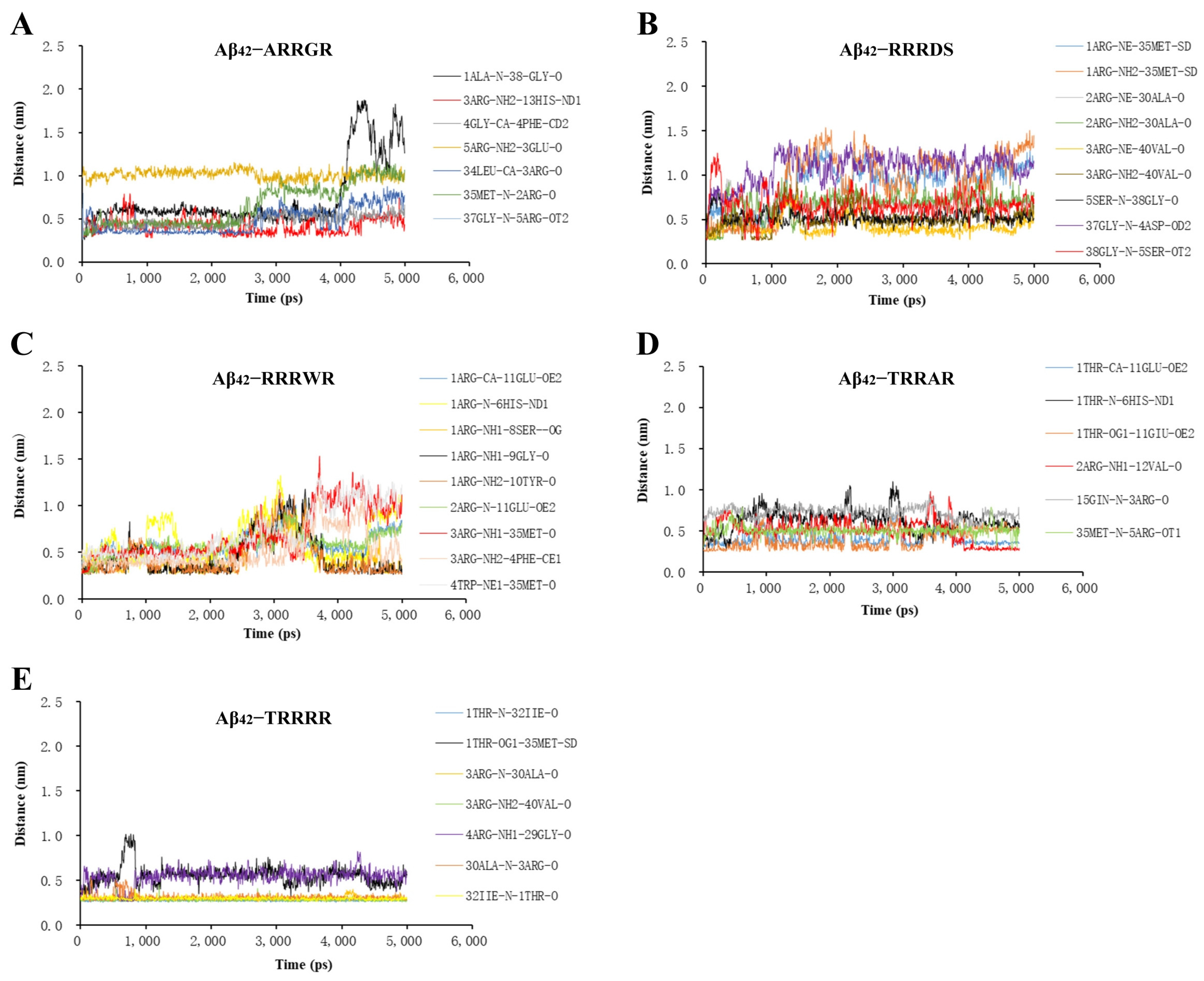
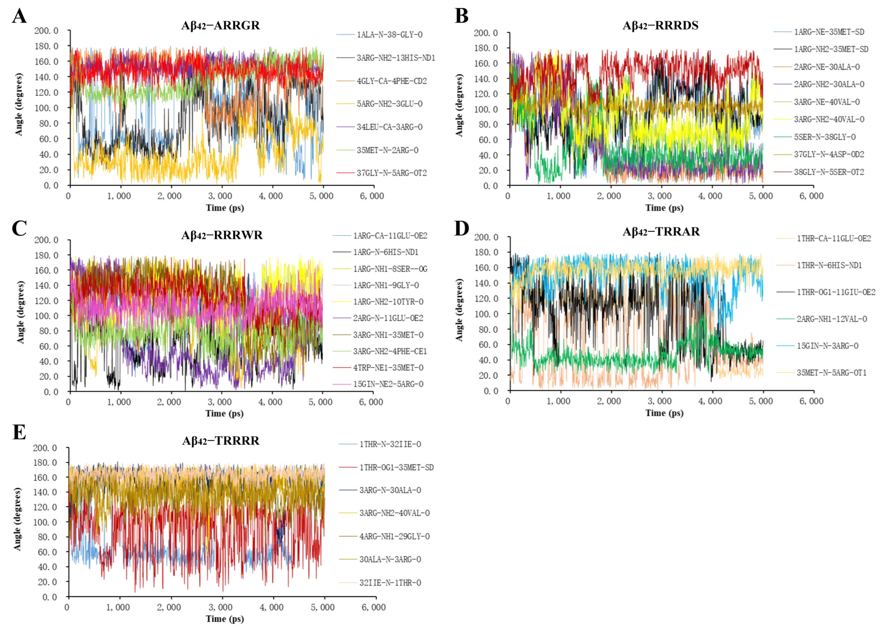
2.3. Molecular Dynamics Analysis of Binding Stability

Molecular dynamic analysis (MDA) has been extensively used in drug design [31] for the physical and chemical depiction of intricate movements between biomolecules [32,33,34]. MDA enables us to have a better understanding of the dynamic attributes of biomolecular systems, such as protein folding and stability, ligand binding, and protein complexing, among others. A key feature of MDA is its ability to imitate both in vitro and in vivo conditions—for example, at different pH conditions, in the presence of water and ions, at different salt or ionic concentrations, and in the presence of a lipid bilayer and other cellular components [35,36]. MDA has been used to study multiple protein-related issues, such as protein-binding and protein–protein interaction and signaling [35,37].
In our research, molecular dynamics studies were carried out with the Groningen Machine for Chemical Simulation (GROMACS, 2020.03) on the Ubuntu (18.06) Linux operating system, and were sped up by NVIDIA Compute Unified Device Architecture (CUDA)-supported parallel computation. The Aβ42 monomer, trimer, or pentamer was centralized in a dodecahedron box with a distance of 3.0 nm from the edge to the Aβ42 molecule. The box was filled with water molecules, and Na+ and Cl counter ions at final concentrations of 0.1 M were added to the box to keep the system overall neutral at physiological pH. Throughout the MD study, we employed the Amber99SB force field (which is optimized for the ab initio calculation of three-dimensional structure of proteins) and the TIP3P explicit water model. Energy minimization and system equilibration was carried out with the same formulae as previously reported by [30].

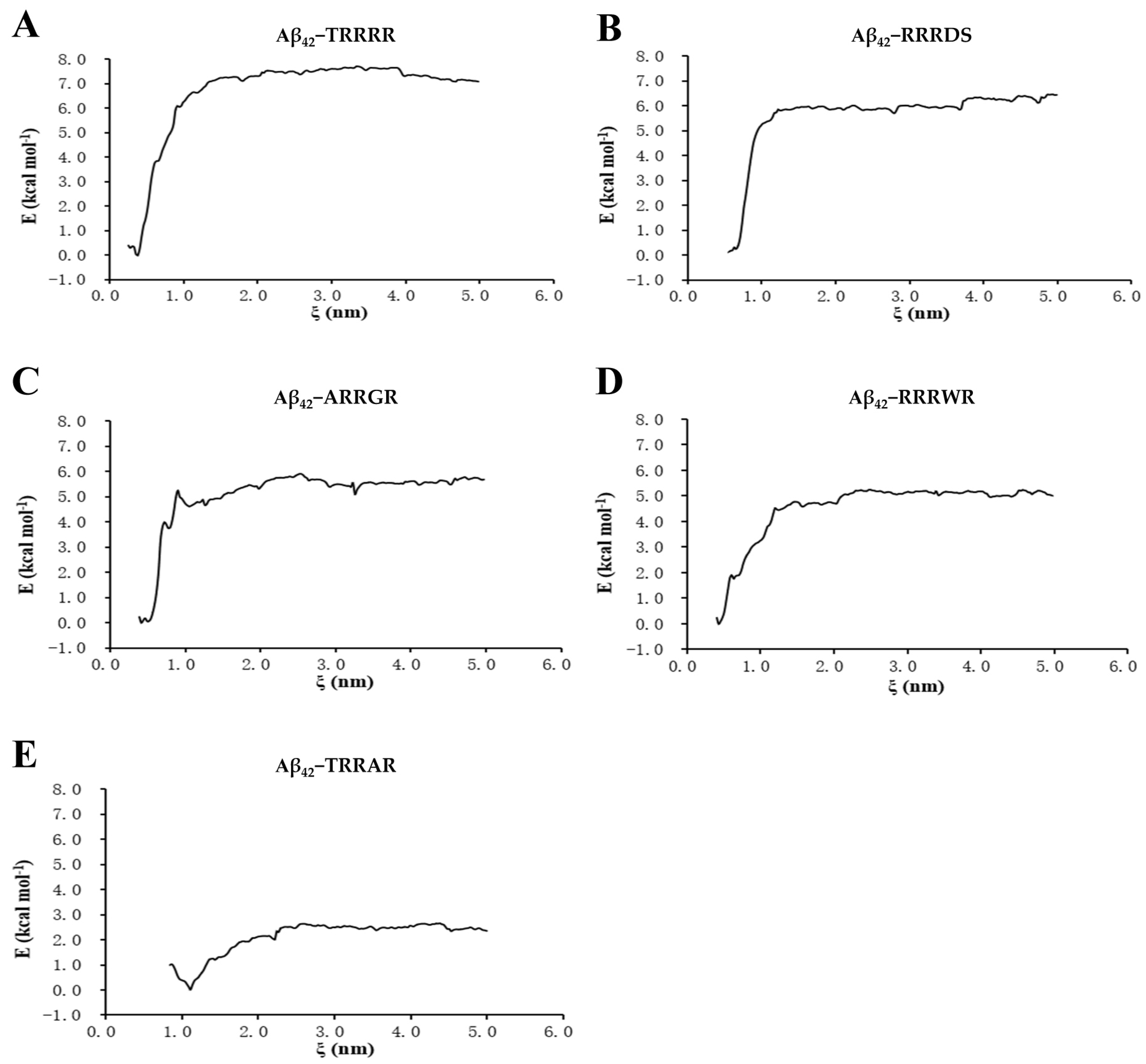
2.4. Umbrella Sampling of Binding Free Energy

The binding free energy is mirrored in alterations in Gibbs free energy (∆G) in an isothermal–isobaric assemblage throughout the procedure of hauling oligopeptides off Aβ42. The Aβ42 protein complex fastened to the pentapeptide was placed at dimensions 3.0 × 3.5 × 1.5 (x, y, z) nm in a cubic box of dimensions 6.0 × 7.0 × 14.0 (x, y, z) nm. The periodic boundary conditions were employed in all simulations. Water molecules were added into the cubic box, and 0.1 M sodium chloride was also added along with the number of Na+ and C counter ions essential to maintain an overall neutral system at physiological pH. The carbonyl carbon atom (Cα) of the 29th glycine and the Cα of every pentapeptides’ second amino acid were chosen as reference atoms. Pressure equilibration was implemented before the pulling and umbrella sampling phases. During the phase of generating configurations, the two proteins were pulled away when a harmonic force at a constant velocity of 0.01 nm/ps over a course of 250,000-time steps was utilized, and 501 coordinate files were saved in the course of the pulling procedure. A total of 23 to 25 umbrella samplings of 10 ns were conducted in each overlapping 0.2 nm spacing sampling window through the reaction axis (ξ), bringing about approximately 450 Gb of data. The GROMACS’s WHAM module was used to determine ∆G.

2.5. ThT Fluorescent Detection of Aggregation of Aβ42

Amyloid aggregation was evaluated using the amyloid dye Thioflavin-T (ThT) [38]. Aβ42 (QYAOBIO, Shanghai, China) was dissolved as previously reported by Yuan et al. [30]. The Aβ42 was adjusted to a final concentration of 10 μM, the pentapeptides (TRRRR and ARRGR) adjusted to a final concentration of 40 μM, and ThT adjusted to a final concentration of 50 μM. These were all carried out in a 96-well plate. The fluorescence intensity was measured at 37 °C using an automated well plate reader (TECAN Infinite 200 PRO, Tecan Asia Pte Ltd., Singapore) at an emission wavelength of 485 at a 5 min interval, with an excitation wavelength of 450 nm. The measurements were performed as independent quintuplicates.

2.6. Transmission Electron Microscope Observation of Aggregated Aβ42

42 (QYAOBIO, Shanghai, China) samples were prepared in PBS at a working concentration of 50 μM. Aβ42 was incubated with TRRRR and ARRGR 37 °C for 48 h. The final concentration of Aβ42 was 10 μM, and that of TRRRR and ARRGR was 40 μM. After incubation, 10 μL of samples to be observed were dropped onto a 300-mesh Formvar–carbon-coated copper grid and stained with 10 μL of 2% phosphotungstic acid solution. Afterwards, the samples were air-dried and observed under a TEM (FEI Inc., Hillsboro, OR, USA) with a voltage of 200 kV.

2.7. Cell Culture

SH-SY5Y cells were expanded in DMEM including 10% fetal bovine serum supplemented with glutamine, and grown at 37 °C with 5% CO2. When the cells reached 80% confluency, they were used for further experiments.

2.8. Transfection and Expression of Secretable Aβ42

Before transfection, the medium was replaced with Opti-MEM (Gibco, Billings, MT, USA) and incubated for 2 h. Then, a mixture of pcDNA3.1-Aβ42 plasmids (4.0 μg, containing the sequence encoding the signal peptide for secretion at the 5′ end of Aβ42 sequence and three consecutive stop codons at the 3′ end; Aβ42 sequence: 5′-ATGCTGCCCGGTTTGGCACTGCTCCTGCTGGCCGCCTGGACGGCTCGGGCGGATGCAGAATTCCGACATGACTCAGGATATGAAGTTCATCATCAAAAATTGGTGTTCTTTGCAGAAGATGTGGGTTCAAACAAAGGTGCAATCATTGGACTCATGGTGGGCGGTGTTGTCATAGCGTGATGATGA-3′). Lipofectamine 2000 (10 μL, Sigma, St. Louis, MI, USA) and Opti-MEM that had been pre-incubated for 15 min at room temperature was added into the plates and incubated for 4 h at 37 °C. Four hours after the transfection, the Opti-MEM was replaced with DMEM (ThermoFisher, Waltham, MA, USA) containing 5% FBS and antibiotics (penicillin and streptomycin). After transfection, G418 was supplemented 24 h later at a final concentration of 100 µg/mL for positive selection.

2.9. ELISA Experiment

TRRRR or ARRGR was added to the cells secreting Aβ42 at final concentrations of 10 µM or 50 µM 4 h after transfection. A total of 24 h later, 100 μL of the supernatant was transferred to 96-well high-absorbent plates and incubated at 4 °C overnight. The wells were then washed four times with PBS, followed by blocking with 5% non-fat milk for 2 h. Primary antibody (1:200) was added and incubated at 4 °C overnight. After four washes with PBS, HRP-conjugated secondary antibody (1:1000) was added. Following treatment with the EL-TME kit (C520026, Sango Biotech, Shanghai, China), chemiluminescence was measured by using a microplate reader.

2.10. Microscopic Observation of Cell Death

TRRRR or ARRGR was added to the cells secreting Aβ42 4 h after transfection at final concentrations of 10 µM or 50 µM, followed by incubation at 37 °C for 24 h. The culture medium was removed, and the cells were fixed with 4% paraformaldehyde at room temperature for 15 min. The cells were gently washed twice with PBS, followed by incubation with EB (1 mg/mL) at 37 °C in the dark for 20 min. The cells were gently washed twice with PBS, then observed under a fluorescence microscope (Axio Observer 7, Zeiss, Germany) (excitation wavelength: 545 nm, and emission wavelength: 590 nm).

2.11. Flow Cytometry Methods to Detect Reactive Oxygen Species and Apoptosis

After the Opti-MEM was replaced with DMEM medium containing 5% FBS and antibiotics (penicillin and streptomycin), SH-SY5Y cells expressing Aβ42 were incubated with TRRRR or ARRGR at final concentration of 10 μM and 50 μM for 24 h. After collecting and washing cells with PBS, they were stained using PI and the Annexin-V kit (UElandy Inc., Suzhou, China), then cell apoptosis was computed by flow cytometry (CytoFLEX LX, Beckman Coulter Life Sciences, Indianapolis, IN, USA).

2.12. Statistical Analysis

The results of experiments were expressed as mean ± SD when applicable. Differences among groups have been evaluated by One-way ANOVA, followed by the Tukey-Kramer test for multiple comparisons using GraphPad Prism 6.0 (Boston, MA, USA). A p value less than 0.05 was considered statistically significant.

3. Results

3.1. Molecular Docking of Pentapeptides

Hydrophobic interaction and electrostatic forces including hydrogen bonds have an immense influence on the conformational alterations of Aβ42 [39]. In this study, a molecular docking experiment was conducted between Aβ42 and five pentapeptides. The pentapeptides which were incorporated in the Aβ42 hydrophobic cluster (Figure 1) were Threonine-arginine-arginine-arginine-arginine (TRRRR), Arginine-arginine-arginine-tryptophan-arginine (RRRWR), Arginine-arginine-arginine-aspartic acid-serine (RRRDS), Alanine-arginine-arginine-glycine-arginine (ARRGR), and Threonine-arginine-arginine-alanine-arginine (TRRAR). The docking free energy of the five peptides are shown in Supplementary Table S1. As illustrated on Figure 1, the binding of pentapeptides to Aβ42 can be seen. The grids in magenta denote the molecular surfaces of the pentapeptides, whereas those in gray denote the range of van der Waals forces.
But for the RRRWR pentapeptide, which did not interact with any of the hydrophobic patches of the Aβ42 monomer, the other four pentapeptides had side-chain interactions with the Aβ42 monomer via hydrophobic clusters through hydrogen bonds including π-type bonding. On the incorporation of RRRWR in the Aβ42 monomer, few side-chain contacts were noted at the N-terminal (6–13) region. However, contacts between CHC (17–22)/mid-domain (26–32) and C-terminal (34–42) regions were reduced. As for the incorporation of RRRDS in the Aβ42 monomer, few side-chain contacts were noted at the hydrophobic patch on the C-terminal (30–35) region. For the incorporation of TRRAR in the Aβ42 monomer, few side-chain contacts were noted at the N-terminal (6–15) and central hydrophobic core (Leucine 17) regions. Also, few side-chain contacts were noted at the N-terminal (13, 15) and the hydrophobic patch at the C-terminal (33–35) regions when ARRGR was incorporated in the Aβ42 molecule. Finally, for TRRRR and Aβ42, few side-chain contacts were noted at the central hydrophobic core of Aβ42 (17–21) and the hydrophobic patch towards the C-terminal (30–33) regions. However, based on the other experiments conducted, TRRRR and ARRGR had better inhibitory effects on the polymerization of Aβ42 when compared to the other three peptides. So, the reports of the remaining experiments focused on these two peptides (TRRRR and ARRGR).
The RMSD fluctuated at a higher value after 1.0 ns and then steadily attained a stable plateau (Figure 2A). The average RMSD of Aβ42 pentamer displayed limited change in the presence of pentapeptides which indicates stable binding of the pentapeptides with the Aβ42 pentamer. Also, the interaction energy of pentapeptides with Aβ42 (Figure 2B) was relatively stable. The conformational change of Aβ42 is mainly affected by electrostatic and hydrophobic interaction forces including hydrogen bonds [40]. The hydrogen bond formed when Aβ42 interacts with ligand can effectively prevent the generation of hydrogen bond between Aβ4242 and inhibit the formation of Aβ42 aggregates [41]. Hydrogen bonds can form hydrogen bonds over long distances in the range of 0.5 nm. The gmx distance calculates the distance between two locations as a function of time. Each selection specifies an independent set of distances to be calculated. The gmx distance instruction was executed in the protein–ligand interaction analysis window of GROMACS (2020.03) to run the distance between atoms of hydrogen bond interaction between tripeptide and pentapeptides ligands and Aβ42 monomer. The results of the hydrogen bond distances are shown in Figure 3 and Figure 4. The distances of the five pentapeptides were all less than 2.0 nm in 5 ns time. The hydrogen bond distance between TRRRR and the atoms of the Aβ42 docking complex (Figure 3E) was maintained at about 0.3 nm and 0.6 nm, and the interacting hydrogen bond was generally stable. The hydrogen bond distance formed between the atoms of ARRGR and Aβ42 bonding complex (Figure 3A) changes by microwave during the running time of 4 ns to 5 ns, showing a tendency to increase the hydrogen bond distance, suggesting that the binding between ARRGR and Aβ42 may be unstable; further molecular biology experiments are needed to verify this.
The gmx angle can be used to calculate the angle distribution of multiple angles or dihedral angles. A hydrogen bond is easier to form when the angle between the donor atom, hydrogen atom, and acceptor atom is greater than 120° [42]. The index file command can be executed to specify hydrogen bond atoms and the gmx angle command can be run to calculate the angle between the atoms of hydrogen bond interacting between a pentapeptide ligand and Aβ42. The results of the angles of the hydrogen bonds are shown in Figure 4. The hydrogen bond angles formed between the atoms of ARRGR or TRRRR and the Aβ42 docking complex (Figure 4A,E) are mostly greater than 120°, which means that hydrogen bond formation between pentapeptide ARRGR or TRRRR and Aβ42 monomer is relatively easy. It may affect the formation of hydrogen bond between Aβ4242 to potentially inhibit Aβ42 aggregation.
From results obtained from the umbrella sampling free analysis of peptides binding to Aβ42, TRRRR had the highest free binding energy curve (Figure 5), indicating that it bound more firmly to Aβ42 compared to the other peptides. The higher energy value indicates that more energy was required to pull TRRRR from Aβ42. After TRRRR, RRRDS and ARRGR also had higher binding energies than RRRWR and TRRAR.

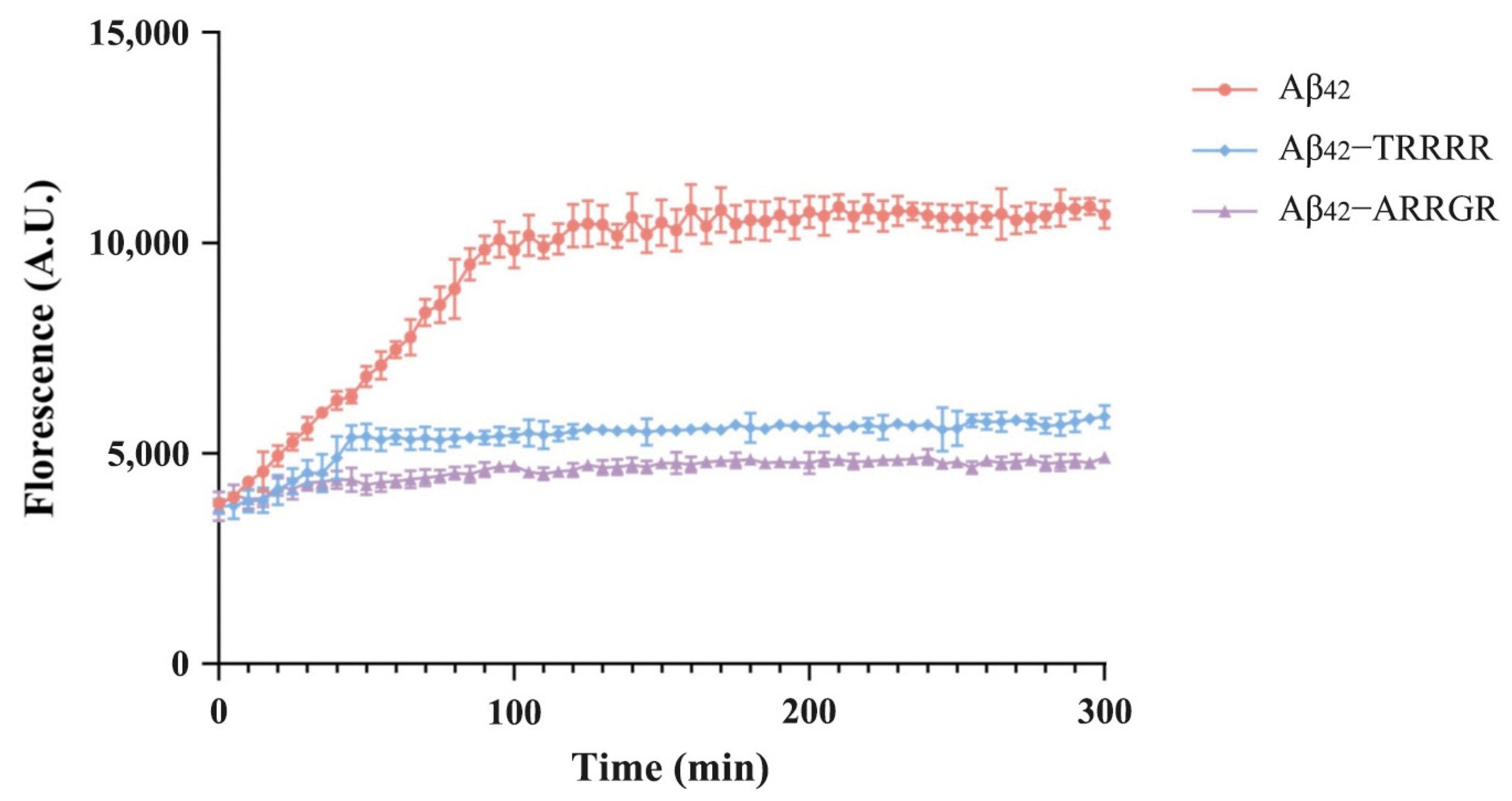
3.2. Effects of Pentapeptides on Aggregation of Aβ42 Detected by ThT Fluorescence Assay

From the results obtained from our computational analysis of the interaction between pentapeptides and Aβ42, TRRRR and ARRGR were chosen for further experiments with the ThT fluorescence assay. The fluorescence intensity of TRRRR and ARRGR were evaluated independently to determine their inhibitory effects on Aβ42 aggregation. Also, there was an Aβ42 group for the control experiment. The fluorescence intensity of the Aβ42 control group was higher than that of TRRRR and ARRGR after it was incubated solo at 37 °C for 48 h (Figure 6). Those of the groups of TRRRR incubated with Aβ42 and ARRGR incubated with Aβ42 were lower. The high fluorescent intensity of the Aβ42 control group could be attributed to the fact that Aβ42 amassed a large quantity of ThT. The decline in fluorescence intensity in the groups containing TRRRR and ARRGR could be attributed to a decrease in the quantity of Aβ42 clusters. These definitely demonstrate that our cationic arginine-rich pentapeptides may be effective at inhibiting the polymerization of Aβ42.

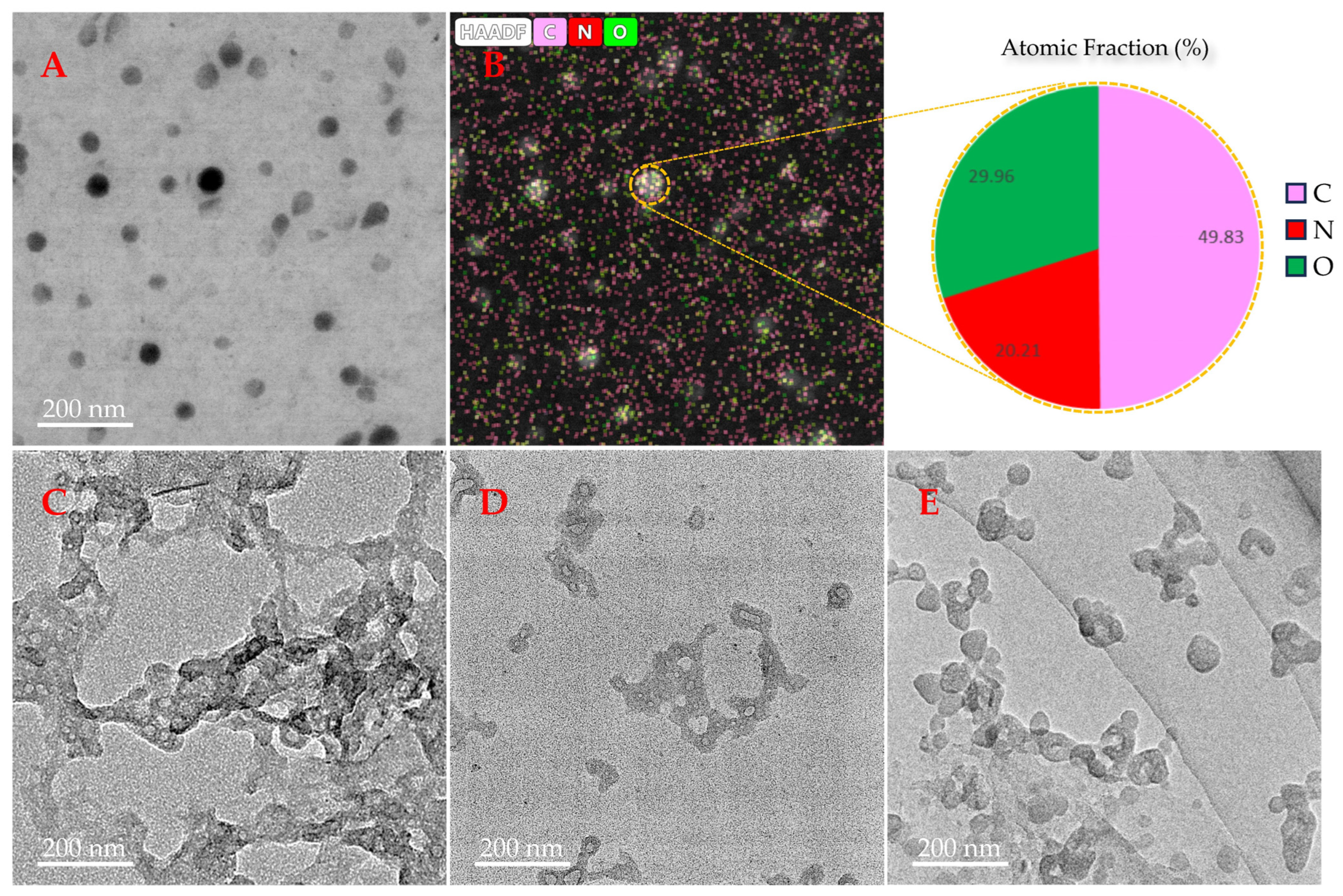
3.3. Observation of the Effect of TRRRR or ARRGR on Aβ42 Aggregation with a Transmission Electron Microscope (TEM)

A transmission electron microscope (TEM) was used to observe the effects of TRRRR or ARRGR on the aggregation of Aβ42. Many fibrils were clearly visible after incubating Aβ42 alone for 48 h (Figure 7C). Aβ42 incubated for 48 h too with TRRRR and ARRGR revealed minute clusters which did not have a well-defined shape (Figure 7D,E). Results from the transmission electron microscope showed that creation of Aβ42 fibrils were inhibited by both pentapeptides. This is supported by the fact that the growth of Aβ42 fibrils was inhibited to an extent.

3.4. ELISA Experiments to Investigate the Effects of TRRRR and ARRGR on Aβ42 Expression

To investigate the effects of TRRRR and ARRGR on Aβ42 protein expression, an ELISA experiment was conducted (as shown in Figure 8). The results indicated that Aβ42 protein endogenously expressed in SH-SY5Y cells was detected in the control group (SH-SY5Y cells not secreting Aβ42), TRRRR 50 μM treatment group, and ARRGR 50 μM treatment group, with no significant differences in expression levels observed among the groups. The Aβ42 levels in the Aβ42 group (SH-SY5Y cells secreting Aβ42) were significantly higher than in the aforementioned three groups. Additionally, no significant differences were observed among the Aβ42 group, Aβ42 with TRRRR 10 μM treatment group, Aβ42 with TRRRR 50 μM treatment group, Aβ42 with ARRGR 10 μM treatment group, and Aβ42 with ARRGR 50 μM treatment group. These findings suggest that pentapeptides do not affect Aβ42 protein expression level in SH-SY5Y cells.

3.5. Effect of Either TRRRR or ARRGR on Cell Death Induced by Secreted Aβ42

Using a fluorescent microscope, the viability results of Aβ42-secreting SH-SY5Y cells in the control group and the pentapeptide treatment groups are presented in Figure 9. Ethidium bromide can incorporate into the DNA of dead cells that have lost their membrane integrity, resulting in the red-stained cells. The morphology of transfected SH-SY5Y cells treated with either TRRRR or ARRGR was not different from the negative control, indicating that they had no obvious cytotoxic effects on SH-SY5Y cells (Figure 9C). As shown in Figure 9B(d), a large number of SH-SY5Y cells that were transfected and secreted Aβ42 died. SH-SY5Y cells treated with 10 µM of the pentapeptides showed improved cell survival (Figure 9B(e,f)). With the treatment of 50 µM pentapeptides, the number of cell deaths was significantly reduced compared to the Aβ42-secreting group (Figure 9B(g,h)), indicating that either TRRRR or ARRGR could well ameliorate the cytotoxicity of Aβ42 secreted from SH-SY5Y cells.

3.6. Effect of Either TRRRR or ARRGR on Reactive Oxygen Species Produced by SH-SY5Y Cells Secreting Aβ42

SH-SY5Y cells secreting Aβ42 and those not secreting Aβ42 were incubated independently with either TRRRR or ARRGR for 24 h. After this, a fluorescent probe DCFH DA on a flow cytometer was used to compute the intracellular ROS levels. According to the results obtained (Figure 10), ROS levels in SH-SY5Y cells expressing secreted Aβ42 were significantly reduced by TRRRR and ARRGR, respectively, compared to non-treated SH-SY5Y cells secreting Aβ42. Also, the influence of TRRRR and ARRGR were dose-dependent.

3.7. Protective Effect of Either TRRRR or ARRGR on Cell Apoptosis of SH-SY5Y Cells Secreting Aβ42

SH-SY5Y cells were categorized by different treatments with an apoptosis kit and then evaluated by flow cytometry, and the results can be seen in Figure 11. Figure 11I shows the quantification of cell apoptosis. In Figure 11, Q1 denotes nude nucleus necrotic cells, Q2 denotes necrotic and late apoptotic cells, Q3 early apoptotic cells, and Q4 living cells. In the group which received drug dosages (pentapeptides), cell apoptosis was significantly reduced when either TRRRR or ARRGR were added (Figure 11I). This was particularly significant when the drug dosage was at 50 μM as compared to the group which did not receive the pentapeptides. These results prove that both TRRRR and ARRGR can effectively protect cells from cytotoxicity.

4. Discussion

Amyloid plaques are generated when Aβ is produced excessively, not cleared after production, and then form clusters. These clusters also promote the inflammation of brain tissues and apoptosis. The first pathological occurrence that happens years before the development of clinical symptoms is probably going to be the accumulation of Aβ in the brain. Aβ is generated when there is proteolytic cleavage of the amyloid precursor protein (APP). APP is a transmembrane glycoprotein, which has a cytoplasmic β-domain with 55 amino acids and an extracellular domain with 590–680 amino acids [43]. When the proteases β- and ϒ-secretases cleave APP, different sizes of Aβ fragments are generated, according to the cleavage sites [44]. Aβ40 which is approximately 90% of the Aβ fragments generated, and Aβ42 which is approximately 5–10% of the Aβ fragments generated, are the most preeminent. However, Aβ42 is more toxic than Aβ40. After the generation of the Aβ fragments, they coalesce to form amyloid clusters. These clusters are in disparate configurations such as low-molecular-weight oligomers, protofibrils, and mature fibrils.
The principal therapeutic targets investigated for AD have been directly or indirectly concomitant to neurofibrillary clusters (tau protein) and Aβ senile plaques (protein) [45,46]. Most of these therapeutic targets are developed with the aim of inhibiting aggregation and fostering the clearance of fibrils. However, investigations on inhibitors such as the β-secretase 1 (BACE-1) and ϒ secretases have been discontinued due to challenges in toxicity [45,47,48,49]. Nevertheless, there have been efforts put into developing metal ion-chelating agents [50], peptides [51], natural compounds [52], and biomolecules [53,54]. Peptides have drawn a lot of scrutinization and the number of approved peptide biotherapeutics has been on the rise over the contemporary decades. This has been an interesting strategy because they have the ability to bind with larger interfacial pockets than small molecules [55]. Also, the agenda of peptide development has been piloted by significant approaches in computational structural prognosis and the augmentation of available chemical refinements to ameliorate affinity, stability, and specificity. Peptides are considered a better option for AD than small molecule-based compounds because of their high affinity for Aβ and low toxicity [56]. With this knowledge, peptides could be potential candidates for inhibiting conformational transitions, self-assembly, and toxicity against neurons, and the promotion of the pathways of the nontoxic fibrillation and early diagnosis of Alzheimer’s disease [57]. Our laboratory previously reported the inhibitory effects of dipeptides [30] and tripeptides [58] on Aβ polymerization. We made a discovery that, through hydrophobic interactions between β strands, the Aβ42 monomer first aggregates into oligopeptides and while they lengthen, clumps are formed through parallel clustering. The whole process of the formation of clusters is piloted by hydrophobic interactions and stabilized by electrostatic forces.
Here, in the present work, we demonstrated the inhibitory effects of the cationic arginine-rich peptides (CARPs) TRRRR and ARRGR on Aβ polymerization [10,59,60], performed a detailed review of peptide-based inhibitors developed for AD, and pointed out that, regardless of their advantages, the lack of membrane permeability and difficulty in penetrating the blood–brain barrier are obstacles to the development of anti-aggregatory peptides. Different schools of thought, however, have shown that cationic arginine-rich peptides (CARPs) can demonstrate a special ability to cross membranes of the cell and the blood–brain barrier [61,62,63], thus making them more commonly used ‘‘carrier’’ molecules for a variety of therapeutic ‘‘cargo’’ such as oligonucleotides, peptides, and proteins (reviewed by [64]).
In addition, it has been suggested that there are multiple potential mechanisms for CARPs to confer cytoprotecting against toxic aggregates of amyloid-beta. As described, the predominant approach has focused on inhibiting aggregation; CARPs which have demonstrated the ability to reduce amyloid-beta-induced cytotoxicity in vitro by inhibiting aggregation include those developed by [65,66,67,68].
Arginine has many attributes that make it truly special, to a great extent due to the peculiar structure and chemistry of the guanidinium group [69]. Not only have arginine monomers long been considered a formidable inhibitor of protein aggregation [70], polypeptides containing many arginine residues also influence the formation and cytotoxicity of protein clusters in a fascinating manner. Arginine residues have several significant bioactive properties that are very effective in regulating protein aggregation [71,72,73], and should therefore be taken into consideration when developing Aβ-targeting therapeutic peptides for treating Alzheimer’s disease. Pentapeptides containing arginine bound more tightly to Aβ42 monomer as compared to histidine-containing pentapeptides due to the electrostatic interactions of arginine with the negatively charged residues of the Aβ42 monomer [74].
Aβ peptide (1–42) fibrils are organized as β-sheets [75,76]. Aβ peptide aggregation pathways are determined by the primary amino acid sequence and intermolecular interactions [30,77]. Aβ monomers can form higher-order fabrications ranging from low-molecular-weight oligomers, including dimers, trimers, tetramers, and pentamers, to midrange-molecular-weight oligomers, including hexamers, nanomers, and dodecamers, to protofibrils and fibrils [77]. When molecular dynamics analysis was used to probe mechanisms underlying the aggregation of Aβ42 [30], it was depicted that the bulk of hydrophobic amino acid residues were confined inside the polypeptide chain in clustered polypeptides, bringing about the formation of three hydrophobic Aβ42 clusters. As these hydrophobic clusters extend through the fibril axis inside of packed subunits to be deposited away from solvents, they principally aid in keeping the protein fibril structure stable. By lessening free energy, hydrophobic contact is said to be the predominant force driving Aβ42 aggregation [30].
In our experiment, our pentapeptide TRRRR had hydrogen bond interactions with Val18 and Phe19 of the central hydrophobic core of the Aβ42 molecule. The pentapeptide ARRGR relatively interacted with 33 Gly, 33 Leu, and 35 Met through hydrogen bonds (Figure 1A,D). The pentapeptides were attached to similar hydrophobic regions as those already described by our laboratory [30,58]. When bound to the hydrophobic regions, they were able to obstruct and lessen the binding free energy between Aβ42 strands. The real inhibitory potencies of both TRRRR and ARRGR were probed by computational studies on Aβ42 and confirmed by in vitro studies. The ThT assay and transmission electron microscopy experiments elucidated that both TRRRR and ARRGR can successfully impede clusters of Aβ42. The fluorescent staining, MTT, and flow cytometric experiments further confirmed that the two pentapeptides reduced Aβ42 cytotoxicity (Figure 10 and Figure 11).

5. Conclusions

The present molecular dynamics and pharmacological experimental results indicate that TRRRR and ARRGR may bind to the hydrophobic clusters of the Aβ42 polymerization core to inhibit aggregation and ameliorate its neurotoxicity.

Supplementary Materials

The following supporting information can be downloaded at: https://www.mdpi.com/article/10.3390/cimb46090606/s1, Supplementary Table S1. Docking free energy of pentapeptides to Aβ42.

Author Contributions

Conceptualization, D.W.; methodology, D.W. and L.Y.; software, Z.W. and N.Y.; validation, Z.X., W.G. and J.Z.; formal analysis, L.Y. and Z.W.; investigation, L.Y., Z.W. and N.Y.; resources, D.W.; data curation, Y.P. and Y.M.; writing—original draft preparation, N.M.C.A.; writing—review and editing, D.W.; visualization, L.Y., N.M.C.A. and Z.W.; supervision, D.W.; project administration, D.W.; funding acquisition, D.W. and Y.M. All authors have read and agreed to the published version of the manuscript.

Funding

This research was funded by the National Natural Science Foundation of China, grant number 32160214 and 31760246 to D.W., the Natural Science Foundation of Hainan Province, grant number 821RC1053 and 822RC651 to D.W. and Y.M., and the Cooperative Innovation Center of Hainan University, grant number XTCX2022JKB07 to D.W.

Informed Consent Statement

Not applicable.

Data Availability Statement

All data related to the results presented in the paper are available upon request.

Acknowledgments

We thank Wanmeng Fu for her assistance in making experiments.

Conflicts of Interest

The authors declare no conflicts of interest.

References

  1. Hansen, R.A.; Gartlehner, G.; Webb, A.P.; Morgan, L.C.; Moore, C.G.; Jonas, D.E. Efficacy and safety of donepezil, galantamine, and rivastigmine for the treatment of Alzheimer’s disease: A systematic review and meta-analysis. Clin. Interv. Aging 2008, 3, 211–225. [Google Scholar] [PubMed]
  2. Alzheimer’s Association. 2023 Alzheimer’s disease facts and figures. Alzheimer’s Dement. 2023, 19, 1598–1695. [Google Scholar] [CrossRef] [PubMed]
  3. Gustavsson, A.; Norton, N.; Fast, T.; Frolich, L.; Georges, J.; Holzapfel, D.; Kirabali, T.; Krolak-Salmon, P.; Rossini, P.M.; Ferretti, M.T.; et al. Global estimates on the number of persons across the Alzheimer’s disease continuum. Alzheimer’s Dement. 2023, 19, 658–670. [Google Scholar] [CrossRef]
  4. Li, C.; Grajales, S.; Shuang, S.; Dong, C.; Nair, M. β-amyloid biomarker detection for Alzheimer’s disease. Zhonghua Yi Xue Za Zhi 2017, 1, 15–25. [Google Scholar] [CrossRef]
  5. Demuro, A.; Mina, E.; Kayed, R.; Milton, S.C.; Parker, I.; Glabe, C.G. Calcium dysregulation and membrane disruption as a ubiquitous neurotoxic mechanism of soluble amyloid oligomers. J. Biol. Chem. 2005, 280, 17294–17300. [Google Scholar] [CrossRef]
  6. Li, S.; Hong, S.; Shepardson, N.E.; Walsh, D.M.; Shankar, G.M.; Selkoe, D. Soluble oligomers of amyloid Beta protein facilitate hippocampal long-term depression by disrupting neuronal glutamate uptake. Neuron 2009, 62, 788–801. [Google Scholar] [CrossRef]
  7. Butterfield, D.A.; Lauderback, C.M. Lipid peroxidation and protein oxidation in Alzheimer’s disease brain: Potential causes and consequences involving amyloid beta-peptide-associated free radical oxidative stress. Free Radic. Biol. Med. 2002, 32, 1050–1060. [Google Scholar] [CrossRef] [PubMed]
  8. Clark, T.A.; Lee, H.P.; Rolston, R.K.; Zhu, X.; Marlatt, M.W.; Castellani, R.J.; Nunomura, A.; Casadesus, G.; Smith, M.A.; Lee, H.G.; et al. Oxidative Stress and its Implications for Future Treatments and Management of Alzheimer Disease. Int. J. Biomed. Sci. IJBS 2010, 6, 225–227. [Google Scholar] [PubMed]
  9. Bhattacharjee, S.; Bhattacharyya, R. PRFF Peptide Mimic Interferes with Toxic Fibrin-Abeta(42) Interaction by Emulating the Abeta Binding Interface on Fibrinogen. ACS Chem. Neurosci. 2021, 12, 4144–4152. [Google Scholar] [CrossRef]
  10. Goyal, D.; Shuaib, S.; Mann, S.; Goyal, B. Rationally Designed Peptides and Peptidomimetics as Inhibitors of Amyloid-beta (Abeta) Aggregation: Potential Therapeutics of Alzheimer’s Disease. ACS Comb. Sci. 2017, 19, 55–80. [Google Scholar] [CrossRef]
  11. Lei, L.; Zou, Z.; Liu, J.; Xu, Z.; Fu, Y.; Tian, Y.; Zhang, W. Multifunctional peptide assembled micelles for simultaneously reducing amyloid–β and reactive oxygen–β species. Chem. Sci. 2021, 12, 6449–6459. [Google Scholar] [CrossRef] [PubMed]
  12. Mondal, P.; Khan, J.; Gupta, V.; Ghosh, S. In Silico Approach for Designing Potent Neuroprotective Hexapeptide. ACS Chem. Neurosci. 2019, 10, 3018–3030. [Google Scholar] [CrossRef] [PubMed]
  13. Ren, B.; Tang, Y.; Zhang, D.; Liu, Y.; Zhang, Y.; Chen, H.; Hu, R.; Zhang, M.; Zheng, J. Conformational-specific self-assembled peptides as dual-mode, multi-target inhibitors and detectors for different amyloid proteins. J. Mater. Chem. B 2022, 10, 1754–1762. [Google Scholar] [CrossRef] [PubMed]
  14. Roy, R.; Pradhan, K.; Khan, J.; Das, G.; Mukherjee, N.; Das, D.; Ghosh, S. Human Serum Albumin-Inspired Glycopeptide-Based Multifunctional Inhibitor of Amyloid-beta Toxicity. ACS Omega 2020, 5, 18628–18641. [Google Scholar] [CrossRef] [PubMed]
  15. Ryan, P.; Patel, B.; Makwana, V.; Jadhav, H.R.; Kiefel, M.; Davey, A.; Reekie, T.A.; Rudrawar, S.; Kassiou, M. Peptides, Peptidomimetics, and Carbohydrate-Peptide Conjugates as Amyloidogenic Aggregation Inhibitors for Alzheimer’s Disease. ACS Chem. Neurosci. 2018, 9, 1530–1551. [Google Scholar] [CrossRef] [PubMed]
  16. Wang, L.; Wang, N.; Zhang, W.; Cheng, X.; Yan, Z.; Shao, G.; Wang, X.; Wang, R.; Fu, C. Therapeutic peptides: Current applications and future directions. Signal Transduct. Target. Ther. 2022, 7, 48. [Google Scholar] [CrossRef]
  17. Delaunay, M.; Ha-Duong, T. Computational Tools and Strategies to Develop Peptide-Based Inhibitors of Protein-Protein Interactions, Computational Peptide Science: Methods and Protocols; Springer: New York, NY, USA, 2021. [Google Scholar]
  18. Rehman, A.U.; Khurshid, B.; Ali, Y.; Rasheed, S.; Wadood, A.; Ng, H.L.; Chen, H.F.; Wei, Z.; Luo, R.; Zhang, J. Computational approaches for the design of modulators targeting protein-protein interactions. Expert Opin. Drug Discov. 2023, 18, 315–333. [Google Scholar] [CrossRef]
  19. Chandrasekhar, G.; Srinivasan, E.; Nandhini, S.; Pravallika, G.; Sanjay, G.; Rajasekaran, R. Computer aided therapeutic tripeptide design, in alleviating the pathogenic proclivities of nocuous alpha-synuclein fibrils. J. Biomol. Struct. Dyn. 2024, 42, 483–494. [Google Scholar] [CrossRef]
  20. Han, Y.; Král, P. Computational design of ACE2-based peptide inhibitors of SARS-CoV-2. ACS Nano 2020, 14, 5143–5147. [Google Scholar] [CrossRef]
  21. Jarmula, A.; Zubalska, M.; Stepkowski, D. Consecutive Aromatic Residues Are Required for Improved Efficacy of beta-Sheet Breakers. Int. J. Mol. Sci. 2022, 23, 5247. [Google Scholar] [CrossRef]
  22. Kanchi, P.K.; Dasmahapatra, A.K. Enhancing the binding of the beta-sheet breaker peptide LPFFD to the amyloid-beta fibrils by aromatic modifications: A molecular dynamics simulation study. Comput. Biol. Chem. 2021, 92, 107471. [Google Scholar] [CrossRef] [PubMed]
  23. Luan, B.; Huynh, T. Crystal-structures-guided design of fragment-based drugs for inhibiting the main protease of SARS-CoV-2. Proteins 2022, 90, 1081–1089. [Google Scholar] [CrossRef] [PubMed]
  24. VK, P.; Rath, S.P.; Abraham, P. Computational designing of a peptide that potentially blocks the entry of SARS-CoV, SARS-CoV-2 and MERS-CoV. PLoS ONE 2021, 16, e0251913. [Google Scholar] [CrossRef]
  25. Abel, R.; Wang, L.; Harder, E.D.; Berne, B.J.; Friesner, R.A. Advancing Drug Discovery through Enhanced Free Energy Calculations. Acc. Chem. Res. 2017, 50, 1625–1632. [Google Scholar] [CrossRef]
  26. Kuhn, B.; Tichý, M.; Wang, L.; Robinson, S.; Martin, R.E.; Kuglstatter, A.; Benz, J.; Giroud, M.; Schirmeister, T.; Abel, R.; et al. Prospective Evaluation of Free Energy Calculations for the Prioritization of Cathepsin L Inhibitors. J. Med. Chem. 2017, 60, 2485–2497. [Google Scholar] [CrossRef]
  27. Meier, K.; Bluck, J.P.; Christ, C.D. Free Energy Methods in Drug Discovery: Current State and Future Directions. In ACS Symposium Series; Amarcost, K.A., Thompson, D.C., Eds.; ACS Publications: Washington, DC, USA, 2021; Chapter 2; pp. 39–66. [Google Scholar]
  28. Ross, G.A.; Lu, C.; Scarabelli, G.; Albanese, S.K.; Houang, E.; Abel, R.; Harder, E.D.; Berne, B.J.; Friesner, R.A. The maximal and current accuracy of rigorous protein-ligand binding free energy calculations. Acc. Chem. Res. 2017, 50, 1625–1632. [Google Scholar] [CrossRef]
  29. Sun, Y.; Wang, B.; Pei, J.; Luo, Y.; Yuan, N.; Xiao, Z.; Wu, H.; Luo, C.; Wang, J.; Wei, S.; et al. Molecular dynamic and pharmacological studies on protein-engineered hirudin variants of Hirudinaria manillensis and Hirudo medicinalis. Br. J. Pharmacol. 2022, 179, 3740–3753. [Google Scholar] [CrossRef]
  30. Yuan, N.; Ye Lz Sun, Y.; Wu, H.; Xiao, Z.; Fu, W.; Chen, Z.; Pei, Y.; Min, Y.; Wang, D. Molecular Integrative Analysis of the Inhibitory Effects of Dipeptides on Amyloid Peptide 1–42 Polymerization. Int. J. Mol. Sci. 2023, 24, 7673. [Google Scholar] [CrossRef]
  31. Summa, V.; Petrocchi, A.; Bonelli, F.; Crescenzi, B.; Donghi, M.; Ferrara, M.; Fiore, F.; Gardelli, C.; Gonzalez Paz, O.; Hazuda, D.J.; et al. Discovery of Raltegravir, a potent, selective orally bioavailable HIV-integrase inhibitor for the treatment of HIV-AIDS infection. J. Med. Chem. 2008, 51, 5843–5855. [Google Scholar] [CrossRef] [PubMed]
  32. Vidal-Limon, A.; Aguilar-Toalá, J.E.; Liceaga, A.M. Integration of Molecular Docking Analysis and Molecular Dynamics Simulations for Studying Food Proteins and Bioactive Peptides. J. Agric. Food Chem. 2022, 70, 934–943. [Google Scholar] [CrossRef]
  33. Guallar, V.; Wallrapp, F.H. QM/MM methods: Looking inside heme proteins biochemisty. Biophys. Chem. 2010, 149, 1–11. [Google Scholar] [CrossRef] [PubMed]
  34. Schames, J.R.; Henchman, R.H.; Siegel, J.S.; Sotriffer, C.A.; Ni, H.; McCammon, J.A. Discovery of a novel binding trench in HIV integrase. J. Med. Chem. 2004, 47, 1879–1881. [Google Scholar] [CrossRef] [PubMed]
  35. Sinha, S.; Tam, B.; Wang, S.M. Applications of Molecular Dynamics Simulation in Protein Study. Membranes 2022, 12, 844. [Google Scholar] [CrossRef]
  36. Van Gunsteren, W.F.; Berendsen, H.J. Computer Simulation of Molecular Dynamics: Methodology, Applications, and Perspectives in Chemistry. Angew. Chem. Int. Ed. Engl. 1990, 29, 992–1023. [Google Scholar] [CrossRef]
  37. Dror, R.O.; Dirks, R.M.; Grossman, J.P.; Xu, H.; Shaw, D.E. Biomolecular simulation: A computational microscope for molecular biology. Annu. Rev. Biophys. 2012, 41, 429–452. [Google Scholar] [CrossRef]
  38. Gera, J.; Szogi, T.; Bozso, Z.; Fulop, L.; Barrera, E.E.; Rodriguez, A.M.; Mendez, L.; Delpiccolo, C.M.L.; Mata, E.G.; Cioffi, F.; et al. Searching for improved mimetic peptides inhibitors preventing conformational transition of amyloid-beta(42) monomer. Bioorg. Chem. 2018, 81, 211–221. [Google Scholar] [CrossRef]
  39. Liu, F.F.; Dong, X.Y.; He, L.; Middelberg, A.P.; Sun, Y. Molecular insight into conformational transition of amyloid beta-peptide 42 inhibited by (-)-epigallocatechin-3-gallate probed by molecular simulations. J. Phys. Chem. B 2011, 115, 11879–11887. [Google Scholar] [CrossRef]
  40. Chang, S.Y.; Liu, F.F.; Dong, X.Y.; Sun, Y. Molecular insight into conformational transmission of human P-glycoprotein. J. Chem. Phys. 2013, 139, 225102–225112. [Google Scholar] [CrossRef] [PubMed]
  41. Reddy, G.; Straub, J.E.; Thirumalai, D. Dynamics of locking of peptides onto growing amyloid fibrils. Proc. Natl. Acad. Sci. USA 2009, 106, 11948–11953. [Google Scholar] [CrossRef]
  42. Tonali, N.; Dodero, V.I.; Kaffy, J.; Hericks, L.; Ongeri, S.S. Real-time BODIPY-Binding assay to screen inhibitors of the early oligomerization process of Aβ1-42 peptide. Chembiochem 2020, 21, 1129–1135. [Google Scholar] [CrossRef]
  43. Zhang, Y.; Chen, H.; Li, R.; Sterling, K.; Song, W. Amyloid beta-based therapy for Alzheimer’s disease: Challenges, successes and future. Signal Transduct. Target. Ther. 2023, 8, 248. [Google Scholar] [CrossRef] [PubMed]
  44. Haass, C.; Selkoe, D.J. Cellular processing of β-amyloid precursor protein and the genesis of amyloid β-peptide. Cell 1993, 75, 1039–1042. [Google Scholar] [CrossRef] [PubMed]
  45. Conti Filho, C.E.; Loss, L.B.; Marcolongo-Pereira, C.; Rossoni Junior, J.V.; Barcelos, R.M.; Chiarelli-Neto, O.; da Silva, B.S.; Passamani Ambrosio, R.; Castro, F.; Teixeira, S.F.; et al. Advances in Alzheimer’s disease’s pharmacological treatment. Front. Pharmacol. 2023, 14, 1101452. [Google Scholar] [CrossRef]
  46. Jokar, S.; Khazaei, S.; Gameshgoli, X.E.; Khafaji, M.; Yarani, B.; Sharifzadeh, M.; Beiki, D.; Bavi, O. Amyloid beta-Targeted Inhibitory Peptides for Alzheimer’s Disease: Current State and Future Perspectives. In Alzheimer’s Disease: Drug Discovery; Huang, X., Ed.; Exon Publications: Brisbane, AU, Australia, 2020. [Google Scholar]
  47. Cebers, G.; Alexander, R.C.; Haeberlein, S.B.; Han, D.; Goldwater, R.; Ereshefsky, L.; Olsson, T.; Ye, N.; Rosen, L.; Russell, M.; et al. AZD3293: Pharmacokinetic and Pharmacodynamic Effects in Healthy Subjects and Patients with Alzheimer’s Disease. J. Alzheimer’s Dis. 2017, 55, 1039–1053. [Google Scholar] [CrossRef]
  48. McDade, E.; Voytyuk, I.; Aisen, P.; Bateman, R.J.; Carrillo, M.C.; De Strooper, B.; Haass, C.; Reiman, E.M.; Sperling, R.; Tariot, P.N.; et al. The case for low-level BACE1 inhibition for the prevention of Alzheimer disease. Nat. Rev. Neurol. 2021, 17, 703–714. [Google Scholar] [CrossRef]
  49. Yang, G.; Zhou, R.; Guo, X.; Yan, C.; Lei, J.; Shi, Y. Structural basis of gamma-secretase inhibition and modulation by small molecule drugs. Cell 2021, 184, 521–533 e514. [Google Scholar] [CrossRef] [PubMed]
  50. Liu, H.; Dong, X.; Liu, F.; Zheng, J.; Sun, Y. Iminodiacetic acid-conjugated nanoparticles as a bifunctional modulator against Zn2+-mediated amyloid protein aggregation and cytotoxicity. J. Colloid Interface Sci. 2017, 505, 973–982. [Google Scholar] [CrossRef]
  51. Wang, W.; Han, Y.; Fan, Y.; Wang, Y. Effects of Gold Nanospheres and Nanocubes on Amyloid-beta Peptide Fibrillation. Langmuir 2019, 35, 2334–2342. [Google Scholar] [CrossRef]
  52. Brahmachari, S.; Paul, A.; Segal, D.; Gazit, E. Inhibition of amyloid oligomerization into different supramolecular architectures by small molecules: Mechanistic insights and design rules. Future Med. Chem. 2017, 9, 797–810. [Google Scholar] [CrossRef]
  53. Jokar, S.; Khazaei, S.; Behnammanesh, H.; Shamloo, A.; Erfani, M.; Beiki, D.; Bavi, O. Recent advances in the design and applications of amyloid-beta peptide aggregation inhibitors for Alzheimer’s disease therapy. Biophys. Rev. 2019, 11, 901–925. [Google Scholar] [CrossRef]
  54. Rahimi, F.; Li, H.; Sinha, S.; Bitan, G. Modulators of amyloid β-protein self-assembly. In Developing Therapeutics for Alzheimer’s Disease; Elsevier: Boston, MA, USA, 2016; pp. 97–191. [Google Scholar] [CrossRef]
  55. Lee, A.C.; Harris, J.L.; Khanna, K.K.; Hong, J.H. A Comprehensive Review on Current Advances in Peptide Drug Development and Design. Int. J. Mol. Sci. 2019, 20, 2383. [Google Scholar] [CrossRef] [PubMed]
  56. Danho, W.; Swistok, J.; Khan, W.; Chu, X.J.; Cheung, A.; Fry, D.; Sun, H.; Kurylko, G.; Rumennik, L.; Cefalu, J.; et al. Opportunities and challenges of developing peptide drugs in the pharmaceutical industry. Adv. Exp. Med. Biol. 2009, 611, 467–469. [Google Scholar] [CrossRef] [PubMed]
  57. Jokar, S.; Erfani, M.; Bavi, O.; Khazaei, S.; Sharifzadeh, M.; Hajiramezanali, M.; Beiki, D.; Shamloo, A. Design of peptide-based inhibitor agent against amyloid-beta aggregation: Molecular docking, synthesis and in vitro evaluation. Bioorg. Chem. 2020, 102, 104050. [Google Scholar] [CrossRef] [PubMed]
  58. Wu, Z.; Ye, L.; Yuan, N.; Che Ajuyo, N.M.; Xiao, Z.; Liu, L.; Chen, Z.; Pei, Y.; Min, Y.; Wang, D. A Molecular Integrative Study on the Inhibitory Effects of WRR and ERW on Amyloid Peptide (1–42) Polymerization and Cell Toxicity. Polymers 2023, 15, 4356. [Google Scholar] [CrossRef]
  59. Funke, S.A.; Willbold, D. Peptides for therapy and diagnosis of Alzheimer’s disease. Curr. Pharm. Des. 2012, 18, 755–767. [Google Scholar] [CrossRef]
  60. Kumar, J.; Sim, V. D-amino acid-based peptide inhibitors as early or preventative therapy in Alzheimer disease. Prion 2014, 8, 119–124. [Google Scholar] [CrossRef]
  61. Allolio, C.; Magarkar, A.; Jurkiewicz, P.; Baxova, K.; Javanainen, M.; Mason, P.E.; Sachl, R.; Cebecauer, M.; Hof, M.; Horinek, D.; et al. Arginine-rich cell-penetrating peptides induce membrane multilamellarity and subsequently enter via formation of a fusion pore. Proc. Natl. Acad. Sci. USA 2018, 115, 11923–11928. [Google Scholar] [CrossRef]
  62. Mitchell, D.J.; Kim, D.T.; Steinman, L.; Fathman, C.G.; Rothbard, J.B. Polyarginine enters cells more efficiently than other polycationic homopolymers. J. Pept. Res. 2000, 56, 318–325. [Google Scholar] [CrossRef]
  63. Schmidt, N.; Mishra, A.; Lai, G.H.; Wong, G.C. Arginine-rich cell-penetrating peptides. FEBS Lett. 2010, 584, 1806–1813. [Google Scholar] [CrossRef]
  64. Habault, J.; Poyet, J.L. Recent Advances in Cell Penetrating Peptide-Based Anticancer Therapies. Molecules 2019, 24, 927. [Google Scholar] [CrossRef]
  65. Cheng, Y.S.; Chen, Z.T.; Liao, T.Y.; Lin, C.; Shen, H.C.H.; Wang, Y.H.; Chang, C.W.; Liu, R.S.; Chen, R.P.Y.; Tu, P.H. An intranasally delivered peptide drug ameliorates cognitive decline in Alzheimer transgenic mice. EMBO Mol. Med. 2017, 9, 703–715. [Google Scholar] [CrossRef] [PubMed]
  66. Henning-Knechtel, A.; Kumar, S.; Wallin, C.; Król, S.; Wärmländer, S.K.; Jarvet, J.; Esposito, G.; Kirmizialtin, S.; Gräslund, A.; Hamilton, A.D.; et al. Designed Cell-Penetrating Peptide Inhibitors of Amyloid-beta Aggregation and Cytotoxicity. Cell Rep. Physic. Sci. 2020, 1, 100014–100035. [Google Scholar] [CrossRef]
  67. Kawasaki, T.; Onodera, K.; Kamijo, S. Identification of novel short peptide inhibitors of soluble 37/48 kDa oligomers of amyloid beta42. Biosci. Biotechnol. Biochem. 2011, 75, 1496–1501. [Google Scholar] [CrossRef]
  68. Parthsarathy, V.; McClean, P.L.; Holscher, C.; Taylor, M.; Tinker, C.; Jones, G.; Kolosov, O.; Salvati, E.; Gregori, M.; Masserini, M.; et al. A novel retro-inverso peptide inhibitor reduces amyloid deposition, oxidation and inflammation and stimulates neurogenesis in the APPswe/PS1DeltaE9 mouse model of Alzheimer’s disease. PLoS ONE 2013, 8, e54769. [Google Scholar] [CrossRef]
  69. Xu, B.; Jacobs, M.I.; Kostko, O.; Ahmed, M. Guanidinium Group Remains Protonated in a Strongly Basic Arginine Solution. Chemphyschem 2017, 18, 1503–1506. [Google Scholar] [CrossRef]
  70. Sharma, S.; Sarkar, S.; Paul, S.S.; Roy, S.; Chattopadhyay, K. A small molecule chemical chaperone optimizes its unfolded state contraction and denaturant like properties. Sci. Rep. 2013, 3, 3525. [Google Scholar] [CrossRef]
  71. Arakawa, T.; Kita, Y.; Ejima, D.; Tsumoto, K.; Fukada, H. Aggregation suppression of proteins by arginine during thermal unfolding. Protein Pept. Lett. 2006, 13, 921–927. [Google Scholar] [CrossRef]
  72. Ghosh, R.; Sharma, S.; Chattopadhyay, K. Effect of arginine on protein aggregation studied by fluorescence correlation spectroscopy and other biophysical methods. Biochemistry 2009, 48, 1135–1143. [Google Scholar] [CrossRef]
  73. Golovanov, A.P.; Hautbergue, G.M.; Wilson, S.A.; Lian, L.Y. A simple method for improving protein solubility and long-term stability. J. Am. Chem. Soc. 2004, 126, 8933–8939. [Google Scholar] [CrossRef]
  74. Kaur, A.; Goyal, B. Identification of new pentapeptides as potential inhibitors of amyloid-beta(42) aggregation using virtual screening and molecular dynamics simulations. J. Mol. Graph. Model. 2023, 124, 108558. [Google Scholar] [CrossRef]
  75. Antzutkin, O.N.; Leapman, R.D.; Balbach, J.J.; Tycko, R. Supramolecular structural constraints on Alzheimer’s beta-amyloid fibrils from electron microscopy and solid-state nuclear magnetic resonance. Biochemistry 2002, 41, 15436–15450. [Google Scholar] [CrossRef] [PubMed]
  76. Balbach, J.J.; Petkova, A.T.; Oyler, N.A.; Antzutkin, O.N.; Gordon, D.J.; Meredith, S.C.; Tycko, R. Supramolecular structure in full-length Alzheimer’s beta-amyloid fibrils: Evidence for a parallel beta-sheet organization from solid-state nuclear magnetic resonance. Biophys. J. 2002, 83, 1205–1216. [Google Scholar] [CrossRef] [PubMed]
  77. Chen, G.F.; Xu, T.H.; Yan, Y.; Zhou, Y.R.; Jiang, Y.; Melcher, K.; Xu, H.E. Amyloid beta: Structure, biology and structure-based therapeutic development. Acta Pharmacol. Sin. 2017, 38, 1205–1235. [Google Scholar] [CrossRef] [PubMed]
Figure 1. Molecular docking of the five pentapeptides with Aβ42. (A) Binding of TRRRR with Aβ42. (B) Binding of RRRWR with Aβ42. (C) Binding of RRRDS with Aβ42. (D) Binding of ARRGR with Aβ42. (E) Binding of TRRAR with Aβ42. For clarity, only three strands taken from Aβ42 pentamer are shown in this figure. The magenta mesh represents the molecular surface of the pentapeptides, and the gray mesh represents the boundary of van der Waal’s force. Arrows indicate the hydrogen bonds. TRRRR: Threonine-arginine-arginine-arginine-arginine; RRRWR: Arginine-arginine-arginine-tryptophan-arginine; RRRDS: Arginine-arginine-arginine-aspartic acid-serine; ARRGR: Alanine-arginine-arginine-glycine-arginine; TRRAR: Threonine-arginine-arginine-alanine-arginine.
Cimb 46 00606 g001
Figure 2. The binding stability and interaction energy of the pentapeptides at different hydrophobic regions. (A) The binding stability of pentapeptides to Aβ42 pentamer. (B) The interaction energy between Aβ42 pentamer and the pentapeptides. RMSD: The root mean square deviation of the positions of the heavy elements of a pentapeptide. The interaction energy is the algebraic sum of Lennard-Jones and Coulombic potential energy. TRRRR: Threonine-arginine-arginine-arginine-arginine; RRRWR: Arginine-arginine-arginine-tryptophan-arginine; RRRDS: Arginine-arginine-arginine-aspartic acid-serine; ARRGR: Alanine-arginine-arginine-glycine-arginine; TRRAR: Threonine-arginine-arginine-alanine-arginine.
Cimb 46 00606 g002
Figure 3. Variation in the lengths of hydrogen bonds formed between the pentapeptides and Aβ42. (A) ARRGR and Aβ42. (B) RRRDS and Aβ42. (C) RRRWR and Aβ42. (D) TRRAR and Aβ42. (E) TRRRR and Aβ42. TRRRR: Threonine-arginine-arginine-arginine-arginine; RRRWR: Arginine-arginine-arginine-tryptophan-arginine; RRRDS: Arginine-arginine-arginine-aspartic acid-serine; ARRGR: Alanine-arginine-arginine-glycine-arginine; TRRAR: Threonine-arginine-arginine-alanine-arginine.
Cimb 46 00606 g003
Figure 4. Variation in the angles of hydrogen bonds formed the pentapeptides and Aβ42. (A) ARRGR and Aβ42. (B) RRRDS and Aβ42. (C) RRRWR and Aβ42. (D) TRRAR and Aβ42. (E) TRRRR and Aβ42. TRRRR: Threonine-arginine-arginine-arginine-arginine; RRRWR: Arginine-arginine-arginine-tryptophan-arginine; RRRDS: Arginine-arginine-arginine-aspartic acid-serine; ARRGR: Alanine-arginine-arginine-glycine-arginine; TRRAR: Threonine-arginine-arginine-alanine-arginine.
Cimb 46 00606 g004
Figure 5. Binding free energy between the pentapeptides and Aβ42. (A) TRRRR and Aβ42. (B) RRRDS and Aβ42. (C) ARRGR and Aβ42. (D) RRRWR and Aβ42. (E) TRRAR and Aβ42. TRRRR: Threonine-arginine-arginine-arginine-arginine; RRRWR: Arginine-arginine-arginine-tryptophan-arginine; RRRDS: Arginine-arginine-arginine-aspartic acid-serine; ARRGR: Alanine-arginine-arginine-glycine-arginine; TRRAR: Threonine-arginine-arginine-alanine-arginine.
Cimb 46 00606 g005
Figure 6. Effects of pentapeptides on aggregation of Aβ42 detected by ThT fluorescence assay. Results are expressed as means ± SD, p < 0.01 among groups, tested by two-way ANOVA, n = 5.
Cimb 46 00606 g006
Figure 7. Effect of pentapeptides on aggregation of Aβ42 detected by transmission electron microscopy. (A) Transmission electron microscopic images of 10 μM Aβ42 before incubation. (B) The Atomic Fraction of Aβ42 detected by HAADF-STEM imaging. (C) Transmission electron microscopic images of 10 μM Aβ42 incubated for 48 h. (D) Transmission electron microscopic images of 10 μM Aβ42 co-incubated with 40 μM TRRRR for 48 h. (E) Transmission electron microscopic image of 10 μM Aβ42 co-incubated with 40 μM ARRGR for 48 h.
Cimb 46 00606 g007
Figure 8. Effects of TRRRR and ARRGR on Aβ42 protein expression. Aβ42: SH-SY5Y cells not secreting Aβ42; Aβ42+: SH-SY5Y cells secreting Aβ42. The results are expressed as means ± SD; ns: insignificant; ** p < 0.01; the results were analyzed by one-way ANOVA, followed by the Tukey–Kramer test for multiple comparisons, with n = 3.
Cimb 46 00606 g008
Figure 9. Effects of TRRRR and ARRGR against cell toxicity of Aβ42 secreted from SH-SY5Y. (A) The morphology of SH-SY5Y cells secreting Aβ42. Round-shaped cells with a bright edge are dying or dead. (B) Damaged cells detected with ethidium bromide, a nuclei acid tracer that cannot pass through an intact cell membrane. (C) Quantification of cell damage by using Image J. Results are expressed as means ± SD with ** p < 0.01, and were analyzed by one-way ANOVA, followed by the Tukey–Kramer test for multiple comparisons, n = 3; ns: insignificant. In (A,B): (a) SH-SY5Y control cells that do not secrete Aβ42. (b) The control cells treated with TRRRR at 50 μM. (c) The control cells treated with ARRGR at 50 μM. (d) SH-SY5Y cells secreting Aβ42. (e) Aβ42-secreting SH-SY5Y cells treated with 10 μM TRRRR. (f) Aβ42-secreting SH-SY5Y cells treated with 10 μM ARRGR. (g) Aβ42-secreting SH-SY5Y cells treated with 50 μM TRRRR. (h) Aβ42-secreting SH-SY5Y cells treated with 50 μM ARRGR.
Cimb 46 00606 g009
Figure 10. Effects of the pentapeptides on ROS levels in SH-SY5Y cells secreting Aβ42. (A) ROS levels in the SH-SY5Y control cells that do not secrete Aβ42. (B) ROS levels in the control cells treated with 50 μM TRRRR. (C) ROS levels in the control cells treated with 50 μM ARRGR. (D) ROS levels in SH-SY5Y cells secreting Aβ42. (E) ROS levels in the Aβ42-secreting cells treated with 10 μM TRRRR. (F) ROS levels in the Aβ42-secreting cells treated with 10 μM ARRGR. (G) ROS levels in the Aβ42-secreting cells treated with 50 μM TRRRR. (H) ROS levels in the Aβ42-secreting cells treated with 50 μM ARRGR. (I) Overlay of the flow cytometry plots (AH). (J) Quantification of the ROS levels in the cells. Results are expressed as means ± SD, ns: not significant, ** p < 0.01, by one-way ANOVA, followed by the Tukey–Kramer test for multiple comparisons, with n = 3.
Cimb 46 00606 g010
Figure 11. Effects of pentapeptide on apoptosis of SY-SY5Y cells secreting Aβ42. (A) Apoptosis in the SH-SY5Y control cells that do not secrete Aβ42. (B) Apoptosis in the control cells treated with 50 μM TRRRR. (C) Apoptosis in the control cells treated with 50 μM ARRGR. (D) Apoptosis in SH-SY5Y cells secreting Aβ42. (E) Apoptosis in the Aβ42-secreting cells treated with 10 μM TRRRR. (F) Apoptosis in the Aβ42-secreting cells treated with 10 μM ARRGR. (G) Apoptosis in the Aβ42-secreting cells treated with 50 μM TRRRR. (H) Apoptosis in the Aβ42-secreting cells treated with 50 μM ARRGR. (I) Overlay of the flow cytometry plots (AH). (J) Quantification of apoptosis in the cells. Results are expressed as means ± SD; ns: not significant, ** p < 0.01; results were analyzed by one-way ANOVA, followed by the Tukey–Kramer test for multiple comparisons, with n = 3.
Cimb 46 00606 g011
Disclaimer/Publisher’s Note: The statements, opinions and data contained in all publications are solely those of the individual author(s) and contributor(s) and not of MDPI and/or the editor(s). MDPI and/or the editor(s) disclaim responsibility for any injury to people or property resulting from any ideas, methods, instructions or products referred to in the content.

