Article

Premature Vertebral Mineralization in hmx1-Mutant Zebrafish

by
Younes El Fersioui
1,2,*,
Gaëtan Pinton
1,
Nathalie Allaman-Pillet
1 and
Daniel F. Schorderet
1,3,4
1
IRO—Institute for Research in Ophthalmology, 1950 Sion, Switzerland
2
Jules-Gonin Eye Hospital, Unit of Gene Therapy and Stem Cell Biology, 1004 Lausanne, Switzerland
3
Faculty of Life Sciences, Swiss Federal Institute of Technology (EPFL), 1015 Lausanne, Switzerland
4
Faculty of Biology and Medicine, University of Lausanne, 1011 Lausanne, Switzerland
*
Author to whom correspondence should be addressed.
Cells 2022, 11(7), 1088; https://doi.org/10.3390/cells11071088
Submission received: 12 February 2022 / Revised: 11 March 2022 / Accepted: 21 March 2022 / Published: 24 March 2022
(This article belongs to the Section Cell Signaling)

Abstract

:
H6 family homeobox 1 (HMX1) regulates multiple aspects of craniofacial development, and mutations in HMX1 are linked to an ocular defect termed oculoauricular syndrome of Schorderet–Munier–Franceschetti (OAS) (MIM #612109). Recently, additional altered orofacial features have been reported, including short mandibular rami, asymmetry of the jaws, and altered premaxilla. We found that in two mutant zebrafish lines termed hmx1mut10 and hmx1mut150, precocious mineralization of the proximal vertebrae occurred. Zebrafish hmx1mut10 and hmx1mut150 report mutations in the SD1 and HD domains, which are essential for dimerization and activity of hmx1. In hmx1mut10, the bone morphogenetic protein (BMP) antagonists chordin and noggin1 were downregulated, while bmp2b and bmp4 were highly expressed and specifically localized to the dorsal region prior to the initiation of the osteogenic process. The osteogenic promoters runx2b and spp1 were also upregulated. Supplementation with DMH1—an inhibitor of the BMP signaling pathway—at the specific stage in which bmp2b and bmp4 are highly expressed resulted in reduced vertebral mineralization, resembling the wildtype mineralization progress of the axial skeleton. These results point to a possible role of hmx1 as part of a complex gene network that inhibits bmp2b and bmp4 in the dorsal region, thus regulating early axial skeleton development.

1. Introduction

The oculoauricular syndrome of Schorderet–Munier–Franceschetti (OAS) (MIM:612109) is caused by defects associated with mutations in the HMX1 transcription factor [1]. HMX1, belonging to a homeobox (HMX) family of transcription factors, presents a phylogenetically conserved 60-amino-acid homeobox domain [2]. OAS patients report aberrant orofacial development resulting in short mandibular rami, asymmetry of the jaws, and altered premaxilla [3].
In zebrafish, hmx1-deficient embryos exhibit increased apoptosis in the eyes and brain, in addition to a delayed withdrawal of retinal progenitors from the cell cycle [4]. The “Dmbo” mouse model showed malformations of the squamous temporal bone and hyperplasia of the gonial bone, and failed to develop somatosensory neurons in the geniculate ganglion [5,6].
The majority of the craniofacial bones are formed via direct ossification, while the cranial base, mammal limbs, and axial skeleton structures are formed via endochondral ossification—a process whereby a cartilage template is replaced by bone [7,8].
The dual process of ossification is conserved in teleost fish, including zebrafish. As early as 3 days post-fertilization (dpf) the cranial cartilage starts forming, followed by perichondral bone elements building up on the existing cartilage structures [9,10,11]. Within the same time window, the first bone elements—including the opercle, cleithrum, mandible, and maxilla—form via direct ossification [12,13]. Teleosts’ vertebral column is formed through direct mineralization of the notochord, and occurs progressively in a craniocaudal direction [14].
BMP gradient is essential for dorsoventral patterning. and is highly modulated through different mechanisms, including extramembranous regulation. Chordin, follistatin, and noggin negatively regulate BMP signaling by binding BMP ligands and preventing their interaction with transducer receptors localized on the cell membranes of target cells [15,16,17]. BMP expression, following dorsoventral axis formation and regulation, is required for the osteogenic process to proceed [18].
In zebrafish, BMP signaling is crucial between 48 and 72 h post-fertilization (hpf) for head cartilage development and bone mineralization [19].
The runx2a and runx2b orthologs initiate both endochondral and intramembranous ossification [20,21,22]. BMP–Runx2 interaction initiates osteoblast differentiation and drives the onset of genes involved in bone extracellular matrix deposition, such as bone gamma-carboxyglutamate (Gla) protein (Bglap) and secreted phosphoprotein 1 (Spp1) [23,24,25,26].
Disruption of BMP signaling is associated with several human diseases [27], and deletion of BMP2 and BMP4 or BMP2 alone results in a severe chondrodysplastic phenotype [28], while craniofacial defects were reported in conditional knockout mice lacking the BMP type I receptor Alk2 [29]. In zebrafish, a mutation in bmp1a resulted in mature bone with a higher mineral content [30].
Despite the recent reports on the phenotypic defects associated with HMX1 mutation, not much is known about its role in transcriptional regulation. HMX1 binds preferentially to the consensus sequence 5′-CAAGTG-3′ located in the promoter region of target genes, and acts as a transcriptional regulator [31]. We previously designed a predictive promoter model [32] and generated two zebrafish models—hmx1mut10 and hmx1mut150 mutant lines—to screen and identify potential hmx1 target genes [33]. The hmx1mut10 and hmx1mut150 mutant lines carry mutations in the SD1 and homeodomain domains, which are implicated in the dimerization and activity of hmx1 [34]. Embryos collected from both mutant zebrafish lines presented eye-related defects in a similar manner to the phenotype observed in human patients, and at present, while there a growing number of articles focus on HMX1-related ocular defects, not much is known about its role during bone formation. With the present work, we aimed at investigating the contribution of hmx1 during skeletogenesis in zebrafish.

2. Material and Methods

2.1. Zebrafish and Mouse Maintenance and Breeding

All animal procedures were carried out in accordance with the policies established by the ARVO (Association for Research in Vision and Ophthalmology) Statement for the Use of Animals, and followed FELASA (Federation of European Laboratory Animal Science Associations) recommendations on the use of zebrafish [35]. Experiments were approved by the Veterinary Service of the State of Valais (Switzerland). Zebrafish (Danio rerio) were maintained in a 14 h/10 h light/dark cycle; embryos were kept at 28.5 °C in E3 medium [35]. All embryos at desired stages were kept in Danieau’s solution with 0.003% 1-phenyl-2-thiourea (Sigma, Buchs, Switzerland) to suppress pigmentation. The zebrafish mutant lines hmx1mut10 and hmx1mut150 were previously generated and described [33]. Briefly, hmx1mut10 zebrafish have a frameshift mutation that generates a termination codon in the SD1 domain, while mutant hmx1mut150 has an indel mutation that replaces the HD domain; dimerization of SD1 and HD is necessary for the proper function of hmx1.

2.2. RNA Extraction, cDNA Synthesis, and RT-PCR

Wildtype and hmx1-knockout embryos at different stages up to 5 dpf were collected and euthanatized. Thirty embryos were obtained from different breeding groups and pooled together. All experiments were repeated three times.
Total RNA was extracted from samples using the RNeasy Micro Kit (Qiagen; Hombrechtikon, Switzerland). Primers specific to the selected genes were designed (Supplementary Table S1), and first-strand cDNA synthesis using the AffinityScript™ Multiple Temperature Reverse Transcriptase Kit (Agilent; Basel, Switzerland) was performed according to the manufacturer’s protocol. cDNA was generated (GoScript Reverse Transcriptase System; Promega; Dübendorf, Switzerland), and real-time RT-PCR (FastStart SYBR Green Master Roche; Rotkreuz, Switzerland) was performed following standard protocols. Gene expression changes were determined using the 2–ΔΔCt method; relative values were normalized with β-actin.

2.3. Statistical Analysis

Averages of the different experiments were expressed as the mean ± SEM.; Student’s t-test was used to express the significance of differences between two groups. Significance was set at 0.05.

2.4. Alcian Blue and Alizarin Red Staining

Zebrafish embryos at different embryonic stages were euthanatized, rinsed with PBS, and fixed with 4% paraformaldehyde overnight. Specimens were preserved in 100% methanol.
Staining for bone was performed with 0.1% alizarin red (Fluka; Buchs, Switzerland) and 0.03% KOH in ddH20 for 30 min. After the washing step, zebrafish embryos were conserved in 1% KOH/50% glycerol.
Staining for cartilage was performed using alcian blue solution (0.1% alcian blue, 1% concentrated hydrochloric acid, 70% ethanol) (Sigma, CAS No: 33864-99-2; Buchs, Switzerland) for 30 min. Embryos were washed and cleared in acidic ethanol (5% hydrochloric acid, 70% ethanol); after dehydration in an ethanol series, they were stored in glycerol. Pictures were taken using an Olympus DP71 camera.

2.5. Whole-Mount in Situ Hybridization

Whole-mount staining was performed as previously described [34]. Sense and antisense probes for bmp2b, bmp4, and runx2b were synthetized by transcription of cDNA clones with T7 and SP6 RNA polymerase, using digoxigenin labeling mix. Zebrafish embryos were fixed at 2 dpf in 4% paraformaldehyde. The hybridization reaction was carried out at 68 °C for 14–18 h. Incubation and washing were performed using the BioLane HTI system (Hölle & Hüttner, Tubingen, Germany).

2.6. DMH1 Treatment

For BMP signaling inhibition, 10 mM DMH1 stock solution (Sigma, CAS. No: 1206711-16-1) was diluted in DMSO. Embryos at 2 dpf were placed in a 6-well plate with the inhibitor diluted in E3 medium at 50 μM or 100 μM overnight; 0.1% DMSO in E3 was used as a control. DMH1 solution was replaced with E3 medium, and at the desired stage the embryos were collected and fixed. All experiments were performed on 30 individuals per zebrafish group and repeated at least 3 times.

3. Results

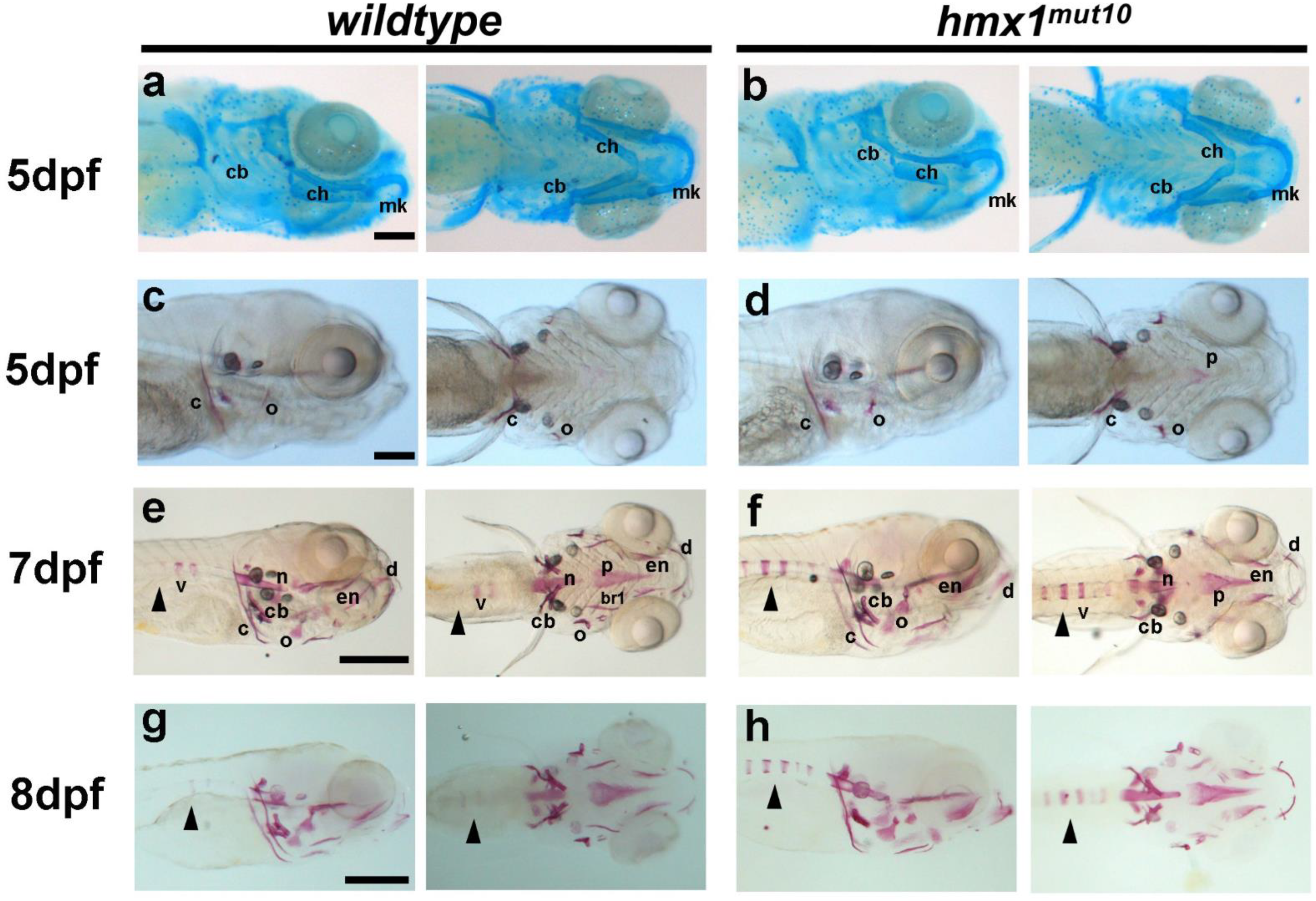
3.1. Hmx1mut10 Zebrafish Present Premature Vertebrae Mineralization

Human patients carrying HMX1 mutations presented defects related to bone development [3]; therefore, we asked whether the mutations generated in hmx1 would affect cranial cartilage and bone development. We performed alcian blue and alizarin red staining on wildtype and hmx1mut10 embryos. At 5 dpf in hmx1mut10, all cranial cartilaginous elements were present and unaffected in their development. Meckel’s cartilage, ceratohyal, and branchial arches were formed and fully developed in both wildtype and hmx1mut10 embryos (Figure 1a,b). Head skeleton formation was initiated subsequently to cartilage formation starting at 3 dpf, and at 5 dpf cranial bones were generally fully formed. Structures formed via intramembranous ossification—such as the cleithrum, anterior notochord, and operculum—were formed and visible at 5 dpf in wildtype and hmx1mut10 embryos. A morphological inspection showed that there were no differences in bone size, shape, or localization (Figure 1c,d).
In wildtype zebrafish, following the formation of the anterior notochord, mineralization of the vertebrae progresses towards the caudal region, with 70% of the embryos presenting one mineralized vertebra, while the remaining assessed embryos either presented two alizarin-red-stained vertebrae or none at 7 dpf (Figure 1e,f). At the same embryonic stage, hmx1mut10 zebrafish presented a precocious mineralization, with several additional vertebrae positive for alizarin red; at 8 dpf, the differential mineralization pattern was still maintained, with approximately 80% of embryos presenting five mineralized vertebrae (Figure 1g,h).

3.2. Hmx1mut150 Zebrafish Recapitulate Premature Vertebral Mineralization Similarly to Hmx1mut10 Mutants

Hmx1 presents three conserved regions: the SD1, SD2, and HD domains. Hmx1mut150 zebrafish carry an indel mutation that replaces the HD domain. Dimerization of HD and SD1 is necessary for hmx1 activity. Alizarin red staining at 5 dpf of hmx1mut150 embryos showed that at 5 dpf both cartilage (Figure 2a,b) and bone (Figure 2c,d) structures were formed. At 7 dpf, hmx1mut150similarly to hmx1mut10presented an early mineralization pattern of the first cranial vertebrae (Figure 2e,f).

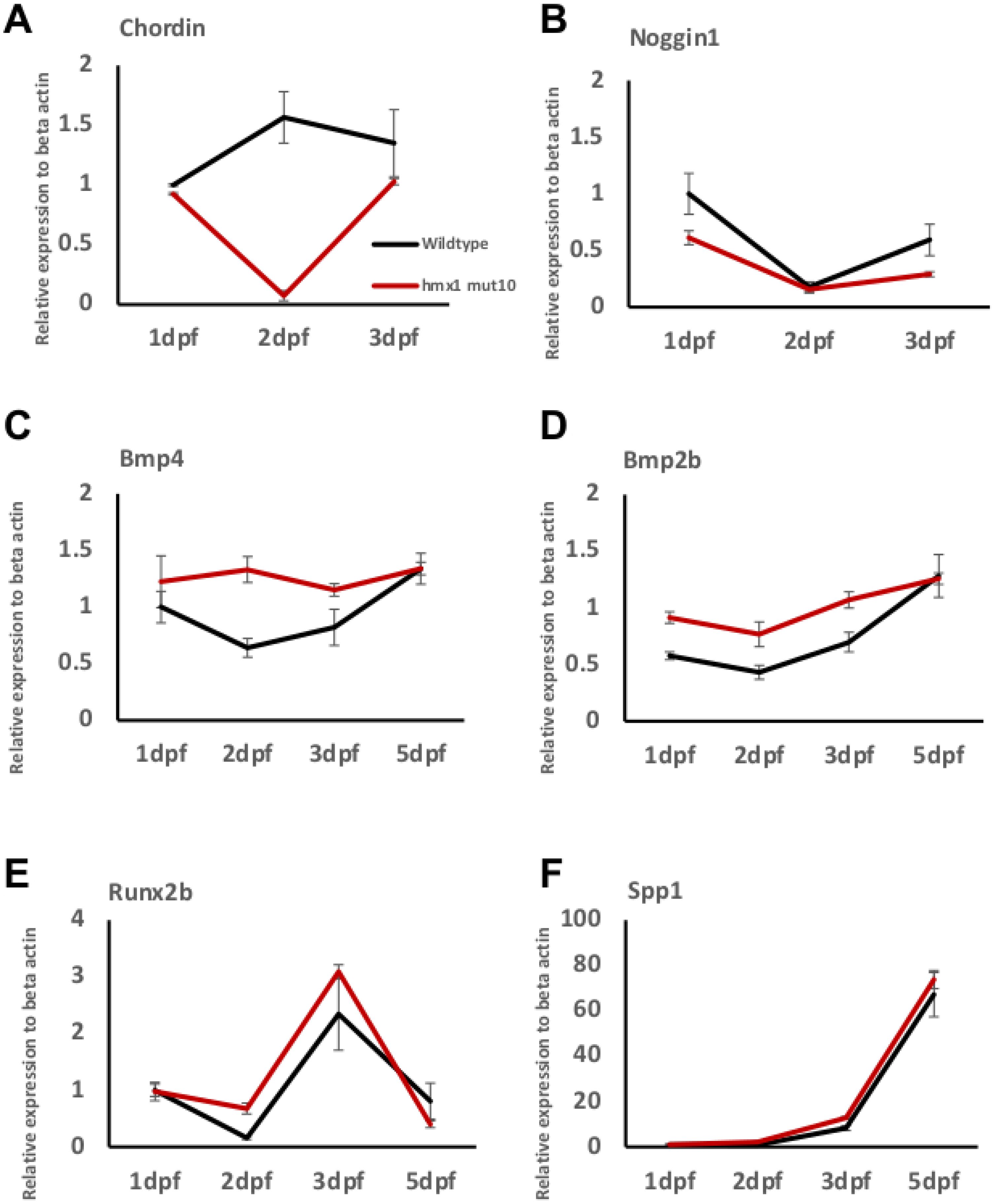
3.3. Osteogenic Signaling during Early Bone Development in Wildtype and Hmx1mut10 Zebrafish

We aimed at describing the time course of early osteogenesis by profiling bone-related genes. We focused on genes encoding for BMP antagonists and BMPs themselves, in addition to a master transcription factor and a gene involved in osteoblast function and mineralization.
Total RNA was isolated from wildtype and mutant zebrafish embryos collected from 1 dpf to 5 dpf, and RT-PCR analysis was performed. During early development, the antagonists chordin and noggin are expressed in the dorsal region and, thus, restrict bmp activity in the dorsal region [16,36]. RT-PCR analysis of chordin indicated that the antagonist expression did not alter from 1 dpf to 3 dpf in wildtype embryos, while in hmx1mut10, chordin expression was greatly reduced at 2 dpf (Figure 3A). Noggin1 expression, on the other hand, was significantly reduced at 1 dpf and 3 dpf in hmx1mut10 in comparison to wildtype embryos (Figure 3B).
The osteogenic differentiation process is initiated upon expression of BMP factors—namely, bmp2 and bmp4—followed by the specific early marker of osteoprogenitor cells runx2b. Starting at 1 dpf, and continuously until 3 dpf, bmp2b and bmp4 transcripts were significantly greater in hmx1mut10 compared to wildtype zebrafish; at 5 dpf, bmp2b and bmp4 transcript quantities were stabilized at the same expression level in both wildtype and hmx1mut10 zebrafish (Figure 3C,D). In regard to the expression of runx2b, the transcription factor was differentially expressed, and presented a higher RNA level at 2 dpf; spp1 expression followed the expression of runx2b, and it was significantly higher at 3 dpf in hmx1mut10 (Figure 3E,F). Finally, runx2b and spp1 expression patterns detected in mutant zebrafish resembled the pattern observed in wildtype embryos.

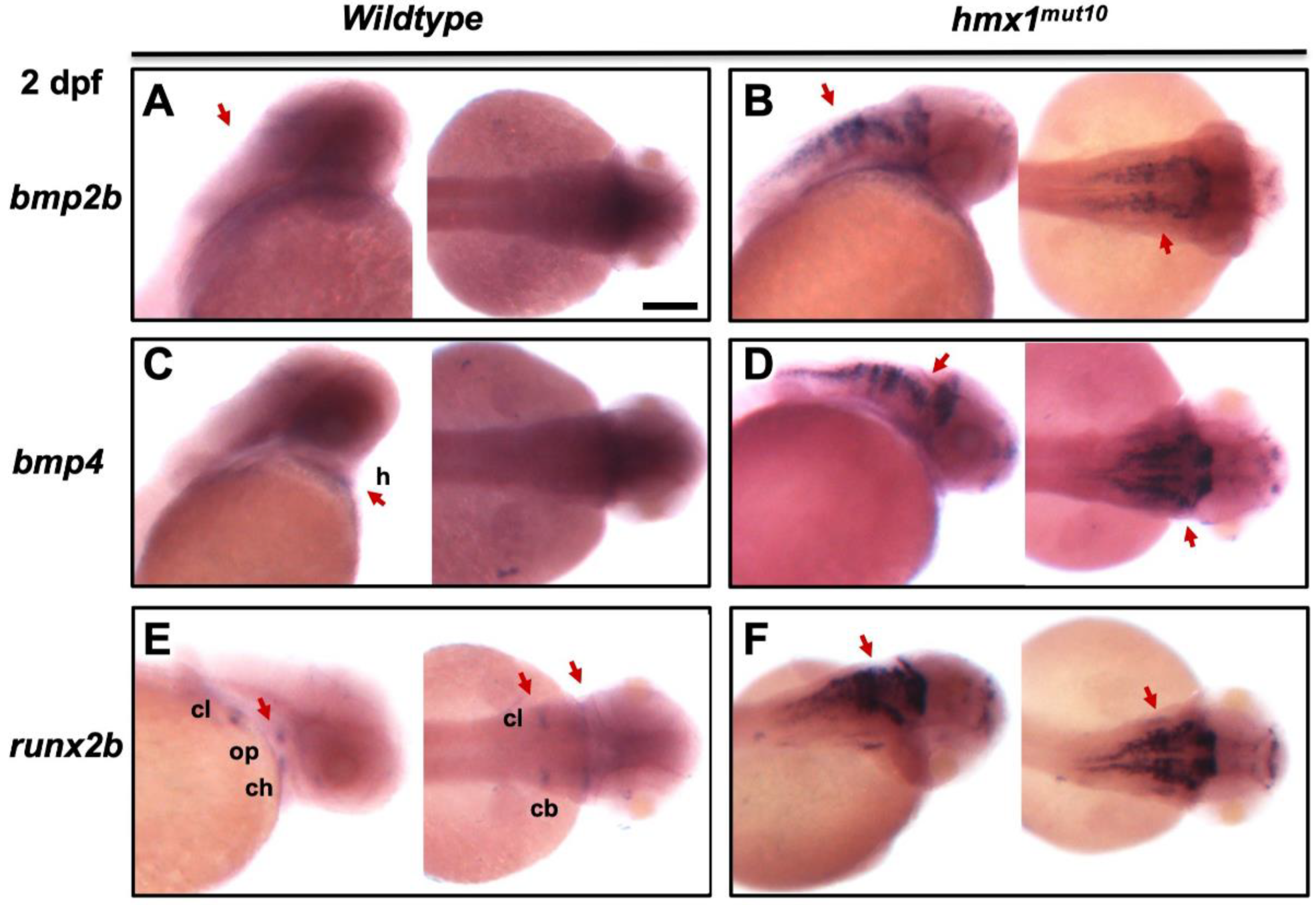
3.4. Bmp2b and bmp4 Are Expressed in the Dorsal Region in Hmx1mut10 Embryos

Given the increased expression of skeletal-associated genes during early development, we performed whole-mount in situ hybridization to track the localization of bmp2b, bmp4, and the osteogenic transcription factor runx2b (Supplementary Figure S1). In wildtype embryos, bmp2b and bmp4 expression is inhibited by the antagonists noggin and chordin [37,38] in the dorsal region during early development. We found that prior to the initiation of the osteogenic process, at 2 dpf, there was no signal detected for bmp2b and bmp4 in the wildtype dorsal region (Figure 4a,c). Inspection of hmx1mut10 evidenced strong and localized signals for bmp2b and bmp4 in the dorsal region in the correspondence of the hind–midbrain, along with an increased signal in specific bone-forming domains of the cranial frontal region (Figure 4b,d). Bmp2 induces runx2b expression; therefore, we analyzed the expression pattern of runx2b in zebrafish embryos at the same stage. In wildtype embryos, as expected, runx2b at 2 dpf was expressed in the opercle, ceratohyal, Meckel’s cartilage, ceratobranchial cartilage, and cleithrum (Figure 4e); hmx1mut10 presented a spread and stronger signal for runx2b in the dorsal region at the same stage. In situ hybridization for runx2b presented a pattern identical to bmp2b and bmp4 expression (Figure 4f).

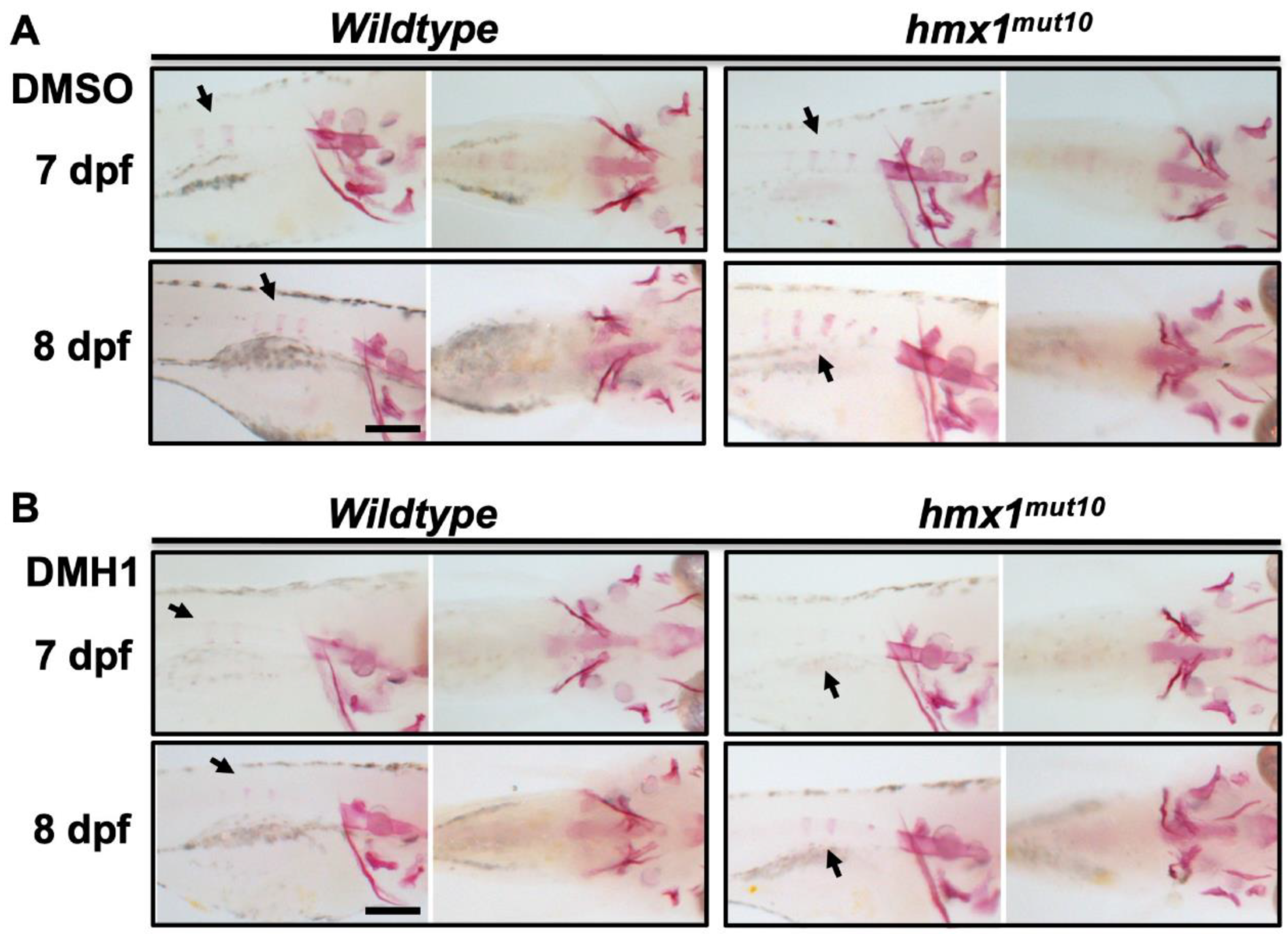
3.5. Inhibition of bmp Signaling at 2 dpf Reduced the Progression of Vertebral Mineralization in Hmx1mut10 Embryos

Mutant embryos presenting precocious mineralization of the vertebrae showed localized BMP signaling in the dorsal region during early embryonic development. To confirm the involvement of bmp2 and bmp4 factors in the developing vertebrae in mutant embryos, we investigated the effects of DMH1—an inhibitor of BMP-specific signaling [39]. Overnight treatment with 50 μM or 100 μM concentrations of DMH1 was performed at 2 dpf. Wildtype and hmx1mut10 were collected at 7 and 8 dpf; alizarin red staining was performed to detect cranial and vertebral mineralization. Wildtype embryos treated with 100 mM DMH1 were viable, and showed reduced mineralization in the cranial region (Supplementary Figure S2A(a,c),B(a–d)), while hmx1mut10 reported aberrant development, including cardiac edema, and resulting in their failure to grow (Supplementary Figure S2A(b–d)). Alizarin red staining of wildtype embryos treated with 50 μM DMH1 at 8 dpf showed that the treatment did not lead to reduced cranial mineralization, and that vertebral bone development continued to progress (Figure 5B). Cranial bone development in hmx1mut10 treated with 50 μM DHM1 was unaffected, while the precocious vertebral mineralization was inhibited, resulting in a mineralization pattern similar to that of wildtype embryos (Figure 5B). Wildtype and hmx1mut10 embryos treated with DMSO did not show altered development or aberrant bone development.

4. Discussion

The HMX gene family is involved in the development of the sensory organs, and mutations in HMX1 result in a set of defects affecting the development of the retina, along with other congenital eye-related defects [1,40]. Patients carrying mutations of HMX1 presented an additional subset of defective maxillomandibular structures and spina bifida [3]. This indicates a possible additional role for HMX1 in the regulation of several developmental processes specific to craniofacial and axial skeleton formation.
We have previously generated two mutated zebrafish lines for hmx1; hmx1mut10 and hmx1mut150 carry mutations affecting the SD1 and homeodomain domains—both essential for an active dimeric hmx1 protein [34]. Inspection of hmx1mut10 at 5 dpf indicated that both maturation of cartilaginous structures and mineralization of the cranial bones were unaffected (Figure 1a–d). In wildtype embryos, vertebral development initiated in a timely manner, while mineralization of the axial skeleton in hmx1mut10 at the same stages was accelerated, with several vertebrae presenting early mineralization at 7 and 8 dpf (Figure 1e–h). Hmx1mut10 and hmx1mut150 zebrafish lines both showed early mineralization of the vertebrae, indicating that mutations affecting either the SD1 or homeodomain domains result in the same altered osteogenic phenotype.
Noggin [41] and chordin activity is required for proper axial skeleton development [16,42]. Whole-embryo RT-PCR analysis showed reduced expression of chordin at 1 dpf and 3 dpf, and of noggin at 2 dpf, in hmx1mut10 embryos (Figure 3A–B). Short-term expression of BMP-2 is necessary to induce bone formation [43] by upregulating the expression of the osteogenic markers runxb2 and spp1 [44], and loss of both BMP-2 and BMP-4 results in severe impairment of osteogenesis [45]. Bmp2b and bmp4 were highly expressed in hmx1mut10 embryos (Figure 3C–D), followed by an increased sequential expression of runxb2 and spp1 at 2 dpf and 3 dpf when compared to wildtype embryos (Figure 3E–F).
In situ hybridization showed a strong and broad signal for bmp2b and bmp4 in the dorsal region in hmx1mut10 embryos. Runx2b presents a dorsal pattern similar to those of bmp2b and bmp4, whereas in wildtype embryos at 2 dpf it was specifically localized to the ventral osteogenic sites (Figure 3).
Treatment with DMH1—a highly selective BMP signaling inhibitor widely used in zebrafish models—limited vertebral mineralization in hmx1mut10 zebrafish, with no effect on cranial bone development in either hmx1mut10 or wildtype zebrafish (Figure 4), confirming that early mineralization of the vertebrae occurred in response to the increased bmp2b and bmp4 transcripts [46,47,48]. Noggin and DMH1 have been shown to have a similar effect in regulating the expression of a set of genes involved in neurogenesis [49].
The question of whether hmx1 directly regulates bmp2b and bmp4 could be assessed by employing the heat-shock zebrafish model that we utilized to validate potential hmx1 target genes in vivo [34]; however, we do not believe that this is the case. By accessing the Eukaryotic Promoter Database (https://epd.epfl.ch; accessed 12 December 2020), and using the predictive promoter model, we screened bmp2b and bmp4, as well the noggin1 and chordin regions upstream of the transcription starting site. We did not identify CAAGTG binding sites in bmp2b and bmp4, but several clusters of CAAG binding sites common to humans, mice, and zebrafish were located on the potential promoter region of noggin1 and chordin, (Supplementary Figure S3). It appears more plausible that hmx1 induces bmp2b and bmp4 expression through an indirect mechanism.
Previously, we identified uhrf1 as an hmx1 target gene, and it has been implicated in the regulation of several developmental and homeostatic processes related to zebrafish development. In situ hybridization in hmx1 transgenic and mutant zebrafish showed that uhrf1 expression is modulated in the hindbrain, eye region, and branchial arches [33]. Potentially, uhrf1 could change the methylation pattern of bmp2b and bmp4 and, therefore, induce gene transcription. Reductions in noggin1 and chordin expression could follow as part of a feedback mechanism. In this view, we could analyze the methylome patterns of bmp2b and bmp4 in the dorsal region at critical stages for axial skeletogenesis.
Another potential indirect mechanism could be linked to the dorsoventral polarization process; it is well known that bmp2b and bmp4 are expressed following a gradient pattern across the dorsoventral axis. Noggin1 and chordin are expressed dorsally and, therefore, inhibit BMP expression. Given that potential noggin1 and chordin promoter regions present several hmx1 binding sites, and that we observed a reduced expression in hmx1mut10 zebrafish, we could assume that in wildtype zebrafish hmx1 contributes to regulating noggin1 and chordin expression, consequently modulating bmp2b and bmp4 expression in the dorsal region. In the mutated hmx1 background, noggin1 and chordin expression is reduced, thus inducing precocious bmp2b, bmp4, and runx2b expression and initiating axial bone formation.
Our work indicates that hmx1 could be a modulator contributing to the maintenance of the BMP gradient in the dorsal region during axial bone development. In vitro studies of hmx1 activity on the chordin and noggin1 promoter regions, coupled with hmx1 heat-shock experiments, could elucidate the contribution of hmx1 during axial development.

Supplementary Materials

The following supporting information can be downloaded at: https://www.mdpi.com/article/10.3390/cells11071088/s1, Figure S1: Alcian blue and alizarin red staining of Wildtype and hmx1mut150 embryos; Figure S2: DMH1 treatment of Wildtype and hmx1mut150 embryos at 2 dpf; Figure S3: Schematic representation of HMX1 predicted promoter regions in human, zebrafish and mouse; Table S1: Primers.

Author Contributions

Y.E.F. and G.P. performed the experiments. Y.E.F., N.A.-P., and D.F.S. analyzed the data and prepared the figures. Y.E.F., N.A.-P., and D.F.S. conceived the project. All authors have read and agreed to the published version of the manuscript.

Funding

This work was supported by a grant from the Fondation pour la recherche sur les maladies héréditaires.

Institutional Review Board Statement

All zebrafish animal experiments performed were reviewed and approved by the Veterinary Service of the Canton of Valais, VS-10. All experiments were regularly evaluated and approved by the Council of the Institute for Research in Ophthalmology (no number).

Informed Consent Statement

Not applicable.

Data Availability Statement

All data analyzed during this study are included in this article or Supplementary Materials.

Acknowledgments

We thank Sylvianne Metrailler and Martine Emery from the Institute for Research in Ophthalmology for their technical help.

Conflicts of Interest

The authors declare no conflict of interest.

References

  1. Schorderet, D.F.; Nichini, O.; Boisset, G.; Polok, B.; Tiab, L.; Mayeur, H.; Raji, B.; de la Houssaye, G.; Abitbol, M.M.; Munier, F.L. Mutation in the human homeobox gene NKX5-3 causes an oculo-auricular syndrome. Am. J. Hum. Genet. 2008, 82, 1178–1184. [Google Scholar] [CrossRef] [PubMed] [Green Version]
  2. Holland, P.W.; Booth, H.A.; Bruford, E.A. Classification and nomenclature of all human homeobox genes. BMC Biol. 2007, 5, 47. [Google Scholar] [CrossRef] [PubMed] [Green Version]
  3. Abdel-Salam, G.M.H.; Abdel-Hamid, M.S.; Mehrez, M.I.; Kamal, A.M.; Taher, M.B.; Afifi, H.H. Further delineation of the oculoauricular syndrome phenotype: A new family with a novel truncating HMX1 mutation. Ophthalmic Genet. 2018, 39, 215–220. [Google Scholar] [CrossRef] [PubMed]
  4. Boisset, G.; Schorderet, D.F. Zebrafish hmx1 promotes retinogenesis. Exp. Eye Res. 2012, 105, 34–42. [Google Scholar] [CrossRef] [PubMed]
  5. Munroe, R.J.; Prabhu, V.; Acland, G.M.; Johnson, K.R.; Harris, B.S.; O’Brien, T.P.; Welsh, I.C.; Noden, D.M.; Schimenti, J.C. Mouse H6 Homeobox 1 (Hmx1) mutations cause cranial abnormalities and reduced body mass. BMC Dev. Biol. 2009, 9, 27. [Google Scholar] [CrossRef] [Green Version]
  6. Quina, L.A.; Tempest, L.; Hsu, Y.W.; Cox, T.C.; Turner, E.E. Hmx1 is required for the normal development of somatosensory neurons in the geniculate ganglion. Dev. Biol. 2012, 365, 152–163. [Google Scholar] [CrossRef] [PubMed] [Green Version]
  7. Berendsen, A.D.; Olsen, B.R. Bone development. Bone 2015, 80, 14–18. [Google Scholar] [CrossRef] [Green Version]
  8. Shahi, M.; Peymani, A.; Sahmani, M. Regulation of Bone Metabolism. Rep. Biochem. Mol. Biol. 2017, 5, 73–82. [Google Scholar]
  9. Bird, N.C.; Mabee, P.M. Developmental morphology of the axial skeleton of the zebrafish, Danio rerio (Ostariophysi: Cyprinidae). Dev. Dyn. 2003, 228, 337–357. [Google Scholar] [CrossRef]
  10. Mork, L.; Crump, G. Zebrafish Craniofacial Development: A Window into Early Patterning. Curr. Top. Dev. Biol. 2015, 115, 235–269. [Google Scholar] [CrossRef] [PubMed] [Green Version]
  11. Talbot, J.C.; Nichols, J.T.; Yan, Y.L.; Leonard, I.F.; BreMiller, R.A.; Amacher, S.L.; Postlethwait, J.H.; Kimmel, C.B. Pharyngeal morphogenesis requires fras1-itga8-dependent epithelial-mesenchymal interaction. Dev. Biol. 2016, 416, 136–148. [Google Scholar] [CrossRef] [PubMed]
  12. Aceto, J.; Nourizadeh-Lillabadi, R.; Maree, R.; Dardenne, N.; Jeanray, N.; Wehenkel, L.; Alestrom, P.; van Loon, J.J.; Muller, M. Zebrafish Bone and General Physiology Are Differently Affected by Hormones or Changes in Gravity. PLoS ONE 2015, 10, e0126928. [Google Scholar] [CrossRef] [PubMed] [Green Version]
  13. Sen, R.; Pezoa, S.A.; Carpio Shull, L.; Hernandez-Lagunas, L.; Niswander, L.A.; Artinger, K.B. Kat2a and Kat2b Acetyltransferase Activity Regulates Craniofacial Cartilage and Bone Differentiation in Zebrafish and Mice. J. Dev. Biol. 2018, 6, 27. [Google Scholar] [CrossRef] [PubMed] [Green Version]
  14. Bensimon-Brito, A.; Cardeira, J.; Cancela, M.L.; Huysseune, A.; Witten, P.E. Distinct patterns of notochord mineralization in zebrafish coincide with the localization of Osteocalcin isoform 1 during early vertebral centra formation. BMC Dev. Biol. 2012, 12, 28. [Google Scholar] [CrossRef] [PubMed] [Green Version]
  15. Branam, A.M.; Hoffman, G.G.; Pelegri, F.; Greenspan, D.S. Zebrafish chordin-like and chordin are functionally redundant in regulating patterning of the dorsoventral axis. Dev. Biol. 2010, 341, 444–458. [Google Scholar] [CrossRef] [PubMed] [Green Version]
  16. Fisher, S.; Halpern, M.E. Patterning the zebrafish axial skeleton requires early chordin function. Nat. Genet. 1999, 23, 442–446. [Google Scholar] [CrossRef]
  17. Laux, D.W.; Febbo, J.A.; Roman, B.L. Dynamic analysis of BMP-responsive smad activity in live zebrafish embryos. Dev. Dyn. 2011, 240, 682–694. [Google Scholar] [CrossRef] [PubMed] [Green Version]
  18. Pomreinke, A.P.; Soh, G.H.; Rogers, K.W.; Bergmann, J.K.; Blassle, A.J.; Muller, P. Dynamics of BMP signaling and distribution during zebrafish dorsal-ventral patterning. eLife 2017, 6, e25861. [Google Scholar] [CrossRef]
  19. Windhausen, T.; Squifflet, S.; Renn, J.; Muller, M. BMP Signaling Regulates Bone Morphogenesis in Zebrafish through Promoting Osteoblast Function as Assessed by Their Nitric Oxide Production. Molecules 2015, 20, 7586–7601. [Google Scholar] [CrossRef] [PubMed] [Green Version]
  20. Komori, T. Regulation of Proliferation, Differentiation and Functions of Osteoblasts by Runx2. Int. J. Mol. Sci. 2019, 20, 1694. [Google Scholar] [CrossRef] [PubMed] [Green Version]
  21. Lee, K.S.; Kim, H.J.; Li, Q.L.; Chi, X.Z.; Ueta, C.; Komori, T.; Wozney, J.M.; Kim, E.G.; Choi, J.Y.; Ryoo, H.M.; et al. Runx2 is a common target of transforming growth factor beta1 and bone morphogenetic protein 2, and cooperation between Runx2 and Smad5 induces osteoblast-specific gene expression in the pluripotent mesenchymal precursor cell line C2C12. Mol. Cell. Biol. 2000, 20, 8783–8792. [Google Scholar] [CrossRef] [Green Version]
  22. van der Meulen, T.; Kranenbarg, S.; Schipper, H.; Samallo, J.; van Leeuwen, J.L.; Franssen, H. Identification and characterisation of two runx2 homologues in zebrafish with different expression patterns. Biochim. Biophys. Acta 2005, 1729, 105–117. [Google Scholar] [CrossRef] [PubMed]
  23. Javed, A.; Afzal, F.; Bae, J.S.; Gutierrez, S.; Zaidi, K.; Pratap, J.; van Wijnen, A.J.; Stein, J.L.; Stein, G.S.; Lian, J.B. Specific residues of RUNX2 are obligatory for formation of BMP2-induced RUNX2-SMAD complex to promote osteoblast differentiation. Cells Tissues Organs 2009, 189, 133–137. [Google Scholar] [CrossRef] [PubMed]
  24. Phimphilai, M.; Zhao, Z.; Boules, H.; Roca, H.; Franceschi, R.T. BMP signaling is required for RUNX2-dependent induction of the osteoblast phenotype. J. Bone Miner. Res. 2006, 21, 637–646. [Google Scholar] [CrossRef] [Green Version]
  25. Si, J.; Wang, C.; Zhang, D.; Wang, B.; Zhou, Y. Osteopontin in Bone Metabolism and Bone Diseases. Med. Sci. Monit. 2020, 26, e919159. [Google Scholar] [CrossRef] [PubMed]
  26. Sodek, J.; Chen, J.; Nagata, T.; Kasugai, S.; Todescan, R., Jr.; Li, I.W.; Kim, R.H. Regulation of osteopontin expression in osteoblasts. Ann. N. Y. Acad. Sci. 1995, 760, 223–241. [Google Scholar] [CrossRef]
  27. Wang, R.N.; Green, J.; Wang, Z.; Deng, Y.; Qiao, M.; Peabody, M.; Zhang, Q.; Ye, J.; Yan, Z.; Denduluri, S.; et al. Bone Morphogenetic Protein (BMP) signaling in development and human diseases. Genes Dis. 2014, 1, 87–105. [Google Scholar] [CrossRef] [Green Version]
  28. Beederman, M.; Lamplot, J.D.; Nan, G.; Wang, J.; Liu, X.; Yin, L.; Li, R.; Shui, W.; Zhang, H.; Kim, S.H.; et al. BMP signaling in mesenchymal stem cell differentiation and bone formation. J. Biomed. Sci. Eng. 2013, 6, 32–52. [Google Scholar] [CrossRef] [Green Version]
  29. Dudas, M.; Sridurongrit, S.; Nagy, A.; Okazaki, K.; Kaartinen, V. Craniofacial defects in mice lacking BMP type I receptor Alk2 in neural crest cells. Mech. Dev. 2004, 121, 173–182. [Google Scholar] [CrossRef]
  30. Asharani, P.V.; Keupp, K.; Semler, O.; Wang, W.; Li, Y.; Thiele, H.; Yigit, G.; Pohl, E.; Becker, J.; Frommolt, P.; et al. Attenuated BMP1 function compromises osteogenesis, leading to bone fragility in humans and zebrafish. Am. J. Hum. Genet. 2012, 90, 661–674. [Google Scholar] [CrossRef] [Green Version]
  31. Amendt, B.A.; Sutherland, L.B.; Russo, A.F. Transcriptional antagonism between Hmx1 and Nkx2.5 for a shared DNA-binding site. J. Biol. Chem. 1999, 274, 11635–11642. [Google Scholar] [CrossRef] [Green Version]
  32. Boulling, A.; Wicht, L.; Schorderet, D.F. Identification of HMX1 target genes: A predictive promoter model approach. Mol. Vis. 2013, 19, 1779–1794. [Google Scholar] [PubMed]
  33. El Fersioui, Y.; Pinton, G.; Allaman-Pillet, N.; Schorderet, D.F. Hmx1 regulates urfh1 expression in the craniofacial region in zebrafish. PLoS ONE 2021, 16, e0245239. [Google Scholar] [CrossRef]
  34. Marcelli, F.; Boisset, G.; Schorderet, D.F. A dimerized HMX1 inhibits EPHA6/epha4b in mouse and zebrafish retinas. PLoS ONE 2014, 9, e100096. [Google Scholar] [CrossRef] [Green Version]
  35. Alestrom, P.; D’Angelo, L.; Midtlyng, P.J.; Schorderet, D.F.; Schulte-Merker, S.; Sohm, F.; Warner, S. Zebrafish: Housing and husbandry recommendations. Lab. Anim. 2019, 54, 213–224. [Google Scholar] [CrossRef] [PubMed] [Green Version]
  36. Stafford, D.A.; Brunet, L.J.; Khokha, M.K.; Economides, A.N.; Harland, R.M. Cooperative activity of noggin and gremlin 1 in axial skeleton development. Development 2011, 138, 1005–1014. [Google Scholar] [CrossRef] [Green Version]
  37. Stottmann, R.W.; Berrong, M.; Matta, K.; Choi, M.; Klingensmith, J. The BMP antagonist Noggin promotes cranial and spinal neurulation by distinct mechanisms. Dev. Biol. 2006, 295, 647–663. [Google Scholar] [CrossRef] [PubMed] [Green Version]
  38. Zakin, L.; Chang, E.Y.; Plouhinec, J.L.; De Robertis, E.M. Crossveinless-2 is required for the relocalization of Chordin protein within the vertebral field in mouse embryos. Dev. Biol. 2010, 347, 204–215. [Google Scholar] [CrossRef] [PubMed] [Green Version]
  39. Lin, T.; Wang, X.L.; Zettervall, S.L.; Cai, Y.; Guzman, R.J. Dorsomorphin homologue 1, a highly selective small-molecule bone morphogenetic protein inhibitor, suppresses medial artery calcification. J. Vasc. Surg. 2017, 66, 586–593. [Google Scholar] [CrossRef] [PubMed]
  40. Gillespie, R.L.; Urquhart, J.; Lovell, S.C.; Biswas, S.; Parry, N.R.; Schorderet, D.F.; Lloyd, I.C.; Clayton-Smith, J.; Black, G.C. Abrogation of HMX1 function causes rare oculoauricular syndrome associated with congenital cataract, anterior segment dysgenesis, and retinal dystrophy. Investig. Ophthalmol. Vis. Sci. 2015, 56, 883–891. [Google Scholar] [CrossRef] [Green Version]
  41. Wijgerde, M.; Karp, S.; McMahon, J.; McMahon, A.P. Noggin antagonism of BMP4 signaling controls development of the axial skeleton in the mouse. Dev. Biol. 2005, 286, 149–157. [Google Scholar] [CrossRef] [PubMed] [Green Version]
  42. Stafford, D.A.; Monica, S.D.; Harland, R.M. Follistatin interacts with Noggin in the development of the axial skeleton. Mech. Dev. 2014, 131, 78–85. [Google Scholar] [CrossRef] [PubMed]
  43. Noel, D.; Gazit, D.; Bouquet, C.; Apparailly, F.; Bony, C.; Plence, P.; Millet, V.; Turgeman, G.; Perricaudet, M.; Sany, J.; et al. Short-term BMP-2 expression is sufficient for in vivo osteochondral differentiation of mesenchymal stem cells. Stem Cells 2004, 22, 74–85. [Google Scholar] [CrossRef] [PubMed]
  44. Stock, M.; Schafer, H.; Fliegauf, M.; Otto, F. Identification of novel genes of the bone-specific transcription factor Runx2. J. Bone Miner. Res. 2004, 19, 959–972. [Google Scholar] [CrossRef] [PubMed]
  45. Bandyopadhyay, A.; Tsuji, K.; Cox, K.; Harfe, B.D.; Rosen, V.; Tabin, C.J. Genetic analysis of the roles of BMP2, BMP4, and BMP7 in limb patterning and skeletogenesis. PLoS Genet. 2006, 2, e216. [Google Scholar] [CrossRef] [Green Version]
  46. Choi, T.Y.; Khaliq, M.; Tsurusaki, S.; Ninov, N.; Stainier, D.Y.R.; Tanaka, M.; Shin, D. Bone morphogenetic protein signaling governs biliary-driven liver regeneration in zebrafish through tbx2b and id2a. Hepatology 2017, 66, 1616–1630. [Google Scholar] [CrossRef] [PubMed]
  47. Esser, J.S.; Steiner, R.E.; Deckler, M.; Schmitt, H.; Engert, B.; Link, S.; Charlet, A.; Patterson, C.; Bode, C.; Zhou, Q.; et al. Extracellular bone morphogenetic protein modulator BMPER and twisted gastrulation homolog 1 preserve arterial-venous specification in zebrafish blood vessel development and regulate Notch signaling in endothelial cells. FEBS J. 2018, 285, 1419–1436. [Google Scholar] [CrossRef] [PubMed] [Green Version]
  48. Hao, J.; Ho, J.N.; Lewis, J.A.; Karim, K.A.; Daniels, R.N.; Gentry, P.R.; Hopkins, C.R.; Lindsley, C.W.; Hong, C.C. In vivo structure-activity relationship study of dorsomorphin analogues identifies selective VEGF and BMP inhibitors. ACS Chem. Biol. 2010, 5, 245–253. [Google Scholar] [CrossRef] [Green Version]
  49. Neely, M.D.; Litt, M.J.; Tidball, A.M.; Li, G.G.; Aboud, A.A.; Hopkins, C.R.; Chamberlin, R.; Hong, C.C.; Ess, K.C.; Bowman, A.B. DMH1, a highly selective small molecule BMP inhibitor promotes neurogenesis of hiPSCs: Comparison of PAX6 and SOX1 expression during neural induction. ACS Chem. Neurosci. 2012, 3, 482–491. [Google Scholar] [CrossRef] [PubMed]
Figure 1. Hmx1mut10 zebrafish embryos develop early mineralized vertebrae: Cartilage structures stained with alcian blue in wildtype and hmx1mut10 embryos (a,b) at 5 dpf; no morphological differences in the developing cranial cartilage structures were detected. Cranial bone structures stained with alizarin red in wildtype and hmx1mut150 at 5 dpf (c,d); wildtype and hmx1mut10 present regular development of bone structures. At 7 dpf and 8 dpf, alizarin red staining detected an early and progressive mineralization of the vertebrae in hmx1mut10 (f,h) in comparison to wildtype zebrafish (e,g). cb, ceratobranchial pairs; ch, ceratohyal; mk, Meckel’s cartilage; v, vertebrae; c, cleithrum; n, notochord; cb, ceratobranchial 5; en, entopterygoid; o, opercle; d, dentary. Black arrowhead: early mineralization of the vertebrae in hmx1mut10 (f,h). Bar, (ac) 250 μm; (eg) 500 μm.
Cells 11 01088 g001
Figure 2. Precocious vertebral bone development in hmx1mut150 zebrafish embryos: Cartilage structures stained with alcian blue in wildtype and hmx1mut150 embryos (a,b) at 5 dpf; the developing cranial cartilage structures develop properly in hmx1-mutant embryos. Cranial bone structures stained with alizarin red in wildtype and hmx1mut150 at 5 and 7 dpf (c,f). Wildtype and hmx1mut150 cranial bones developed regularly at 5 dpf (c,d), while at 7 dpf early mineralization of the vertebrae was detected in hmx1mut150, similarly to hmx1mut10 embryos (e,f). Bar, (ac) 250 μm; (ef) 500 μm.
Cells 11 01088 g002
Figure 3. Hmx1mut10 embryos present enhanced expression of osteogenic factors: RT-PCR analysis of the BMP antagonist chordin and noggin1 relative to β-actin, the osteogenic inducers bmp2b and bmp4, and the osteogenic markers runx2b and spp1. In hmx1mut10 embryos, chordin expression is reduced at 2 dpf (A), while noggin1 expression is lower at 1 dpf and 3 dpf (B). Bmp2b and bmp4 transcripts are significantly different in hmx1mut10 embryos when compared to wildtype zebrafish from 1 dpf to 3 dpf (C,D). The osteogenic markers runx2b and spp1 are differentially expressed, and are higher in hmx1mut10 at 2 dpf and 3 dpf, respectively (E,F). Data are expressed as mean of three or more experiments.
Cells 11 01088 g003
Figure 4. In situ hybridization detected bmp2b and bmp4 the dorsal region in Hmx1mut10 embryos: In situ hybridization in wildtype and hmx1mut10 embryos at 2 dpf. Staining for bmp2b, bmp4, and runx2b. No signal was detected for bmp2b and bmp4 in the dorsal region of wildtype zebrafish, while bmp4 was detected in the heart region ((A,C) red arrows). In hmx1mut10, bmp2b and bmp4 are expressed in the dorsal region (B,D). Runx2b in wildtype embryos at 2 dpf is expressed in the bone-forming regions (E). In hmx1mut10 embryos, runx2b is expressed in the dorsal region, and presents a similar pattern to bmp2b and bmp4 (F). Cl, cleithrum; ch, ceratohyal; cb, ceratobranchial; h, heart; op, opercle. Bar, A–F 100 μm.
Cells 11 01088 g004
Figure 5. DMH1-mediated inhibition of BMP signaling reduces the progression of vertebral mineralization: Wildtype and hmx1mut10 embryos were treated with 50 μM DMH1 at 2 dpf and placed in E3 medium at 3 dpf. Alizarin red staining to detect mineralized structures was performed at 7 and 8 dpf (left, lateral view, right ventral view). Wildtype and hmx1mut10 control embryos treated with DMSO (A). Precocious vertebral mineralization was inhibited in DMH1-treated hmx1mut10 embryos at 7 dpf (B). At 8 dpf, treated hmx1mut10 embryos still presented a vertebral mineralization pattern similar to wildtype embryos (B). Bar, (A,B) 500 μm.
Cells 11 01088 g005
Publisher’s Note: MDPI stays neutral with regard to jurisdictional claims in published maps and institutional affiliations.

