Case Report

Spinal Intradural Hematoma after Spinal Anesthesia in a Young Male Patient: Case Report and Review of the Literature

1
Department of Anesthesiology and Pain Medicine, Soonchunhyang University Hospital Cheonan, 31, Suncheonhyang 6-gil, Dongam-gu, Cheonan 31151, Korea
2
Department of Neurosurgery, Soonchunhyang University Hospital Cheonan, 31, Suncheonhyang 6-gil, Dongam-gu, Cheonan 31151, Korea
3
Department of Orthopaedic Surgery, Soonchunhyang University Hospital Cheonan, 31, Suncheonhyang 6-gil, Dongam-gu, Cheonan 31151, Korea
4
Department of Radiology, Soonchunhyang University Hospital Cheonan, 31, Suncheonhyang 6-gil, Dongam-gu, Cheonan 31151, Korea
*
Author to whom correspondence should be addressed.
These authors contributed equally to this work.
Int. J. Environ. Res. Public Health 2022, 19(8), 4845; https://doi.org/10.3390/ijerph19084845
Submission received: 28 March 2022 / Revised: 12 April 2022 / Accepted: 13 April 2022 / Published: 16 April 2022
(This article belongs to the Section Traumas)

Abstract

:
Spinal intradural hematoma (SIH) is a rare condition which can cause neurological sequelae such as permanent motor weakness and sensory loss in the lower extremities. Herein, we describe a case of SIH following spinal anesthesia. The patient was a 30-year-old man who underwent treatment for accessory navicular syndrome at our department. The patient was not receiving anticoagulation therapy, and spinal anesthesia was thus selected. No symptoms of hematoma were observed in the immediate postoperative period, but the patient complained of pain in both buttocks on postoperative day 5. However, neither motor weakness nor sensory loss were observed. Additionally, as the radiating pain extending to the lower extremities typical of neurological pain was not observed, musculoskeletal pain was suspected. Magnetic resonance imaging revealed intradural hematomas at L4-5 and S1. Conservative treatment and follow-up evaluations were performed to ensure that additional neurological sequelae did not occur. Six months after symptom onset, his pain Numeric Rating Scale score was 0, and no other neurological findings were observed. However, in patients who undergo spinal anesthesia, localized pain in the back without other neurological symptoms and lack of radiating pain may be associated with more than musculoskeletal pain. Such patients must be continuously monitored.

1. Introduction

Spinal intradural (subdural and subarachnoid) hematoma (SIH) is caused by lumbar spinal punctures, underlying neoplasm, old age, infection, spinal surgery, anticoagulation therapy, and bleeding disorders (hemophilia, leukemia, thrombocytopenia) [1,2,3,4]. Neuraxial hematoma after spinal anesthesia, which is commonly performed for lower extremity anesthesia, is the rarest cause, with an incidence of <1%. Epidural hematoma is the most commonly observed (75%) cause, and the incidences of subarachnoid hematoma and subdural hematoma are 15.7% and 4.1%, respectively [5,6]. Although SIH is rare, it can cause neurological sequelae such as permanent motor weakness and sensory loss in the lower extremities. Such complications must be carefully monitored after spinal anesthesia [7]. Herein, we describe the case of a 30-year-old patient with no specific medical history who developed SIH following spinal anesthesia. We report the details of the case and review the possible causes of SIH and adequate countermeasures.

2. Case Presentation

This study was approved by the Institutional Review Board (IRB No. 2022-02-009), and consent was obtained from the patient for the publication of this report.
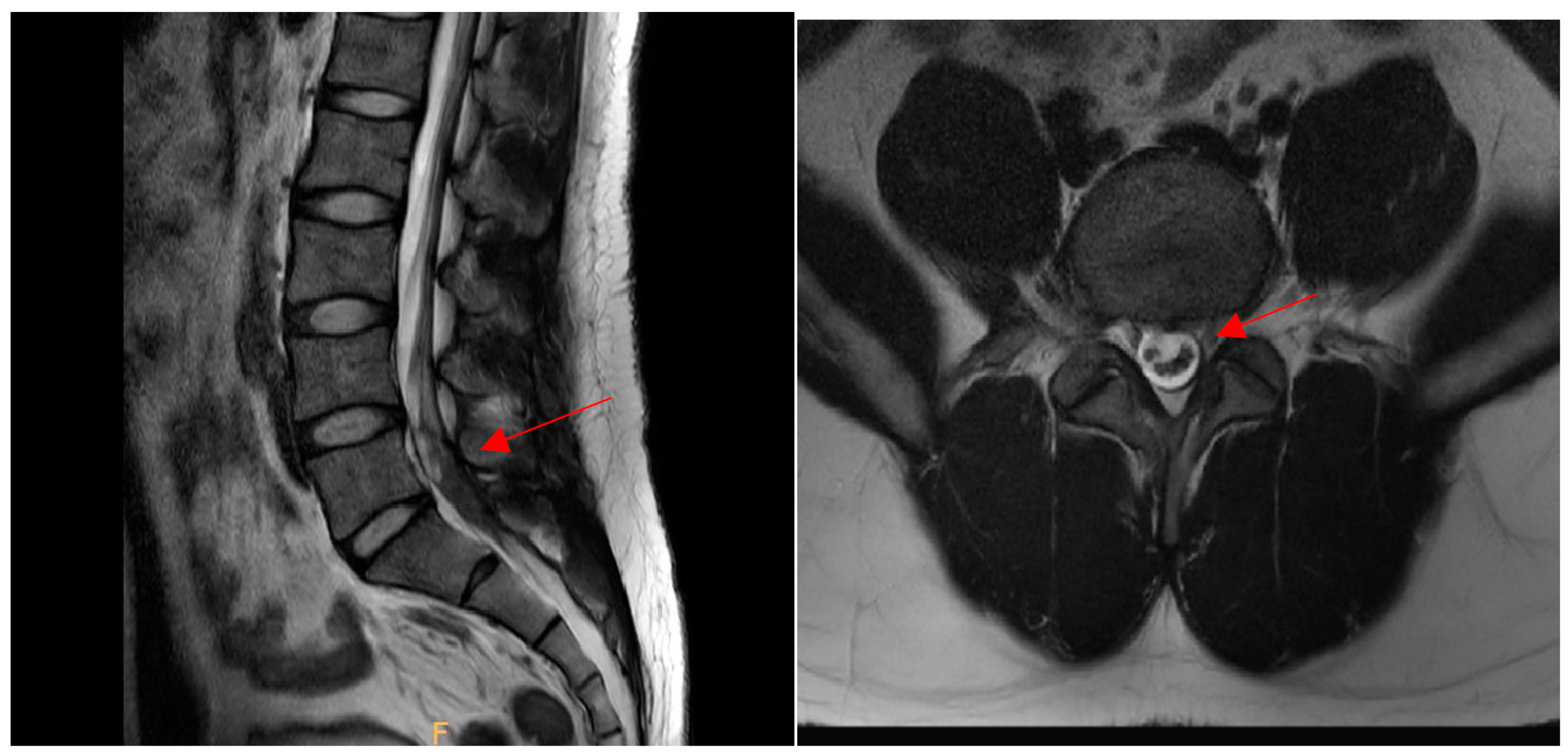
This case was a 30-year-old male patient who works as a motorcycle deliveryman. His body mass index in the historical record was 26.7, and he had no specific medical history or drug use, except that he had stage 1 obesity. He injured himself by falling while walking on the street, and the subsequent pain persisted to the extent that it interfered with walking, with pain on the inside of the foot, and the patient visited the hospital. On radiological findings, an accessory bone was found in the navicular bone (Figure 1), so the patient decided to undergo a modified Kidner procedure. A blood test performed 1 day preoperatively revealed a platelet count of 256,000/µL, a prothrombin time (international normalized ratio) of 1.10, and an activated partial thromboplastin clotting time of 35.9 s. The patient had not received anticoagulation therapy for 6 months prior to the operation, and thus, spinal anesthesia was selected. After the patient was placed in the lateral position, a 24-gauge Quincke needle was inserted into the midline between the fifth lumbar vertebra and first sacral vertebra. The first insertion failed, as the bone was touched. On the second attempt, penetration of the dura mater was detected by a pop sign. As cerebrospinal fluid (CSF) was withdrawn, 10 mg of bupivacaine was administered. Blood was not observed through the needle in either attempt. Upon physical examination, a sensory block was confirmed at thoracic dermatome level 6, and the operation commenced. The patient exhibited no specific symptoms until he was discharged from the recovery room. Immediately after the operation, the patient complained of pain in the surgical site, which was somewhat relieved after intravenous injections of Nefopam 60 mg and Propacetamol 1 g per day for two days. Dressing at the surgical site was performed daily, and antibiotics, Flomoxef sodium 500 mg, was administered. There were no symptoms or signs of infection at the surgical site. Two days after surgery, the patient was able to walk using crutches. On the fifth day postoperatively, the patient complained of burning and sharp pain in both buttocks, which worsened at night. Physical examination revealed motor grade 5 for both legs, which did not indicate motor weakness. The patellar and Achilles tendon reflexes were normal, and neurological abnormalities such as urinary retention and sensory loss in the lower extremities were not observed. Additionally, as the radiating pain extending to the lower extremities typical of neurological pain was not observed, musculoskeletal pain was suspected. Thus, the nonsteroidal anti-inflammatory drug (NSAID), Celebrex, 200 mg, and muscle relaxant, Eperisone, 50 mg, were each administered twice a day, and extracorporeal shock wave therapy (ESWT, 0.23–0.45 mJ/mm2, 1500 to 2000 times/region, 7 Hz) was provided; however, the symptoms were only temporarily improved and later worsened again. The patient complained of pain in the bilateral sacrum, and a Patrick’s test was conducted. Although the result was negative, a joint injection was conducted on both sides to evaluate inflammation of the sacroiliac joint [8]. However, the patient’s symptoms did not improve. Although there are no clear neurological symptoms, since symptoms appear after spinal anesthesia, it was necessary to reliably exclude spinal intradural hematoma. Thus, magnetic resonance imaging (MRI) was conducted to allow accurate diagnosis. Non-contrast MR images revealed intradural hematomas at L4-5 and S1. T1 images showed hyperintensity and T2 images showed hypointensity, suggesting early subacute hematomas. It was assumed that the hematomas developed after spinal anesthesia (Figure 2 and Figure 3).
After MRI, a conventional conservative treatment and continuous physical examination were provided to ensure that additional neurological sequelae did not occur. As no serious complications such as motor weakness, sensory loss, urinary distension, or any neurological sequelae were observed and the pain was relieved, it was decided to monitor the patient’s progress with follow-ups periodically after discharge. Three months after discharge, contrast-enhanced MR images were obtained to determine the cause of the hematoma and assess the improvement of the condition. MRI revealed that the intradural hematoma at L4-5 had decreased in size and that the nerves were displaced to the left side. There were no findings of tumor or vascular malformation (Figure 4).
Six months after symptom onset, the pain Numeric Rating Scale (NRS) score was 0, and no other neurological findings were observed (Figure 5, flow diagram).

3. Discussion

Although the patient showed findings indicative of SIH and pain in the bilateral sacrum, paralysis of the lower extremities or serious neurological findings such as sphincter dysfunction were not observed. His symptoms improved over time. In patients who develop SIH after spinal anesthesia, the symptoms generally manifest 3–4 days later [7]. In the current patient, no symptoms were immediately observed after spinal anesthesia, and buttock pain was first observed five days postoperatively. As tumors and vascular malformation are possible causes of SIH [9], contrast-enhanced spinal MRI was conducted three months later. However, no specific findings were observed.
In spinal hematoma, although blood does not press on the nerves, it may have neurotoxic effects on the spinal cord [3], causing pain. Once blood clots have formed, the spinal cord may be damaged, and arachnoid fibrosis may occur [2]. Therefore, persistent complaints of pain after spinal anesthesia must be diagnosed and treated promptly to identify possible hematoma.
SIH is caused by various factors; however, the pathogenesis of hematoma has not been clearly established [10,11]. Additionally, even if there is bleeding in the subarachnoid space, it is highly likely that the blood is diluted by the flow of CSF, causing it to disappear. Therefore, hematoma is rarely observed in the subarachnoid space, and the case reported here is thus quite rare [12].
In other reported cases of SIH at the lumbar level (), hematomas were observed after spinal anesthesia in patients without any relevant medical history, as in our case [6,13]. Moreover, in other patients, the hematoma was caused by coagulopathy [3,14,15].
In one unique case, it was reported that bleeding of a cranial hematoma may have descended toward the spine following gravity, and that this may have caused a spinal hematoma. Therefore, it was recommended that brain imaging also be conducted if the patient complained of a headache [16] As in the cases shown in Table 1, hematoma is commonly diagnosed by physical examination or spinal MRI after observing neurological symptoms suggestive of spinal abnormalities. However, as in the current patient who exhibited no signs of coagulopathy, complained of localized pain in the sacrum or lower back after spinal anesthesia, and did not exhibit neurological symptoms, hematoma may be easily overlooked.
A total of 33 previous cases of spinal hematoma have been reported between 1911 and 1981. Eleven patients had underlying diseases and died, and nine patients achieved complete neurological recovery, indicating that the prognosis of spinal hematoma is generally poor [7]. However, in several recent cases [1,3,17], early diagnosis following the improvement of diagnostic and surgical techniques led to complete recovery without sequelae (Table 1).
In cases in which it was difficult to determine the definite cause, it is possible that the rapid increase in abdominal pressure of the large radiculomedullary vessels and abundant capillary network in the subarachnoid space may rupture and form a hematoma, spreading to the subdural space [10,18]. Other causes include bleeding from aggressive needling or technical issues related to the piercing of the anterior portion of the subarachnoid space.
In patients who exhibit hematoma-induced neuropathy symptoms and do not undergo surgery, there is a 63.6% possibility of developing serious sequelae such as permanent paralysis of the lower extremities and sphincter dysfunction. However, in those who undergo emergency surgery immediately after symptom onset, the risk of serious sequelae is significantly lower, at 17.9% [2]. Therefore, if serious neurological damage is suspected, prompt surgical treatment is required.
However, when a patient who is taking anticoagulants or suffering from coagulopathy develops hematoma due to spinal anesthesia, and a decompression surgery is urgently performed on the spine, difficulties in bleeding control due to surgery and intraoperative bleeding may further compress the spinal cord, leading to additional damage [19]. Therefore, for patients with coagulopathy, general anesthesia is preferred over spinal anesthesia.
When there are no serious neurological complications caused by SIH, conservative treatment is commonly provided. Provided complications such as motor weakness are absent, conservative treatment is associated with a good prognosis of pain symptom recovery within a few hours at the earliest [19]. In the current patient, MRI demonstrated that the hematoma was not partially confined to the intradural space but was spread widely from L4 to S3, and that there were risks of additional neurological complications. However, in the early period after hematoma was identified, no symptoms other than pain were observed. The symptoms were relieved after three months of conservative treatment. At six months postoperatively, the pain NRS score was 0, and no other neurological sequelae were observed.

4. Conclusions

In this report, a young patient with no underlying disease who was not receiving anticoagulant therapy developed SIH after spinal anesthesia. Although patients may not be considered to be at high risk of spinal hematoma, the possibility of a hematoma must be carefully monitored after surgery. Furthermore, in patients who undergo spinal anesthesia, localized pain in the back without other neurological symptoms and lack of radiating pain from the lumbar level may be associated with more than simple musculoskeletal pain. Such patients must be continuously monitored for potentially lethal or permanent neurological complications and undergo MRI for an accurate diagnosis.

Author Contributions

Conceptualization, J.Y.J. and J.H.C.; methodology, N.S.K.; software, J.Y.J. and H.C.J.; validation, Y.H.S.; investigation, J.Y.J. and H.S.J.; resources, H.R.C.; writing—original draft preparation, J.Y.J.; writing—review and editing, W.J.K. and J.M.A.; visualization, J.S.P. and C.H.P.; supervision, J.S.C.; project administration, J.Y.J.; funding acquisition, J.Y.J. All authors have read and agreed to the published version of the manuscript.

Funding

This work was supported by the Soonchunhyang University Research Fund for financial support (No. 1022-0012).

Institutional Review Board Statement

The study was conducted in accordance with the guidelines of the Declaration of Helsinki, and approved by the Institutional Review Board and Human Research Ethics Committee of Soonchunhyang University Cheonan Hospital (IRB No. 2022-02-009).

Informed Consent Statement

Written informed consent was obtained from the patient(s) to publish this paper.

Data Availability Statement

Data sharing is not applicable to this article as no datasets were generated or analyzed during the current study.

Conflicts of Interest

The authors declare no conflict of interest.

Abbreviations

SIHSpinal intradural hematoma
MRIMagnetic resonance imaging
NRSNumeric Rating Scale

References

  1. Avecillas-Chasin, J.; Matias-Guiu, J.A.; Gómez, G.; Saceda-Gutierrez, J. A case of acute spinal intradural hematoma due to spinal anesthesia. J. Acute Dis. 2015, 4, 341–343. [Google Scholar] [CrossRef] [Green Version]
  2. Domenicucci, M.; Ramieri, A.; Paolini, S.; Russo, N.; Occhiogrosso, G.; Di Biasi, C.; Delfini, R. Spinal subarachnoid hematomas: Our experience and literature review. Acta Neurochir. 2005, 147, 741–750. [Google Scholar] [CrossRef] [PubMed]
  3. Bruce-Brand, R.A.; Colleran, G.C.; Broderick, J.M.; Lui, D.F.; Smith, E.M.; Kavanagh, E.C.; Poynton, A.R. Acute Nontraumatic Spinal Intradural Hematoma in a Patient on Warfarin. J. Emerg. Med. 2013, 45, 695–697. [Google Scholar] [CrossRef] [PubMed]
  4. Maddali, P.; Walker, B.; Fisahn, C.; Page, J.; Diaz, V.; Zwillman, M.E.; Oskouian, R.J.; Tubbs, R.S.; Moisi, M. Subdural Thoracolumbar Spine Hematoma after Spinal Anesthesia: A Rare Occurrence and Literature Review of Spinal Hematomas after Spinal Anesthesia. Cureus 2017, 9, e1032. [Google Scholar] [CrossRef] [PubMed] [Green Version]
  5. Jang, W.; Cho, Y.-H.; Lee, D.-H.; Kim, S.-H. Subarachnoid hematoma after spinal anesthesia—A case report. Anesthesia Pain Med. 2018, 13, 154–157. [Google Scholar] [CrossRef] [Green Version]
  6. Jeon, S.B.; Ham, T.-I.; Kang, M.-S.; Shim, H.-Y.; Park, S.L. Spinal subarachnoid hematoma after spinal anesthesia. Korean J. Anesthesiol. 2013, 64, 388–389. [Google Scholar] [CrossRef] [PubMed]
  7. Lam, D.H. Subarachnoid haematoma after spinal anaesthesia mimicking transient radicular irritation: A case report and review. Anaesthesia 2008, 63, 423–427. [Google Scholar] [CrossRef] [PubMed]
  8. Kurosawa, D.; Murakami, E.; Aizawa, T. Referred pain location depends on the affected section of the sacroiliac joint. Eur. Spine J. 2014, 24, 521–527. [Google Scholar] [CrossRef] [PubMed]
  9. Kreppel, D.; Antoniadis, G.; Seeling, W. Spinal hematoma: A literature survey with meta-analysis of 613 patients. Neurosurg. Rev. 2003, 26, 1–49. [Google Scholar] [CrossRef] [PubMed]
  10. Yang, N.-R.; Kim, S.J.; Cho, Y.J.; Cho, D.S. Spontaneous Resolution of Nontraumatic Acute Spinal Subdural Hematoma. J. Korean Neurosurg. Soc. 2011, 50, 268–270. [Google Scholar] [CrossRef] [PubMed]
  11. Kyriakides, A.E.; Lalam, R.K.; Masry, W.S.E. Acute spontaneous spinal subdural hematoma presenting as paraplegia: A rare case. Spine 2007, 32, E619–E622. [Google Scholar] [CrossRef] [PubMed]
  12. Wang, Y.; Zheng, H.; Ji, Y.; Lu, Q.; Li, X.; Jiang, X. Idiopathic Spinal Subdural Hematoma: Case Report and Review of the Literature. World Neurosurg. 2018, 116, 378–382. [Google Scholar] [CrossRef] [PubMed]
  13. Cui, Z.; Zhong, Z.; Wang, B.; Sun, Q.; Zhong, C.; Bian, L. Coexistence of Spontaneous Spinal and Undiagnosed Cranial Subdual Hematomas. J. Craniofacial Surg. 2015, 26, e118–e119. [Google Scholar] [CrossRef] [PubMed]
  14. Boukobza, M.; Haddar, D.; Boissonet, M.; Merland, J. Spinal Subdural Haematoma: A Study of Three Cases. Clin. Radiol. 2001, 56, 475–480. [Google Scholar] [CrossRef] [PubMed]
  15. Koyama, S.; Tomimatsu, T.; Kanagawa, T.; Sawada, K.; Tsutsui, T.; Kimura, T.; Chang, Y.S.; Wasada, K.; Imai, S.; Murata, Y. Spinal subarachnoid hematoma following spinal anesthesia in a patient with HELLP syndrome. Int. J. Obstet. Anesth. 2010, 19, 87–91. [Google Scholar] [CrossRef] [PubMed]
  16. Moon, W.; Joo, W.; Chough, J.; Park, H. Spontaneous spinal subdural hematoma concurrent with cranial subdural hematoma. J. Korean Neurosurg. Soc. 2013, 54, 68–70. [Google Scholar] [CrossRef] [PubMed]
  17. Basaran, R.; Efendioglu, M.; Bölükbaşi, F.H.; Aslan, S.; Isik, N.; Kaner, T. Spinal Intradural Hematoma and Permanent Paraparesis after a Lumboperitoneal Shunt Operation: An Unusual Complication. Asian Spine J. 2014, 8, 516–520. [Google Scholar] [CrossRef] [PubMed] [Green Version]
  18. Domenicucci, M.; Ramieri, A.; Ciappetta, P.; Delfini, R. Nontraumatic acute spinal subdural hematoma: Report of five cases and review of the literature. J. Neurosurg. 1999, 91 (Suppl. 1), 65–73. [Google Scholar] [CrossRef] [PubMed]
  19. Maeda, M.; Mochida, J.; Toh, E.; Nishimura, K.; Nomura, T. Nonsurgical treatment of an upper thoracic spinal subdural hemorrhage. Spinal Cord. 2001, 39, 657–661. [Google Scholar] [CrossRef] [PubMed] [Green Version]
  20. Mashiko, R.; Noguchi, S.; Uemura, K.; Takada, T.; Matsumura, A. Lumbosacral Subdural Hematoma-Case Report. Neurol. Med. Chir. 2006, 46, 258–261. [Google Scholar] [CrossRef] [PubMed] [Green Version]
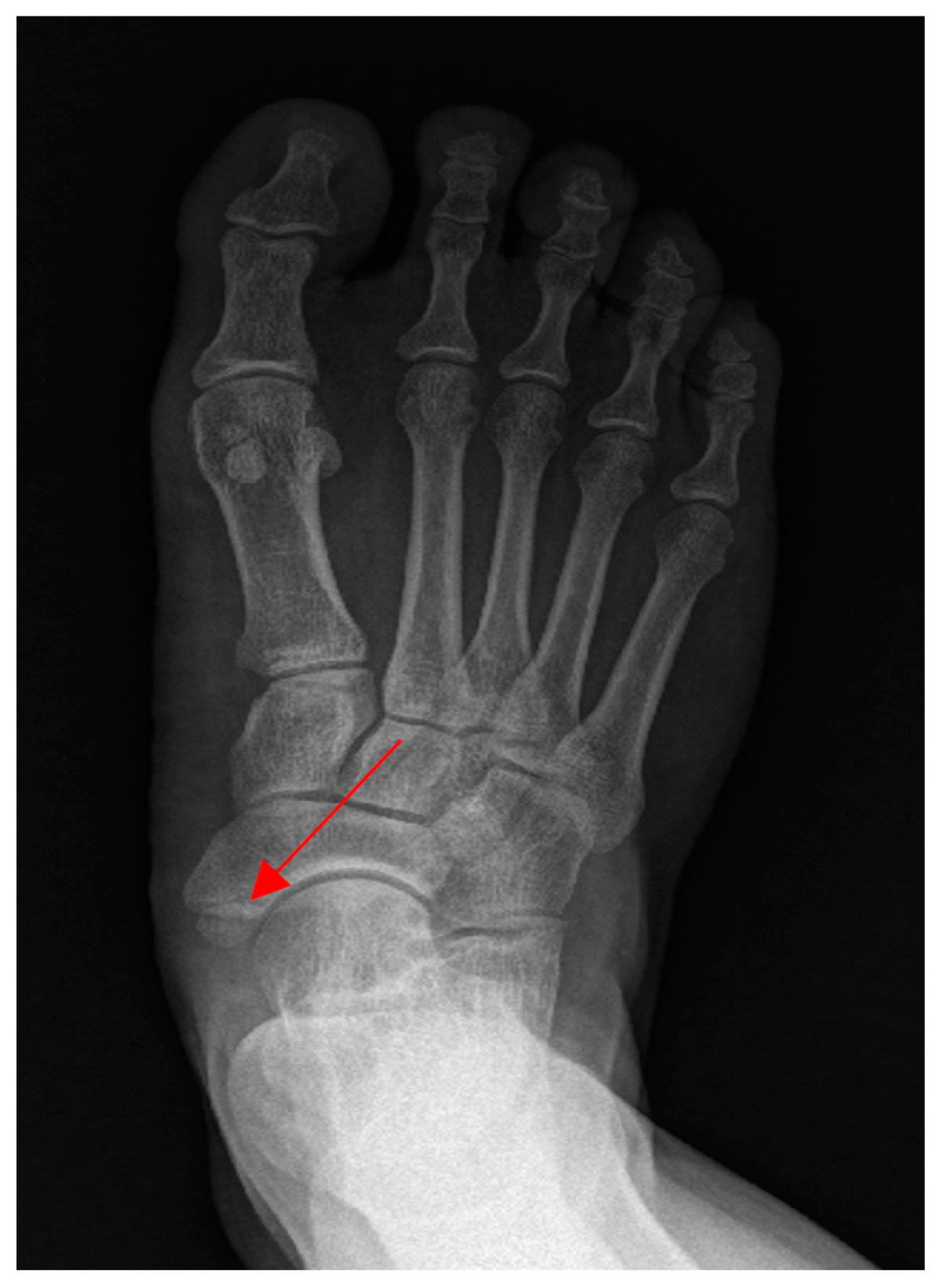
Figure 1. Patient’s accessory navicular bone (Arrow).
Ijerph 19 04845 g001
Figure 2. Hematoma (arrow) with a hyperintense signal at L4-5 and S1 on sagittal magnetic resonance imaging (MRI) (T1) on the left, and hematoma (arrow) with a hypointense signal on sagittal MRI (T2) on the right.
Ijerph 19 04845 g002
Figure 3. Hematoma (arrow) on the intradural side on magnetic resonance imaging (T2). In the horizontal section, the hematoma was observed on the spinal intradural side.
Ijerph 19 04845 g003
Figure 4. Magnetic resonance imaging (T2) showed a slight decrease in the size of the hematoma on the left side (arrow). The intradural hematoma on the right side was also decreased in size (arrow).
Ijerph 19 04845 g004
Figure 5. Flow diagram.
Ijerph 19 04845 g005
Table 1. Spinal intradural (subarachnoid or subdural) hematoma cases including the lumbar level.
Case StudyAge
(Years)
CauseHematoma TypeSymptomsTreatmentOutcome
Boukobza et al. (2001) [14]54 Antivitamin K treatment
Spontaneous
Subdural (T9-L1)Lower back pain, saddle pain, urinary retentionLaminectomy (T10-L4)Complete recovery
Mashiko et al. (2006) [20]18Trauma Subdural (L5-S2)Headache, lower back painObservationComplete recovery
Lam et al. (2008) [7] ElderlySpinal anesthesiaSubarachnoid (L4)Lower back pain, buttock pain, right leg painConservative treatmentComplete recovery
Koyama et al. (2009) [15] 39HELLP (Hemolysis, Elevated Liver enzymes, and Low platelet count) syndrome, Spinal anesthesiaSubarachnoid (L2-S1)Numbness of the thigh and toes, urinary retentionConservative treatmentComplete recovery
Moon et al. (2013) [16]39Chronic cranial subdural hematomaSubdural (L4-S2)Headache, lower back pain, pain radiating down both legsEvacuation of cranial hematomaComplete recovery
Bruce–Brand et al. (2013) [3]76Warfarin treatment
Spontaneous
Intradural (L1-L4)Flaccid paraparesis in both legs, sensory weakness below T12Laminectomy (T12-L4), intradural hematoma evacuationRemaining motor weakness (ASIA grade C)
Jeon et al. (2013) [6]33Unknown
Spinal anesthesia
Subarachnoid (L5)Lower back pain, numbness of the lower limbsObservationComplete recovery
Basaran et al. (2014) [17]27Lumboperitoneal shunt operationIntradural (L2-3)Urinary and fecal incontinence with paraparesisL2-L3 total laminectomy Permanent paraparesis
Avecillas-Chasin et al. (2015) [1]79Spinal anesthesiaIntradural (L1-L3)Motor and sensory weaknessLaminectomy (L1-L3), intradural hematoma evacuationUnable to walk without assistance
Cui et al. (2015) [13]45UnknownSubdural (L4-S3)Saddle pain and dysuriaHematoma evacuationComplete recovery
Jang et al. (2017) [5]59UnknownSubarachnoid (L3-L4)Headache, nausea, vomiting, neck stiffness, lower back painLaminectomy (L3-4), hematoma evacuationComplete recovery
Publisher’s Note: MDPI stays neutral with regard to jurisdictional claims in published maps and institutional affiliations.