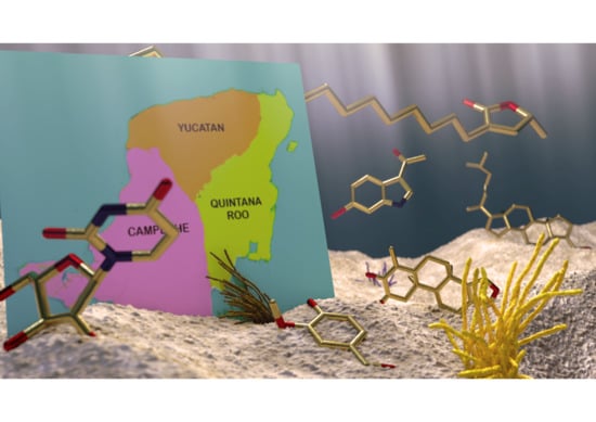
Marine Natural Products from the Yucatan Peninsula


 and
and 
Abstract
:
1. Introduction
2. Marine Natural Products from the Yucatan Peninsula
2.1. Polyketides
2.1.1. Aliphatic Polyketides
2.1.2. Glycolipids
2.1.3. Aromatic Acids
2.2. Terpenoids
2.2.1. Diterpenes and Sesterterpenes
2.2.2. Steroids
2.2.3. Triterpenoid Saponins
2.3. Nitrogen Compounds
2.3.1. Indole Derivatives
2.3.2. Nucleosides and Nitrogenous Bases
2.3.3. Conotoxins
2.4. Biopolymers
3. Bioprospecting Overview
4. Conclusions
Author Contributions
Funding
Conflicts of Interest
References
- Jiménez, C. Marine Natural Products in Medicinal Chemistry. ACS Med. Chem. Lett. 2018, 9, 959–961. [Google Scholar] [CrossRef] [Green Version]
- Altmann, K.H. Drugs from the Oceans: Marine Natural Products as Leads for Drug Discovery. Chimia 2017, 71, 646–652. [Google Scholar] [CrossRef]
- Mayer, A.M.S.; Rodríguez, A.D.; Taglialatela-Scafati, O.; Fusetani, N. Marine Pharmacology in 2012–2013: Marine Compounds with Antibacterial, Antidiabetic, Antifungal, Anti-Inflammatory, Antiprotozoal, Antituberculosis, and Antiviral Activities; Affecting the Immune and Nervous Systems, and Other Miscellaneous Mechanisms of Action. Mar. Drugs 2017, 15, 273. [Google Scholar]
- Barrera, A. La Península de Yucatán Como Provincia Biótica. Rev. Soc. Mex. Hist. Nat. 1962, 23, 71–105. [Google Scholar]
- Hernández-Bolio, G.I.; Ruiz-Vargas, J.A.; Peña-Rodríguez, L.M. Natural Products from the Yucatecan Flora: Structural Diversity and Biological Activity. J. Nat. Prod. 2019, 82, 647–656. [Google Scholar] [CrossRef] [PubMed]
- Pech Pool, D.; Ardisson Herrera, P.L. Diversidad en el Bentos Marino-Costero. In Biodiversidad y Desarrollo Humano en Yucatán; Duran, R., Méndez, M., Eds.; CICY; PPD-FMAM; CONABIO; SEDUMA: Mérida, Yucatán, Mexico, 2010; pp. 144–146. ISBN 978-607-7823-08-7. [Google Scholar]
- Garza Pérez, J.R.; Simões, N.; Chiappa Carrara, X.; Cucio, C.; Mascaró Miquelajáuregui, M.; Oseguera Cruz, M.; Lozano Aburto, M.; Acosta González, G. Comunidades Coralinas de las Bajas de Sisal. In Biodiversidad y Desarrollo Humano en Yucatán; Duran, R., Méndez, M., Eds.; CICY; PPD-FMAM; CONABIO; SEDUMA: Mérida, Yucatán, Mexico, 2010; pp. 148–149. ISBN 978-607-7823-08-7. [Google Scholar]
- Torruco Gómez, D.; González Solís, A. Las Esponjas y Su Importancia. In Biodiversidad y Desarrollo Humano en Yucatán; Duran, R., Méndez, M., Eds.; CICY; PPD-FMAM; CONABIO; SEDUMA: Mérida, Yucatán, Mexico, 2010; pp. 202–203. ISBN 978-607-7823-08-7. [Google Scholar]
- Acosta González, G.; Arias González, J.E. Comunidades Bentónicas Del Arrecife Alacranes. In Biodiversidad y Desarrollo Humano en Yucatán; Duran, R., Méndez, M., Eds.; CICY; PPD-FMAM; CONABIO; SEDUMA: Mérida, Yucatán, Mexico, 2010; p. 147. ISBN 978-607-7823-08-7. [Google Scholar]
- Palomino-Alvarez, L.A.; Moreira Rocha, R.; Simões, N. Checklist of Ascidians (Chordata, Tunicata) from the Southern Gulf of Mexico. Zookeys 2019, 832, 1–33. [Google Scholar] [CrossRef] [Green Version]
- Guo, Y.W.; Gavagnin, M.; Mollo, E.; Trivellone, E.; Cimino, G. Three New Butenolide Lipids from the Caribbean Gorgonian Pterogorgia anceps. J. Nat. Prod. 1999, 62, 1194–1196. [Google Scholar] [CrossRef]
- Medina-Gómez, S.; Mirón-López, G.; Quijano-Quiñones, R. Caracterización Estructural de Compuestos Obtenidos a Partir de Esponja de Mar Empleando Resonancia Magnética Nuclear y Cálculos Teóricos. In 11° Foro en Ciencias Químicas y Bioquímicas; Posgrado en Ciencias Químicas y Bioquímicas: Mérida, Mexico, 2018; pp. 23–24. [Google Scholar]
- Fattorusso, E.; Mangoni, A. Progress in the Chemistry of Organic Natural Products; Herz, W., Kirby, G.W., Moore, R.E., Steglich, W., Tamm, C., Eds.; Springer: Berlin, Germany, 1997; pp. 215–301. ISBN 978-3-7091-7456-2. [Google Scholar]
- Cantillo-Ciau, Z.; Moo-Puc, R.; Quijano, L.; Freile-Pelegrín, Y. The Tropical Brown Alga Lobophora variegata: A Source of Antiprotozoal Compounds. Mar. Drugs 2010, 8, 1292–1304. [Google Scholar] [CrossRef] [Green Version]
- Carroll, A.R.; Copp, B.R.; Davis, R.A.; Keyzers, R.A.; Prinsep, M.R. Marine Natural Products. Nat. Prod. Rep. 2019, 36, 122–173. [Google Scholar] [CrossRef] [PubMed] [Green Version]
- Pech-Puch, D.; Rodríguez, J.; Cautain, B.; Sandoval-Castro, C.A.; Jiménez, C. Cytotoxic Furanoditerpenes from the Sponge Spongia tubulifera Collected in the Mexican Caribbean. Mar. Drugs 2019, 17, 416. [Google Scholar] [CrossRef] [PubMed] [Green Version]
- Caamal-Fuentes, E.; Moo-Puc, R.; Freile-Pelegrín, Y.; Robledo, D. Cytotoxic and Antiproliferative Constituents from Dictyota ciliolata, Padina sanctae-crucis and Turbinaria tricostata. Pharm. Biol. 2014, 52, 1244–1248. [Google Scholar] [CrossRef] [Green Version]
- Bohlin, L.; Sjöstrand, U.; Djerassi, C.; Sullivan, B. Minor and Trace Sterols in Marine Invertebrates. Part 20. 3E-Hydroxy-methyl-A-nor-Patinosterol and 3E-Hydroxymethyl-A-nor-dinosterol. Two New Sterols with Modified Nucleus and Side-Chain from the Sponge Teichaxinella morchella. J. Chem. Soc. Perkin Trans. 1981. [Google Scholar] [CrossRef]
- Khotimchenko, Y. Pharmacological Potential of Sea Cucumbers. Int. J. Mol. Sci. 2018, 19, 1342. [Google Scholar] [CrossRef] [Green Version]
- Graniel-Sabido, M.J.; Mirón-López, G.; León-Deniz, L.V.; Moo-Puc, R.E.; Quintal-Novelo, C.J.; Quijano, L.; Mena-Rejón, G.J. Total NMR Assignment of a New Antiproliferative Triterpene Oligoglycoside from the Sea Cucumber Astichopus multifidus. Tetrahedron Lett. 2016, 57, 4375–4378. [Google Scholar] [CrossRef]
- Salazar-Mendoza, J.; Padilla-Montaño, N.; León-Deniz, L.V.; Mena-Rejón, G.J.; Quijano, L. Actividad Antifúngica de Metabolitos Aislados de la Pared Corporal de Holoturia Floridana. In Foro en Ciencias Químicas y Bioquímicas. Posgrado Institucional en Ciencias Químicas y Bioquímicas; Universidad Autónoma de Yucatán: Mérida, Mexico, 2013; pp. 19–20. [Google Scholar]
- Olguin-Uribe, G.; Abou-Mansour, E.; Boulander, A.; Débard, H.; Francisco, C.; Combaut, G. 6-Bromoindole-3-Carbaldehyde, from an Acinetobacter sp. Bacterium Associated with the Ascidian Stomozoa murrayi. J. Chem. Ecol. 1997, 23, 2507–2521. [Google Scholar] [CrossRef]
- Pech-Puch, D.; Cruz-López, H.; Canche-Ek, C.; Campos-Espinosa, G.; García, E.; Mascaro, M.; Rosas, C.; Chávez-Velasco, D.; Rodríguez-Morales, S. Chemical Tools of Octopus maya During Crab Predation Are Also Active on Conspecifics. PLoS ONE 2016, 11, e0148922. [Google Scholar] [CrossRef]
- Salazar Mendoza, J.; Mirón López, G.; Mena Rejón, G.J. Estudio Químico de Halichondria Magniconulosa (Porifera: Demospongiae) del Litoral del Estado de Yucatán. In 11° Foro en Ciencias Químicas y Bioquímicas; Posgrado Institucional en Ciencias Químicas y Bioquímicas: Mérida, Mexico, 2018; pp. 5–6. [Google Scholar]
- Aguilar, M.B.; López-Vera, E.; Ortiz, E.; Becerril, B.; Possani, L.D.; Olivera, B.M.; Heimer de la Cotera, E.P. A Novel Conotoxin from Conus delessertii with Posttranslationally Modified Lysine Residues. Biochemistry 2005, 44, 11130–11136. [Google Scholar] [CrossRef] [PubMed]
- Aguilar, M.B.; López-Vera, E.; Imperial, J.S.; Falcón, A.; Olivera, B.M.; Heimer de la Cotera, E.P. Putative γ-Conotoxins in Vermivorous Cone Snails: The Case of Conus delessertii. Peptides 2005, 26, 23–27. [Google Scholar] [CrossRef]
- Aguilar, M.B.; Flores-Torres, A.; Batista, C.V.F.; Falcón, A.; López-Vera, E.; Heimer de la Cotera, E.P. Structural Characterization of Five Post-translationally Modified Isomorphs of a Novel Putative δ-Conotoxin from the Vermivorous Snail Conus delessertii from the Mexican Caribbean Sea. Peptides 2009, 30, 458–466. [Google Scholar] [CrossRef]
- Aguilar, M.B.; Ortiz, E.; Kaas, Q.; López-Vera, E.; Becerril, B.; Possani, L.D.; Heimer de la Cotera, E.P. Precursor De13.1 from Conus delessertii Defines the Novel G Gene Superfamily. Peptides 2013, 41, 17–20. [Google Scholar] [CrossRef]
- Aguilar, M.B.; Lezama-Monfil, L.; Maillo, M.; Pedraza-Lara, H.; López-Vera, E.; Heimer de la Cotera, E.P. A biologically active hydrophobic T-1-Conotoxin from the Venom of Conus spurius. Peptides 2006, 27, 500–505. [Google Scholar] [CrossRef] [PubMed]
- López-Vera, E.; Aguilar, M.B.; Schiavon, E.; Marinzi, C.; Ortiz, E.; Restano Cassulini, R.; Batista, C.V.F.; Possani, L.D.; Heimer de la Cotera, E.P.; Peri, F.; et al. Novel α-Conotoxins from Conus spurius and the α-Conotoxin EI Share High-Affinity Potentiation and Low-Affinity Inhibition of Nicotinic Acetylcholine Receptors. FEBS J. 2007, 274, 3972–3985. [Google Scholar] [CrossRef] [PubMed]
- Aguilar, M.B.; López-Vera, E.; Heimer de la Cotera, E.P.; Falcón, A.; Olivera, B.M.; Maillo, M. I-Conotoxins in Vermivorous Species of the West Atlantic: Peptide Sr11a from Conus spurius. Peptides 2007, 28, 18–23. [Google Scholar] [CrossRef]
- Luna-Ramírez, K.S.; Aguilar, M.B.; Falcón, A.; Heimer de la Cotera, E.P.; Olivera, B.M.; Maillo, M. An O-Conotoxin from the Vermivorous Conus spurius Active on Mice and Mollusks. Peptides 2007, 28, 24–30. [Google Scholar] [CrossRef] [PubMed]
- Aguilar, M.B.; Luna-Ramírez, K.S.; Echeverría, D.; Falcón, A.; Olivera, B.M.; Heimer de la Cotera, E.P.; Maillo, M. Conorfamide-Sr2, a gamma-carboxyglutamate-containing FMRFamide-related Peptide from the Venom of Conus spurius with Activity in Mice and Mollusks. Peptides 2008, 29, 186–195. [Google Scholar] [CrossRef] [PubMed] [Green Version]
- Zamora-Bustillos, R.; Aguilar, M.B.; Falcón, A.; Heimer de la Cotera, E.P. Identification, by RT-PCR, of Four Novel T-1-Superfamily Conotoxins from the Vermivorous Snail Conus spurius from the Gulf of Mexico. Peptides 2009, 30, 1396–1404. [Google Scholar] [CrossRef]
- Zamora-Bustillos, R.; Aguilar, M.B.; Falcón, A. Identification, by Molecular Cloning, of a Novel Type of I2-Superfamily Conotoxin Precursor and Two Novel I2-Conotoxins from the Worm-Hunter Snail Conus spurius from the Gulf of México. Peptides 2010, 31, 384–393. [Google Scholar] [CrossRef]
- Aguilar, M.B.; Chan de la Rosa, R.A.; Falcón, A.; Olivera, B.M.; Heimer de la Cotera, E.P. Peptide pal9a from the Venom of the Turrid Snail Polystira Albida from the Gulf of Mexico: Purification, Characterization, and Comparison with P-Conotoxin-like (framework IX) Conoidean Peptides. Peptides 2009, 30, 467–476. [Google Scholar] [CrossRef] [Green Version]
- Peñuela, A.; Robledo, D.; Bourgougnon, N.; Bedoux, G.; Hernández-Núñez, E.; Freile-Pelegrín, Y. Environmentally Friendly Valorization of Solieria filiformis (Gigartinales, Rhodophyta) from IMTA Using a Biorefinery Concept. Mar. Drugs 2018, 16, 487. [Google Scholar] [CrossRef] [Green Version]
- Parera-Valadez, Y.; Yam-Puc, A.; López-Aguiar, L.K.; Borges-Argáez, R.; Figueroa-Saldivar, M.A.; Cáceres-Farfán, M.; Márquez-Velázquez, N.A.; Prieto-Davó, A. Ecological Strategies Behind the Selection of Cultivable Actinomycete Strains from the Yucatan Peninsula for the Discovery of Secondary Metabolites with Antibiotic Activity. Microb. Ecol. 2019, 77, 839–851. [Google Scholar] [CrossRef]
- Pless, D.D.; Aguilar, M.B.; Falcón, A.; Lozano-Alvarez, E.; Heimer de la Cotera, E.P. Latent Phenoloxidase Activity and N-Terminal Amino Acid Sequence of Hemocyanin from Bathynomus giganteus, a Primitive Crustacean. Arch. Biochem. Biophys. 2003, 409, 402–410. [Google Scholar] [CrossRef]
- Méndez Alpuche, A.A. Caracterización Fisicoquímica de Biopolímeros Obtenidos del Exoesqueleto de la Cacerolita de Mar, Limulus polyphemus. Master’s Thesis, Centro de Investigacion Científica de Yucatán A. C., Yucatán, Mexico, 2017. [Google Scholar]
- Rinehart, K.L.; Kishore, V.; Bible, K.C.; Sakai, R.; Sullins, D.W.; Li, K. Didemnins and Tunichlorin: Novel Natural Products from the Marine Tunicate Trididemnum solidum. J. Nat. Prod. 1988, 51, 1–21. [Google Scholar] [CrossRef] [PubMed]
- Cruz-Hernández, E.; Rodríguez-Morales, S. Sea Anemones from Yucatan Peninsula as Source of Bioactive Compounds. Toxicol. Lett. 2016, 259, S198. [Google Scholar] [CrossRef]
- Sánchez-Rodriguez, J.; Zugasti-Cruz, A.; Burnnet, J.W. Cutaneous Stings from Bartholomea annulata. Contact Dermat. 2001, 44, 314–315. [Google Scholar] [CrossRef]
- Santamaría, A.; Sánchez-Rodríguez, J.; Zugasti, A.; Martínez, A.; Galván-Arzate, S.; Segura-Puertas, L. A Venom Extract from the Sea Anemone Bartholomea annulata Produces Haemolysis and Lipid Peroxidation in Mouse Erythrocytes. Toxicology 2002, 173, 221–228. [Google Scholar] [CrossRef]
- Sánchez-Rodríguez, J.; Zugasti, A.; Santamaría, A.; Galván-Arzate, S.; Segura-Puertas, L. Isolation, Partial Purification and Characterization of Active Polypeptide from the Sea Anemone Bartholomea annulata. Basic Clin. Pharmacol. Toxicol. 2006, 99, 116–121. [Google Scholar] [CrossRef]
- Morales-Landa, J.L.; Zapata-Pérez, O.; Cedillo-Rivera, R.; Segura-Puertas, L.; Simá-Alvarez, R.; Sánchez-Rodríguez, J. Antimicrobial, Antiprotozoal, and Toxic Activities of Cnidarian Extracts from the Mexican Caribbean Sea. Pharm. Biol. 2007, 45, 37–43. [Google Scholar] [CrossRef]
- Fenton-Navarro, B.; Arreguín, L.B.; García-Hernández, E.; Heimer, E.; Aguilar, M.B.; Rodríguez, A.C.; Arreguín-Espinosa, R. Purification and Structural Characterization of Lectins from the Cnidarian Bunodeopsis antillienis. Toxicon 2003, 42, 525–532. [Google Scholar] [CrossRef]
- Monroy-Estrada, H.I.; Chirino, Y.I.; Soria-Mercado, I.E.; Sánchez-Rodríguez, J. Toxins from the Caribbean Sea Anemone Bunodeopsis globulifera Increase Cisplatin-Induced Cytotoxicity of Lung Adenocarcinoma Cells. J. Venom. Anim. Toxins Trop. Dis. 2013, 19, 12. [Google Scholar] [CrossRef] [Green Version]
- Flores-Pérez, A.J.; Sánchez-Rodríguez, J. Isolation and Purification of Neurotoxins from the Caribbean Sea Anemone Bunodeopsis globulifera. Toxicol. Lett. 2016, 259, S198. [Google Scholar] [CrossRef]
- Santos, Y.; Martínez, M.; Sandoval, A.; Rodríguez, A.A.; Falcón, A.; Heimer de la Cotera, E.P.; Aguilar, M.B.; Flores, P.; Felix, R.; Arreguín, R. Arrhythmogenic Effect of a Crude Extract from Sea Anemone Condylactis gigantea: Possible Involvement of rErg1 Channels. Toxicon 2013, 67, 47–54. [Google Scholar] [CrossRef] [PubMed]
- Sánchez-Rodríguez, J.; Cruz-Vazquez, K. Isolation and Biological Characterization of Neurotoxic Compounds from the Sea Anemone Lebrunia danae (Duchassaing and Michelotti, 1860). Arch. Toxicol. 2006, 80, 436–441. [Google Scholar] [CrossRef] [PubMed]
- Monroy-Estrada, H.I.; Segura-Puertas, L.; Galván-Arzate, S.; Santamaría, A.; Sánchez-Rodríguez, J. The Crude Venom from the Sea Anemone Stichodactyla helianthus Induces Haemolysis and Slight Peroxidative Damage in Rat and Human Erythrocytes. Toxicol. In Vitro 2007, 21, 398–402. [Google Scholar] [CrossRef] [PubMed]
- García-Arredondo, A.; Rojas-Molina, A.; Ibarra-Alvarado, C.; Iglesias-Prieto, R. Effects of Bleaching on the Pharmacological and Toxicological Activities Elicited by the Aqueous Extracts Prepared from Two “Fire Corals” Collected in the Mexican Caribbean. J. Exp. Mar. Biol. Ecol. 2011, 396, 171–176. [Google Scholar] [CrossRef]
- Hernández-Matehuala, R.; Vuelvas-Solórzano, A.A.; Zepeda-Rodríguez, A.; Palma, L.; Rojas, A. Acute Toxicity and Brine Shrimp Cytotoxicity Induced by the Venom of the Fire Coral M. alcicornis Collected in the Mexican Caribbean. Toxicon 2012, 60, 156–157. [Google Scholar] [CrossRef]
- García-Arredondo, A.; Rojas, A.; Ibarra-Alvarado, C.; Iglesias-Prieto, R. A Comparison of the Structural Characteristics of the Nematocysts of the “Fire Corals” Millepora alcicornis and M. complanata, and Their Hemolytic and Vasoconstrictor Effects. Toxicon 2012, 60, 150. [Google Scholar] [CrossRef]
- Hernández-Matehuala, R.; Rojas-Molina, A.; Vuelvas-Solórzano, A.A.; Garcia-Arredondo, A.; Ibarra Alvarado, C.; Olguín-López, N.; Aguilar, M. Cytolytic and Systemic Toxic Effects Induced by the Aqueous Extract of the Fire Coral Millepora alcicornis Collected in the Mexican Caribbean and Detection of Two Types of Cytolisins. J. Venom. Anim. Toxins Trop. Dis. 2015, 21, 36. [Google Scholar] [CrossRef] [Green Version]
- Olguín-López, N.; Hérnandez-Elizárraga, V.H.; Hernández-Matehuala, R.; Cruz-Hernández, A.; Guevara-González, R.; Caballero-Pérez, J.; Ibarra-Alvarado, C.; Rojas-Molina, A. Impact of El Niño-Southern Oscillation 2015–2016 on the Soluble Proteomic Profile and Cytolytic Activity of Millepora alcicornis (“Fire Coral”) from the Mexican Caribbean. PeerJ 2019, 7, 1–26. [Google Scholar] [CrossRef] [Green Version]
- Rojas, A.; Torres, M.; Rojas, J.I.; Feregrino, A.; Heimer-de la Cotera, E.P. Calcium-Dependent Smooth Muscle Excitatory Effect Elicited by the Venom of the Hydrocoral Millepora complanata. Toxicon 2002, 40, 777–785. [Google Scholar] [CrossRef]
- Ibarra-Alvarado, C.; García, J.A.; Aguilar, M.B.; Rojas, A.; Falcón, A.; Heimer de la Cotera, E.P. Biochemical and Pharmacological Characterization of Toxins Obtained from the Fire Coral Millepora complanata. Comp. Biochem. Phys. 2007, 146, 511–518. [Google Scholar] [CrossRef]
- García-Arredondo, A.; Rojas, A.; Ibarra-Alvarado, C.; Bah, M. Systemic Toxicity of the “Fire Coral” Millepora complanata: Isolation of a Non-Protein Vasoconstrictor Fraction with Lethal Activity in Mice. Toxicon 2012, 60, 153–154. [Google Scholar] [CrossRef]
- García-Arredondo, A.; Rojas-Molina, A.; Bah, M.; Ibarra-Alvarado, C.; Gallegos-Corona, M.A.; García-Servín, M. Systemic Toxic Effects Induced by the Aqueous Extract of the Fire Coral Millepora complanata and Partial Purification of Thermostable Neurotoxins with Lethal Effects in Mice. Comp. Biochem. Phys. 2015, 169, 55–64. [Google Scholar] [CrossRef] [PubMed]
- Hernández-Elizárraga, V.H.; Olguín-López, N.; Hernández-Matehuala, R.; Ocharán-Mercado, A.; Cruz-Hernández, A.; Guevara-González, R.G.; Caballero-Pérez, J.; Ibarra-Alvarado, C.; Sánchez-Rodríguez, J.; Rojas-Molina, A. Comparative Analysis of the Soluble Proteome and the Cytolytic Activity of Unbleached and Bleached Millepora complanata (“Fire Coral”) from the Mexican Caribbean. Mar. Drugs 2019, 17, 393. [Google Scholar] [CrossRef] [PubMed] [Green Version]
- García-Arredondo, A.; Rojas-Molina, A.; Ibarra-Alvarado, C.; Lazcano-Pérez, F.; Arreguín-Espinosa, R.; Sánchez-Rodríguez, J. Composition and Biological Activities of the Aqueous Extracts of Three Scleractinian Corals from the Mexican Caribbean: Pseudodiploria strigosa, Porites astreoides and Siderastrea siderea. J. Venom. Anim. Toxins Trop. Dis. 2016, 22, 32. [Google Scholar] [CrossRef] [PubMed] [Green Version]
- Segura-Puertas, L.; Avila-Soria, G.; Sánchez-Rodríguez, J.; Ramos-Aguilar, M.E.; Burnett, J.W. Some Toxinological Aspects of Aurelia aurita (Linné) from the Mexican Caribbean. J. Venom. Anim. Toxins 2002, 8, 269–282. [Google Scholar] [CrossRef]
- Ponce, D.; López-Vera, E.; Aguilar, M.B.; Sánchez-Rodríguez, J. Preliminary Results of the in Vivo and in Vitro Characterization of a Tentacle Venom Fraction from the Jellyfish Aurelia aurita. Toxins 2013, 5, 2420–2433. [Google Scholar] [CrossRef] [Green Version]
- Sánchez-Rodríguez, J.; Torrens, E.; Segura-Puertas, L. Partial Purification and Characterization of a Novel Neurotoxin and Three Cytolysins from Box Jellyfish (Carybdea marsupialis) Nematocyst Venom. Arch. Toxicol. 2006, 80, 163–168. [Google Scholar] [CrossRef]
- Lazcano-Pérez, F.; Arellano, R.O.; Garay, E.; Arreguín-Espinosa, R.; Sánchez-Rodríguez, J. Electrophysiological Activity of a Neurotoxic Fraction from the Venom of Box Jellyfish Carybdea marsupialis. Comp. Biochem. Phys. 2017, 191, 177–182. [Google Scholar] [CrossRef]
- Torres, M.; Aguilar, M.B.; Falcón, A.; Sánchez, L.; Radwan, F.F.Y.; Burnett, J.W.; Heimer-de la Cotera, E.P.; Arellano, R.O. Electrophysiological and Hemolytic Activity Elicited by the Venom of the Jellyfish Cassiopea xamachana. Toxicon 2001, 39, 1297–1307. [Google Scholar] [CrossRef]
- Orduña-Novoa, K.; Segura-Puertas, L.; Sánchez-Rodríguez, J.; Meléndez, A.; Nava-Ruíz, C.; Rembao, D.; Santamaria, A.; Galván-Arzate, S. Possible Antitumoral Effect of the Crude Venom of Cassiopea xamachana (Cnidaria: Scyphozoa) on Tumors of the Central Nervous System Induced by N-Ethyl-N-Nitrosourea (ENU) in Rats. Proc. West. Pharmacol. Soc. 2003, 46, 85–87. [Google Scholar]
- Sánchez-Rodríguez, J.; Lucio-Martínez, N.L. Isolation and Prepurification of Active Compounds in Venom from Pelagia noctiluca (Scyphozoa: Pelagiidae) from the Caribbean Sea. Cienc. Mar. 2011, 37, 369–377. [Google Scholar] [CrossRef] [Green Version]
- González Vásquez, J.M.; Mena Rejón, G.J.; Quijano, L. Obtención de Saponinas Triterpénicas Potencialmente Citotóxicas a Partir de la Pared Corporal de Holothuria Mexicana. In Foro en Ciencias Químicas y Bioquímicas; Posgrado Institucional en Ciencias Químicas y Bioquímicas: Mérida, Mexico, 2013; pp. 65–66. [Google Scholar]
- Pérez-Vega, J.A.; Olivera-Castillo, L.; Gómez-Ruiz, J.A.; Hernández-Ledesma, B. Release of Multifunctional Peptides by Gastrointestinal Digestion of Sea Cucumber (Isostichopus badionotus). J. Funct. Foods 2013, 5, 869–877. [Google Scholar] [CrossRef]
- Pérez Espadas, A.R. Evaluación de la Actividad Citotóxica y Componentes del Pepino de Mar Isostichopus Badionotus (Selenka, 1867) del Litoral de la Península de Yucatán, Mexico. Ph.D. Thesis, Universidad Autónoma de Nuevo León, Nuevo Leon, Mexico, 2014. [Google Scholar]
- Pérez-Espadas, A.R.; Verde-Star, M.J.; Rivas-Morales, C.; Oranday-Cárdenas, A.; Morales-Rubio, M.E.; León-Deniz, L.V.; Canul-Canché, J.; Quijano, L. In Vitro Cytotoxic Activity of Isostichopus badionotus, A Sea Cucumber from Yucatan Peninsula Coast. J. Pharm. Nutr. Sci. 2014, 4, 183–186. [Google Scholar]
- Rojas, A.; Feregrino, A.; Ibarra-Alvarado, C.; Aguilar, M.B.; Falcon, A.; Heimer de la Cotera, E.P. Pharmacological Characterization of Venoms Obtained from Mexican Toxoglossate Gastropods on Isolated Guinea Pig Ileum. J. Venom. Anim. Toxins Trop. Dis. 2008, 14, 497–513. [Google Scholar] [CrossRef] [Green Version]
- Aguilar, M.B.; Pérez-Reyes, L.I.; López, Z.; Heimer de la Cotera, E.P.; Falcón, A.; Ayala, C.; Galván, M.; Salvador, C.; Escobar, L.I. Peptide sr11a from Conus spurius is a Novel Peptide Blocker for Kv1 Potassium Channels. Peptides 2010, 31, 1287–1291. [Google Scholar] [CrossRef] [PubMed]
- López-Vera, E.; Heimer De La Cotera, E.P.; Maillo, M.; Riesgo-Escovar, J.R.; Olivera, B.M.; Aguilar, M.B. A Novel Structural Class of Toxins: The Methionine-Rich Peptides from the Venoms of Turrid Marine Snails (Mollusca, Conoidea). Toxicon 2004, 43, 365–374. [Google Scholar] [CrossRef]
- Tello Cetina, J.; Chan Pat, A.; Rivera Muñoz, G.; Tamayo Cortes, J.; Jimenez Suaste, N.; Loria Sunsa, H. Uso de la Melanina del Pulpo (Octopus maya) de Yucatán como Agente Antibacteriano. Revista Cubana Investigaciones Pesqueras 2018, 35, 13–17. [Google Scholar]
- Zubia, M.; Robledo, D.; Freile-Pelegrin, Y. Antioxidant Activities in Tropical Marine Macroalgae from the Yucatan Peninsula, Mexico. J. Appl. Phycol. 2007, 19, 449–458. [Google Scholar] [CrossRef]
- Freile-Pelegrin, Y.; Robledo, D.; Chan-Bacab, M.J.; Ortega-Morales, B.O. Antileishmanial Properties of Tropical Marine Algae Extracts. Fitoterapia 2008, 79, 374–377. [Google Scholar] [CrossRef]
- Moo-Puc, R.; Robledo, D.; Freile-Pelegrin, Y. Evaluation of Selected Tropical Seaweeds for in Vitro Anti-Trichomonal Activity. J. Ethnopharmacol. 2008, 120, 92–97. [Google Scholar] [CrossRef]
- Moo-Puc, R.; Robledo, D.; Freile-Pelegrín, Y. Actividad Citotóxica y Antiproliferativa in Vitro de Macroalgas Marinas de Yucatán, Mexico. Cienc. Mar. 2009, 35, 345–358. [Google Scholar] [CrossRef] [Green Version]
- León-Deniz, L.V.; Dumonteil, E.; Moo-Puc, R.; Freile-Pelegrin, Y.A. Antitrypanosomal in Vitro Activity of Tropical Marine Algae Extracts. Pharm. Biol. 2009, 47, 864–871. [Google Scholar] [CrossRef]
- Morales, J.L.; Cantillo-Ciau, Z.O.; Sánchez-Molina, I.; Mena-Rejón, G.J. Screening of Antibacterial and Antifungal Activities of Six Marine Macroalgae from Coasts of Yucatán Peninsula. Pharm. Biol. 2006, 44, 632–635. [Google Scholar] [CrossRef]
- Freile-Pelegrin, Y.; Morales, J.L. Antibacterial Activity in Marine Algae from the Coast of Yucatan, Mexico. Bot. Mar. 2004, 47, 140–146. [Google Scholar] [CrossRef]
- De Lara-Isassi, G.; Álvarez-Hernández, S.; Collado-Vides, L. Ichtyotoxic Activity of Extracts from Mexican Marine Macroalgae. J. Appl. Phycol. 2000, 12, 45–52. [Google Scholar] [CrossRef]
- García Granados, R.U. Efecto Hipoglucémico, Hipolipidémico y Citotóxico de Macroalgas y Pastos Marinos del Golfo de México. Master’s Thesis, Universidad Autónoma Metropolitana Unidad Iztapalapa, Mexico City, Mexico, 2015. [Google Scholar]
- Robledo, D.; Freile Pelegrín, Y. Chemical and Mineral Composition of Six Potentially Edible Seaweed Species of Yucatan. Bot. Mar. 1997, 40, 301–306. [Google Scholar] [CrossRef]
- Gomez Hernandez, M. Actividad Antifúngica de Extractos de Macroalgas Marinas de La Costa de Yucatán. Master’s Thesis, Centro de Investigación Científica de Yucatán, A.C., Yucatán, Mexico, 2018. [Google Scholar]
- Moo-Puc, R.; Robledo, D.; Freile-Pelegrin, Y. Improved Antitumoral Activity of Extracts Derived from Cultured Penicillus dumetosus. Trop. J. Pharm. Res. 2011, 10, 177–185. [Google Scholar] [CrossRef] [Green Version]
- Moo-Puc, R.; Robledo, D.; Freile-Pelegrin, Y. Enhanced Antitumoral Activity of Extracts Derived from Cultured Udotea flabellum (Chlorophyta). Evid.-Based Complement. Altern. Med. 2011. [Google Scholar] [CrossRef] [Green Version]
- García-Ríos, V.; Ríos-Leal, E.; Robledo, D.; Freile-Pelegrin, Y. Polysaccharides Composition from Tropical Brown Seaweeds. Phycol. Res. 2012, 60, 305–315. [Google Scholar] [CrossRef]
- Caamal-Fuentes, E.; Chale-Dzul, J.; Moo-Puc, R.; Freile-Pelegrin, Y.; Robledo, D. Bioprospecting of Brown Seaweed (Ochrophyta) from the Yucatan Peninsula: Cytotoxic, Antiproliferative, and Antiprotozoal Activities. J. Appl. Phycol. 2014, 26, 1009–1017. [Google Scholar] [CrossRef]
- Chale-Dzul, J.; Freile-Pelegrín, Y.; Robledo, D.; Moo-Puc, R. Protective Effect of Fucoidans from Tropical Seaweeds against Oxidative Stress in HepG2 Cells. J. Appl. Phycol. 2017, 29, 2229–2238. [Google Scholar] [CrossRef]
- Bedoux, G.; Caamal-Fuentes, E.; Boulho, R.; Marty, C.; Bourgougnon, N.; Freile-Pelegrín, Y.; Robledo, D. Antiviral and Cytotoxic Activities of Polysaccharides Extracted from Four Tropical Seaweed Species. Nat. Prod. Commun. 2017, 12, 807–811. [Google Scholar] [CrossRef] [Green Version]
- Quintal-Novelo, C.; Rangel-Méndez, J.; Ortiz-Tello, Á.; Graniel-Sabido, M.; Pérez-Cabeza de Vaca, R.; Moo-Puc, R. A Sargassum Fluitans Borgesen Ethanol Extract Exhibits a Hepatoprotective Effect in Vivo in Acute and Chronic Liver Damage Models. BioMed Res. Int. 2018. [Google Scholar] [CrossRef] [PubMed] [Green Version]
- Chale-Dzul, J.; Moo-Puc, R.; Robledo, D.; Freile-Pelegrín, Y. Hepatoprotective Effect of the Fucoidan from the Brown Seaweed Turbinaria tricostata. J. Appl. Phycol. 2014, 27, 2123–2135. [Google Scholar] [CrossRef]
- Freile-Pelegrín, Y.; Robledo, D.; Azamar, J.A. Carrageenan of Eucheuma isiforme (Solieriaceae, Rhodophyta) from Yucatán, Mexico. I. Effect of Extraction Conditions. Bot. Mar. 2006, 49, 65–71. [Google Scholar] [CrossRef]
- Freile-Pelegrín, Y.; Murano, E. Agars from Three Species of Gracilaria (Rhodophyta) from Yucatán Peninsula. Bioresour. Technol. 2005, 96, 295–302. [Google Scholar] [CrossRef]
- Espinoza-Avalos, J.; Hernández-Garibay, E.; Zertuche-González, J.A.; Meave del Castillo, M.E. Agar de Dos Especies Coexistentes de Gracilaria (Gracilariaceae) del Caribe Mexicano. Cienc. Mar. 2003, 29, 211–228. [Google Scholar] [CrossRef] [Green Version]
- Zubia, M.; Freile-Pelegrín, Y.; Robledo, D. Photosynthesis, Pigment Composition and Antioxidant Defences in the Red Alga Gracilariopsis tenuifrons (Gracilariales, Rhodophyta) under Environmental Stress. J. Appl. Phycol. 2014, 26, 2001–2010. [Google Scholar] [CrossRef]
- Freile-Pelegrín, Y.; Azamar, J.A.; Robledo, D. Preliminary Characterization of Carrageenan from the Red Seaweed Halymenia floresii. J. Aquat. Food Prod. Technol. 2011, 20, 73–83. [Google Scholar] [CrossRef]
- Godínez Ortega, J.L.; Robledo Ramírez, D.; Freile Pelegrín, Y.; Ríos Castillo, T. Seasonal Fatty Acid Composition of Halymenia floresii (Rhodophyta) in Yucatan, Mexico. Revista Latinoamericana Quimica 2012, 40, 99–105. [Google Scholar]
- Vázquez-Delfín, E.; Robledo, D.; Freile-Pelegrín, Y. Microwave-assisted Extraction of the Carrageenan from Hypnea musciformis (Cystocloniaceae, Rhodophyta). J. Appl. Phycol. 2014, 26, 901–907. [Google Scholar] [CrossRef]
- Peralta-García, E.; Caamal-Fuentes, E.; Robledo, D.; Hernández-Núñez, E.; Freile-Pelegrín, Y. Lipid Characterization of Red Alga Rhodymenia pseudopalmata (Rhodymeniales, Rhodophyta). Phycol. Res. 2016, 65, 58–68. [Google Scholar] [CrossRef] [Green Version]
- Pliego-Cortés, H.; Caamal-Fuentes, E.; Montero-Muñoz, J.; Freile-Pelegrín, Y.; Robledo, D. Growth, Biochemical and Antioxidant Content of Rhodymenia pseudopalmata (Rhodymeniales, Rhodophyta) Cultivated under Salinity and Irradiance Treatments. J. Appl. Phycol. 2017, 29, 2595–2603. [Google Scholar] [CrossRef]
- Morán-Santibañez, K.; Cruz-Suárez, L.E.; Ricque-Marie, D.; Robledo, D.; Freile-Pelegrín, Y.; Peña-Hernández, M.A.; Rodríguez-Padilla, C.; Trejo-Avila, L.M. Synergistic Effects of Sulfated Polysaccharides from Mexican Seaweeds against Measles Virus. Biomed. Res. Int. 2016. [Google Scholar] [CrossRef] [PubMed] [Green Version]
- Caamal-Fuentes, E.; Robledo, D.; Freile-Pelegrín, Y. Physicochemical Characterization and Biological Activities of Sulfated Polysaccharides from Cultivated Solieria filiformis Rhodophyta. Nat. Prod. Commun. 2017, 12, 803–806. [Google Scholar] [CrossRef] [Green Version]
- WoRMS Editorial Board. World Register of Marine Species. 2019. Available online: http://www.marinespecies.org (accessed on 3 December 2019).


| Phylum | Species | Compounds Isolated and Biogenetic Origin | References |
|---|---|---|---|
| Proteobacteria | Acinetobacter sp. | 42, 43 (Nitrogen compounds) | [22] |
| Chordata (Ascidian) | Stomozoa murrayi (now Stomozoa roseola) | 42, 43 (Nitrogen compounds) | [22] |
| Cnidaria (Coral) | Pterogorgia anceps | 1–3 (Polyketides) | [11] |
| Echinodermata (Sea cucumbers) | Astichopus multifidus | 38–40 (Terpenoids) | [20] |
| Holothuria floridana (now H. (Halodeima) floridana) | 41 (Terpenoid) | [21] | |
| Mollusca (Mollusks) | Conus delessertii (now Conasprella delessertii) | 49–52 (Nitrogen compounds) | [25,26,27,28] |
| Conus spurius | 53–64 (Nitrogen compounds) | [29,30,31,32,33,34,35] | |
| Octopus maya | 44 (Nitrogen compound) | [23] | |
| Polystira albida | 65 (Nitrogen compound) | [36] | |
| Porifera (Sponges) | Halichondria magniconulosa (now H. (Halichondria) magniconulosa) | 45–47 (Nitrogen compounds) | [24] |
| Haliclona tubifera (now H. (Reniera) tubifera) | 6, 10–14 (Polyketides) and 48 (Nitrogen compound) | [12] | |
| Spongia tubulifera (now S. (Spongia) tubulifera) | 15–21 (Terpenoids) | [16] | |
| Teichaxinella morchella (now Axinella corrugata) | 24–35 (Terpenoids) | [18] | |
| Phaeophyta (Brown algae) | Dictyota ciliolata | 22, 23, 36 (Terpenoids) | [17] |
| Lobophora variegata | 7–9 (Polyketides) | [14] | |
| Padina sanctae-crucis | 36, 37 (Terpenoids) | [17] | |
| Turbinaria tricostata | 36, 37 (Terpenoids) | [17] | |
| Rhodophyta (Red algae) | Solieria filiformis | 66 (Biopolymer) | [37] |
| Domain: Bacteria | ||
| Kingdom: Bacteria | ||
| Phylum | Genus or Species | References |
| Actinobacteria | Streptomyces | [38] |
| Saccharomonospora | ||
| Dietzia | ||
| Nocardiopsis | ||
| Pseudonocardia | ||
| Verrucosispora | ||
| Brachybacterium | ||
| Jiangella | ||
| Salinispora | ||
| Domain: Eukarya | ||
| Kingdom: Animalia | ||
| Arthropoda (Crustaceans) | Bathynomus giganteus | [39] |
| Limulus Polyphemus | [40] | |
| Chordata (Ascidians) | Trididemnum solidum | [41] |
| Cnidaria (Anemones) | Actiinidae Gen. sp. nov *. | [42] |
| Anthopleura texaensis | ||
| Bartholomea annulata | [43,44,45,46] | |
| Bunodeopsis antilliensis | [47] | |
| Bunodeopsis globulifera (now Viatrix globulifera) | [48,49] | |
| Condylactis gigantea | [50] | |
| Lebrunia danae (now L. neglecta) | [47,51] | |
| Stichodactyla helianthus | [47,52] | |
| Telmatactis bernoni* | [43] | |
| Cnidaria (Corals) | Millepora alcicornis | [53,54,55,56,57] |
| Millepora complanata | [53,55,58,59,60,61,62] | |
| Porites astreoides | [63] | |
| Pseudodiploria strigosa | [63] | |
| Siderastrea siderea | [63] | |
| Cnidaria (Jellyfish) | Aurelia aurita | [64,65] |
| Carybdea marsupialis | [47,66,67] | |
| Cassiopea xamachana (now Cassiopea andromeda) | [47,68,69] | |
| Linuche unguiculata | [47] | |
| Pelagia noctiluca | [70] | |
| Echinodermata (Sea cucumbers) | Holothuria mexicana (now H. (Halodeima) mexicana) | [71] |
| Isostichopus badionotus | [72,73,74] | |
| Mollusca (Mollusks) | Conus austini (now C. cancellatus) | [75] |
| Conus spurius | [75,76] | |
| Gemmula periscelida | [77] | |
| Octopus maya | [78] | |
| Polystira albida | [75,77] | |
| Domain: Eukarya | ||
| Kingdom: Plantae | ||
| Chlorophyta (Green algae) | Acetabularia schenckii (now A. subg. Acicularia schenckii) | [79] |
| Avrainvillea cf digitata sp. | [80,81,82,83] | |
| Avrainvillea longicaulis | [79] | |
| Avrainvillea nigricans | [84] | |
| Caulerpa ashmeadii | [79,85] | |
| Caulerpa cupressoides | [79,85,86] | |
| Caulerpa mexicana | [85] | |
| Caulerpa paspaloides | [79,85] | |
| Caulerpa prolifera | [79,85,87] | |
| Caulerpa racemosa | [85] | |
| Caulerpa racemosa var. racemosa | [88] | |
| Caulerpa sertularioides | [79,87] | |
| Caulerpa taxifolia | [79] | |
| Cladophora prolifera | [79] | |
| Cladophora vagabunda | [79] | |
| Codium decorticatum | [79,83] | |
| Codium isthmocladum | [85,88,89] | |
| Enteromorpha intestinalis (now Ulva intestinalis) | [79] | |
| Halimeda incrassata | [80,81,82,83,85] | |
| Halimeda monile | [79] | |
| Halimeda opuntia | [86] | |
| Halimeda tuna | [79,80,81,82,83,86] | |
| Penicillus capitatus | [85,86] | |
| Penicillus dumetosus | [79,80,81,82,83,90] | |
| Penicillus lamourouxii | [80,81,82,83] | |
| Penicillus pyriformis | [79] | |
| Rhipocephalus phoenix | [81,82] | |
| Rhipocephalus phoenix f brevifolius | [80,83] | |
| Udotea conglutinata | [79,80,81,82,83] | |
| Udotea flabellum | [80,81,82,83,86,91] | |
| Udotea occidentalis | [85] | |
| Phaeophyta (Brown algae) | Dictyopteris jamaicensis | [83] |
| Dictyota bartayresiana | [86] | |
| Dictyota caribaea | [80,81,82,83,92] | |
| Dictyota cervicornis (now Canistrocarpus cervicornis) | [79,86] | |
| Dictyota ciliolata | [79,93,94] | |
| Dictyota crenulata | [79] | |
| Dictyota dichotoma | [89] | |
| Dictyota menstrualis | [85] | |
| Lobophora variegata | [79,80,81,82,83] | |
| Padina boergesenii | [86] | |
| Padina durvillaei* | [86] | |
| Padina gymnospora | [79,86,88] | |
| Padina pavonica | [83] | |
| Padina perindusiata | [80,81,82,83,92] | |
| Padina sanctae-crucis | [93,94] | |
| Sargassum cymosun | [86] | |
| Sargassum filipendula | [84,85,86,88,92] | |
| Sargassum fluitans | [80,81,82,83,86,93,94,95,96] | |
| Sargassum hystrix | [84,86] | |
| Sargassum polyceratium | [86] | |
| Sargassum pteropleuron | [79,86] | |
| Sargassum ramifolium | [79] | |
| Sargassum vulgare | [86] | |
| Turbinaria tricostata | [79,86,93,97] | |
| Turbinaria turbinata | [80,81,82,83,86,92] | |
| Rhodophyta (Red algae) | Acanthophora spicifera | [79] |
| Agardhiella sp. | [80,81,82,83] | |
| Agardhiella subulata | [89] | |
| Bryothamnion triquetrum (now Alsidium triquetrum) | [79,80,81,82,83,85,86,89] | |
| Ceramium nitens | [79,80,81,82,83,85] | |
| Champia salicornioides | [79,80,82,83] | |
| Chondria atropurpurea | [79] | |
| Chondria baileyana | [79] | |
| Chondrophycus papillosus (now Palisada perforata) | [79] | |
| Chondrophycus poiteaui (now Yuzurua poteaui) | [79] | |
| Digenea simplex | [79,85,86] | |
| Eucheuma isiforme (now Eucheumatopsis isiformis) | [79,80,81,82,83,85,88,98] | |
| Gracilaria blodgettii | [99] | |
| Gracilaria bursa-pastoris | [79] | |
| Gracilaria caudata (now Crassiphycus caudatus) | [79,80,81,82,83,85] | |
| Gracilaria cervicornis | [80,81,82,83,99] | |
| Gracilaria cornea (now Crassiphycus corneus) | [79,85,88,100] | |
| Gracilaria crassissima (now Crassiphycus crassissimus) | [99,100] | |
| Gracilaria cylindrica | [79] | |
| Gracilaria damaecornis* | [80,81,82,83] | |
| Gracilaria sp. | [80,81,82,83] | |
| Gracilaria tikvahiae | [79] | |
| Gracilariopsis tenuifrons | [79,101] | |
| Halymenia floresia (now H. floresii) | [79,80,82,83,84,85,89,102,103] | |
| Heterosiphonia gibbesii | [79,80,81,82,83] | |
| Hydropuntia cornea (now Crassiphycus corneus) | [80,81,82,83,95] | |
| Hypnea musciformis | [104] | |
| Hypnea spinella | [79] | |
| Jania capillacea | [80,81,82,83] | |
| Laurencia intricata | [79,86,87] | |
| Laurencia microcladia | [80,81,82,83] | |
| Laurencia obtusa | [79,84,85,86] | |
| Laurencia papillosa (now Palisada perforata) | [86] | |
| Laurencia poiteaui (now Palisada poiteaui) | [85,86] | |
| Liagora ceranoides | [79] | |
| Nemalion helmintoides* | [79] | |
| Rhodymenia pseudopalmata | [95,105,106] | |
| Solieria filiformis | [95,107,108] | |
| Spyridia filamentosa | [87] | |
| Tracheophyta (Seagrasses) | Syringodium filiforme | [87] |
| Thalassia testudinum | [87] | |
© 2020 by the authors. Licensee MDPI, Basel, Switzerland. This article is an open access article distributed under the terms and conditions of the Creative Commons Attribution (CC BY) license (http://creativecommons.org/licenses/by/4.0/).
Share and Cite
Pech-Puch, D.; Pérez-Povedano, M.; Lenis-Rojas, O.A.; Rodríguez, J.; Jiménez, C. Marine Natural Products from the Yucatan Peninsula. Mar. Drugs 2020, 18, 59. https://doi.org/10.3390/md18010059
Pech-Puch D, Pérez-Povedano M, Lenis-Rojas OA, Rodríguez J, Jiménez C. Marine Natural Products from the Yucatan Peninsula. Marine Drugs. 2020; 18(1):59. https://doi.org/10.3390/md18010059
Chicago/Turabian StylePech-Puch, Dawrin, Mar Pérez-Povedano, Oscar A. Lenis-Rojas, Jaime Rodríguez, and Carlos Jiménez. 2020. "Marine Natural Products from the Yucatan Peninsula" Marine Drugs 18, no. 1: 59. https://doi.org/10.3390/md18010059