Article

Antibacterial Meroterpenoids, Merochlorins G–J from the Marine Bacterium Streptomyces sp.

1
Department of Chemistry and Nanoscience, Ewha Womans University, Seoul 03760, Korea
2
Laboratories of Marine New Drugs, Redone Seoul, Seoul 08594, Korea
3
Natural Products Research Institute, College of Pharmacy, Seoul National University, Seoul 08826, Korea
4
Department of Convergence Study on the Ocean Science and Technology, Korea Maritime and Ocean University, Busan 49112, Korea
5
Center for Marine Biotechnology and Biomedicine, Scripps Institution of Oceanography, University of California San Diego, La Jolla, CA 92093-0204, USA
*
Authors to whom correspondence should be addressed.
Mar. Drugs 2021, 19(11), 618; https://doi.org/10.3390/md19110618
Submission received: 12 October 2021 / Revised: 28 October 2021 / Accepted: 28 October 2021 / Published: 30 October 2021

Abstract

:
Four new chlorinated meroterpenoids, merochlorins G−J (14), and 10, a dihydronaphthalenedione precursor, along with known merochlorins A (5) and C−F (69), were obtained from cultivation of the bacterium strain Streptomyces sp. CNH-189, which was isolated from marine sediment. The planar structures of compounds 14 and 10 were elucidated by interpretation of MS, UV, and NMR spectroscopic data. The relative configurations of compounds 14 were determined via analysis of nuclear Overhauser effect (NOE) spectroscopic data, after which their absolute configurations were established by comparing the experimental electronic circular dichroism (ECD) spectra of compounds 14 to those of previously reported possible enantiomer models and DP4 calculations. Compound 3 displayed strong antibacterial activities against Bacillus subtilis, Kocuria rhizophila, and Staphylococcus aureus, with MIC values of 1, 2, and 2 μg/mL, respectively, whereas compound 1 exhibited weak antibacterial effects on these three strains, with a 16−32 μg/mL MIC value range.

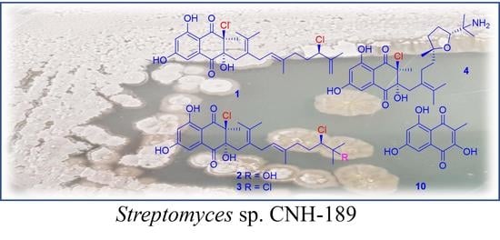
Graphical Abstract

1. Introduction

Marine actinomycetes are remarkable sources of novel secondary metabolites with wide chemical diversity [1]. Actinomycetales are supposed to be productive producers of a wide variety of biologically active secondary metabolites, including antibiotics, enzyme inhibitors, antivascular compounds, and antitumor compounds [2,3,4,5,6,7]. Among the many genera of marine actinomycetes, members of the genus Streptomyces have garnered increasing attention due to their capacity to produce novel compounds with unique structural and pharmacological properties [8,9,10]. The genus Streptomyces is common and bountiful in terrestrial habitats and has also been reported in marine ecosystems [11]. Lately, the investigation of chemical components for Streptomyces species isolated from diverse marine environments has led to the discovery of diverse bioactive secondary metabolites, such as enzyme inhibitors, anticancer agents, and antibiotics [2,12,13,14,15]. Our intensive chemical screening of the marine actinomycete strain Streptomyces sp. CNH-189 also resulted in the discovery of novel meroterpenoids, such as ansalactams A–D, merochlorins A–F, and meroindenon [5,6,7,8]. Ansalactams possess a distinctive spiro γ-lactam moiety with a modified polyketide chain, which exhibits biosynthetic plasticity between strains [16]. Ansalactams B−D exert antibacterial properties against methicillin-resistant Staphylococcus aureus (MRSA) with minimum inhibitory concentration (MIC) values of 31.2, 31.2, and 62.5 μg/mL, respectively [17]. Merochlorins are polyketide−terpenoid hybrid products with chlorination, which are biosynthesized from tetrahydroxynaphthalene (THN) linked to a modified C15 isoprene unit [18]. Merochlorins A and B possess polycyclic ring systems derived from a C15 isoprene unit, such as a bicyclo[3.2.1]octanone or a bicyclo[6-5-5]-fused ring system, where merochlorins C and D are formed in a linear C15 isoprene chain. The chemical structures of merochlorins E and F are closely related to those of merochlorins C and D, where merochlorins E and F share the same planar structure with different stereochemistry of their gem-dimethylmethylcyclohexane ring system. Merochlorin A reportedly achieves MIC values of 1-4 μg/mL against MRSA, whereas merochlorins E and F exhibit stronger antibacterial activity against pathogenic Gram-positive strains, with MIC values of 1−2 μg/mL. The observation of this activity suggested that the gem-dimethylmethylcyclohexane ring in merochlorins E and F plays an important role in the antibacterial activity [18,19]. Therefore, strain CNH-189 is a promising source for the discovery and isolation of novel antibacterial meroterpenoids.
Continuous efforts to discover novel compounds from strain CNH-189 resulted in the isolation of merochlorins G–J (14) and 10 from crude extracts of this bacterium, along with other known compounds, such as merochlorins A (5) and C–F (69) (Figure 1). Analyses of LC/MS, the UV profile, and characteristic MS isotopic patterns for chlorine atoms were then conducted to characterize these compounds. Here, we describe the isolation and structural characterization of merochlorins G–J (14) and 10, as well as their antibacterial activity against Gram-positive bacteria.

2. Results and Discussion

Merochlorin G (1) was isolated as a white powder, and its molecular formula was determined to be C26H3235Cl2O5 based on HRESIMS spectral data analysis (a pseudomolecular ion peak at m/z 517.1517 [M + Na]+, calcd for C26H3235Cl2O5Na, 517.1524) and interpretation of 13C NMR data. The 1H NMR spectrum of 1 exhibited two aromatic protons at δH 7.01 and 6.71; one olefinic proton at δH 4.87; 10 methylenes at δH 4.99, 4.90, 2.84, 2.61, 2.45, 2.37, 2.05, 1.95, 1.92, and 1.87; five methyl singlets at δH 1.94, 1.82, 1.58, 1.51, and 1.20; and three exchangeable protons at δH 11.70, 5.77, and 4.08. The 13C NMR and HSQC spectra displayed two carbonyls at δC 196.1 and 194.8; 10 quaternary carbons at δC 165.3, 162.9, 144.3, 134.7, 134.0, 131.6, 125.1, 109.9, 84.0, and 74.5; four methine carbons at δC 123.5, 108.9, 107.4, and 66.4; five methylene carbons at δC 114.2, 38.6, 36.6, 34.8, and 31.3; and five methyl singlets at δC 20.9, 20.6, 18.1, 17.0, and 15.9.
Analysis of the 2D NMR spectroscopic data of 1 allowed for the construction of two distinct fragments. Analysis of a 1H−1H coupling constant and HMBC correlations enabled the construction of the first fragment of 1. The meta-coupled aromatic protons at δH 7.01 (H-3, 1H, d, J = 2.0 Hz) and 6.71 (H-5, 1H, d, J = 2.0 Hz), along with the observation of HMBC correlations from H-3 to C-2 (δC 134.7), C-4 (δC 162.9), C-5 (δC 108.9), and C-7 (δC 109.9) and from H-5 to C-3 (δC 107.4), C-4, C-6 (δC 165.3), and C-7, indicated the presence of a 1,2,3,5-tetra-substituted benzene moiety. Carbon chemical shifts at δC 162.9 and 165.3 suggested the substitutions of two aromatic hydroxy groups at C-4 and C-6, respectively. Further, HMBC correlations from the aromatic proton H-3 to C-1 (δC 196.1), from the methyl singlet at 1.51 (H-26, 3H, s) to C-8 (δC 194.8), C-9 (δC 74.5), and C-10 (δC 84.0), and from the exchangeable proton at 10-OH (δH 4.08, 1H, s) to C-9, C-10, and C-11 supported the presence of a dihydronaphthalenedione moiety (Table 1).
The second fragment, the C15 rearranged sesquiterpene moiety, was assigned via COSY crosspeak analysis and HMBC correlations. The two methyl singlets H3-23 (δH 1.20, 3H, s) and H3-24 (δH 1.58, 3H, s) were attached at the same carbon C-22, which was further supported by the observation of the HMBC correlations between two methyl singlets (H3-23 and H3-24) and C-12 (δC 131.6) and C-22 (δC 125.1). Furthermore, long-range HMBC correlations from the methylene protons H-11 (δH 2.61, d, J = 14.0 Hz, δH 2.37, d J = 14.0 Hz) to C-12 (δC 131.6) and C-22 (δC 125.1) enabled the identification of an isoprene unit. The COSY crosspeaks between H-13 (δH 2.84, 1H, dd, J = 15.2, 6.7 Hz, δH 2.45, 1H, dd, J = 15.2, 6.7 Hz) and H-14 (δH 4.87, 1H, t, J = 7.0 Hz) and HMBC correlations from a methyl singlet H3-25 (δH 1.94, 3H, s) to C-14 (δC 123.5), C-15 (δC 134.0), and C-16 (δC 36.6) allowed for the isolation of another isoprene unit. The 25E geometry of the trisubstituted double bond was assigned based on the carbon chemical shift of 25-Me (δC 18.1) [10]. The last isoprene unit in the C15 rearranged sesquiterpene side chain was established by the COSY crosspeaks for the connectivity of H-17 (δH 1.92, 1H, m, δH 1.87, 1H, m) and H-18 (δH 4.33, 1H, t, J = 7.0 Hz), by the HMBC correlations from an olefinic proton H-20 (δH 4.99, 1H, s, δH 4.90, 1H, quint, J = 1.4 Hz) to C-18 (δC 66.4) and C-19 (δC 144.3), and by the HMBC correlations from a methyl singlet H-21 (δH 1.82, 3H, s) to C-18 and C-19. The connectivity between the three isoprene units was determined by the HMBC correlations from H-13 to C-12, and C-22 and the COSY crosspeaks between H-16 (δH 2.05, 1H, m, δH 1.95, and 1H, m) and H-17 (Table 1).
Lastly, the connection of these two fragments was secured by the interpretation of the HMBC correlations. Correlations from the methylene protons H-11 (δH 2.61, 1H, d, J = 14.0 Hz, δH 2.37, 1H, d, J =14.0 Hz) to C-1 and from an exchangeable proton 10-OH to C-9, C-10, and C-11 allowed for the attachment of C-10/C-11 and the placement of the hydroxy group at C-10. The HMBC correlations from a methyl singlet H3-26 (δH 1.51, 3H, s) to C-8, C-9, and C-10 allowed for the placement of the methyl substituent at C-9. Carbon chemical shifts at δC 74.5 and 66.4; the isotope ratio (9:6:1) of three pseudomolecular ion peaks [M + H]+, [M + H + 2]+, and [M + H + 4]+ in the LR-ESI-MS spectroscopic data; and the fact that there is no other available heteroatom besides two chlorines allowed for the attachment of two chlorine atoms at C-9 and C-18, respectively, thus completing the structural assignment of 1 (Figure 2).
Merochlorin H (2) was isolated as a pale-yellow oil, and its molecular formula was assigned as C26H3435Cl2O6 based on HRESIMS data analysis (a pseudomolecular ion peak at m/z 535.1627 [M +Na]+, calcd for C26H3435Cl2O6 Na, 535.1630). The isotope ratio (9:6:1) of three pseudomolecular ion peaks [M + H]+, [M + H + 2]+, and [M + H + 4]+ in the LR-ESI-MS spectrum also supported the presence of two chlorine atoms in the molecule. The 1H NMR spectrum of 2 was almost identical to that of 1, except for the terminal structure of the rearranged sesquiterpene moiety in the terminal fragment. The largest difference in the 1H NMR spectrum was attributed to the methyl singlet proton signal H3-20 (δH 1.12, 3H, s) instead of the geminal olefinic protons in 1. The long-ranged HMBC correlations from H3-20 and H3-21 (δH 1.18, 6H, s) to carbons C-18 (δC 69.8) and C-19 (δC 71.7) and the carbon chemical shift of C-19 indicated that 2 has a hydroxy group at C-19 (Table 1).
Merochlorin I (3) was isolated as a pale-yellow oil, and its molecular formula was assigned as C26H3335Cl3O5 based on the observation of a pseudomolecular ion peak at m/z 553.1281 [M + Na]+ in the HRESIMS spectroscopic data (calcd for C26H3435Cl3O5Na, 553.1291). The 1H and 13C NMR data for 3 were almost identical to those of 2, suggesting that they share the same carbon backbone. Comparisons and interpretation of the 2D NMR spectroscopic data of 3 to those of 2 indicated that 3 also possesses dihydronaphthalenedione and rearranged sesquiterpene moieties. The isotope ratio (27:27:9:1) of the four pseudomolecular ion peaks [M + H]+, [M + H + 2]+, [M + H + 4]+, and [M + H + 6]+ in the LR-ESI-MS clearly indicated the presence of three chlorines in 3. The consideration of the carbon chemical shift of C-19 (δC 72.9) and the molecular formula of 3 allowed for the attachment of a chlorine atom at C-19 (Table 1).
The molecular formula of merochlorin J (4) was determined to be C26H3635ClNO6 based on a pseudomolecular ion peak (m/z 494.2311 [M + H]+ (calcd for C26H3535ClNO6, 494.2309)) in the HRESIMS spectrum data and interpretation of 13C NMR data. The 1H NMR spectrum of 4 displayed a pair of meta-coupled aromatic protons at δH 6.81 and 6.62; nine methylenes at δH 2.45, 2.31, 2.23, 1.88, 1.65, 1.64, 1.56, 1.42, and 1.17; one methine at δH 3.98; six methyl singlets at δH 1.81, 1.47, 1.16, 1.14, 1.06, and 1.05; and two exchangeable protons at δH 6.16 and 3.98. The 13C and HSQC NMR spectroscopic data revealed two carbonyls at δC 195.3 and 193.6; 10 quaternary carbons at δC 164.1, 134.9, 129.2, 126.4, 107.8, 107.7, 84.0, 83.4, 76.1, and 55.5; three methine carbons at δC 107.7, 107.2, and 81.0; five methylene carbons at δC 39.7, 37.9, 36.3, 26.3, and 25.9; and six methyl carbons at δC 24.7, 22.8, 20.0, 20.0, 19.7, and 18.1.
Analysis of 2D NMR spectroscopic data indicated that 4 has a dihydronaphthalenedione and the common rearranged sesquiterpene side chain. The first fragment, the dihydronaphthalenedione moiety, was constructed by the interpretation of the HMBC correlation from H-3 (δH 6.81, 1H, d, J = 2.0 Hz) to C-1 (δC 195.3), C-2 (δC 134.9), C-4 (δC 164.1), C-5 (δC 107.7), and C-7 (δC 107.8); from H-5 (δH 6.62, 1H, d, J = 2.0 Hz) to C-3 (δC 107.2), C-4, C-6 (δC 165.2), and C-7; from H3-26 (δH 1.81, 3H, s) to C-8 (δC 193.6), C-9 (δC 76.1), and C-10 (δC 84.0); and from the exchangeable proton at 10-OH (δH 6.16, 1H, br s) to C-1, C-9, C-10 and C-11 (δC 37.9).
However, the 1H and 13C chemical shifts of the sesquiterpene component in 4 were substantially different from those of 13. The observation of the COSY crosspeaks for H-16 (δH 1.56, 2H, m)/H-17 (δH 1.88, 1H, m, 1.64, 1H, m)/H-18 (δH 3.98, 1H, t, J = 7.6 Hz) and of the HMBC correlations from H3-25 (δH 1.06, 3H, s) to C-15 (δC 83.4) and C-16 (δC 36.3); from H3-24 (δH 1.47, 3H, s) to C-12 (δC 126.4), C-22 (δC 129.2), and C-23 (δC 20.0); from H3-23 (δH 1.05, 3H, s) to C-12, C-23, and C-24 (δC 20.0); from H3-21 (δH 1.16, 3H, s) to C-18 (δC 81.0), C-19 (δC 55.5), and C-20 (δC 19.7); from H3-20 (δH 1.14, 3H, s) to C-18, C-19, and C-21 (δC 22.8); from H-14 (δH 1.42, 1H, m/δH 1.17, 1H, m) to C-15 and C-25 (δC 24.7); and from H-11 (δH 2.45, 1H, d, J = 16.0 Hz/δH 2.31, 1H, d, J = 16.0 Hz) to C-12 and the carbon chemical shifts of C-15 and C-18 (δC 81.0) allowed for the construction of the ether linkage between C-15 and C-18 of the second isoprene unit in the sesquiterpene component (Figure 3). Lastly, the consideration of the carbon chemical shift of C-19 (δC 55.5) and the molecular formula of 4 allowed for the attachment of an NH2 group at C-19 (Table 2).
Compound 10 was isolated as a dark-yellow oil, and its molecular formula was determined as C11H8O5 based on analysis of HRESIMS data (a pseudomolecular ion peak at m/z 220.0373 [M + H]+, calcd for C11H8O5, 220.0372). The 1H NMR spectrum of 10 was almost identical to that of the first fragment of 1, except for a methyl singlet at δH 1.89 for H-11. The 1H NMR spectrum of 10 displayed two aromatic protons at δH 6.97 (1H, d, J = 2.0 Hz, H-3) and 6.54 (1H, d, J = 2.0 Hz, H-5), one methyl singlet at δH 1.89 (3H, s, H3-11), and two exchangeable protons at δH 12.59 (1H, s, 4-OH) and 10.96 (1H, s, 6-OH). The 13C NMR and HSQC spectra of 10 displayed two carbonyl carbons at δC 189.6 (C-8) and 180.0 (C-1); six quaternary carbons at δC 163.4 (C-6), 162.7 (C-4), 155.6 (C-10), 131.8 (C-2), 119.2 (C-9), and 107.2 (C-7); two aromatic carbons at δC 107.9 (C-5) and 107.8 (C-3); and one methyl singlet carbon at δC 7.87 (C-11). Therefore, 10 was identified as 2,5,7-trihydroxy-3-methylnaphthalene-1,4-dione based on the comparison of NMR data in the literature [20]. This compound has been previously reported as a synthesis compound, but it is the first report as a natural product.
The relative stereochemistry of 14 was assigned by analysis of NOE spectroscopic data. The NOE crosspeak between 10-OH and the methyl singlet protons H3-26 suggested that these protons should be located at the same side on the dihydronaphthalenedione moiety. The chemical structures of 14 indicated that they are modified biosynthetic products of 7. This was supported by the conclusion that the C-9 and C-10 stereochemistry of 14 was likely the same as that of 7. To determine the absolute configurations of C-9 and C-10 in 14, the experimental ECD spectral data of 14 were measured (Figure 4). The ECD spectra of 14 exhibited almost identical patterns, suggesting that C-9 and C-10 of the dihydronaphthalenedione moiety in 14 have the same absolute configurations. Further, comparing their ECD spectra to those of previously reported enantiomer models by our group allowed us to suggest that C-9 and C-10 in 14 are in 9S, 10R configurations [19]. Furthermore, compounds 1, 2, 3, and 6 shared the same chemical scaffold and were isolated from the same bacterial strain. These observations suggested that the four compounds likely share the same absolute configuration for C-18. The absolute configurations of C-18 in 1 and of C-15 and C-18 in 4 were determined via DP4 calculations [21]. DP4 calculation is a computational method based on quantum mechanics for confirmation of the absolute configuration using NMR chemical shifts. It was required to design the models of two possible diastereomers of 1 (18R and 18S) and four possible diastereomer models of 4 (15R/18R, 15R/18S, 15S/18R, and 15S/18S). As a result, DP4 analysis indicated that 1 had an 18R configuration with 86% probability (Figure S22), whereas 4 had 15R, 18R configurations with a 100% probability (Figure S23).
Previous studies have proposed plausible biosynthetic pathways for tetrahydroxynaphthalene (THN)-derived meroterpenes from strain CNH-189, such as merochlorins, ansalactams, and meroindenon [17,18,19]. These pathways started from THN coupled with a C15 isoprene unit with atypical modifications, including a Baeyer‒Villiger-style oxidation, a Paterno‒Büchi-type 2 + 2 cycloaddition, and a pinacol-type contraction. All instances of atypical enzymatic biosynthesis are good examples of the biosynthetic potential of this marine microorganism.
There are two classes of bacterial THN-derived meroterpenoids, depending on the attachment of an isoprene chain to the THN frame [22]. Class I includes neomarinone, merochlorin A (5), and merochlorin B, which have a modified isoprene unit at the C-2 or C-4 carbon of the THN [23]. Class II compounds include merochlorins G–J (14) and are characterized by the attachment of an isoprene unit at the C-3 carbon of THN. Compounds 1, 2, and 3 are chlorinated products of merochlorin D (7), and compound 4 is an intramolecular cyclization product with further amination at C-19. Interestingly, chlorination at the isoprene chain of 13 occurred specifically at C-18. A tetrahydrofuran ring moiety identified in 4 was likely a biosynthesis product of an epoxide-opening reaction that starts with the selective enzymatic epoxidation of the double bonds [23,24,25]. Based on an AntiMarin2013 database search, the amine group at the tail of the isoprene chain of 4 was the first reported marine microbial natural product to possess this modification. It is also a rare case of an amination product from a tertiary alcohol that was produced by an epoxide-opening reaction. There are some synthetic examples of amination in the isopropyl group [26]. Lastly, compound 10 could be synthesized from the reduction of THN to hydroxynaphthoquinone, followed by methylation at C-3 [18,27].
Compounds 110 were tested for their antibacterial activities against six pathogenic bacteria, including three Gram-positive bacteria (Bacillus subtilis KCTC 1021, Kocuria rhizophila KCTC 1915, and Staphylococcus aureus KCTC 1927) and three Gram-negative bacteria (Escherichia coli KCTC 2441, Salmonella typhimurium KCTC 2515, and Klebsiella pneumoniae KCTC 2690). Merochlorin I (3) displayed strong antibacterial activities against B. subtilis, K. rhizophila, and S. aureus, with MIC values of 1, 2, and 2 μg/mL, respectively. Merochlorin G (1) was found to exhibit moderate antibacterial activities against the pathogenic strains, with a 16–32 μg/mL MIC range. Compound 10 exhibited strong antibacterial activity against K. rhizophila and moderate activity against B. subtilis, with MIC values of 2 and 32 μg/mL, respectively. Meanwhile, merochlorins H (2) and J (4) did not show any significant antibacterial activities against the six tested pathogens, with MIC values up to 128 μg/mL (Table 3). The presence of a polar moiety at the isoprene chain (C-19) abrogated the antibacterial properties of merochlorins, which was demonstrated with the hydroxy group in 2 and the amine group in 4. This was also supported by the strong antibacterial activity of 79, which possess nonpolar isoprene chain moieties. Therefore, the antibacterial activity against Gram-positive bacteria of these compounds depended on the hydroxy group at the THN core [28,29]. Interestingly, merochlorin A (5) exhibited no antibacterial activity against K. rhizophila, with MIC values up to 128 μg/mL, but exerted a strong antibacterial activity against two other Gram-positive bacteria (B. subtilis and S. aureus) [30].

3. Materials and Methods

3.1. General Experimental Procedures

Optical rotations were acquired using a Kruss Optronic P-8000 polarimeter with a 5 cm cell. UV spectra were recorded in a Varian Cary UV-visible spectrophotometer with a path length of 1 cm, and IR spectra were recorded on a Perkin-Elmer 1600 FT-IR spectrometer. Low-resolution LC/MS measurements were performed using the Agilent Technologies 1260 quadrupole and Waters Micromass ZQ LC/MS system using a reversed-phase column (Phenomenex Luna C18 (2) 100 Å, 50 mm × 4.6 mm, 5 µm) at a flow rate of 1.0 mL/min at the National Research Facilities and Equipment Center (NanoBioEnergy Materials Center) at Ewha Womans University. CD spectra were recorded using an Applied Photophysics Chirascan-Plus circular dichroism spectrometer (Applied Photophysics Ltd., Leatherhead, Surrey, UK). 1H and 2D NMR spectra data were recorded at 400 and 800 MHz in DMSO-d6 and CDCl3 solutions containing Me4Si as an internal standard on Varian Inova spectrometers. 13C NMR spectra were acquired at 100 or 200 MHz on a Varian Inova spectrometer. High-resolution EI-MS spectra were acquired using a JEOL JMS-AX505WA mass spectrometer at Seoul National University.

3.2. Collection and Phylogenetic Analysis of Strain CNH-189

The marine-derived actinomycete strain CNH-189 was isolated from a marine sediment sample collected near Oceanside, California. The strain was identified as a Streptomyces sp. based on 16S rRNA gene sequence analysis (accession no. HQ214120).

3.3. Fermentation

The bacterium strain CNH-189 was cultured in 60 2.8 L Fernbach flasks, each containing 1 L of a deionized water-based medium (M1: 10 g/L glucose, 20 g/L Grandma’s molasses, 5 g/L peptone, 2 g/L CaCO3, 40 mg/L Mg(SO4)·4H2O, 200 mg/L KCl, 200 mg/L KBr, and 40 mg/L Fe2(SO4)3·4H2O) at 27 °C.

3.4. Extraction and Isolation

Sterilized XAD7HP resin (20 g/L) was added after 24 h of cultivation, and the culture was incubated for an additional 5 days at 27 °C. Once the bacteria were cultured in the presence of XAD7HP resin, the resin was collected on cheesecloth, washed with deionized water, and eluted with acetone. The acetone was removed under reduced pressure, and the resulting aqueous layer was extracted with ethyl acetate (3 × 500 mL). The ethyl-acetate-soluble fraction was dried in vacuo to yield 4.5 g of crude extract. The crude extract was fractionated by open-column chromatography on silica gel (25 g) and then eluted with a step gradient of dichloromethane and methanol to obtain seven fractions. The first fraction was purified by reversed-phase HPLC (Phenomenex Luna C-18(2), 250 × 100 mm2, 2.0 mL/min, 5 μm, 100 Å, UV = 254 nm) using an isocratic solvent system to 90% CH3CN in water to render merochlorins G (1, 4.2 mg, tR = 26.1 min) and H (2, 3.0 mg, tR = 23.2 min). The fourth fraction was subjected to reversed-phase HPLC chromatography eluting with 82% CH3CN in water to obtain merochlorin I (3, 5.8 mg, tR = 30.2 min). The third fraction was purified using 64% CH3CN in water to obtain merochlorin J (4, 2.0 mg, tR = 31.2 min). To isolate compound 10, strain CNH-189 was regrown in 80 L scale. The treatment of the cultivation and crude extract fractionation was the same as the first culture. The first fraction was purified by reversed-phase HPLC (Phenomenex Luna C-18(2), 250 × 100 mm2, 2.0 mL/min, 5 μm, 100 Å, UV = 254 nm) using an isocratic solvent system from 82% CH3CN in water to obtain compound 10 (3.3 mg, tR = 6.5 min). Merochlorins A (5, 2.6 mg, tR = 29.1 min), C (6, 2.1 mg, tR = 19.8 min), D (7, 2.2 mg, tR = 35.6 min), E (8, 3.1 mg, tR = 20.3 min), and F (9, 2.3 mg, tR = 21.1 min) were isolated from the crude extract using a previously reported isolation procedure [19].
Merochlorin G (1): white powder; α D 21   +30 (c 0.30, MeOH); UV (MeOH) λmax (log ε) 224 (4.70), 239 (4.30), 296 (4.30), 334 (4.30) nm; IR (KBr) νmax 3625, 3191, 2924, 2361, 1643, 1376, 1266, 1165, 757 cm−1; 1H and 13C NMR (800 MHz and 200 MHz, CDCl3), see Table 1; HR-FAB-MS m/z 517.1517 [M + Na]+ (calcd for C26H3235Cl2O5Na, 517.1524).
Merochlorin H (2): pale-yellow oil; α D 21 +42 (c 0.40, MeOH); UV (MeOH) λmax (log ε) 224 (4.60), 239 (4.70), 296 (4.30), 334 (4.30) nm; IR (KBr) νmax 3562, 3483, 2361, 2066, 1623, 678 cm−1; 1H and 13C NMR (400 MHz and 100 MHz, DMSO-d6), see Table 1; HR-FAB-MS m/z 535.1627 [M + Na]+ (calcd for C26H3435Cl2O6 Na, 535.1630).
Merochlorin I (3): pale-yellow oil; α D 21 +85 (c 0.60, MeOH); UV (MeOH) λmax (log ε) 224 (4.60), 239 (4.70), 296 (4.30), 334 (4.30) nm; IR (KBr) νmax 3461, 2932, 2348, 1630, 1369, 1272, 1168, 858, 754 cm−1; 1H and 13C NMR (400 MHz and 100 MHz, DMSO-d6), see Table 1; HR-FAB-MS m/z 553.1281 [M + Na]+ (calcd for C26H3435Cl3O5Na, 553.1291).
Merochlorin J (4): pale-yellow oil; α D 21 +68 (c 0.275, MeOH); UV (MeOH) λmax (log ε) 224 (4.60), 239 (4.70), 296 (4.30), 334 (4.30) nm; IR (KBr) νmax 2958, 2357, 1630, 1180, 753 cm−1; 1H and 13C NMR (400 MHz and 100 MHz, DMSO-d6), see Table 2; HR-FAB-MS m/z 494.2311 [M + H]+ (calcd for C26H3535ClNO6, 494.2309).
Merochlorin A (5): 1H (500 MHz, DMSO-d6); δH 11.9 (br s, 3-OH), 6.16 (d, J = 2.0 Hz, H-4), 6.38 (d, J = 2.0 Hz, H-6), 2.24 (dd, J = 9.4, 4.0 Hz, H-9), 2.87 (d, J = 13.0 Hz, H-13)/2.65 (d, J = 13.0 Hz, H-13), 2.36 (dd, J = 14.0, 4.0 Hz, H-15)/2.33 (dd, J = 14.0, 9.4 Hz, H-15), 1.14 (q, J = 6.0 Hz, H-16)/1.40 (dt, J = 14.8, 4.8 Hz, H-16), 2.03 (m, H-17)/1.75 (m, H-17), 4.92 (t, J = 6.5 Hz, H-18), 1.45 (s, H-20), 1.53 (s, H-21), 1.56 (s, H-22), 1.65 (s, H-24), 0.81 (s, H-25), 13C NMR (125 MHz, DMSO-d6); δC 200.1 (C-12), 193.2 (C-1), 166.5 (C-5), 165.4 (C-3), 150.5 (C-7), 132.1 (C-14), 131.6 (C-19), 124.2 (C-18), 123.1 (C-23), 109.8 (C-2), 102.1 (C-4), 103.7 (C-6), 91.3 (C-11), 61.5 (C-8), 58.8 (C-9), 45.3 (C-10), 39.2 (C-16), 31.9 (C-15), 29.3 (C-13), 26.1 (C-21), 22.8 (C-17), 21.1 (C-24), 20.9 (C-22), 18.1 (C-20), 16.5 (C-25), LR-ESI-MS m/z = 429.18 [M + H]+.
Merochlorin C (6): 1H (500 MHz, DMSO-d6); δH 11.2 (s, 6-OH), 5.82 (s, 10-OH), 7.19 (d, J = 2.0 Hz, H-3), 6.91 (d, J = 2.0 Hz, H-5), 4.36 (t, J = 4.8 Hz, H-14), 3.84 (d, J = 10.2 Hz, H-18), 2.88 (d, J = 16.0 Hz, H-11)/2.11 (d, J = 16.0 Hz, H-11), 2.79 (d, J = 14.0 Hz, H-13)/1.81 (m, H-13), 1.96 (m, H-16), 1.76 (s, H-22), 1.74 (s, H-26), 1.70 (s, H-21), 1.61 (m, H-17)/1.54 (m, H-17), 1.59 (s, H-24), 1.58 (s, H-20), 0.98 (s, H-25), 13C NMR (125 MHz, DMSO-d6); δC 196.4 (C-1), 187.1 (C-8), 164.0 (C-6), 159.0 (C-4), 136.2 (C-2), 135.1 (C-15), 132.5 (C-23), 124.9 (C-14), 124.5 (C-12), 118.9 (C-5), 118.8 (C-7), 110.6 (C-3), 87.8 (C-19), 84.2 (C-10), 78.0 (C-9), 66.4 (C-18), 38.2 (C-11), 35.8 (C-16), 31.8 (C-17), 30.5 (C-13 & C-21), 25.4 (C-20), 22.6 (C-22), 21.1 (C-24), 19.9 (C-26), 16.5 (C-25), LR-ESI-MS m/z = 517.15 [M + Na]+.
Merochlorin D (7): 1H (500 MHz, DMSO-d6); δH 11.5 (s, 6-OH), 6.09 (s, 10-OH), 6.83 (d, J = 2.0 Hz, H-3), 6.60 (d, J = 2.0 Hz, H-5), 4.97 (d, J = 6.0 Hz, H-18), 4.77 (t, J = 6.0 Hz, H-14), 2.82 (dd, J = 14.0, 6.0 Hz, H-13)/2.35 (dd, J = 14.0, 6.0 Hz, H-13), 2.42 (d, J = 16.0 Hz, H-11)/2.24 (d, J = 16.0 Hz, H-11), 1.95 (m, H-17), 1.84 (m, H-16), 1.77 (s, H-26), 1.59 (s, H-20), 1.51 (s, H-21), 1.47 (s, H-22), 1.42 (s, H-25), 1.08 (s, H-24), 13C NMR (125 MHz, DMSO-d6); δC 195.9 (C-1), 194.1 (C-8), 166.0 (C-4), 165.0 (C-6), 135.7 (C-2), 134.9 (C-15), 131.3 (C-19), 130.3 (C-23), 126.5 (C-12), 124.6 (C-18), 123.1 (C-14), 108.6 (C-7), 108.4 (C-5), 108.1 (C-3), 84.5 (C-10), 77.0 (C-9), 39.0 (C-11), 39.6 (C-16), 31.1 (C-13), 26.8 (C-20), 26.7 (C-17), 21.3 (C-24), 21.0 (C-22), 18.9 (C-26), 18.8 (C-21), 16.8 (C-25), LR-ESI-MS m/z = 483.19 [M + Na]+.
Merochlorin E (8): 1H (500 MHz, DMSO-d6); δH 11.52 (s, 4-OH), 11.47 (s, 6-OH), 5.95 (s, 10-OH), 6.85 (d, J = 2.0 Hz, H-3), 6.63 (d, J = 2.0 Hz, H-5), 5.02 (t, J = 4.8 Hz, H-16), 2.69 (d, J = 14.1 Hz, H-11)/2.12 (d, J = 14.1 Hz, H-11), 2.23 (dd, J = 14.0, 4.8 Hz, H-13)/1.22 (m, H-13), 1.80 (s, H-26), 1.78 (m, H-17)/1.63 (m, H-17), 1.52 (s, H-24), 1.36 (s, H-25), 1.33 (s, H-22), 1.05 (dd, J = 14.0, 14.0 Hz, H-18)/0.88 (dd, J = 14.0, 14.0 Hz, H-18), 0.82 (s, H-20), 0.72 (s, H-21), 13C NMR (125 MHz, DMSO-d6); δC 195.2 (C-1), 193.4 (C-8), 165.6 (C-6), 164.1 (C-4), 136.6 (C-15), 135.2 (C-2), 130.9 (C-23), 125.1 (C-12), 119.4 (C-16), 107.8 (C-7), 107.6 (C-5), 107.3 (C-3), 83.9 (C-10), 78.0 (C-9), 47.0 (C-14), 39.5 (C-11), 34.4 (C-13), 32.0 (C-19), 28.6 (C-18), 28.1 (C-20), 26.3 (C-21), 24.8 (C-25), 22.5 (C-17), 20.8 (C-22 & C-24), 18.1 (C-26), LR-ESI-MS m/z = 461.20 [M + H]+.
Merochlorin F (9): 1H (500 MHz, DMSO-d6); δH 11.52 (s, 4-OH), 11.47 (s, 10-OH), 5.99 (s, 10-OH), 6.83 (d, J = 2.0 Hz, H-3), 6.62 (d, J = 2.0 Hz, H-5), 5.19 (t, J = 4.8 Hz, H-16), 2.56 (d, J = 14.0 Hz, H-11)/2.32 (d, J = 14.0 Hz, H-11), 2.14 (dd, J = 14.8, 14.8 Hz, H-13)/1.78 (m, H-13), 1.91 (m, H-17), 1.48 (m, H-18)/1.01 (dd, J = 7.2, 7.2 Hz, H-18), 1.79 (s, H-26), 1.49 (s, H-24), 1.43 (s, H-25), 1.17 (s, H-22), 0.71 (s, H-20), 0.62 (s, H-21), 13C NMR (125 MHz, DMSO-d6); δC 195.1 (C-1), 193.2 (C-8), 165.7 (C-6), 164.1 (C-4), 136.8(C-15), 134.9 (C-2), 130.5 (C-23), 125.7 (C-12), 119.9 (C-16), 107.9 (C-7), 107.7 (C-5), 107.4 (C-3), 83.9 (C-10), 76.0 (C-9), 46.9 (C-14), 38.4 (C-11), 33.7 (C-13), 32.1 (C-19), 29.1 (C-18), 27.9 (C-20), 26.4 (C-21), 24.7 (C-25), 22.6 (C-17), 20.7 (C-22 & C-24), 18.0 (C-26), LR-ESI-MS m/z = 461.20 [M + H]+.
Compound 10: 1H (400 MHz, DMSO-d6); δH 12.59 (s, 4-OH), 10.96 (s, 6-OH), 6.97 (d, J = 2.0 Hz, H-3), 6.54 (d, J = 2.0 Hz, H-5), 1.89 (s, H-11), 13C NMR (100 MHz, DMSO-d6); δC 189.6 (C-8), 180.0 (C-1), 163.4 (C-6), 162.7 (C-4), 155.6 (C-10), 131.8 (C-2), 119.2 (C-9), 107.9 (C-5), 107.8 (C-3), 107.2 (C-7), 7.8 (C-11), HR-FAB-MS m/z 220.0373 [M + H]+ (calcd for C11H8O5, 220.0372).

3.5. Conformational Search and DP4 Calculations

A conformational search of merochlorins G (1) and J (4) was carried out by MacroModel with the Merck molecular force field (gas phase), a 10 kJ/mol upper energy limit, and a 0.001 kJ (mol Å)−1 convergence threshold on the rms gradient to minimize computational complexity and expense. In the case of 1, 66 conformers were obtained for the 18R diastereomer and 37 conformers were obtained for the 18S model under the 10 kJ/mol limit of molecular potential energy. In addition, in the case of 4, 10 conformers were obtained for the 15R/18R diastereomer, 5 conformers for the 15R/18S diastereomer, 2 conformers for the 15S/18R diastereomer, and 7 conformers for the 15S/18S diastereomer. The Boltzmann population was calculated based on the potential energy of each conformer. Ground-state geometry optimization was performed by density functional theory (DFT) modeling with TurbomoleX 4.3.2. The basis set was def-SV(P) for all atoms, and the level of theory was B3-LYP at the functional level in the gas phase. Calculated chemical shifts of 1H and 13C were averaged by the Boltzmann populations. The experimental chemical shifts compared to these Boltzmann-averaged chemical shifts and the DP4 analyses indicated the 18R configuration of 1 with 86.6% and 15R/18R configurations of 4 with 100.0% probability.

3.6. Antibacterial Assay

Antibacterial susceptibility was tested against three Gram-positive bacteria (Bacillus subtilis KCTC 1021, Kocuria rhizophila KCTC 1915, and Staphylococcus aureus KCTC 1927) and three Gram-negative bacteria (Escherichia coli KCTC 2441, Salmonella typhimurium KCTC 2515, and Klebsiella pneumoniae KCTC 2690), as described in a previous study with modifications [16]. These bacteria were grown in Mueller–Hinton broth at 37 °C and 225 rpm for 24 h. Compounds 110 and positive controls were dissolved in DMSO, and 100 μL of each solution was dispensed into the wells of 96-well plates starting at 128 μg/mL concentration. Compounds 110 and positive controls were serially diluted, and Mueller–Hinton broth was added to a final concentration of 0.5% McFarland standard. The 96-well microtiter plates were cultivated for 24 h at 37 °C. The minimal inhibitory concentration (MIC) was defined as the lowest positive control concentration that visibly inhibited bacterial growth [31].

4. Conclusions

In conclusion, an intensive chemical screening of the Streptomyces sp. strain CNH-189 resulted in the discovery of new merochlorin derivatives (14) and a proposed biosynthetic merochlorin precursor 10, along with known congeners 59. Further, compound 3 exhibited strong antibacterial activities against Gram-positive strains such as B. subtilis, K. rhizophila, and S. aureus, with MIC values of 1–2 μg/mL.

Supplementary Materials

The following are available online at www.mdpi.com/article/10.3390/md19110618/s1, Figures S1–S21: 1D NMR and 2D NMR of compounds 14, 10; Figures S22 and S23; DP4 analysis of NMR calculation of compounds 1 and 4; Figures S24–S32; LRESIMS and HRESIMS spectra of compounds 14, 10; Figures S33 and S34; Simulated conformer models of possible diastereomers of compounds 1 and 4; Tables S1 and S2: Experimental and calculation chemical shift values of compounds 1 and 4.

Author Contributions

M.-J.R. purified the compounds and contributed to NMR data collection and interpretation; P.F.H. purified the compounds, contributed to NMR data and antibacterial activity assay data collection, solved their structure, and contributed to manuscript writing; J.L. contributed to manuscript writing; S.H. contributed to ECD and DP4 data collection; E.-Y.L. contributed to manuscript writing; S.-S.C., I.Y., D.-C.O. and W.F. contributed to review and editing; and S.-J.N. contributed to supervision and conceptualization. All authors have read and agreed to the published version of the manuscript.

Funding

This research was supported by the National Research Foundation of Korea grant funded by the Korean Government (Ministry of Science and ICT; no. 2021R1A4A2001251 to S.-J.N.) and in part by the project titled “Development of Potential Antibiotic Compounds Using Polar Organism Re-sources” (15250103, KOPRI grant PM21030 to S.-S.C.) funded by the Ministry of Oceans and Fish-eries, Korea. Isolation of the bacterium strain Streptomyces sp. CNH-189 was a result of the financial support from the US National Cancer Institute (grant CA R37044848 (to W.F.)).

Institutional Review Board Statement

Not applicable.

Data Availability Statement

The data presented in this study are available on request from the corresponding author.

Conflicts of Interest

The authors declare no conflict of interest.

References

  1. Nett, M.; Ikeda, H.; Moore, B.S. Genomic Basis for Natural Product Biosynthetic Diversity in the Actinomycetes. Nat. Prod. Rep. 2009, 26, 1362–1384. [Google Scholar] [CrossRef]
  2. Almasi, F.; Mohammadipanah, F.; Adhami, H.R.; Hamedi, J. Introduction of Marine-Derived Streptomyces sp. UTMC 1334 as A Source of Pyrrole Derivatives with Anti-Acetylcholinesterase Activity. J. Appl. Microbiol. 2018, 125, 1370–1382. [Google Scholar] [CrossRef]
  3. Mohammadipanah, F.; Matasyoh, J.; Hamedi, J.; Klenk, H.P.; Laatsch, H. Persipeptides A and B, Two Cyclicpeptides from Streptomyces sp. UTMC 1154. Bioorganic Med. Chem. 2012, 20, 335–339. [Google Scholar] [CrossRef] [PubMed]
  4. Imada, C. Enzyme Inhibitors and Other Bioactive Compounds from Marine Actinomycetes. Antonie Van Leeuwenhoek 2005, 87, 59–63. [Google Scholar] [CrossRef] [PubMed]
  5. Salimi, F.; Hamedi, J.; Motevaseli, E.; Mohammadipanah, F. Isolation and Screening of Rare-Actinobacteria, A New Insight for Finding Natural Products with Anti-Vascular Calcification Activity. J. Appl. Microbiol. 2017, 124, 254–266. [Google Scholar] [CrossRef] [PubMed]
  6. Salcedo, R.l.G.a.; Olano, C.; Gomez, C.; Fernandez, R.; Brana, A.F.; Mendez, C.; Calle, F.; Salas, J.A. Characterization and Engineering of the Biosynthesis Gene Cluster for Antitumor Macrolides PM100117 and PM100118 from A Marine Actinobacteria: Generation of A Novel Improved Derivative. Microb. Cell Fact. 2017, 124, 254–266. [Google Scholar] [CrossRef] [PubMed] [Green Version]
  7. Azarakhsh, Y.; Mohammadipanah, F.; Nassiri, S.M.; Siavashi, V.; Hamedi, J. Isolation and Screening of Proangiogenic and Antiangiogenic Metabolites Producing Rare Actinobacteria from Soil. J. Appl. Microbiol. 2017, 122, 1595–1602. [Google Scholar] [CrossRef] [PubMed]
  8. Dharmaraj, S. Marine Streptomyces as a Novel Source of Bioactive Substances. World J. Microbiol. Biotechnol. 2010, 26, 2123–2139. [Google Scholar] [CrossRef]
  9. Prieto-Davó, A.; Fenical, W.; Jensen, P.R. Comparative Actinomycete Diversity in Marine Sediments. Aquat. Microb. Ecol. 2008, 52, 1–11. [Google Scholar] [CrossRef]
  10. Udwary, D.W.; Zeigler, L.; Asolkar, R.N.; Singan, V.; Lapidus, A.; Fenical, W.; Jensen, P.R.; Moore, B.S. Genome Sequencing Reveals Complex Secondary Metabolome in the Marine Actinomycete Salinispora Tropica. Proc. Natl. Acad. Sci. USA 2007, 104, 10376–10381. [Google Scholar] [CrossRef] [Green Version]
  11. Nam, S.J.; Kauffman, C.A.; Paul, L.A.; Jensen, P.R.; Fenical, W. Actinoranone, A Cytotoxic Meroterpenoid of Unprecedented Structure from A Marine Adapted Sterptomyces sp. Org. Lett. 2013, 15, 5400–5403. [Google Scholar] [CrossRef] [Green Version]
  12. Sproule, A.; Correa, H.; Decken, A.; Haltli, B.; Berrue, F.; Overy, D.P.; Kerr, R.G. Terrosamycins A and B, Bioactive Polyether Ionophores from Streptomyces sp. RKND004 from Prince Edward Island Sediment. Mar. Drug. 2019, 17, 347. [Google Scholar] [CrossRef] [Green Version]
  13. Kim, M.C.; Cullum, R.; Hebishy, A.M.S.; Mohamed, H.A.; Faraag, A.H.I.; Salah, N.M.; Abdelfattah, M.S.; Fenical, W. Mersaquinone, A New Tetracene Derivative from The Marine-Derived Streptomyces sp. EG1 Exhibiting Activity Against Methicillin-Resistance Staphylococcus aureus (MRSA). Antibiotics 2020, 9, 252. [Google Scholar] [CrossRef]
  14. Paderog, M.J.V.; Suarez, A.F.L.; Sabido, E.M.; Low, Z.J.; Saludes, J.P.; Dalisay, D.S. Anthracycline Shunt Metabolites from Philippine Marine Sediment-Derived Streptomyces Destroy Cell Membrane Integrity of Multidrug-Resistant Staphylococcus aureus. Front. Microbiol. 2020, 11, 743. [Google Scholar] [CrossRef] [Green Version]
  15. Song, Y.; Yang, J.; Yu, J.; Li, J.; Yuan, J.; Wong, N.; Ju, J. Chlorinated Bis-indole Alkaloids from Deep-Sea Derived Streptomyces sp. SCSIO 11791 with Antibacterial and Cytotoxic Activities. J. Antibiot. 2020, 73, 542–547. [Google Scholar] [CrossRef]
  16. Wilson, M.C.; Nam, S.J.; Gulder, T.A.M.; Kauffman, C.A.; Jensen, P.R.; Fenical, W.; Moore, B.S. Structure and Biosynthesis of the Marine Streptomycete Ansamycin Ansalactam A and Its Distinctive Branched Chain Polyketide Extender Unit. J. Am. Chem. Soc. 2011, 133, 1971–1977. [Google Scholar] [CrossRef] [Green Version]
  17. Le, T.C.; Yang, I.; Yoon, Y.J.; Nam, S.J.; Fenical, W. Ansalactams B-D Illustrate Further Biosynthetic Plasticity within the Ansamycin Pathway. Org. Lett. 2016, 18, 2256–2259. [Google Scholar] [CrossRef]
  18. Kaysser, L.; Bernhardt, P.; Nam, S.J.; Loesgen, S.; Ruby, J.G.; Skewes-Cox, P.; Jensen, P.R.; Fenical, W.; Moore, B.S. Merochlorins A-D, Cyclic Meroterpenoid Antibiotics Biosynthesized in Divergent Pathways with Vanadium-Dependent Chloroperoxidases. J. Am. Chem. Soc. 2012, 134, 11988–11991. [Google Scholar] [CrossRef] [Green Version]
  19. Ryu, M.J.; Hwang, S.; Kim, S.; Yang, I.; Oh, D.C.; Nam, S.J.; Fenical, W. Meroindenon and Merochlorins e and F, Antibacterial Meroterpenoids from a Marine-Derived Sediment Bacterium of the Genus Streptomyces. Org. Lett. 2019, 21, 5779–5783. [Google Scholar] [CrossRef]
  20. Sabutskii, Y.E.; Polonik, S.G.; Denisenko, V.A.; Dmitrenok, P.S. A New Method for Thiomethylation of Hydroxy-1,4-naphthoquinones with N-Acetyl-L-cystein; First Synthesis of Fibrostatin, B,C, and D. Synthesis 2014, 46, 2763–2770. [Google Scholar]
  21. Zanardi, M.M.; Suarez, A.G.; Sarotti, A.M. Determination of the Relative Configuration of Terminal and Spiroepoxides by Computational Methods. Advantages of the Inclusion of Unscaled Data. J. Org. Chem. 2020, 2020. 85, 11566–11570. [Google Scholar] [CrossRef]
  22. Maxwell, A. and Rampersad, D. Novel Prenylated Hydroxybenzoic Acid Derivatives from Piper Saltuum. J. Nat. Prod. 1989, 52, 614–618. [Google Scholar] [CrossRef]
  23. Miles, Z.D.; Diethelm, S.; Pepper, H.P.; Huang, D.M.; George, J.H.; Moore, B.S. A Unifying Paradigm for Naphthoquinone-Based Meroterpenoid (Bio)Synthesis. Nat. Chem. 2017, 9, 1235–1242. [Google Scholar] [CrossRef] [PubMed]
  24. Morimoto, Y.; Iwai, T.; Kinoshita, T. Revised Structure of Squalene-Derived PentaTHF Polyether, Glabrescol, through Its Enantioselective Total Synthesis: Biogenetically Intriguing C(s) vs C2 Symmetric Relationships. J. Am. Chem. Soc. 2000, 122, 7124–7125. [Google Scholar] [CrossRef]
  25. Lorente, A.; Lamariano-Merketegi, J.; Albericio, F.; Álvarez, M. Tetrahydrofuran-Containing Macrolides: A Fascinating Gift from the Deep Sea. Chem. Rev. 2013, 113, 4567–4610. [Google Scholar] [CrossRef] [PubMed]
  26. Bahn, S.; Imm, S.; Neubert, L.; Zhang, M.; Neumann, H.; Beller, M. The Catalytic amination of Alcohols. ChemCatChem. 2011, 3, 1853–1864. [Google Scholar] [CrossRef]
  27. Saha, N.; Muller, M.; Husain, S.S. Asymmetric Synthesis of Natural cis-Dihydroarenediols Using Tetrahydroxynaphthalene Reductase and Its Biosynthetic Implications. Org. Lett. 2019, 21, 2204–2208. [Google Scholar] [CrossRef] [PubMed]
  28. López-Pérez, B.; Pepper, H.P.; Ma, R.; Fawcett, B.J.; Pehere, A.D.; Wei, Q.; Ji, Z.; Polyak, S.W.; Dai, H.; Song, F.; et al. Biosynthetically Guided Structure–Activity Relationship Studies of Merochlorin A, an Antibiotic Marine Natural Product. ChemMedChem 2017, 12, 1969–1976. [Google Scholar] [CrossRef]
  29. Yang, H.; Liu, X.; Li, Q.; Li, L.; Zhang, J.R.; Tang, Y. Total Synthesis and Preliminary SAR Study of (±)-Merochlorins A and B. Org. Biomol. Chem. 2015, 14, 198–205. [Google Scholar] [CrossRef] [Green Version]
  30. Sakoulas, G.; Nam, S.J.; Loesgen, S.; Fenical, W.; Jensen, P.R.; Nizet, V.; Hensler, M. Novel Bacterial Metabolite Merochlorin A Demonstrates in Vitro Activity against Multi-Drug Resistant Methicillin-Resistant Staphylococcus aureus. PLoS ONE 2012, 7, 1–6. [Google Scholar] [CrossRef]
  31. Wiegand, I.; Hilpert, K.; Hancock, R.E.W. Agar and Broth Dilution Methods to Determine the Minimal Inhibitory Concentration (MIC) of Antimicrobial Substances. Nat. Protoc. 2008, 3, 163–175. [Google Scholar] [CrossRef]
Figure 1. Structures of merochlorins G–J (14), A (5), C–F (69), and 2,5,7-trihydroxy-3-methylnaphthalene-1,4-dione (10).
Marinedrugs 19 00618 g001
Figure 2. COSY and key HMBC correlations of merochlorin G (1).
Marinedrugs 19 00618 g002
Figure 3. COSY and key HMBC correlations for merochlorin J (4).
Marinedrugs 19 00618 g003
Figure 4. Experimental ECD spectra of 14 in methanol.
Marinedrugs 19 00618 g004
Table 1. NMR spectroscopic data for merochlorin G (1) in CDCl3 and 1H and 13C NMR spectroscopic data for merochlorins H and I (2 and 3) in DMSO-d6.
No.1 a2 b3 b
δC, Mult. cδH (J in Hz)COSYHMBCδC, Mult. cδH (J in Hz)δC, Mult. cδH (J in Hz)
1196.1, C 195.2, C 195.1, C
2134.7, C 135.1, C 135.0, C
3107.4, CH7.01, d (2.0)H-51, 2, 4, 5, 7107.3, CH6.84, d (2.0)107.3, CH6.94, d (2.0)
4162.9, C 165.7, C 164.2, C
5108.9, CH6.71, d (2.0)H-33, 4, 6, 7107.8, CH6.64, d (2.0)107.7, CH6.62, d (2.0)
6165.3, C 164.2, C 165.7, C
7109.9, C 108.0, C 108.0, C
8194.8, C 193.4, C 193.3, C
974.5, C 76.2, C 76.2, C
1084.0, C 84.0, C 84.0, C
1138.6, CH22.61, d (14.0), 2.37, d (14.0) 1, 1330.9, CH21.38, s31.1, CH22.13, m
12131.6, C 125.9, C 125.7, C
1331.3, CH22.84, dd (15.2, 6.7), 2.45, dd (15.2, 6.7)H-1411, 14, 2430.3, CH22.88, dd (15.2, 7.2), 2.42, dd (15.2, 7.2)30.3, CH22.86, dd (15.2, 7.2), 2.40, dd (15.2, 7.2)
14123.5, CH4.87, t (7.0)H-1313, 16, 24, 26123.2, CH4.89, t (7.0)123.9, CH4.90, t (7.0)
15134.0, C 133.5, C 132.9, C
1636.6, CH22.05, m d, 1.95, m dH-1714, 17, 18, 2636.8, CH22.15, m d, 1.95, m d36.3, CH22.18, m d, 2.00, m d
1734.8, CH21.92, m d, 1.87, m dH-16, H-1816, 1831.3, CH21.28, d (9.2)39.2, CH22.45, d (14.6), 2.27, d (14.6)
1866.4, CH4.33, t (7.0)H-1717, 20, 2169.8, CH3.51, t (2.0)70.7, CH3.93, t (2.0)
19144.3, C 71.7, C 72.9, C
20114.2, CH24.99, s, 4.90, quint (1.4) 18, 2124.4, CH31.12, s29.1, CH31.59, s
2117.0, CH31.82, s 18, 1927.6, CH31.18, s30.3, CH31.63, s
22125.1, C 129.5, C 129.7, C
2320.9, CH31.20, s 10, 11, 12, 13, 14, 22, 2420.3, CH31.05, s20.7, CH31.09, s
2420.6, CH31.58, s 10, 11, 12, 22, 2320.2, CH31.49, s20.3, CH31.49, s
2518.1, CH31.94, s 1715.6, CH31.47, s15.4, CH31.45, s
2615.9, CH31.51, s 8, 9, 1018.0, CH31.80, s18.1, CH31.79, s
4-OH 5.77, s 3, 4, 5 11.43, s 11.48, s
6-OH 11.70, s 4, 5, 6, 7, 8 11.52, s 11.52, s
10-OH 4.08, s 9, 10, 11 6.14, s 6.13, s
a 800 MHz for 1H NMR and 200 MHz for 13C NMR. b 400 MHz for 1H NMR and 100 MHz for 13C NMR. c Numbers of attached protons were determined by analysis of 2D spectra. d Signals were overlapping.
Table 2. NMR spectroscopic data for merochlorin J (4) in DMSO-d6 a.
No.4
δC, Mult. bδH (J in Hz)COSYHMBC
1195.3, C
2134.9, C
3107.2, CH6.81, d (2.0)H-51, 2, 4, 5, 7
4164.1, C
5107.7, CH6.62, d (2.0)H-33, 4, 6, 7
6165.2, C
7107.8, C
8193.6, C
976.1, C
1084.0, C
1137.9, CH22.45, d (16.0), 2.31, d (16.0) 9, 10, 12, 13, 23
12126.4, C
1326.3, CH22.23, d (14.0), 1.65, m cH-1411, 23
1439.7, CH21.42, m c, 1.17, m cH-1315, 25
1583.4, C
1636.3, CH21.56, m cH-1714, 15, 17, 18, 25
1725.9, CH21.88, m c 1.64, m cH-16, H-1815, 16, 18, 19
1881.0, CH3.82, t (7.6)H-1720, 21
1955.5, C
2019.7, CH31.14, s 18, 19, 21
2122.8, CH31.16, s 18, 19, 20
22129.2, C
2320.0, CH31.05, s 12, 22, 24
2420.0, CH31.47, s 12, 22, 23
2524.7, CH31.06, s 14, 15, 16
2618.1, CH31.81, s 8, 9, 10
10-OH 6.16, br s 1, 9, 10, 11
a 400 MHz for 1H NMR and 100 MHz for 13C NMR. b Numbers of attached protons were determined by analysis of 2D spectra. c Signals were overlapping.
Table 3. Antibacterial activities of merochlorins (110) a.
MIC (μg/mL)
Gram-Positive BacteriaGram-Negative Bacteria
B. subtilis KCTC 1021K. rhizophila KCTC 1915S. aureus KCTC 1927 E. coli KCTC 2441S. typhimurium KCTC 2515K. pneumonia KCTC 2690
1163216 >128>128>128
264>128>128 >128>128>128
3122 >128>128>128
4>128>128>128 >128>128>128
52>1284 >128>128>128
6 b163232 >128>128>128
710.51 >128>128>128
8 b122 >128>128>128
9 b121 >128>128>128
10322>128>128>128>128>128
Ampicillin0.250.250.25 42>128
Vancomycin0.250.50.5 >128>128>128
a Each sample was tested in triplicate and repeated three times. b Tested in the previous study.
Publisher’s Note: MDPI stays neutral with regard to jurisdictional claims in published maps and institutional affiliations.