
Marine-Bioinspired Nanoparticles as Potential Drugs for Multiple Biological Roles

 , , ,
, , ,  and
and
Abstract
:1. Introduction
2. Green Synthesis of Nanoparticles (NPs) for Its Application in the Field of Medicine
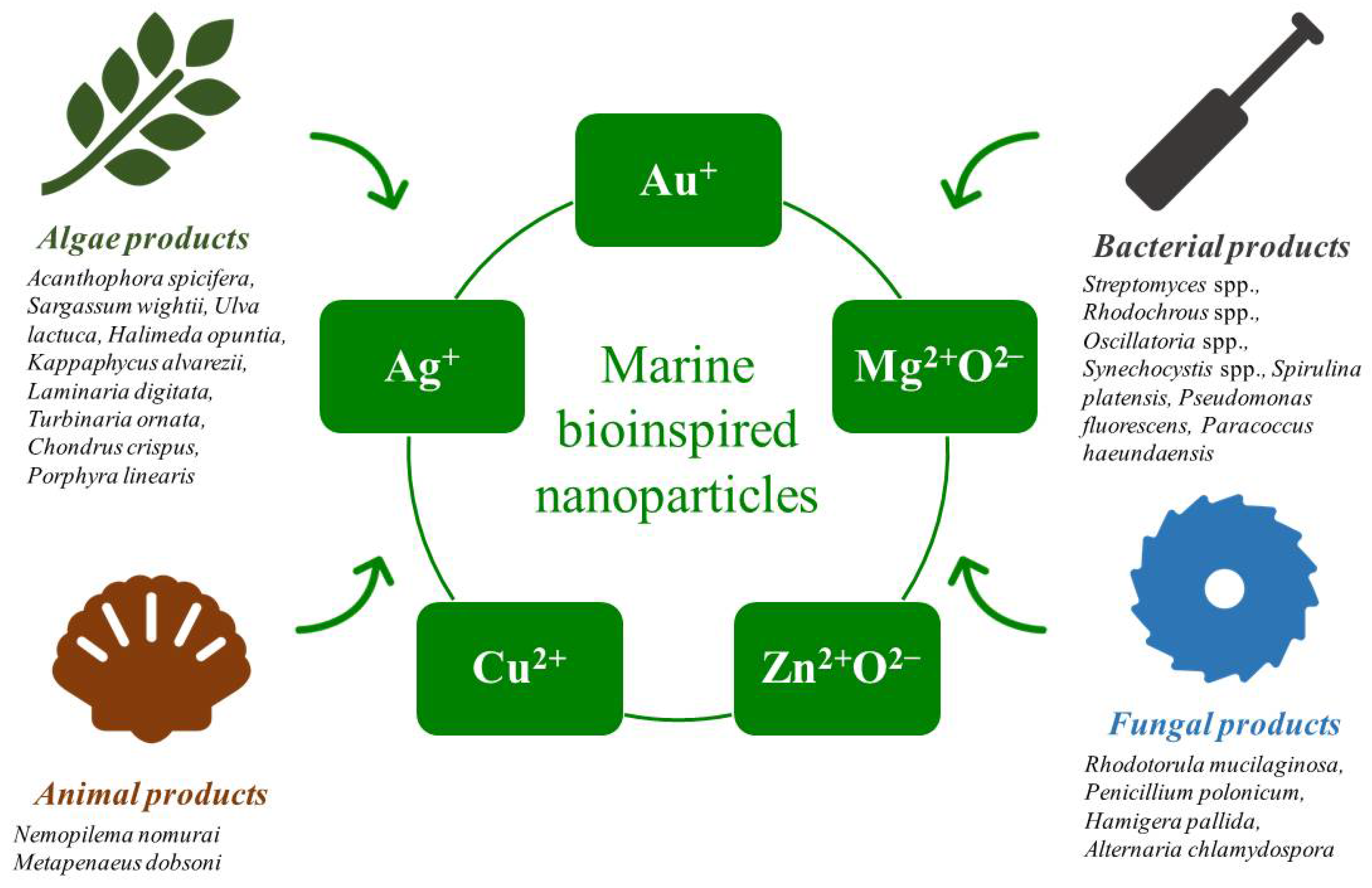
3. Marine Organisms and Compounds for the Green Synthesis of NPs
4. Marine Bioinspired NPs Used for Bacterial Infection
5. Marine Bioinspired NPs Used for Fungal Infection

| Name of Marine-Derived Compound/Product | Sources/Organism | Name of NPs | Size Range of MNPs | Shape/Morphology | Antimicrobial Types | Microbial Pathogens | References |
|---|---|---|---|---|---|---|---|
| Extracts |
| AgNPs | 12 nm | Spherical | Antibacterial |
| [78] |
| Extracts | U. lactuca | AgNPs | 20–50 nm | - | Antibacterial |
| [79] |
| Extracts |
| SeNPs | 30, 80 nm | Spherical | Antibacterial |
| [80] |
| Extracts | Spirulina platensis | SNPs | 200–450 nm | Spherical | Antibacterial | V. parahaemolyticus | [81] |
| Extracts | Chroococcus minutus | AgNPs | - | - | Antibacterial |
| [56] |
| Extracts | U. lactuca | SeNP | 85 nm | Spherical | Antibacterial |
| [82] |
| Extracts | Sargassum muticum | AgNPs | 20–54 nm | Spherical | Antibacterial |
| [57] |
| Extracts | S. swartzii | AgNPs | 20–40 nm | Spherical | Antibacterial | E. coli | [58] |
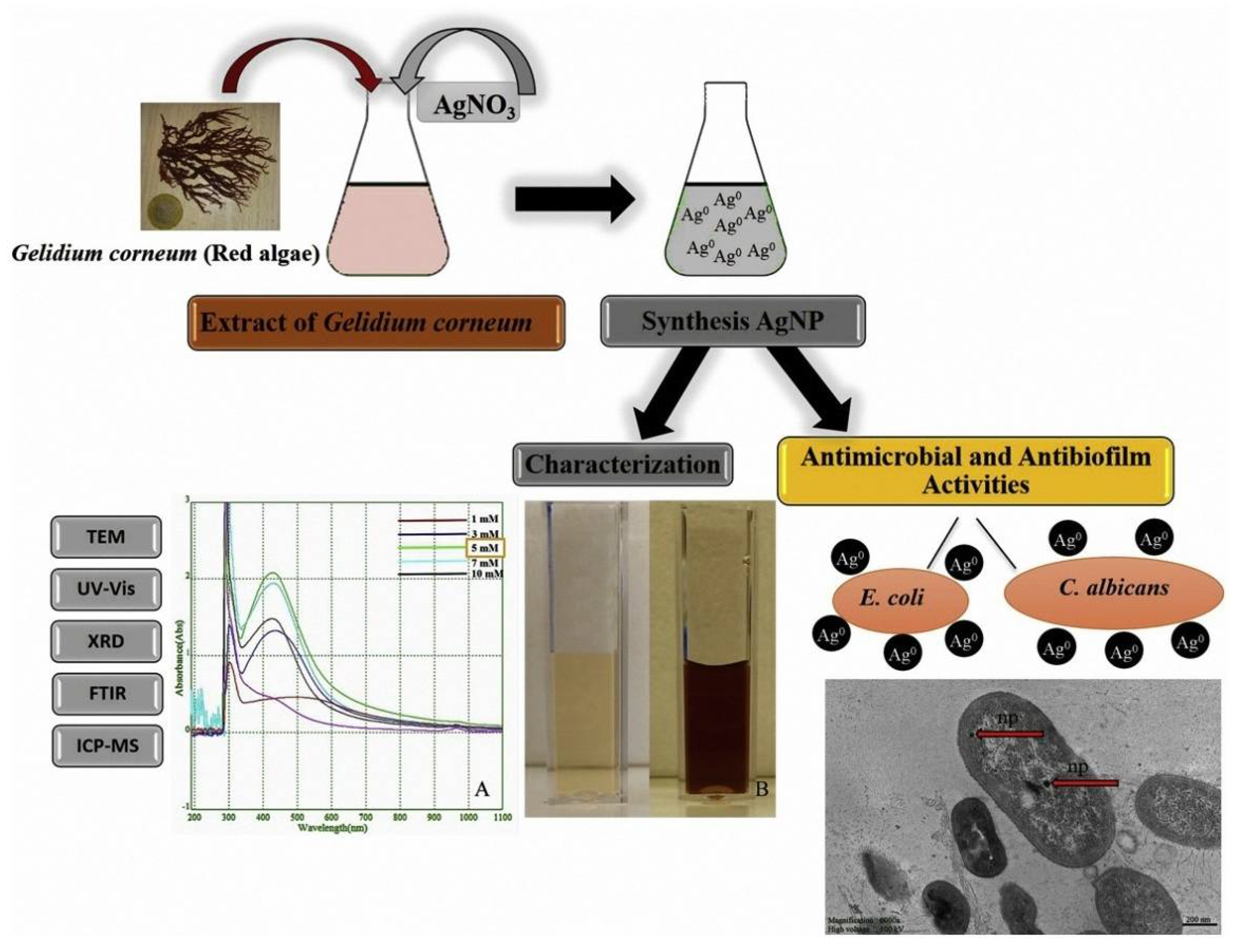
| Extracts | Gelidium corneum | AgNPs | 20–50 nm | Spherical | Antibacterial | E. coli | [83] |
| Extracts | Laminaria ochroleuca | AgNPs | 10–20 nm | Spherical | Antibacterial |
| [84] |
| Extracts | Streptomyces sp. Al-Dhabi-87 | AgNPs | 10–17 nm | Spherical | Antibacterial |
| [61] |
| Extracts |
| AgNPs | 30–40, 55–70 nm | - | Antibacterial |
| [59] |
| Extracts |
| AgNPs | 5.52, 35 nm | Spherical | Antibacterial |
| [85] |
| Extracts | Gelidiella acerosa | AgNPs | - | - | Antibacterial |
| [60] |
| Extracts | Acanthophora spicifera | AuNPs | <20 nm | Spherical | Antibacterial |
| [86] |
| Extracts | G. amansii | AgNPs | 27–54 nm | Spherical | Antibacterial |
| [87] |
| Extracts | S. wighitii | MgONPs | 68.06 nm | Flower | Antibacterial |
| [88] |
| Extracts | Oscillatoria princeps | AgNPs | 3.30–17.97 nm | Spherical | Antibacterial |
| [89] |
| Extracts | Nocardiopsis dassonvillei-DS013 | AgNPs | 30–80 nm | Circular | Antibacterial |
| [90] |
| Extracts | Streptomyces sp. Al-Dhabi-87 | AgNPs | 11–21 nm | Cubic | Antibacterial |
| [91] |
| Extracts | Penicillium polonicum | AgNPs | 10 nm | Spherical | Antibacterial | A. baumanii | [44] |
| Chitosan | Marine Seafood | AgNPs | 5–20 nm | Spherical | Antibacterial |
| [92] |
| Chitosan |
|
|
| Spherical | Antibacterial | S. aureus | [93] |
| Extracts |
| AgNPs | 10.69, 12.83 nm | Spherical | Antibacterial |
| [94] |
| Extracts | Cymodocea serrulata | AgNPs | 40.49–66.44 nm | - | Antibacterial | V. parahaemolyticus | [95] |
| Extracts | S. longifolium | CuONPs | 40–60 nm | - | Antibacterial |
| [62] |
| Extracts | C. crinita | ZnONPs | 23–200 nm | Rectangular | Antibacterial |
| [96] |
| Extracts | Synechocystis sp. | AgNPs | 10–35 nm | Spherical | Antibacterial | MRSA | [97] |
| Extracts | O. limnetica | AgNPs | 3.30–17.97 nm | Quasi-spherical | Antibacterial |
| [98] |
| Extracts | Red algae | Co3O4NPs | 29.8 ± 8.6 nm | Spherical | Antibacterial |
| [99] |
| Extracts | U. lactuca | AgNPs | 20–50 nm | - | Antiviral |
| [79] |
| Extracts |
|
|
|
| Antiviral | HSV-1 | [100] |
| Extracts |
| AgNPs | 5.52–35.00 nm | Spherical | Antiviral | Poliovirus | [85] |
| Extracts |
| AgNPs | 50–65, 15–30, and 40–50 nm | Spherical | Antiviral | Newcastle disease virus | [101] |
| Extracts | U. rigida | AgNPs | 12 nm | Spherical | Antifungal |
| [78] |
| Extracts | S. griseus | AgNPs | 14.54 nm | Spherical | Antifungal | C. albicans | [102] |
| Extracts | G. corneum | AgNPs | 20–50 nm | Spherical | Antifungal | C. albicans | [83] |
| Extracts | P. fluorescens | AgNPs | - | - | Antifungal |
| [103] |
| Extracts |
| AgNPs | - | - | Antifungal |
| [104] |
| Extracts |
| AgNPs | 10.69,12.83 nm | Spherical | Antifungal |
| [94] |
6. Marine Bioinspired NPs for Treating Viral Infection
7. Marine Bioinspired NPs for Treating Non-Infectious Diseases
8. Conclusions and Future Perspectives
Author Contributions
Funding
Institutional Review Board Statement
Informed Consent Statement
Data Availability Statement
Conflicts of Interest
References
- World Health Organization. Global Health Estimates 2015: Deaths by Cause, Age, Sex, by Country and by Region, 2000–2015; WHO: Geneva, Switzerland, 2016. [Google Scholar]
- Hoffmann, A.R.; Proctor, L.; Surette, M.; Suchodolski, J. The microbiome: The trillions of microorganisms that maintain health and cause disease in humans and companion animals. Vet. Pathol. 2016, 53, 10–21. [Google Scholar] [CrossRef]
- Deen, J.; Mengel, M.; Clemens, J. Epidemiology of Cholera. Vaccine 2020, 38 (Suppl. S1), A31–A40. [Google Scholar] [CrossRef] [PubMed]
- Pappas, P.G.; Lionakis, M.S.; Arendrup, M.C.; Ostrosky-Zeichner, L.; Kullberg, B.J. Invasive candidiasis. Nat. Rev. Dis. Primers 2018, 4, 18026. [Google Scholar] [CrossRef] [PubMed]
- World Health Organization. Responding to Community Spread of COVID-19. Reference WHO/COVID-19/Community_Transmission/2020.1; WHO: Geneva, Switzerland, 2020. [Google Scholar]
- World Health Organization. Global Antimicrobial Resistance and Use Surveillance System (GLASS) Report: 2021; WHO: Geneva, Switzerland, 2021. [Google Scholar]
- Chakraborty, S.; Rahman, T. The difficulties in cancer treatment. Ecancermedicalscience 2012, 6, ed16. [Google Scholar] [CrossRef] [PubMed]
- Hurt, R.T.; Kulisek, C.; Buchanan, L.A.; McClave, S.A. The obesity epidemic: Challenges, health initiatives, and implications for gastroenterologists. Gastroenterol. Hepatol. 2010, 6, 780. [Google Scholar]
- Drożdżal, S.; Lechowicz, K.; Szostak, B.; Rosik, J.; Kotfis, K.; Machoy-Mokrzyńska, A.; Białecka, M.; Ciechanowski, K.; Gawrońska-Szklarz, B. Kidney damage from nonsteroidal anti-inflammatory drugs—Myth or truth? Review of selected literature. Pharmacol. Res. Perspect. 2021, 9, e00817. [Google Scholar] [CrossRef]
- Hoffman, P.S. Antibacterial discovery: 21st century challenges. Antibiotics 2020, 9, 213. [Google Scholar] [CrossRef]
- Sedeek, A.M.; Ismail, M.M.; Elsayed, T.R.; Ramadan, M.A. Recent methods for discovering novel bioactive metabolites, specifically antimicrobial agents, from marine-associated microorganisms. Lett. Appl. Microbiol. 2022. [Google Scholar] [CrossRef]
- Patil, M.P.; Kim, G.-D. Marine microorganisms for synthesis of metallic nanoparticles and their biomedical applications. Colloids Surf. B Biointerfaces 2018, 172, 487–495. [Google Scholar] [CrossRef]
- Montuori, E.; de Pascale, D.; Lauritano, C. Recent Discoveries on Marine Organism Immunomodulatory Activities. Mar. Drugs 2022, 20, 422. [Google Scholar] [CrossRef]
- Yosri, N.; Khalifa, S.A.; Guo, Z.; Xu, B.; Zou, X.; El-Seedi, H.R. Marine organisms: Pioneer natural sources of polysaccharides/proteins for green synthesis of nanoparticles and their potential applications. Int. J. Biol. Macromol. 2021, 193, 1767–1798. [Google Scholar] [CrossRef] [PubMed]
- Khan, F.; Jeong, G.-J.; Khan, M.S.A.; Tabassum, N.; Kim, Y.-M. Seaweed-Derived Phlorotannins: A Review of Multiple Biological Roles and Action Mechanisms. Mar. Drugs 2022, 20, 384. [Google Scholar] [CrossRef] [PubMed]
- Fang, Y.; Li, H.; Ji, B.; Cheng, K.; Wu, B.; Li, Z.; Zheng, C.; Hua, H.; Li, D. Renieramycin-type alkaloids from marine-derived organisms: Synthetic chemistry, biological activity and structural modification. Eur. J. Med. Chem. 2021, 210, 113092. [Google Scholar] [CrossRef]
- Bayda, S.; Adeel, M.; Tuccinardi, T.; Cordani, M.; Rizzolio, F. The history of nanoscience and nanotechnology: From chemical–physical applications to nanomedicine. Molecules 2019, 25, 112. [Google Scholar] [CrossRef] [PubMed]
- Khalil, M.A.; El-Shanshoury, A.E.-R.R.; Alghamdi, M.A.; Alsalmi, F.A.; Mohamed, S.F.; Sun, J.; Ali, S.S. Biosynthesis of silver nanoparticles by Marine actinobacterium Nocardiopsis dassonvillei and exploring their therapeutic potentials. Front. Microbiol. 2021, 12, 705673. [Google Scholar] [CrossRef]
- Vala, A.; Shah, S. Rapid synthesis of silver nanoparticles by a marine-derived fungus Aspergillus niger and their antimicrobial potentials. Int. J. Nanosci. Nanotechnol. 2012, 8, 197–206. [Google Scholar]
- Hammami, I.; Alabdallah, N.M. Gold nanoparticles: Synthesis properties and applications. J. King Saud Univ. Sci. 2021, 33, 101560. [Google Scholar] [CrossRef]
- Spirescu, V.A.; Chircov, C.; Grumezescu, A.M.; Vasile, B.Ș.; Andronescu, E. Inorganic nanoparticles and composite films for antimicrobial therapies. Int. J. Mol. Sci. 2021, 22, 4595. [Google Scholar] [CrossRef]
- Balakumaran, M.; Ramachandran, R.; Balashanmugam, P.; Mukeshkumar, D.; Kalaichelvan, P. Mycosynthesis of silver and gold nanoparticles: Optimization, characterization and antimicrobial activity against human pathogens. Microbiol. Res. 2016, 182, 8–20. [Google Scholar] [CrossRef]
- Gnanajobitha, G.; Paulkumar, K.; Vanaja, M.; Rajeshkumar, S.; Malarkodi, C.; Annadurai, G.; Kannan, C. Fruit-mediated synthesis of silver nanoparticles using Vitis vinifera and evaluation of their antimicrobial efficacy. J. Nanostructure Chem. 2013, 3, 67. [Google Scholar] [CrossRef]
- Song, X.; Shi, X.; Yang, M. Dual application of Shewanella oneidensis MR-1 in green biosynthesis of Pd nanoparticles supported on TiO2 nanotubes and assisted photocatalytic degradation of methylene blue. IET Nanobiotechnol. 2018, 12, 441–445. [Google Scholar] [CrossRef] [PubMed]
- Kumar, D.A.; Palanichamy, V.; Roopan, S.M. Green synthesis of silver nanoparticles using Alternanthera dentata leaf extract at room temperature and their antimicrobial activity. Spectrochim. Acta Part A Mol. Biomol. Spectrosc. 2014, 127, 168–171. [Google Scholar] [CrossRef]
- Jin, R. The impacts of nanotechnology on catalysis by precious metal nanoparticles. Nanotechnol. Rev. 2012, 1, 31–56. [Google Scholar] [CrossRef]
- Nagarajan, R. Nanoparticles: Building Blocks for Nanotechnology; ACS Publications: Washington, DC, USA, 2008. [Google Scholar]
- Dash, D.K.; Panik, R.K.; Sahu, A.K.; Tripathi, V. Role of nanobiotechnology in drug discovery, development and molecular diagnostic. In Applications of Nanobiotechnology; IntechOpen: London, UK, 2020. [Google Scholar]
- Mahmood Ansari, S.; Saquib, Q.; De Matteis, V.; Awad Alwathnani, H.; Ali Alharbi, S.; Ali Al-Khedhairy, A. Marine Macroalgae Display Bioreductant Efficacy for Fabricating Metallic Nanoparticles: Intra/Extracellular Mechanism and Potential Biomedical Applications. Bioinorg. Chem. Appl. 2021, 2021, 5985377. [Google Scholar] [CrossRef] [PubMed]
- Ponnuchamy, K.; Jacob, J.A. Metal nanoparticles from marine seaweeds—A review. Nanotechnol. Rev. 2016, 5, 589–600. [Google Scholar] [CrossRef]
- Daraio, C.; Jin, S. Synthesis and patterning methods for nanostructures useful for biological applications. In Nanotechnology for Biology and Medicine; Springer: Berlin/Heidelberg, Germany, 2012; pp. 27–44. [Google Scholar]
- Sardar, M.; Mazumder, J.A. Biomolecules assisted synthesis of metal nanoparticles. In Environmental Nanotechnology; Springer: Berlin/Heidelberg, Germany, 2019; pp. 1–23. [Google Scholar]
- Asmathunisha, N.; Kathiresan, K. A review on biosynthesis of nanoparticles by marine organisms. Colloids Surf. B Biointerfaces 2013, 103, 283–287. [Google Scholar] [CrossRef]
- Ehrlich, H. Marine Biological Materials of Invertebrate Origin; Springer: Berlin/Heidelberg, Germany, 2019. [Google Scholar]
- Mora, C.; Tittensor, D.P.; Adl, S.; Simpson, A.G.; Worm, B. How many species are there on Earth and in the ocean? PLoS Biol. 2011, 9, e1001127. [Google Scholar] [CrossRef] [PubMed]
- Russo, P.; Kisialiou, A.; Lamonaca, P.; Moroni, R.; Prinzi, G.; Fini, M. New drugs from marine organisms in Alzheimer’s disease. Mar. Drugs 2015, 14, 5. [Google Scholar] [CrossRef]
- Blunt, J.W.; Copp, B.R.; Keyzers, R.A.; Munro, M.H.; Prinsep, M.R. Marine natural products. Nat. Prod. Rep. 2016, 33, 382–431. [Google Scholar] [CrossRef]
- Cardoso, J.; Nakayama, D.G.; Sousa, E.; Pinto, E. Marine-derived compounds and prospects for their antifungal application. Molecules 2020, 25, 5856. [Google Scholar] [CrossRef]
- Šimat, V.; Elabed, N.; Kulawik, P.; Ceylan, Z.; Jamroz, E.; Yazgan, H.; Čagalj, M.; Regenstein, J.M.; Özogul, F. Recent advances in marine-based nutraceuticals and their health benefits. Mar. Drugs 2020, 18, 627. [Google Scholar] [CrossRef] [PubMed]
- Suárez Quintana, W.H.; García-Rico, R.O.; García-Martínez, J.B.; Urbina-Suarez, N.A.; López-Barrera, G.L.; Barajas-Solano, A.F.; Zuorro, A. Enhancement of Metabolite Production in High-Altitude Microalgal Strains by Optimized C/N/P Ratio. Appl. Sci. 2022, 12, 6779. [Google Scholar] [CrossRef]
- Priyadarshini, E.; Priyadarshini, S.S.; Pradhan, N. Heavy metal resistance in algae and its application for metal nanoparticle synthesis. Appl. Microbiol. Biotechnol. 2019, 103, 3297–3316. [Google Scholar] [CrossRef] [PubMed]
- Oukarroum, A.; Bras, S.; Perreault, F.; Popovic, R. Inhibitory effects of silver nanoparticles in two green algae, Chlorella vulgaris and Dunaliella tertiolecta. Ecotoxicol. Environ. Saf. 2012, 78, 80–85. [Google Scholar] [CrossRef] [PubMed]
- Venkatesan, J.; Kim, S.-K.; Shim, M.S. Antimicrobial, antioxidant, and anticancer activities of biosynthesized silver nanoparticles using marine algae Ecklonia cava. Nanomaterials 2016, 6, 235. [Google Scholar] [CrossRef]
- Neethu, S.; Midhun, S.J.; Radhakrishnan, E.; Jyothis, M. Green synthesized silver nanoparticles by marine endophytic fungus Penicillium polonicum and its antibacterial efficacy against biofilm forming, multidrug-resistant Acinetobacter baumanii. Microb. Pathog. 2018, 116, 263–272. [Google Scholar] [CrossRef]
- Wang, L.; Liu, C.-C.; Wang, Y.-Y.; Xu, H.; Su, H.; Cheng, X. Antibacterial activities of the novel silver nanoparticles biosynthesized using Cordyceps militaris extract. Curr. Appl. Phys. 2016, 16, 969–973. [Google Scholar] [CrossRef]
- Srivastava, S.K.; Yamada, R.; Ogino, C.; Kondo, A. Biogenic synthesis and characterization of gold nanoparticles by Escherichia coli K12 and its heterogeneous catalysis in degradation of 4-nitrophenol. Nanoscale Res. Lett. 2013, 8, 70. [Google Scholar] [CrossRef]
- Ahn, E.-Y.; Hwang, S.J.; Choi, M.-J.; Cho, S.; Lee, H.-J.; Park, Y. Upcycling of jellyfish (Nemopilema nomurai) sea wastes as highly valuable reducing agents for green synthesis of gold nanoparticles and their antitumor and anti-inflammatory activity. Artif. Cells Nanomed. Biotechnol. 2018, 46 (Suppl. S2), 1127–1136. [Google Scholar] [CrossRef]
- Yan, N.; Chen, X. Sustainability: Don’t waste seafood waste. Nature 2015, 524, 155–157. [Google Scholar] [CrossRef]
- Doron, S.; Gorbach, S. Bacterial infections: Overview. Int. Encycl. Public Health 2008, 273–282. [Google Scholar] [CrossRef]
- Slavin, Y.N.; Asnis, J.; Häfeli, U.O.; Bach, H. Metal nanoparticles: Understanding the mechanisms behind antibacterial activity. J. Nanobiotechnol. 2017, 15, 65. [Google Scholar] [CrossRef] [PubMed]
- Shannon, E.; Abu-Ghannam, N. Antibacterial derivatives of marine algae: An overview of pharmacological mechanisms and applications. Mar. Drugs 2016, 14, 81. [Google Scholar] [CrossRef] [PubMed]
- Nalini, S.; Richard, D.S.; Riyaz, S.M.; Kavitha, G.; Inbakandan, D. Antibacterial macro molecules from marine organisms. Int. J. Biol. Macromol. 2018, 115, 696–710. [Google Scholar] [CrossRef]
- Gram, L.; Melchiorsen, J.; Bruhn, J.B. Antibacterial activity of marine culturable bacteria collected from a global sampling of ocean surface waters and surface swabs of marine organisms. Mar. Biotechnol. 2010, 12, 439–451. [Google Scholar] [CrossRef]
- Qi, S.-H.; Xu, Y.; Gao, J.; Qian, P.-Y.; Zhang, S. Antibacterial and antilarval compounds from marine bacterium Pseudomonas rhizosphaerae. Ann. Microbiol. 2009, 59, 229–233. [Google Scholar] [CrossRef]
- Cox, S.; Abu-Ghannam, N.; Gupta, S. An assessment of the antioxidant and antimicrobial activity of six species of edible Irish seaweeds. Int. Food Res. J. 2010, 17, 205–220. [Google Scholar]
- Sahoo, C.R.; Maharana, S.; Mandhata, C.P.; Bishoyi, A.K.; Paidesetty, S.K.; Padhy, R.N. Biogenic silver nanoparticle synthesis with cyanobacterium Chroococcus minutus isolated from Baliharachandi sea-mouth, Odisha, and in vitroantibacterial activity. Saudi J. Biol. Sci. 2020, 27, 1580–1586. [Google Scholar] [CrossRef]
- Trivedi, S.; Alshehri, M.A.; Aziz, A.T.; Panneerselvam, C.; Al-Aoh, H.A.; Maggi, F.; Sut, S.; Dall’Acqua, S. Insecticidal, antibacterial and dye adsorbent properties of Sargassum muticum decorated nano-silver particles. S. Afr. J. Bot. 2021, 139, 432–441. [Google Scholar] [CrossRef]
- Stalin Dhas, T.; Sowmiya, P.; Parthasarathy, K.; Natarajan, A.; Narendrakumar, G.; Kumar, R.; Samrot, A.V.; Riyaz, S.U.M.; Ganesh, V.K.; Karthick, V.; et al. In vitro antibacterial activity of biosynthesized silver nanoparticles against gram negative bacteria. Inorg. Nano-Met. Chem. 2022, 1–10. [Google Scholar] [CrossRef]
- Selvaraj, P.; Neethu, E.; Rathika, P.; Jayaseeli, J.P.R.; Jermy, B.R.; AbdulAzeez, S.; Borgio, J.F.; Dhas, T.S. Antibacterial potentials of methanolic extract and silver nanoparticles from marine algae. Biocatal. Agric. Biotechnol. 2020, 28, 101719. [Google Scholar] [CrossRef]
- Thiruchelvi, R.; Jayashree, P.; Mirunaalini, K. Synthesis of silver nanoparticle using marine red seaweed Gelidiella acerosa—A complete study on its biological activity and its characterisation. Mater. Today Proc. 2021, 37, 1693–1698. [Google Scholar] [CrossRef]
- Al-Dhabi, N.A.; Mohammed Ghilan, A.-K.; Arasu, M.V. Characterization of Silver Nanomaterials Derived from Marine Streptomyces sp. Al-Dhabi-87 and Its In Vitro Application against Multidrug Resistant and Extended-Spectrum Beta-Lactamase Clinical Pathogens. Nanomaterials 2018, 8, 279. [Google Scholar] [CrossRef] [PubMed]
- Rajeshkumar, S.; Nandhini, N.T.; Manjunath, K.; Sivaperumal, P.; Krishna Prasad, G.; Alotaibi, S.S.; Roopan, S.M. Environment friendly synthesis copper oxide nanoparticles and its antioxidant, antibacterial activities using Seaweed (Sargassum longifolium) extract. J. Mol. Struct. 2021, 1242, 130724. [Google Scholar] [CrossRef]
- Pacheco-Quito, E.M.; Ruiz-Caro, R.; Veiga, M.D. Carrageenan: Drug Delivery Systems and Other Biomedical Applications. Mar. Drugs 2020, 18, 583. [Google Scholar] [CrossRef]
- Campo, V.L.; Kawano, D.F.; Silva, D.B.D.; Carvalho, I. Carrageenans: Biological properties, chemical modifications and structural analysis—A review. Carbohydr. Polym. 2009, 77, 167–180. [Google Scholar] [CrossRef]
- Vijayakumar, S.; Saravanakumar, K.; Malaikozhundan, B.; Divya, M.; Vaseeharan, B.; Durán-Lara, E.F.; Wang, M.-H. Biopolymer K-carrageenan wrapped ZnO nanoparticles as drug delivery vehicles for anti MRSA therapy. Int. J. Biol. Macromol. 2020, 144, 9–18. [Google Scholar] [CrossRef]
- Khan, F.; Manivasagan, P.; Lee, J.-W.; Pham, D.T.N.; Oh, J.; Kim, Y.-M. Fucoidan-stabilized gold nanoparticle-mediated biofilm inhibition, attenuation of virulence and motility properties in Pseudomonas aeruginosa PAO1. Mar. Drugs 2019, 17, 208. [Google Scholar] [CrossRef]
- Halder, U.; Roy, R.K.; Biswas, R.; Khan, D.; Mazumder, K.; Bandopadhyay, R. Synthesis of copper oxide nanoparticles using capsular polymeric substances produced by Bacillus altitudinis and investigation of its efficacy to kill pathogenic Pseudomonas aeruginosa. Chem. Eng. J. Adv. 2022, 11, 100294. [Google Scholar] [CrossRef]
- Barakat, K.M.; Abouelwafa, A.; El-Sayed, H.S.; Beltagy, E.A. Silver Nanoparticles Mediated-Marine Fungal Amylase Improving Dicentrarchus Labrax Larvae Growth Performance; Research Square: Durham, NC, USA, 2022. [Google Scholar] [CrossRef]
- Khan, F.; Lee, J.-W.; Manivasagan, P.; Pham, D.T.N.; Oh, J.; Kim, Y.-M. Synthesis and characterization of chitosan oligosaccharide-capped gold nanoparticles as an effective antibiofilm drug against the Pseudomonas aeruginosa PAO1. Microb. Pathog. 2019, 135, 103623. [Google Scholar] [CrossRef]
- Khan, F.; Manivasagan, P.; Pham, D.T.N.; Oh, J.; Kim, S.-K.; Kim, Y.-M. Antibiofilm and antivirulence properties of chitosan-polypyrrole nanocomposites to Pseudomonas aeruginosa. Microb. Pathog. 2019, 128, 363–373. [Google Scholar] [CrossRef] [PubMed]
- Khoury, M.K.; Heid, C.A.; Cripps, M.W.; Pickett, M.L.; Nagaraj, M.B.; Johns, M.; Lee, F.; Hennessy, S.A. Antifungal Therapy in Fungal Necrotizing Soft Tissue Infections. J. Surg. Res. 2020, 256, 187–192. [Google Scholar] [CrossRef] [PubMed]
- Kainz, K.; Bauer, M.A.; Madeo, F.; Carmona-Gutierrez, D. Fungal infections in humans: The silent crisis. Microb. Cell 2020, 7, 143–145. [Google Scholar] [CrossRef] [PubMed]
- El-Hossary, E.M.; Cheng, C.; Hamed, M.M.; Hamed, A.N.E.-S.; Ohlsen, K.; Hentschel, U.; Abdelmohsen, U.R. Antifungal potential of marine natural products. Eur. J. Med. Chem. 2017, 126, 631–651. [Google Scholar] [CrossRef] [PubMed]
- Cheung, R.C.F.; Wong, J.H.; Pan, W.L.; Chan, Y.S.; Yin, C.M.; Dan, X.L.; Wang, H.X.; Fang, E.F.; Lam, S.K.; Ngai, P.H.K. Antifungal and antiviral products of marine organisms. Appl. Microbiol. Biotechnol. 2014, 98, 3475–3494. [Google Scholar] [CrossRef]
- Lopes, G.; Pinto, E.; Andrade, P.B.; Valentao, P. Antifungal activity of phlorotannins against dermatophytes and yeasts: Approaches to the mechanism of action and influence on Candida albicans virulence factor. PLoS ONE 2013, 8, e72203. [Google Scholar] [CrossRef]
- Cassilly, C.D.; Maddox, M.M.; Cherian, P.T.; Bowling, J.J.; Hamann, M.T.; Lee, R.E.; Reynolds, T.B. SB-224289 antagonizes the antifungal mechanism of the marine depsipeptide papuamide A. PLoS ONE 2016, 11, e0154932. [Google Scholar] [CrossRef]
- Xu, T.; Feng, Q.; Jacob, M.R.; Avula, B.; Mask, M.M.; Baerson, S.R.; Tripathi, S.K.; Mohammed, R.; Hamann, M.T.; Khan, I.A. The marine sponge-derived polyketide endoperoxide plakortide F acid mediates its antifungal activity by interfering with calcium homeostasis. Antimicrob. Agents Chemother. 2011, 55, 1611–1621. [Google Scholar] [CrossRef]
- Algotiml, R.; Gab-Alla, A.; Seoudi, R.; Abulreesh, H.H.; El-Readi, M.Z.; Elbanna, K. Anticancer and antimicrobial activity of biosynthesized Red Sea marine algal silver nanoparticles. Sci. Rep. 2022, 12, 2421. [Google Scholar] [CrossRef]
- Aziz, A.T. Toxicity of Ulva lactuca and green fabricated silver nanoparticles against mosquito vectors and their impact on the genomic DNA of the dengue vector Aedes aegypti. IET Nanobiotechnol. 2022, 16, 145–157. [Google Scholar] [CrossRef]
- Rajashree, R.; Gayathri, S.; Gobalakrishnan, M. Marine Biomolecule Mediated Synthesis of Selenium Nanoparticles and Their Antimicrobial Efficiency against Fish and Crustacean Pathogens; Research Square: Durham, NC, USA, 2022. [Google Scholar]
- Raja, S.; Anjali, G.; Anitha, J.; Ephsy, D.; Sahana, K.; Mohan, K. Optimization of Silver Nanocrystals Reduced from the Functional Molecules Enriched Spirulina—A Potent Antibiotic against Human and Marine Pathogen Vibrio parahaemolyticus; Research Square: Durham, NC, USA, 2022. [Google Scholar]
- Vikneshan, M.; Saravanakumar, R.; Mangaiyarkarasi, R.; Rajeshkumar, S.; Samuel, S.R.; Suganya, M.; Baskar, G. Algal biomass as a source for novel oral nano-antimicrobial agent. Saudi J. Biol. Sci. 2020, 27, 3753–3758. [Google Scholar] [CrossRef] [PubMed]
- Yılmaz Öztürk, B.; Yenice Gürsu, B.; Dağ, İ. Antibiofilm and antimicrobial activities of green synthesized silver nanoparticles using marine red algae Gelidium corneum. Process Biochem. 2020, 89, 208–219. [Google Scholar] [CrossRef]
- Kaidi, S.; Belattmania, Z.; Bentiss, F.; Jama, C.; Reani, A.; Sabour, B. Synthesis and Characterization of Silver Nanoparticles Using Alginate from the Brown Seaweed Laminaria ochroleuca: Structural Features and Antibacterial Activity. Biointerface Res. Appl. Chem. 2021, 12, 6046–6057. [Google Scholar]
- Alam, A.; Tanveer, F.; Khalil, A.T.; Zohra, T.; Khamlich, S.; Alam, M.M.; Salman, M.; Ali, M.; Ikram, A.; Shinwari, Z.K.; et al. Silver nanoparticles biosynthesized from secondary metabolite producing marine actinobacteria and evaluation of their biomedical potential. Antonie Van Leeuwenhoek 2021, 114, 1497–1516. [Google Scholar] [CrossRef]
- Babu, B.; Palanisamy, S.; Vinosha, M.; Anjali, R.; Kumar, P.; Pandi, B.; Tabarsa, M.; You, S.; Prabhu, N.M. Bioengineered gold nanoparticles from marine seaweed Acanthophora spicifera for pharmaceutical uses: Antioxidant, antibacterial, and anticancer activities. Bioprocess Biosyst. Eng. 2020, 43, 2231–2242. [Google Scholar] [CrossRef]
- Pugazhendhi, A.; Prabakar, D.; Jacob, J.M.; Karuppusamy, I.; Saratale, R.G. Synthesis and characterization of silver nanoparticles using Gelidium amansii and its antimicrobial property against various pathogenic bacteria. Microb. Pathog. 2018, 114, 41–45. [Google Scholar] [CrossRef]
- Pugazhendhi, A.; Prabhu, R.; Muruganantham, K.; Shanmuganathan, R.; Natarajan, S. Anticancer, antimicrobial and photocatalytic activities of green synthesized magnesium oxide nanoparticles (MgONPs) using aqueous extract of Sargassum wightii. J. Photochem. Photobiol. B Biol. 2019, 190, 86–97. [Google Scholar] [CrossRef]
- Bishoyi, A.K.; Sahoo, C.R.; Sahoo, A.P.; Padhy, R.N. Bio-synthesis of silver nanoparticles with the brackish water blue-green alga Oscillatoria princeps and antibacterial assessment. Appl. Nanosci. 2021, 11, 389–398. [Google Scholar] [CrossRef]
- Dhanaraj, S.; Thirunavukkarasu, S.; Allen John, H.; Pandian, S.; Salmen, S.H.; Chinnathambi, A.; Alharbi, S.A. Novel marine Nocardiopsis dassonvillei-DS013 mediated silver nanoparticles characterization and its bactericidal potential against clinical isolates. Saudi J. Biol. Sci. 2020, 27, 991–995. [Google Scholar] [CrossRef]
- Al-Dhabi, N.A.; Ghilan, A.-K.M.; Arasu, M.V.; Duraipandiyan, V. Green biosynthesis of silver nanoparticles produced from marine Streptomyces sp. Al-Dhabi-89 and their potential applications against wound infection and drug resistant clinical pathogens. J. Photochem. Photobiol. B Biol. 2018, 189, 176–184. [Google Scholar] [CrossRef]
- Alharthi, S.S.; Gomathi, T.; Joseph, J.J.; Rakshavi, J.; Florence, J.A.K.; Sudha, P.N.; Rajakumar, G.; Thiruvengadam, M. Biological activities of chitosan-salicylaldehyde schiff base assisted silver nanoparticles. J. King Saud Univ. Sci. 2022, 34, 102177. [Google Scholar] [CrossRef]
- Mostafa, E.M.; Abdelgawad, M.A.; Musa, A.; Alotaibi, N.H.; Elkomy, M.H.; Ghoneim, M.M.; Badawy, M.S.E.M.; Taha, M.N.; Hassan, H.M.; Hamed, A.A. Chitosan Silver and Gold Nanoparticle Formation Using Endophytic Fungi as Powerful Antimicrobial and Anti-Biofilm Potentialities. Antibiotics 2022, 11, 668. [Google Scholar] [CrossRef] [PubMed]
- Dixit, S.; Vishnoi, N.; Tripathi, N.M.; Singh, D.P.; Sharma, Y.K. Biosynthesis of nanostructured silver by green algae and evaluation of its microbicidal property against pathogenic microbes. Environ. Sustain. 2022, 5, 197–206. [Google Scholar] [CrossRef]
- RathnaKumari, P.; Kolanchinathan, P.; Siva, D.; Abirami, B.; Masilamani, V.; John, G.; Achiraman, S.; Balasundaram, A. Antibacterial efficacy of seagrass Cymodocea serrulata-engineered silver nanoparticles against prawn pathogen Vibrio parahaemolyticus and its combative effect on the marine shrimp Penaeus monodon. Aquaculture 2018, 493, 158–164. [Google Scholar] [CrossRef]
- Elrefaey, A.A.K.; El-Gamal, A.D.; Hamed, S.M.; El-belely, E.F. Algae-mediated biosynthesis of zinc oxide nanoparticles from Cystoseira crinite (Fucales; Sargassaceae) and it’s antimicrobial and antioxidant activities. Egypt J. Chem. 2022, 65, 231–240. [Google Scholar] [CrossRef]
- Younis, N.S.; Mohamed, M.E.; El Semary, N.A. Green Synthesis of Silver Nanoparticles by the Cyanobacteria Synechocystis sp.: Characterization, Antimicrobial and Diabetic Wound-Healing Actions. Mar. Drugs 2022, 20, 56. [Google Scholar] [CrossRef]
- Hamouda, R.A.; Hussein, M.H.; Abo-elmagd, R.A.; Bawazir, S.S. Synthesis and biological characterization of silver nanoparticles derived from the cyanobacterium Oscillatoria limnetica. Sci. Rep. 2019, 9, 13071. [Google Scholar] [CrossRef] [PubMed]
- Ajarem, J.S.; Maodaa, S.N.; Allam, A.A.; Taher, M.M.; Khalaf, M. Benign Synthesis of Cobalt Oxide Nanoparticles Containing Red Algae Extract: Antioxidant, Antimicrobial, Anticancer, and Anticoagulant Activity. J. Clust. Sci. 2022, 33, 717–728. [Google Scholar] [CrossRef]
- El-Sheekh, M.M.; Shabaan, M.T.; Hassan, L.; Morsi, H.H. Antiviral activity of algae biosynthesized silver and gold nanoparticles against Herps Simplex (HSV-1) virus in vitro using cell-line culture technique. Int. J. Environ. Health Res. 2022, 32, 616–627. [Google Scholar] [CrossRef]
- Khalid, M.; Khalid, N.; Ahmed, I.; Hanif, R.; Ismail, M.; Janjua, H.A. Comparative studies of three novel freshwater microalgae strains for synthesis of silver nanoparticles: Insights of characterization, antibacterial, cytotoxicity and antiviral activities. J. Appl. Phycol. 2017, 29, 1851–1863. [Google Scholar] [CrossRef]
- El-Enain, I.A.; Abed, N.; Helal, E.; Abdelkhalek, E.; Suleiman, W. Ecofriendly Biosynthesis of Ag-NPs by Streptomyces Griseus Eradicating Candida albicans associated with Different Types of Dysspermatism; Research Square: Durham, NC, USA, 2022. [Google Scholar]
- Sodimalla, T.; Yalavarthi, N. Biosynthesis of silver nanoparticles from Pseudomonas fluorescens and their antifungal activity against Aspergillus niger and Fusarium udum. Ann. Appl. Biol. 2022, 1–11. [Google Scholar] [CrossRef]
- Kailasam, S.; Sundaramanickam, A.; Tamilvanan, R.; Kanth, S.V. Macrophytic waste optimization by synthesis of silver nanoparticles and exploring their agro-fungicidal activity. Inorg. Nano-Met. Chem. 2022, 1–10. [Google Scholar] [CrossRef]
- Kitazato, K.; Wang, Y.; Kobayashi, N. Viral infectious disease and natural products with antiviral activity. Drug Discov. Ther. 2007, 1, 14–22. [Google Scholar] [PubMed]
- Riccio, G.; Ruocco, N.; Mutalipassi, M.; Costantini, M.; Zupo, V.; Coppola, D.; de Pascale, D.; Lauritano, C. Ten-year research update review: Antiviral activities from marine organisms. Biomolecules 2020, 10, 1007. [Google Scholar] [CrossRef] [PubMed]
- Wang, W.; Wang, S.-X.; Guan, H.-S. The antiviral activities and mechanisms of marine polysaccharides: An overview. Mar. Drugs 2012, 10, 2795–2816. [Google Scholar] [CrossRef]
- Conte, R.; Marturano, V.; Peluso, G.; Calarco, A.; Cerruti, P. Recent advances in nanoparticle-mediated delivery of anti-inflammatory phytocompounds. Int. J. Mol. Sci. 2017, 18, 709. [Google Scholar] [CrossRef]
- Assali, M.; Shawahna, R.; Shareef, M.; Alhimony, I.-A. Dexamethasone-diclofenac loaded polylactide nanoparticles: Preparation, release and anti-inflammatory activity. Eur. J. Pharm. Sci. 2018, 122, 179–184. [Google Scholar] [CrossRef]
- Kaulmann, A.; Bohn, T. Carotenoids, inflammation, and oxidative stress—implications of cellular signaling pathways and relation to chronic disease prevention. Nutr. Res. 2014, 34, 907–929. [Google Scholar] [CrossRef] [PubMed]
- Yatoo, M.; Gopalakrishnan, A.; Saxena, A.; Parray, O.R.; Tufani, N.A.; Chakraborty, S.; Tiwari, R.; Dhama, K.; Iqbal, H. Anti-inflammatory drugs and herbs with special emphasis on herbal medicines for countering inflammatory diseases and disorders-a review. Recent Pat. Inflamm. Allergy Drug Discov. 2018, 12, 39–58. [Google Scholar] [CrossRef]
- David, L.; Moldovan, B.; Vulcu, A.; Olenic, L.; Perde-Schrepler, M.; Fischer-Fodor, E.; Florea, A.; Crisan, M.; Chiorean, I.; Clichici, S. Green synthesis, characterization and anti-inflammatory activity of silver nanoparticles using European black elderberry fruits extract. Colloids Surf. B Biointerfaces 2014, 122, 767–777. [Google Scholar] [CrossRef]
- Azeem, M.N.A.; Ahmed, O.M.; Shaban, M.; Elsayed, K.N.M. In vitro antioxidant, anticancer, anti-inflammatory, anti-diabetic and anti-Alzheimer potentials of innovative macroalgae bio-capped silver nanoparticles. Environ. Sci. Pollut. Res. 2022, 1–18. [Google Scholar] [CrossRef] [PubMed]
- Grewal, S.S. Why should cancer biologists care about tRNAs? tRNA synthesis, mRNA translation and the control of growth. Biochim. Et Biophys. Acta (BBA)-Gene Regul. Mech. 2015, 1849, 898–907. [Google Scholar] [CrossRef]
- Rajeshkumar, S.; Malarkodi, C.; Vanaja, M.; Annadurai, G. Anticancer and enhanced antimicrobial activity of biosynthesizd silver nanoparticles against clinical pathogens. J. Mol. Struct. 2016, 1116, 165–173. [Google Scholar] [CrossRef]
- Maharani, V.; Sundaramanickam, A.; Balasubramanian, T. In vitro anticancer activity of silver nanoparticle synthesized by Escherichia coli VM1 isolated from marine sediments of Ennore southeast coast of India. Enzym. Microb. Technol. 2016, 95, 146–154. [Google Scholar] [CrossRef] [PubMed]
- Hassabo, A.A.; Ibrahim, E.I.; Ali, B.A.; Emam, H.E. Anticancer effects of biosynthesized Cu2O nanoparticles using marine yeast. Biocatal. Agric. Biotechnol. 2022, 39, 102261. [Google Scholar] [CrossRef]
- Saeed, A.F.; Su, J.; Ouyang, S. Marine-derived drugs: Recent advances in cancer therapy and immune signaling. Biomed. Pharmacother. 2021, 134, 111091. [Google Scholar] [CrossRef]
- Zhang, D.; Ramachandran, G.; Mothana, R.A.; Siddiqui, N.A.; Ullah, R.; Almarfadi, O.M.; Rajivgandhi, G.; Manoharan, N. Biosynthesized silver nanoparticles using Caulerpa taxifolia against A549 lung cancer cell line through cytotoxicity effect/morphological damage. Saudi J. Biol. Sci. 2020, 27, 3421–3427. [Google Scholar] [CrossRef]
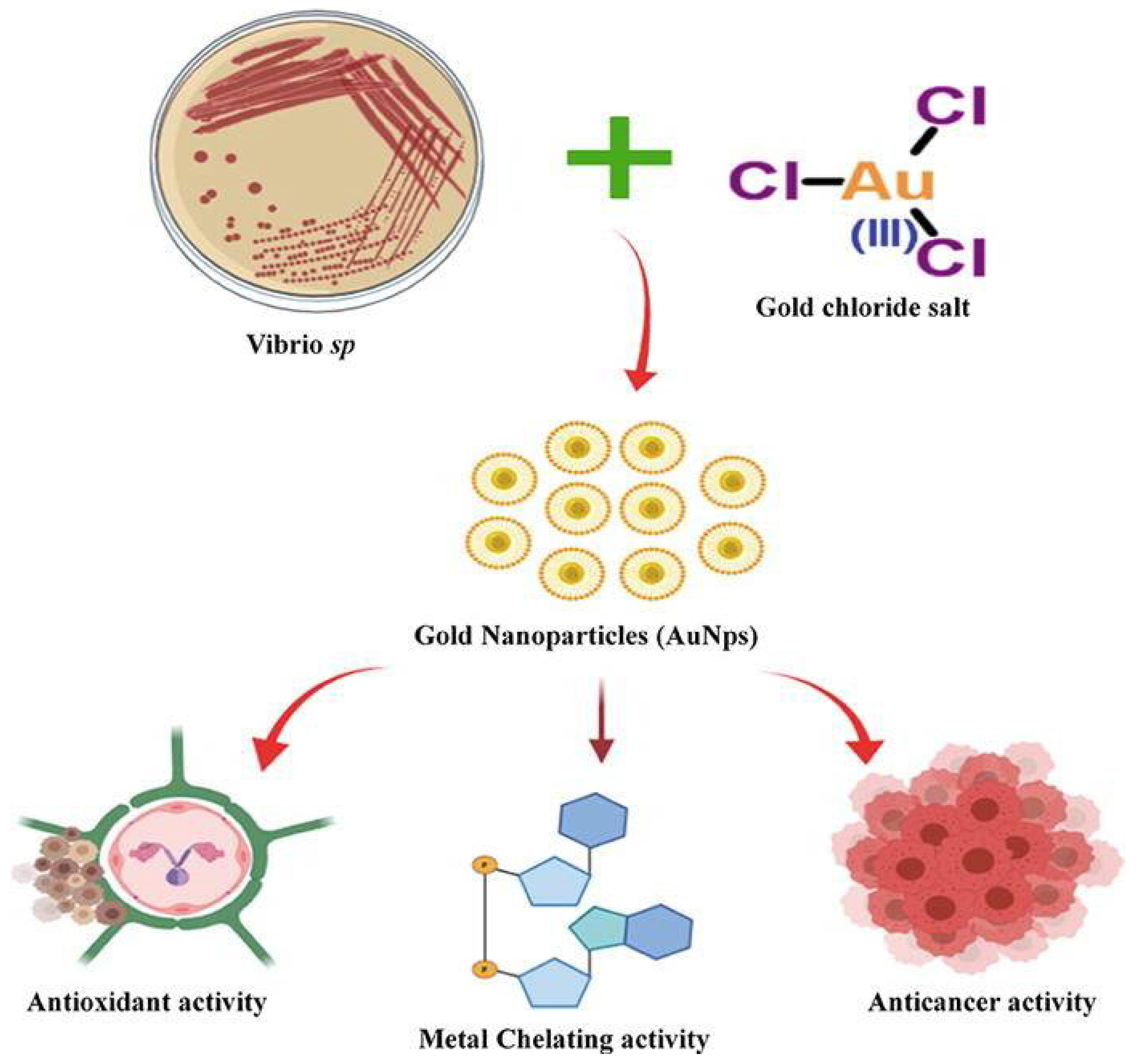
- Shunmugam, R.; Renukadevi Balusamy, S.; Kumar, V.; Menon, S.; Lakshmi, T.; Perumalsamy, H. Biosynthesis of gold nanoparticles using marine microbe (Vibrio alginolyticus) and its anticancer and antioxidant analysis. J. King Saud Univ. Sci. 2021, 33, 101260. [Google Scholar] [CrossRef]
- Aboeita, N.M.; Fahmy, S.A.; El-Sayed, M.M.H.; Azzazy, H.M.E.-S.; Shoeib, T. Enhanced Anticancer Activity of Nedaplatin Loaded onto Copper Nanoparticles Synthesized Using Red Algae. Pharmaceutics 2022, 14, 418. [Google Scholar] [CrossRef]
- Vijayakumar, S.; Chen, J.; Kalaiselvi, V.; Tungare, K.; Bhori, M.; González-Sánchez, Z.I.; Durán-Lara, E.F. Marine polysaccharide laminarin embedded ZnO nanoparticles and their based chitosan capped ZnO nanocomposites: Synthesis, characterization and in vitro and in vivo toxicity assessment. Environ. Res. 2022, 213, 113655. [Google Scholar] [CrossRef]
- Mistry, H.; Thakor, R.; Bariya, H. Biogenesis and Characterization of Proficient Silver Nanoparticles Employing Marine Procured Fungi Hamigera Pallida and Assessment of Their Antioxidative, Antimicrobial and Anticancer Potency on Human Breast Cancer (MCF-7) Cell Line; Research Square: Durham, NC, USA, 2022. [Google Scholar]
- Acharya, D.; Satapathy, S.; Yadav, K.K.; Somu, P.; Mishra, G. Systemic Evaluation of Mechanism of Cytotoxicity in Human Colon Cancer HCT-116 Cells of Silver Nanoparticles Synthesized Using Marine Algae Ulva lactuca Extract. J. Inorg. Organomet. Polym. Mater. 2022, 32, 596–605. [Google Scholar] [CrossRef]
- Ameen, F.; Al-Maary, K.S.; Almansob, A.; AlNadhari, S. Antioxidant, antibacterial and anticancer efficacy of Alternaria chlamydospora-mediated gold nanoparticles. Appl. Nanosci. 2022, 1–18. [Google Scholar] [CrossRef]
- González-Ballesteros, N.; Diego-González, L.; Lastra-Valdor, M.; Grimaldi, M.; Cavazza, A.; Bigi, F.; Rodríguez-Argüelles, M.C.; Simón-Vázquez, R. Immunomodulatory and Antitumoral Activity of Gold Nanoparticles Synthesized by Red Algae Aqueous Extracts. Mar. Drugs 2022, 20, 182. [Google Scholar] [CrossRef] [PubMed]
- Chen, X.; Han, W.; Zhao, X.; Tang, W.; Wang, F. Epirubicin-loaded marine carrageenan oligosaccharide capped gold nanoparticle system for pH-triggered anticancer drug release. Sci. Rep. 2019, 9, 6754. [Google Scholar] [CrossRef] [PubMed]
- Patil, M.P.; Kang, M.-J.; Niyonizigiye, I.; Singh, A.; Kim, J.-O.; Seo, Y.B.; Kim, G.-D. Extracellular synthesis of gold nanoparticles using the marine bacterium Paracoccus haeundaensis BC74171T and evaluation of their antioxidant activity and antiproliferative effect on normal and cancer cell lines. Colloids Surf. B Biointerfaces 2019, 183, 110455. [Google Scholar] [CrossRef]
- Vijayakumar, M.; Priya, K.; Ilavenil, S.; Janani, B.; Vedarethinam, V.; Ramesh, T.; Arasu, M.V.; Al-Dhabi, N.A.; Kim, Y.-O.; Kim, H.-J. Shrimp shells extracted chitin in silver nanoparticle synthesis: Expanding its prophecy towards anticancer activity in human hepatocellular carcinoma HepG2 cells. Int. J. Biol. Macromol. 2020, 165, 1402–1409. [Google Scholar] [CrossRef]
- Kumar, P.; Senthamilselvi, S.; Govindaraju, M. Phloroglucinol-encapsulated starch biopolymer: Preparation, antioxidant and cytotoxic effects on HepG2 liver cancer cell lines. RSC Adv. 2014, 4, 26787–26795. [Google Scholar] [CrossRef]
- Kumar, H.; Bhardwaj, K.; Nepovimova, E.; Kuča, K.; Singh Dhanjal, D.; Bhardwaj, S.; Bhatia, S.K.; Verma, R.; Kumar, D. Antioxidant functionalized nanoparticles: A combat against oxidative stress. Nanomaterials 2020, 10, 1334. [Google Scholar] [CrossRef]
- Nandi, A.; Yan, L.-J.; Jana, C.K.; Das, N. Role of catalase in oxidative stress-and age-associated degenerative diseases. Oxidative Med. Cell. Longev. 2019, 2019, 9613090. [Google Scholar] [CrossRef]
- Munteanu, I.G.; Apetrei, C. Analytical methods used in determining antioxidant activity: A review. Int. J. Mol. Sci. 2021, 22, 3380. [Google Scholar] [CrossRef]
- Valko, M.; Rhodes, C.; Moncol, J.; Izakovic, M.; Mazur, M. Free radicals, metals and antioxidants in oxidative stress-induced cancer. Chem. Biol. Interact. 2006, 160, 1–40. [Google Scholar] [CrossRef] [PubMed]
- Zhong, Q.; Wei, B.; Wang, S.; Ke, S.; Chen, J.; Zhang, H.; Wang, H. The antioxidant activity of polysaccharides derived from marine organisms: An overview. Mar. Drugs 2019, 17, 674. [Google Scholar] [CrossRef]
- Liu, C.; Fu, Y.; Li, C.-E.; Chen, T.; Li, X. Phycocyanin-functionalized selenium nanoparticles reverse palmitic acid-induced pancreatic β cell apoptosis by enhancing cellular uptake and blocking reactive oxygen species (ROS)-mediated mitochondria dysfunction. J. Agric. Food Chem. 2017, 65, 4405–4413. [Google Scholar] [CrossRef] [PubMed]
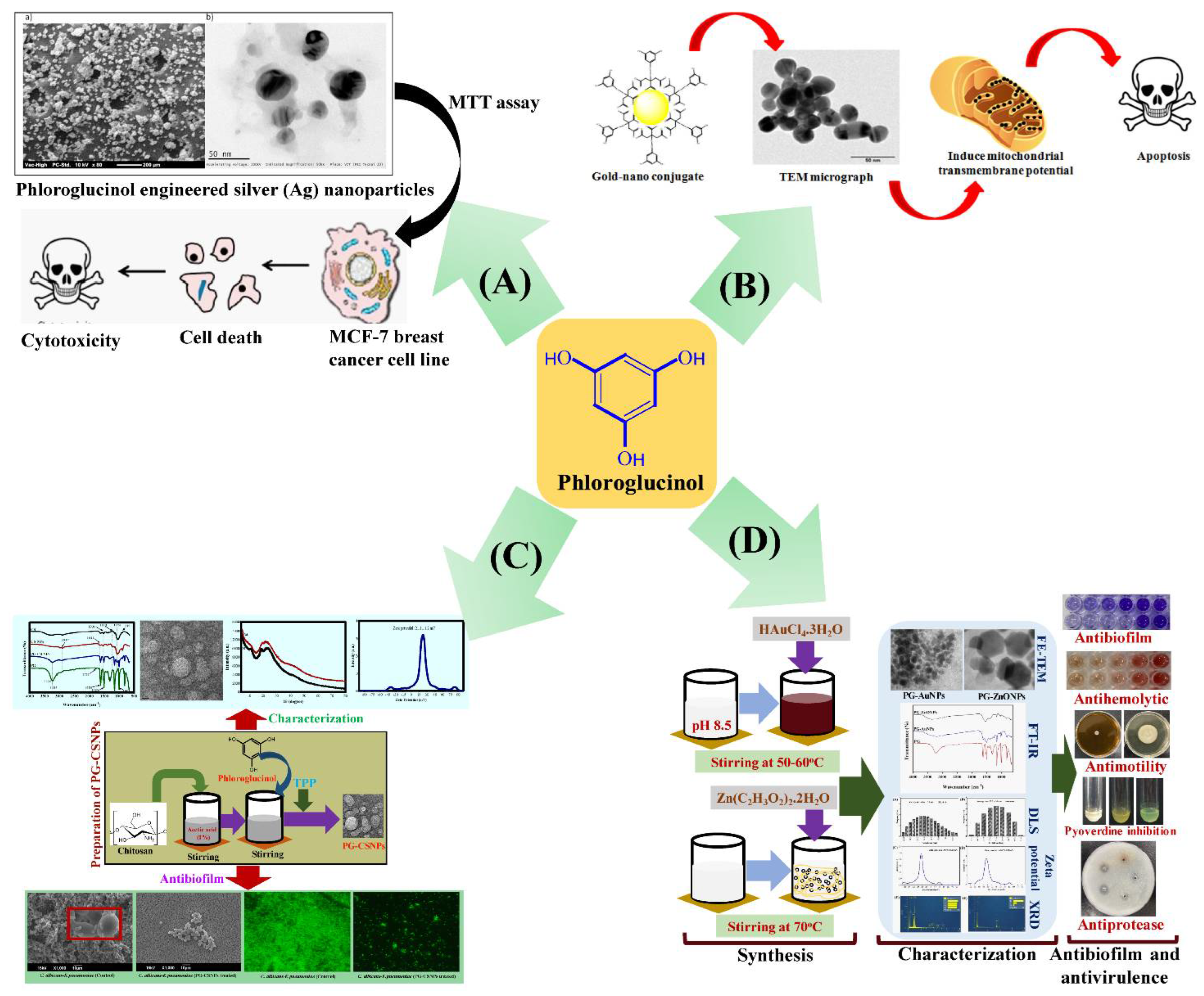
- Kumar, P.; Yuvakkumar, R.; Vijayakumar, S.; Vaseeharan, B. Cytotoxicity of phloroglucinol engineered silver (Ag) nanoparticles against MCF-7 breast cancer cell lines. Mater. Chem. Phys. 2018, 220, 402–408. [Google Scholar] [CrossRef]
- Mahalakshmi, M.; Kumar, P. Phloroglucinol-conjugated gold nanoparticles targeting mitochondrial membrane potential of human cervical (HeLa) cancer cell lines. Spectrochim. Acta Part A Mol. Biomol. Spectrosc. 2019, 219, 450–456. [Google Scholar] [CrossRef]
- Khan, F.; Oh, D.; Chandika, P.; Jo, D.M.; Bamunarachchi, N.I.; Jung, W.K.; Kim, Y.M. Inhibitory activities of phloroglucinol-chitosan nanoparticles on mono- and dual-species biofilms of Candida albicans and bacteria. Colloids Surf. B Biointerfaces 2022, 211, 112307. [Google Scholar] [CrossRef] [PubMed]
- Khan, F.; Kang, M.-G.; Jo, D.-M.; Chandika, P.; Jung, W.-K.; Kang, H.W.; Kim, Y.-M. Phloroglucinol-gold and-zinc oxide nanoparticles: Antibiofilm and antivirulence activities towards Pseudomonas aeruginosa PAO1. Mar. Drugs 2021, 19, 601. [Google Scholar] [CrossRef] [PubMed]
- Jena, J.; Pradhan, N.; Dash, B.P.; Panda, P.K.; Mishra, B.K. Pigment mediated biogenic synthesis of silver nanoparticles using diatom Amphora sp. and its antimicrobial activity. J. Saudi Chem. Soc. 2015, 19, 661–666. [Google Scholar]
- Chandika, P.; Khan, F.; Heo, S.-Y.; Kim, Y.-M.; Yi, M.; Jung, W.-K. Enhanced wound-healing capability with inherent antimicrobial activities of usnic acid incorporated poly (ε-caprolactone)/decellularized extracellular matrix nanofibrous scaffold. Biomater. Adv. 2022, 140, 213046. [Google Scholar] [CrossRef]
- Abdelwahab, G.M.; Mira, A.; Cheng, Y.-B.; Abdelaziz, T.A.; Lahloub, M.F.I.; Khalil, A.T. Acetylcholine esterase inhibitory activity of green synthesized nanosilver by naphthopyrones isolated from marine-derived Aspergillus niger. PLoS ONE 2021, 16, e0257071. [Google Scholar] [CrossRef]
- Dai, S.; Wang, B.; Song, Y.; Xie, Z.; Li, C.; Li, S.; Huang, Y.; Jiang, M. Astaxanthin and its gold nanoparticles mitigate cadmium toxicity in rice by inhibiting cadmium translocation and uptake. Sci. Total Environ. 2021, 786, 147496. [Google Scholar] [CrossRef] [PubMed]
- Manivasagan, P.; Khan, F.; Hoang, G.; Mondal, S.; Kim, H.; Doan, V.H.M.; Kim, Y.-M.; Oh, J. Thiol chitosan-wrapped gold nanoshells for near-infrared laser-induced photothermal destruction of antibiotic-resistant bacteria. Carbohydr. Polym. 2019, 225, 115228. [Google Scholar] [CrossRef] [PubMed]


| Name of Marine-Derived Compound/Product | Organisms/Sources | Name of NPs | Size Range of MNPs | Shape/Morphology | Types of Non-Infectious Disease Treatment | Effects/Activities | References |
|---|---|---|---|---|---|---|---|
| Extracts |
| AgNPs | 12 nm | Spherical | Anticancer | Human breast adenocarcinoma cell line | [78] |
| Extracts |
| AgNPs | 5.52, 35 nm | Spherical |
|
| [85] |
| Extracts | Acanthophora spicifera | AuNPs | <20 nm | Spherical | Anticancer | Human colon adenocarcinoma (HT-29) cells | [86] |
| Extracts | Sargassum wighitii | MgONPs | 68.06 nm | Flower | Anticancer | A549 | [88] |
| Extracts | Rhodotorula mucilaginosa | Cu2ONPs | 51.6–111.4 nm | Spherical | Anticancer |
| [117] |
| Extracts | Pterocladia capillacea | CuONPs | 62 nm | Spherical | Anticancer | Breast cancer, ovarian cancer, and hepatocellular carcinoma cell lines | [121] |
| Extracts | Laminaria digitata | ZnONPs | 100–350 nm | Spindle | Anticancer | Fibroblasts cells and human colon cancer cells | [122] |
| Extracts | Hamigera pallidass | AgNPs | 5.85 ± 0.84, 3.69–16.11 nm | Spherical |
|
| [123] |
| Extracts |
| AgNPs | 30–90, 20–60, 30–90 nm | Spherical |
|
| [113] |
| Extracts | U. lactuca | AgNPs | 8–14 nm | Spherical | Anticancer | Human colon cancer | [124] |
| Extracts | Alternaria chlamydospora | AuNPs | - | Spherical |
|
| [125] |
| Extracts |
| AuNPs | 16.9 ± 2.5, 15.0 ± 3.0, 44.2 ± 6.1 nm | Spherical |
|
| [126] |
| Extracts | C. crinita | ZnONPs | 23–200 nm | Rectangular | Antioxidant | DPPH | [96] |
| Extracts | Synechocystis sp. | AgNPs | 10–35 nm | Spherical | Wound-healing | Diabetic wounded animals | [97] |
| Carrageenan &Carrageenan oligosaccharide | Marine red algae | AuNPs | 141 ± 6 nm | Spherical | Anticancer | HCT-116 and HepG2 cells | [127] |
| Extracts | Paracoccus haeundaensis | AuNPs | 20.93 ± 3.46 nm | Spherical |
|
| [128] |
| Extracts | Caulerpa taxifolia | AgNPs | - | - | Anticancer | A549 lung cancer cells | [119] |
| Extracts | Nemopilema nomurai | AuNPs | 35.2 ± 8.7 nm | Spherical | Anticancer | HeLa cancer cells | [47] |
| Extracts | Oscillatoria limnetica | AgNPs | 3.30–17.97 nm | Quasi-spherical | Anticancer |
| [98] |
| Extracts | Red algae | Co3O4NPs | 29.8 ± 8.6 nm | Spherical | Anticancer | HepG2 cancer cells | [99] |
| Extracts | Vibrio alginolyticus | AuNPs | 50–100 nm | Monodispersed, irregular shape | Anticancer | HCA-7 cells | [120] |
| Classification of Sources | Natural Pure Compounds | Types of Nanomaterial | Size | Morphology | Biological Activity | Action Mechanism | References |
|---|---|---|---|---|---|---|---|
| Algae | Fucoidan | AuNPs | ~53 nm | Spherical | Antibacterial activity against Pseudomonas aeruginosa |
| [66] |
| Algae | Phloroglucinol | AuNPs and ZnONPs | 41.6 ± 3.9, 52.7 ± 3.8 nm | Spherical and hexagonal | Antibacterial activity against P. aeruginosa |
| [140] |
| Algae | Phycocyanin | SeNPs | 165, 235, 371, 815 nm | Spherical | Antioxidant | Protected INS-1E cells against palmitic acid-induced cell death by reducing oxidative stress and signaling pathways downstream | [136] |
| Algae | Fucoxanthin | AgNPs | 20–25 nm | Spherical | Antibacterial activity against Escherichia coli, Bacillus stearothermophilus, and Streptococcus mutans | - | [141] |
| Algae | Phloroglucinol | Starch biopolymer | 1–100 nm | Spherical | Anticancer | Adhesion and adsorption on the surfaces of cancer cells are enhanced | [130] |
| Algae | Phloroglucinol | CSNPs | 414.0 ± 48.5 nm | Spherical | Antibiofilm activity against Klebsiella pneumoniae, Staphylococcus aureus, Candida albicans, S. mutans, and mixed-species such as C. albicans-S. aureus/K. pneumoniae/S. mutans | The positive charge of CSNPs allows for easy biofilm penetration and binding | [139] |
| Algae | Usnic acid | Nanofibrous poly(ε-caprolactone)/decellularized extracellular matrix scaffolds | 3.89 ± 2.52, 4.95 ± 2.19, 5.00 ± 2.05 μm | Fusion of the fiber junctions |
|
| [142] |
| Algae | Carrageenan | ZnONPs | 97.03 ± 9.05 nm | Hexagonal wurtzite phase |
|
| [65] |
| Bacteria | Mannose | CuONPs | 108 nm | Spherical | Antibacterial activity against P. aeruginosa | Entered the cell membrane, causing lysis and cell rupture | [67] |
| Fungi |
| AgNPs | 8–30 nm | Spherical | Acetylcholine esterase inhibitory activity | Enzyme structural alterations | [143] |
| Fungi | α-amylase | AgNPs | 22.88–26.35 nm | Spherical | Antibacterial activity against Aeromonas hydrophila, P. aeruginosa, Vibrio anguillarum, S. faecium, S. agalactiae, and Listeria spp. | Damage to cell membranes, oxidative stress, and protein and DNA damage | [68] |
| Animal | Chitin | AgNPs | 17–49 nm | Spherical | Anticancer activity in human hepatocellular carcinoma HepG2 cells |
| [129] |
| Animal | Astaxanthin | AuNPs | 58.2 ± 4.6 nm | Polygonal and spherical | Antioxidant | Reduced ROS and increased antioxidant enzyme activity in rice plants treated to Cd to alleviate oxidative stress | [144] |
| Animal | Chitosan oligosaccharide | AuNPs | 56.01 ± 3.48 nm | Spherical | Antibacterial activity against P. aeruginosa |
| [69] |
| Animal | Thiol chitosan | AuNSs | 185 ± 19 nm | Spherical | Antibacterial activity against E. coli, P. aeruginosa, and S. aureus | - | [145] |
| Animal | Chitosan | Polypyrrole nanocomposites | 55.77 ± 3.48 nm | Spherical | Antibiofilm activity against P. aeruginosa |
| [70] |
Publisher’s Note: MDPI stays neutral with regard to jurisdictional claims in published maps and institutional affiliations. |
© 2022 by the authors. Licensee MDPI, Basel, Switzerland. This article is an open access article distributed under the terms and conditions of the Creative Commons Attribution (CC BY) license (https://creativecommons.org/licenses/by/4.0/).
Share and Cite
Jeong, G.-J.; Khan, S.; Tabassum, N.; Khan, F.; Kim, Y.-M. Marine-Bioinspired Nanoparticles as Potential Drugs for Multiple Biological Roles. Mar. Drugs 2022, 20, 527. https://doi.org/10.3390/md20080527
Jeong G-J, Khan S, Tabassum N, Khan F, Kim Y-M. Marine-Bioinspired Nanoparticles as Potential Drugs for Multiple Biological Roles. Marine Drugs. 2022; 20(8):527. https://doi.org/10.3390/md20080527
Chicago/Turabian StyleJeong, Geum-Jae, Sohail Khan, Nazia Tabassum, Fazlurrahman Khan, and Young-Mog Kim. 2022. "Marine-Bioinspired Nanoparticles as Potential Drugs for Multiple Biological Roles" Marine Drugs 20, no. 8: 527. https://doi.org/10.3390/md20080527
APA StyleJeong, G.-J., Khan, S., Tabassum, N., Khan, F., & Kim, Y.-M. (2022). Marine-Bioinspired Nanoparticles as Potential Drugs for Multiple Biological Roles. Marine Drugs, 20(8), 527. https://doi.org/10.3390/md20080527