Article

Diversity of Bacterial Secondary Metabolite Biosynthetic Gene Clusters in Three Vietnamese Sponges

1
Mientrung Institute for Scientific Research, Vietnam National Museum of Nature, Vietnam Academy of Science and Technology (VAST), 321 Huynh Thuc Khang, Hue City 531600, Vietnam
2
Laboratory of Microbiology, Wageningen University & Research, Stippeneng 4, 6708 WE Wageningen, The Netherlands
3
Institute of Marine Biochemistry, Vietnam Academy of Science and Technology (VAST), 18 Hoang Quoc Viet, Cau Giay, Hanoi 10072, Vietnam
*
Authors to whom correspondence should be addressed.
Mar. Drugs 2023, 21(1), 29; https://doi.org/10.3390/md21010029
Submission received: 22 November 2022 / Revised: 23 December 2022 / Accepted: 27 December 2022 / Published: 29 December 2022

Abstract

:
Recent reviews have reinforced sponge-associated bacteria as a valuable source of structurally diverse secondary metabolites with potent biological properties, which makes these microbial communities promising sources of new drug candidates. However, the overall diversity of secondary metabolite biosynthetic potential present in bacteria is difficult to access due to the fact that the majority of bacteria are not readily cultured in the laboratory. Thus, use of cultivation-independent approaches may allow accessing “silent” and “cryptic” secondary metabolite biosynthetic gene clusters present in bacteria that cannot yet be cultured. In the present study, we investigated the diversity of secondary metabolite biosynthetic gene clusters (BGCs) in metagenomes of bacterial communities associated with three sponge species: Clathria reinwardti, Rhabdastrella globostellata, and Spheciospongia sp. The results reveal that the three metagenomes contain a high number of predicted BGCs, ranging from 282 to 463 BGCs per metagenome. The types of BGCs were diverse and represented 12 different cluster types. Clusters predicted to encode fatty acid synthases and polyketide synthases (PKS) were the most dominant BGC types, followed by clusters encoding synthesis of terpenes and bacteriocins. Based on BGC sequence similarity analysis, 363 gene cluster families (GCFs) were identified. Interestingly, no GCFs were assigned to pathways responsible for the production of known compounds, implying that the clusters detected might be responsible for production of several novel compounds. The KS gene sequences from PKS clusters were used to predict the taxonomic origin of the clusters involved. The KS sequences were related to 12 bacterial phyla with Actinobacteria, Proteobacteria, and Firmicutes as the most predominant. At the genus level, the KSs were most related to those found in the genera Mycolicibacterium, Mycobacterium, Burkholderia, and Streptomyces. Phylogenetic analysis of KS sequences resulted in detection of two known ‘sponge-specific’ BGCs, i.e., SupA and SwfA, as well as a new ‘sponge-specific’ cluster related to fatty acid synthesis in the phylum Candidatus Poribacteria and composed only by KS sequences of the three sponge-associated bacterial communities assessed here.

1. Introduction

Bacterial secondary metabolites, also referred to as natural products or specialized metabolites, are an important source of biologically-active compounds for healthcare and agriculture applications [1,2,3,4]. These valuable secondary metabolites represent a group of low-molecular-weight structurally diverse compounds including polyketides (PKs), non-ribosomal peptides (NRPs), ribosomally synthesized and post-translationally modified peptides (RiPPs), terpenoids, saccharides, and a plethora of hybrids. The genes responsible for the biosynthesis of secondary metabolites are physically co-located in a biosynthetic gene cluster (BGC) with a size from a few to more than 100 kb [5,6].
In bacteria, non-ribosomal peptide synthase (NRPS) and polyketide synthase (PKS) gene clusters are the two main clusters responsible for the biosynthesis of secondary metabolites [7]. Polyketides are typically produced by PKS assembly lines that are composed of different functional modules. Each module is responsible for the incorporation and tailoring of a two-carbon unit into the final pathway product [8]. The minimal domain of a PKS module consists of an acyltransferase (AT), which selects and loads coenzyme-A (CoA) thioester-derived extender units; an acyl carrier protein (ACP), which functions as site for the covalent tethering of both the growing product chain and AT-selected extender units; and a ketosynthase (KS) domain, required for catalysis of carboncarbon bond formation between the downstream product chain and the upstream extender unit [9]. Non-ribosomal peptide synthetases (NRPS) also form multienzyme assembly lines that are similar to those of PKS and produce peptide-containing compounds. In the NRPS module, an adenylation (A) domain activates an amino acid, and loads it onto a peptidyl carrier protein (PCP). Subsequently, a condensation (C) domain generates an amide bond with the growing chain tethered to the upstream PCP domain [10]. Interestingly, many bacteria have been found to possess the hybrid NRPS-PKS enzymes [11,12,13,14,15] and show that both PKS and NRPS function simultaneously in the same assembly line, but the products synthesized by most of the hybrid PKS/NRPS enzymes are still unknown. With the advances in metagenome sequencing, numerous uncharacterized BGCs have been found in uncultivated bacteria, which are regarded as an untapped source for the discovery of new drugs [16,17,18,19].
Marine sponges are well known to be a particularly rich source of structurally unique secondary metabolites with a broad spectrum of biological properties [20,21,22], many of which are probably produced by their associated bacteria. For example, it is now known that polybrominated biphenyl ether antibiotics isolated from the sponge Dysidea herbacea are actually produced by the endosymbiotic cyanobacterium Oscillatoria spongeliae [23]. In addition, the antifungal peptide theopalauamide isolated from the marine sponge Theonella swinhoei has been found to be contained in a δ-proteobacterial symbiont [24]. Subsequent metagenomic analysis of sponge microbiomes revealed the bacterial origin of many polyketide and modified peptide secondary metabolites [17,25,26,27,28,29,30,31,32,33]. Although the chemical role of many symbiont-derived compounds in the sponge holobionts remains unclear, accumulated evidence shows that symbiont-derived secondary metabolites play a role in chemical defense against predators, biofouling, and competition for space [25,26,28,34,35,36]. These biological properties make these sponge-associated microbial communities promising sources of new drug candidates [37,38,39,40]. However, the overall diversity of secondary metabolite biosynthetic potential present in sponge-associated bacteria is difficult to access because the large majority of these bacteria are not readily cultured in the laboratory environment [41]. Thus, the use of cultivation-independent approaches (e.g., metagenomics, cloning, heterologous expression) for the investigation of the chemistry of sponge symbiotic bacteria may allow access to secondary metabolite biosynthetic gene clusters of the currently uncultivable symbiotic bacteria of sponges.
We have previously investigated the bacterial and archaeal diversity of the 18 marine sponge species and found that three species in particular Spheciospongia sp., Rhabdastrella globostellata and Clathria reinwardti harbored diverse communities of bacteria [42]. Furthermore, recent chemical investigations have revealed diverse natural products derived from these sponge species, i.e., C. reinwardti [43,44,45], R. globostellata [46,47,48,49,50,51,52,53], and Spheciospongia spp. [53,54,55,56,57]. However, it has remained unclear whether/which sponge-associated bacteria are responsible for the production of these natural products. Here, we investigate the diversity of secondary metabolite biosynthetic gene clusters in metagenomes of the bacterial communities associated with these three sponge species.

2. Results

2.1. Metagenomes of the Sponge-Associated Bacteria

The metagenomes of bacteria associated with the three sponge species Spheciospongia sp., Rhabdastrella globostellata, and Clathria reinwardti were sequenced. The numbers of paired-end reads obtained from the samples were in a range of 30,414,932 to 66,835,559 with average G+C content between 61% and 62 % (Table S1). Quality filtering of the reads resulted in a range of 27,783,369 to 62,722,556 high-quality reads that were used in downstream analyses. Assembling reads per sample generated 607,208, 466,098, and 504,262 contigs for the metagenomes of C. reinwardti, R. globostellata, and Spheciospongia sp., respectively. The N50 values of the contigs ranged from 21,342 to 56,925 and the number of protein-coding sequences ranged from 782,060 to 903,985 for the three samples.

2.2. Diversity of Secondary Metabolite Biosynthetic Gene Clusters from the Metagenomes of the Sponge-Associated Bacteria

For the prediction of secondary metabolite biosynthetic gene clusters (BGCs) from the metagenomes from sponge-associated bacteria, only contigs longer than 1000 bp were analyzed. These yielded a total of 1116 BGCs as identified by antiSMASH (Tables S2–S4). The number of BGCs identified per metagenome ranged from 282 to 463 (Table 1). Diverse types of BGCs were observed with 12 different cluster types. Cf fatty acid and type I PKS were the most dominant BGC types with 86–164 and 83–125 clusters per sample, respectively, followed by terpenes (58–97 clusters per sample), and bacteriocins (12–16 clusters per sample), whereas the remaining cluster types only accounted for less than 11 clusters per sample. Surprisingly, not a single NRPS cluster was detected from metagenomes of bacteria associated with three sponge species and interestingly, the use of the KnownClusterBlast function in antiSMASH suggested that none the identified BGCs was encoding for biosynthetic pathways of known compounds.
To identify orthologous clusters among BGCs across the different sponge metagenomes, we reconstructed a BGC sequence similarity network (Figure 1). The analysis of BGC sequence similarity showed that the connected components corresponded to 363 gene cluster families (GCFs), including 86 GCFs of type I PKSs, 5 GCFs of other PKSs, 63 GCFs of terpenes, 8 GCFs of ribosomally synthesized and post-translationally modified peptides (RiPPs), and 201 GCFs of other BGCs, including cf fatty acid (Figure 1). Several larger networks were composed of multiple GCFs because these networks could not be resolved into single GCF networks using default similarity cutoffs.
In general, approximately one-third (103) of GCFs were found in all specimens, 89 GCFs in 2 specimens, and 171 GCFs only in individual specimens (singletons) (Figure 2A). It appears that Type I PKSs are more often unique to a sponge species/sample, while terpenes, other PKS, and RiPPs are more often shared between species/samples (Figure 2B–E). Comparison of the BGCs identified with BGCs that encode known compounds in the MIBiG (Minimum Information about a Biosynthetic Gene cluster) reference database [58] showed that no GCFs from the metagenomes were assigned to pathways responsible for the production of known compounds (Tables S2–S4).

2.3. Ketosynthase Phylogeny of BGCs

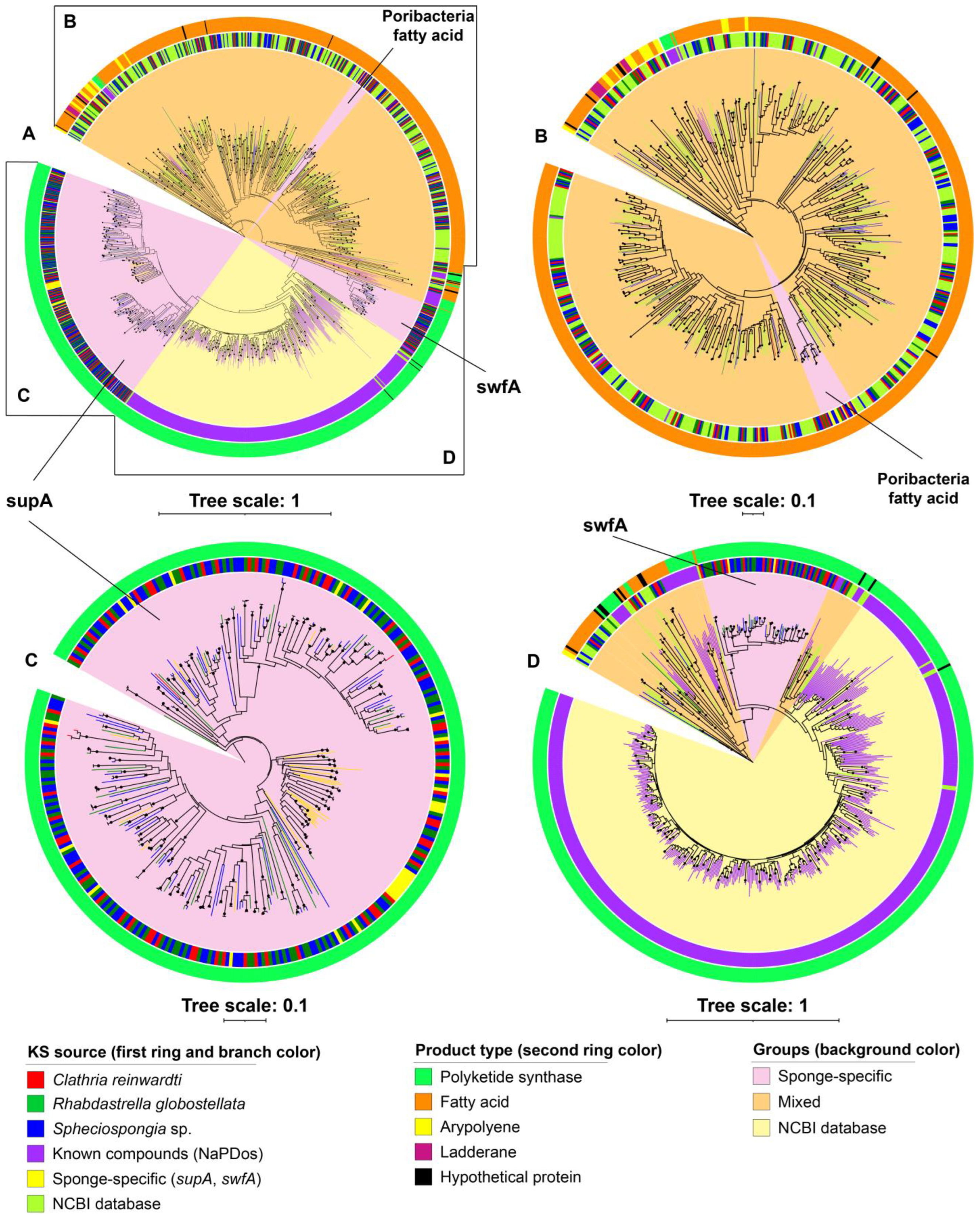
The ketosynthase (KS) domain is the most conserved catalytic domain of the PKS gene cluster and involved in the tailoring the polyketide by catalyzing chain condensation. Therefore, the KS sequences extracted from the identified BGCs were used for phylogenetic analysis (Figure 3). The KS sequences had low identity (21–66%) to the KS sequences of the characterized pathways from the NaPDos database (Tables S5–S7) as could be expected as BGCs from our study did not encode known pathways. In addition, many KS sequences of the identified BGCs from the metagenomes showed low identities (30–70%) to KS sequences from NCBI GenBank (Tables S8–S13).
Phylogenetic analysis of KS sequences showed that most of the KS sequences of the cf_fatty acid BGCs identified from the sponge bacteria metagenomes clustered together with previously found KS sequences from fatty acid biosynthesis gene clusters retrieved from NCBI GenBank (Figure 3B). Several KS sequences from fatty acid BGCs predicted from the sponge-associated bacteria metagenomes had KS sequences from fatty acid BGCs from Candidatus Poribacteria metagenome-assembled genomes as best hit and formed a phylogenetically distant clade from other KS sequences from fatty acid BGCs derived from other sources (Figure 3B). KS sequences from the aryl polyene and ladderane BGCs identified from the metagenomes formed subclusters within clades of KS sequences belonging to fatty acid synthesis pathways retrieved from NCBI Genbank. For PKS BGCs obtained from the sponge-associated bacteria metagenomes in the present study, only a few KS sequences were placed in the same clades as reference non-sponge KS sequences retrieved from NCBI GenBank and NaPDos, and the large majority of KS sequences had best hits with sponge-derived KS sequences and formed two separate clades that were phylogenetically distant to non-sponge-derived KS sequences (supA and swfA) (Figure 3C,D).
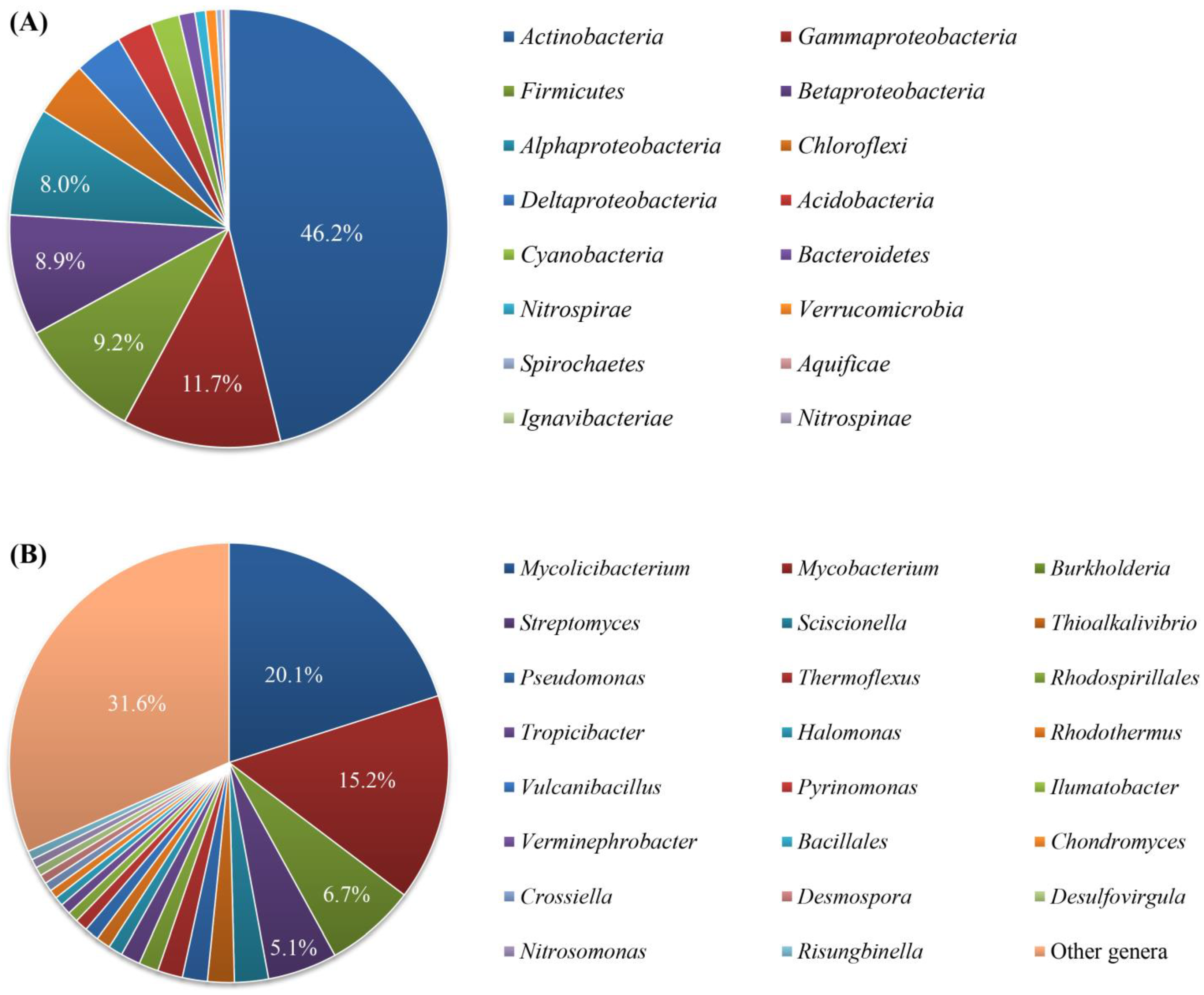
In addition, we used the BLASTX searches of the KS sequences to predict the bacterial taxa harboring these sequences and they were predicted to be related to KS sequences from diverse bacteria represented by 12 phyla, including Actinomycetes, Proteobacteria, Firmicutes, Chloroflexi, Acidobacteria, Cyanobacteria, Bacteroidetes, Nitrospirae, Verrucomicrobia, Spirochaetes, Aquificae, Nitrospinae, and Ignavibacteriae (Figure 4A, Tables S11–S13). Among them, Actinobacteria was the most predominant phylum (46.2% of the KS sequences), followed by the classes of Proteobacteria (3.5–11.7%) and the phylum Firmicutes (9.1%). Each of remaining phyla accounted for less than 5%. At the genus level, the KS sequences of the identified BGCs from the metagenomes were linked to 142 genera (Figure 4B). Of these, the genera Mycolicibacterium, Mycobacterium, Burkholderia, and Streptomyces were most dominant and accounted for 20.1, 15.2, 6.7, and 5.1% of the total KS sequences, respectively.
Except for the KS sequences retrieved from fatty acid BGCs, which were found from a wide range of bacterial phyla (Figure S1), KS sequences from other BGC types were non-uniformly distributed across sponge-associated bacterial taxa (Figures S2–S4). The KS sequences retrieved from PKS BGCs were predominantly related to those from Actinobacteria (80% of the PKS KS sequences), particularly the genera Mycolicibacterium (37%), Mycobacterium (28%), Burkholderia (12%), Streptomyces (7%), and Sciscionella (5%) (Figure S2). On the other hand, the KS sequences from aryl propylene BGCs were predominantly related to those from Proteobacteria (> 80% of the sequences), including Betaproteobacteria (45%), Gammaproteobacteria (26%), and Deltaproteobacteria (11%) with Verminephrobacter, Cellvibrio, Syntrophus, Cupriavidus, Lechevalieria, and Thiobacillus as main genera (Figure S3). In case of ladderane BGCs, the KS sequences were mainly related to only two classes of Proteobacteria (Alpha- and Delta-) and three genera Tropicibacter, Sandaracinus, and Pseudovibrio (Figure S4).

3. Discussion

3.1. Diversity of Secondary Metabolite Biosynthetic Gene Clusters from the Metagenomes of Sponge-Associated Bacteria

Gene clusters related to putative fatty acid synthesis is the most predominant BGC type identified from the metagenomes of sponge-associated bacteria in our study. This is expected because fatty acids are essential components in cell membranes of bacteria. Of particular interest is the detection of a separate cluster of KS genes related to fatty acid synthase from Candidatus Poribacteria, a sponge-specific bacterial candidate phylum. Members of the phylum Candidatus Poribacteria were not detected in the same sponge specimens by 16S rRNA amplicon sequencing [42]. However, this discrepancy between 16S rRNA sequencing and metagenomics has frequently been observed and the lack of Candidatus Poribacteria in the PCR amplicon-based study may be attributed to a bias of the specificity of primers [59]. Except for central roles in the integrity of cell membranes and energy storage [60], many fatty acids have been recently suggested to be potential antimicrobial, antifouling, and antivirulence agents [61,62,63]. Several reports have also shown fatty acids to enhance the antibacterial activities of antibiotics [64,65,66,67,68].
PKSs are among the most dominant BGC types in the metagenomes of sponge-associated bacteria. Of these, PKS type I is most dominant, while only a few PKS type II and type III clusters were detected in the metagenomes. Almost all type I PKSs detected in the metagenomes of sponge-associated bacteria in our study were the non-iterative type I PKSs. Previous investigations have revealed that the distribution of PKSs appears to be origin-dependent. The non-iterative type I PKSs are often found in bacteria, whereas iterative type I PKSs are mainly found in fungi [69,70,71]. Bacterial type I PKSs are particularly attractive targets for exploration and exploitation of their bioactive compounds because they are defined by a modular organization, remarkable versatility, and amenability for pathway engineering [72]. Several antibiotics derived from these multicatalytic enzymes include macrolides (erythromycin), polyenes (nystatin), as well as linear polyketides (tautomycetin) [73,74,75]. In our study type I PKSs from the sponge-associated bacteria metagenomes had a best BLAST hit to clusters encoding for the production of diverse bioactive compounds, such as puwainaphycins, ajudazol, polycyclic tetramate macrolactams, glycopeptidolipid, epothilone, phenalamide, crocacin, curacin, microsclerodermins, nostophycin, jamaicamide, chondrochloren, gephyronic acid, cylindrospermopsin, soraphen, amphotericin, reveromycin, stigmatellin, candicidin, myxalamid, gulmirecin, heat-stable antifungal factor, myxothiazol, tautomycin, and ECO-02301 (a new class of antifungal agent) (Tables S14–S16). Notably, several trans-AT PKSs were also detected in the sponge-associated bacteria metagenomes. Trans-AT PKSs lack integrated AT domains. Instead, a free-standing AT domain acts in trans to load acyl building blocks into the assembly line [76]. Nearly 40% of all bacterial modular PKSs belong to the trans-AT type, suggesting that the polyketides of these enzymes constitute a major natural product class [77]. Indeed, several drugs which are used as antibiotics were identified as trans-AT PKS products (e.g., mupirocin, streptogramins) [78,79]. Many compounds from sponges and their symbionts are known to be biosynthesized by trans-AT PKS pathways (e.g., diffusomycin, psymberin, onnamide A, calyculin A, swinhoeiamide A, swinhodile A, clavosine A, geoetricin A, misakinolide A, oocydin A, and mycalamide A) [80,81,82]. The trans-AT PKS gene clusters detected in the sponge-associated bacteria metagenomes in our study had a best BLAST hit to gene clusters involved in the production of puwainaphycins and epothilones (Tables S14–S16). However, none of the clusters in our dataset could be reliably assigned to a cluster encoding for the production of a known compounds (MiBIG database). These findings imply that the type I PKSs detected in our study are likely involved in the biosynthesis of currently unknown compounds.
Type II PKSs are found exclusively in bacteria and are responsible for producing aromatic polyketides such as the antibiotics actinorhodin, tetracycline or doxorubicin [83]. Although none of the type II PKSs detected in the metagenomes of sponge-associated bacteria in our study could be confidently assigned to gene clusters encoding known compounds, some BGCs had best BLAST hits related to clusters encoding compounds belonging to the class angucyclines (lomaiviticin) and benzoisochromanequinones (frenolicin) (Tables S14–S16). Type III PKSs are relatively small proteins that are mainly involved in the production of important plant compounds such as flavonoids and stilbenes (phenolic), but they are also found in bacteria and fungi [84]. All type III PKSs detected in the metagenomes of sponge-associated bacteria in our study had the cluster encoding for the proteins producing the alkylresorcinols as best BLAST hit (Tables S14–S16). Alkylresorcinols are synthesized by plants, fungi, and bacteria and have diverse biological activities including antioxidant, antibacterial, cytotoxic, and signaling properties [85,86,87].
Terpene BGCs were the third most predominant among BGCs predicted from the three metagenomes. Originally, terpenes were considered to be plant and fungal products. However, extensive bacterial genome analysis has shown that terpene synthase genes are also widespread in bacteria [88,89,90]. Recent genome analyses also detected BGCs of terpenes in genomes of sponge-associated bacteria [91,92,93], and several terpenes have been isolated from sponges and their associated symbionts [94,95]. Terpenes are among targets for drug development because they exhibit various biological properties, such as antimicrobial, anticancer, and antirespiratory activities [94]. Notably, the terpene gene clusters detected in the sponge-associated bacteria metagenomes in our study were, again, not closely related to gene clusters associated to known compounds. Best BLAST hits were clusters encoding the synthesis of diverse known terpenes, such as hopene, thalianol, lupeol, sioxanthin, tirucalla, brasilicardin A, clavaric acid, squalestatin, isorenieratene, arabidiol, baruol, and astaxanthin dideoxyglycoside (Tables S14–S16). Therefore, the terpene gene clusters are likely involved in the biosynthesis of similar, but still different terpenes.
Several RiPP BGC types were detected in the metagenome of sponge-associated bacteria in our study, including 1216 bacteriocin BGCs and 13 lantipeptide BGCs per metagenome. Bacteriocins are ribosomally synthetized peptides or proteins that act as antimicrobial compounds against other Gram-positive and Gram-negative bacteria [96,97]. Genome analyses have detected BGCs of bacteriocins in sponge-associated bacteria [92,93,98]. Interestingly, Phelan, et al. [99] isolated a new bacteriocin (subtilomycin) from Bacillus subtilis MMA7 associated with the sponge Haliclona simulans and found that the subtilomycin biosynthetic cluster is widespread among B. subtilis strains isolated from different shallow and deep water marine sponges. However, the generally large discrepancy between bacteria isolated from sponges and the dominant bacteria present in sponges [41] is also translated to biosynthetic gene clusters present in isolates from sponges and those present in metagenomes from sponges. Indeed, no BGC related to subtilomycin biosynthesis was observed and, in fact, none of the bacteriocin gene clusters detected in the sponge-associated bacteria metagenomes was closely related to clusters encoding the proteins that catalyze biosynthesis of known bacteriocins. Best BLAST hits were obtained for clusters related to the production of bacteriocins, such as patellins, goadsporin, aeruginosamide, microcyclamide, trichamide, pheganomycin, patellamides, tenuecyclamides, and SRO15-3108. Lantipeptides are widely distributed among bacteria [100,101,102] and were previously found in genomes of sponge-associated bacteria [93,103,104,105,106]. A number of known lantipeptides have shown activity against microbial pathogens. Of these, several lantipeptides have been in pre-clinical trials as antibiotics for treatment of Clostridium difficile infections [107,108,109] and other Gram-positive bacterial infections, such as microbisporicin NAI-107, mutacin 1140, duramycin, NVB302, and lactacins 3147 α and β [110,111,112]. The best BLAST hits of lantipeptide BGCs were related to actagardine, SRO15-3108, and labyrinthopeptins biosynthesis but with only low identity scores.
Interestingly, no NRPS-like BGCs were detected in three sponge bacterial metagenomes in the present study. This may be attributed to several reasons. Firstly, an investigation of PKS and NRPS from microbes associated with Discodermia dissoluta from three metagenomic libraries (i.e., whole sponge tissue, and fractions enriched for unicellular or filamentous bacteria) revealed that NRPS sequences were only detected in the fraction enriched by filamentous bacteria, where no NRPS sequences were found in whole sponge tissue and the unicellular-enriched fraction [113]. During the enrichment for bacteria in our study prior to DNA extraction and metagenomic sequencing, most filamentous bacteria (if present) were probably lost and hence their BGCs not included in our dataset. In addition, some NRPS BGCs may be difficult to recover by next-generation sequencing approaches (e.g., from low-GC or high-AT species) due to biases of sequencing or assembly methods [114,115,116,117,118].
Previous chemical investigations have reported diverse natural products isolated from the three sponges C. reinwardti [43,44,45], R. globostellata [46,47,48,49,50,51,52], and Spheciospongia spp. [53,54,55,56,57]. The natural products from the reported sponge species and their associated cultivated bacteria included mainly terpenes and lipids and a few peptides, aromatic, phenolic, and indole compounds, which are products resulting from the main BGC classes (terpenes, PKSs, fatty acids, RiPPs) detected in sponge metagenomes in our study. The decadal challenge is now to link the striking novelty in sponge and other environmental BGCs to the natural products they produce.

3.2. Predicted Taxa of the Secondary Metabolite Biosynthetic Gene Clusters

Previous extensive investigations of BGCs in bacterial genomes revealed non-uniform distribution of BGCs in bacterial taxa. The phyla known to possess a high number of BGCs in their genomes include Actinobacteria, Proteobacteria, Bacteroidetes, Firmicutes, and Cyanobacteria [119]. Since the large majority of the BGCs in our sponge-derived metagenomes contained KS sequences (cf_fatty acids, PKSs, aryl polyenes and ladderanes), the KS sequences can be used to identify the major taxa that harbor BCGs in sponge-associated bacteria. Unsurprisingly, genera of the phylum Actinobacteria, such as Mycolicibacterium, Mycobacterium, and Streptomyces, were most dominant as predicted hosts of the KS sequences. Apart from Streptomyces and Bacillus, which are rich sources of natural products [120], other bacterial taxa are receiving increased attention as novel target genera with a high biosynthetic potential, including Pseudomonas, Clostridium, Burkholderia, Pseudonocardia, Photorhabdus, Xenorhabdus, Chitinophaga, Herpetosiphon, and Planctomyces [121,122]. Particularly in sponges, the genus “Candidatus Entotheonella” has been recognized for its unusually rich and varied arsenal of BGCs [33]. In our study, Streptomyces, Pseudomonas, and Burkholderia were among the prominent genera to which KS sequences were assigned, but “Candidatus Enthotheonella” was not identified, which corroborates with its absence in these three sponge species based on 16S rRNA gene analysis [42]. In addition, other genera, such as Pseudovibrio and Bacillus, that are frequently isolated from sponges and linked to a high prevalence of antimicrobial activity [39] were not identified here based on cultivation-independent means.
Generally, the predicted taxa of bacteria harboring the BGCs in the present study corresponded to 16S rRNA gene analysis in a previous study at the phylum level, but their relative abundances are different [43]. This is likely due to different frequencies of BGCs among bacteria taxa [33,119].

3.3. Putative ‘Sponge-Specific’ Clusters

It is well known that marine sponges harbor specific microbial communities [123]. Interestingly, the KS sequence analysis also revealed the presence of several ‘sponge-specific’ PKS groups. In a systematic analysis of 150 KS sequences from metagenomic DNA from 20 different demosponges through PCR screenings of KS genes, Fieseler et al. [124] detected a cluster of sponge-specific PKS sequences, termed “symbiont ubiquitous type I PKS” (Sup). In another study, Sala et al. [125] detected another sponge-specific KS group, named “sponge (symbiont) widespread fatty acid synthase” (Swf), in several high-microbial-abundance sponges. Notably, both SupA and SwfA are involved in the synthesis of methyl branched fatty acids [125,126]. Phylogenetic reconstruction of KS sequences derived from the metagenomes of sponge-associated bacteria in our study showed three monophyletic sponge-specific clusters that were phylogenetically distant from KS sequences from other sources. Two sponge-specific KS groups were affiliated to the known sponge-specific KS groups, i.e., SupA and SwfA.
The third sponge-specific KS group is involved in fatty acids synthesis in Candidatus Poribacteria. This sponge-specific KS group includes KS sequences from our study and KS sequences retrieved from Candidatus Poribacteria metagenome-assembled genomes [59]. Borchert et al. [127] investigated the diversity of NRPS and PKS genes in microbiome of three deep-sea sponges Inflatella pellicula, Poecillastra compressa, and Stelletta normani through PCR screening of the KS domain and also found several sponge-specific KS groups. Additionally, other recent studies have identified other ‘sponge-specific’ gene clusters related to the biosynthesis of RIPP proteusins, bromotyrosine alkaloids, and ether lipids [19,32,128]. These findings imply that the sponge-specific specialized metabolites are prevalent and may play an important role in sponge holobiont defense or communication.

4. Materials and Methods

4.1. Collection and Identification of Sponges

Sponge specimens of Spheciospongia sp., Rhabdastrella globostellata, and Clathria reinwardti were collected by scuba diving from May to September 2015 from the central coastal region of Vietnam at 5–10 m depth and identified using molecular markers (18S rRNA and COI genes) in a previous study [42].

4.2. Enrichment of Sponge-Associated Bacteria

The specimens were rinsed three times with sterile artificial seawater to remove any debris attached to the sponge. Then the specimens were further cleaned with a sterile scalpel in order to remove sediment and other organisms more strongly attached to the sponges. Sponge-associated bacterial cells were enriched by stepwise centrifugation as described by [129]. In brief, the sponge tissue was cut into small pieces and ground in TEN buffer (3.5% sodium chloride, 10 mM tris-hydroxymethyl-aminomethane, 50 mM ethylenediaminetetraacetic acid, pH 8.5) with a sterilized mortar and pestle. The cell suspensions were then filtered through a large nylon mesh (20 µm) to further remove sediment particles and clumps of cells. Then the sponge cells and bacteria were separated by step-wise centrifugation. The cell suspension was first centrifuged at 500× g for 5 min at 4 °C. The supernatant was then transferred to another tube and centrifuged at 1000× g for 15 min at 4 °C. Next, the supernatant was transferred to another tube and centrifuged at 3000× g for 15 min at 4 °C. Finally, the supernatant was then transferred to another tube and centrifuged at 8000× g for 15 min at 4 °C. The pellet was washed twice with TEN buffer and centrifuged at 8000× g for 20 min at 4 °C. This cell pellet was used for metagenomic DNA extraction.

4.3. Metagenomic DNA Extraction, Sequencing and Assembly

Metagenomic DNA of sponge-associated bacteria was extracted using the ZymoBeadTM Genomic DNA Kit (Zymo Research, Irvine, CA, USA) according to the manufacturer’s protocol. The concentration of the extracted DNA was determined with a Nanodrop 1000 spectrophotometer (Nanodrop Technologies, Wilmington, DE, USA), and its integrity was examined by gel electrophoresis on a 1% (w/v) agarose gel. Metagenomic DNA was sequenced on an Illumina HiSeq2500 platform at BaseClear BV (Leiden, The Netherlands). Quality control of FASTQ files was done with FastQC v.0.11.7 (https://www.bioinformatics.babraham.ac.uk/projects/fastqc/, accessed on 25 January 2018). The raw reads were checked and the adapters, low-quality reads, artifacts, and PhiX contamination were removed using the command bbduk.sh of the BBmap v.34 (https://sourceforge.net/projects/bbmap/, accessed on 25 January 2018). Reads with a low Phred quality score (< 30) and reads shorter than 70 base pairs were filtered out. Next, reads were coverage-normalized with the command bbnorm.sh of BBMap v.34 at default settings. Reads were then assembled with SPAdes v.3.5.0 [130]. The non-normalized Illumina reads were mapped to the contigs with Bowtie2 v.2.2.2 at default settings [131]. The resulting SAM files were converted to BAM, sorted, and indexed with SAMtools v.0.1.18 [132]. HTSeq v.0.6.1 was used to calculate the average coverage of each contig [133]. Contigs longer than 1000 base pairs were used for further analysis. Open reading frames (ORFs) were called with Prodigal v.2.6.1 with -m and -p meta options enabled [134].

4.4. Prediction of Secondary Metabolite Gene Biosynthetic Clusters and Ketosynthase Phylogeny

The predicted protein-coding genes were subjected to a search for secondary metabolite biosynthetic gene clusters (BGCs) using antiSMASH v.4.1.0 (bacterial version). The KnownClusterBlast function in antiSMASH was enabled to identify BGCs that are responsible for the biosynthesis of known compounds in the metagenomes of the sponge-associated bacteria. The predicted biosynthetic gene clusters were also searched for known biosynthetic pathways using the natural product domain seeker (NaPdoS) (http://napdos.ucsd.edu/, accessed on 15 August 2018). Biosynthetic gene cluster similarity networks and gene cluster families were generated using BiG-SCAPE with default settings. The networks were visualized using Cytoscape v. 3.6.0 (https://cytoscape.org, accessed on 28 August 2018) [135].
For ketosynthase (KS) phylogeny, KS domain fragments of the biosynthetic gene clusters were extracted and trimmed using NaPdoS (http://napdos.ucsd.edu/, accessed on 15 August 2018) [136]. The KS sequences were used to search for homologous KS sequences in the NCBI Genbank database using blastp (http://www.ncbi.nlm.nih.gov/, accessed on 5 November 2020) against the nr database. KS sequences with a short length < 200 amino acids were excluded from the phylogenetic analysis. The KS sequences (from this study, their three most similar sequences, and sequences of known biosynthetic pathways from NaPdoS server) were aligned using the MAFFT (v.7.504) program with the FFT-NS-i strategy [137]. Poorly aligned positions and non-conserved regions were removed from the alignments using the trimAl v.1.2 [138]. The ketosynthase phylogeny was created using MEGA version 11.0.11 [139] with the neighbor-joining (NJ) method, the Poisson model and 1,000 bootstrap replicates. The phylogeny was visualized using iTOL v.5 [140] via the online server (https://itol.embl.de/, accessed on 23 April 2021). Taxonomy of the bacteria harboring the KS sequences were predicted based on the most homologous KS sequences (the first hit) by a BLAST search of the KS sequences against the refseq_protein database (v.2018) at NCBI using blastp (http://www.ncbi.nlm.nih.gov/, accessed on 5 May 2021).

5. Conclusions

The metagenomes of associated bacteria of three sponges contained a high number of predicted BGCs, ranging from 282 to 463 BGCs per metagenome, with 12 different BGC types. After cf_fatty_acid, PKSs were the most dominant BGCs, followed by terpene BGCs and bacteriocin BGCs. The BGCs were grouped into 363 gene cluster families (GCFs) based on sequence similarity. Interestingly, not a single GCF from the metagenomes was assigned to pathways responsible for the production of known compounds, implying that they might be responsible for production of novel compounds. Based on taxa of the closest reference KS sequences retrieved from the NCBI database, the KS sequences of the identified BGCs from the metagenomes related to 12 bacterial phyla, of which Actinobacteria, Proteobacteria, and Firmicutes were the most predominant. At the genus level, the KS were related to 142 genera, of which Mycolicibacterium, Mycobacterium, Burkholderia, and Streptomyces were the most predominant. Notably, a large number of KS sequences retrieved from PKS BGCs was present in two known ‘sponge-specific’ clusters, i.e., SupA and SwfA, whereas a part of KS sequences retrieved from fatty acid BGCs formed a new sponge-specific KS cluster related to the fatty acid synthesis in Candidatus Poribacteria. Our study reinforces that sponge-associated bacteria are a rich source of novel BGCs and as such most likely, bioactive compounds.

Supplementary Materials

The following supporting information can be downloaded at: https://www.mdpi.com/article/10.3390/md21010029/s1, Figure S1: Taxonomy of the most homologous KS sequences with the KS sequences of cf_fatty acid BGCs identified from the metagenomes of the sponge-associated bacteria retrieved from NCBI Genbank at phylum/class level (A) and genus level (B); Figure S2: Taxonomy of the most homologous KS sequences with the KS sequences of PKS BGCs identified from the metagenomes of the sponge-associated bacteria retrieved from NCBI Genbank at phylum/class level (A) and genus level (B); Figure S3: Taxonomy of the most homologous KS sequences with the KS sequences of aryl propylene BGCs identified from the metagenomes of the sponge-associated bacteria retrieved from NCBI Genbank at phylum/class level (A) and genus level (B); Figure S4: Taxonomy of the most homologous KS sequences with the KS sequences of ladderane BGCs identified from the metagenomes of the sponge-associated bacteria retrieved from NCBI Genbank at phylum/class level (A) and genus level (B); Table S1: Basic data of the three metagenomes of sponge-associated bacteria; Table S2: BGCs identified from the metagenome of C. reinwardti by antiSMASH; Table S3: BGCs identified from the metagenome of R. globostellata by antiSMASH; Table S4: BGCs identified from the metagenome of Spheciospongia sp. by antiSMASH; Table S5: BLAST search of the KS sequences from the metagenome of C. reinwardti against the KS sequences of known products from the NaPDos database; Table S6: BLAST search of the KS sequences from the metagenome of R. globostellata against the KS sequences of known products from the NaPDos database; Table S7: BLAST search of the KS sequences from the metagenome of Spheciospongia sp. against the KS sequences of known products from the NaPDos database; Table S8: BLAST search of the KS sequences from the metagenome of C. reinwardti against the nr database in NCBI; Table S9: BLAST search of the KS sequences from the metagenome of R. globostellata against the nr database in NCBI; Table S10: BLAST search of the KS sequences from the metagenome of Spheciospongia sp. against the nr database in NCBI; Table S11: BLAST search of the KS sequences from the metagenome of C. reinwardti against the refseq_protein database in NCBI; Table S12: BLAST search of the KS sequences from the metagenome of R. globostellata against the refseq_protein database in NCBI; Table S13: BLAST search of the KS sequences from the metagenome of Spheciospongia sp. against refseq_protein database in NCBI; Table S14: BLAST search of the BGCs identified from the metagenome of Clathria reinwardti against the MiBIG database; Table S15: BLAST search of the BGCs identified from the metagenome of Rhabdastrella globostellata against the MiBIG database; Table S16: BLAST search of the BGCs identified from the metagenome of Spheciospongia sp. against the MiBIG database.

Author Contributions

Conceptualization, T.T.H.D., G.S., N.T.K.C., P.V.C., H.S. and D.S.; data curation, T.T.H.D. and G.S.; formal analysis, T.T.H.D. and G.S.; funding acquisition, N.T.K.C. and D.S.; methodology, T.T.H.D. and G.S.; project administration, N.T.K.C. and D.S.; supervision, N.T.K.C. and H.S.; visualization, T.T.H.D.; writing—original draft, T.T.H.D.; writing—review and editing, T.T.H.D., G.S., N.T.K.C., P.V.C., H.S. and D.S. All authors have read and agreed to the published version of the manuscript.

Funding

This research was funded by the Vietnam Ministry of Science and Technology, grant number ÐTÐLCN.17/14; BE-Basic Foundation-FES grant from the Dutch Ministry of Economic Affairs, grant number: F07.003.06; and MC-ITN-BluePharmTrain funded through the European Union Programme FP7/2007-2013/ under REA grant agreement No. 607786.

Institutional Review Board Statement

Not applicable.

Data Availability Statement

Metagenome datasets have been deposited in the NCBI Sequence Read Archive under the BioProject accession number: PRJNA766414.

Conflicts of Interest

The authors declare no conflict of interest.

References

  1. Katz, L.; Baltz, R.H. Natural product discovery: Past, present, and future. J. Ind. Microbiol. Biotechnol. 2016, 43, 155–176. [Google Scholar] [CrossRef] [PubMed]
  2. Newman, D.J.; Cragg, G.M. Natural products as sources of new drugs from 1981 to 2014. J. Nat. Prod. 2016, 79, 629–661. [Google Scholar] [CrossRef] [PubMed] [Green Version]
  3. Butler, M.S.; Robertson, A.A.B.; Cooper, M.A. Natural product and natural product derived drugs in clinical trials. Nat. Prod. Rep. 2014, 31, 1612–1661. [Google Scholar] [CrossRef] [PubMed]
  4. Singh, R.; Kumar, M.; Mittal, A.; Mehta, P.K. Microbial metabolites in nutrition, healthcare and agriculture. 3 Biotech 2017, 7, 15. [Google Scholar] [CrossRef] [Green Version]
  5. Smanski, M.J.; Zhou, H.; Claesen, J.; Shen, B.; Fischbach, M.A.; Voigt, C.A. Synthetic biology to access and expand nature’s chemical diversity. Nat. Rev. Microbiol. 2016, 14, 135–149. [Google Scholar] [CrossRef] [Green Version]
  6. Osbourn, A. Secondary metabolic gene clusters: Evolutionary toolkits for chemical innovation. Trends Genet. 2010, 26, 449–457. [Google Scholar] [CrossRef]
  7. Weissman, K.J. The structural biology of biosynthetic megaenzymes. Nat. Chem. Biol. 2015, 11, 660–670. [Google Scholar] [CrossRef]
  8. Sánchez, C.; Méndez, C.; Salas, J.A. Indolocarbazole natural products: Occurrence, biosynthesis, and biological activity. Nat. Prod. Rep. 2006, 23, 1007–1045. [Google Scholar] [CrossRef]
  9. Robbins, T.; Liu, Y.-C.; Cane, D.E.; Khosla, C. Structure and mechanism of assembly line polyketide synthases. Curr. Opin. Struct. Biol. 2016, 41, 10–18. [Google Scholar] [CrossRef] [Green Version]
  10. Miller, B.R.; Gulick, A.M. Structural Biology of Nonribosomal Peptide Synthetases. In Nonribosomal Peptide and Polyketide Biosynthesis; Methods in Molecular Biology; Evans, B., Ed.; Humana Press: New York, NY, USA, 2016; Volume 1401, pp. 3–29. [Google Scholar] [CrossRef]
  11. Komaki, H.; Ichikawa, N.; Hosoyama, A.; Takahashi-Nakaguchi, A.; Matsuzawa, T.; Suzuki, K.-I.; Fujita, N.; Gonoi, T. Genome based analysis of type-I polyketide synthase and nonribosomal peptide synthetase gene clusters in seven strains of five representative Nocardia species. BMC Genom. 2014, 15, 323. [Google Scholar] [CrossRef] [Green Version]
  12. Masschelein, J.; Mattheus, W.; Gao, L.-J.; Moons, P.; Van Houdt, R.; Uytterhoeven, B.; Lamberigts, C.; Lescrinier, E.; Rozenski, J.; Herdewijn, P.; et al. A PKS/NRPS/FAS Hybrid Gene Cluster from Serratia plymuthica RVH1 Encoding the Biosynthesis of Three Broad Spectrum, Zeamine-Related Antibiotics. PLoS ONE 2013, 8, e54143. [Google Scholar] [CrossRef] [PubMed] [Green Version]
  13. Rischer, M.; Raguž, L.; Guo, H.; Keiff, F.; Diekert, G.; Goris, T.; Beemelmanns, C. Biosynthesis, Synthesis, and Activities of Barnesin A, a NRPS-PKS Hybrid Produced by an Anaerobic Epsilonproteobacterium. ACS Chem. Biol. 2018, 13, 1990–1995. [Google Scholar] [CrossRef] [PubMed]
  14. Mizuno, C.M.; Kimes, N.E.; López-Pérez, M.; Ausó, E.; Rodríguez-Valera, F.; Ghai, R. A Hybrid NRPS-PKS Gene Cluster Related to the Bleomycin Family of Antitumor Antibiotics in Alteromonas macleodii Strains. PLoS ONE 2013, 8, e76021. [Google Scholar] [CrossRef] [PubMed]
  15. Komaki, H.; Sakurai, K.; Hosoyama, A.; Kimura, A.; Igarashi, Y.; Tamura, T. Diversity of nonribosomal peptide synthetase and polyketide synthase gene clusters among taxonomically close Streptomyces strains. Sci. Rep. 2018, 8, 6888. [Google Scholar] [CrossRef] [PubMed] [Green Version]
  16. Rego, A.; Fernandez-Guerra, A.; Duarte, P.; Assmy, P.; Leão, P.N.; Magalhães, C. Secondary metabolite biosynthetic diversity in Arctic Ocean metagenomes. Microb. Genom. 2021, 7, 000731. [Google Scholar] [CrossRef] [PubMed]
  17. Storey, M.A.; Andreassend, S.K.; Bracegirdle, J.; Brown, A.; Keyzers, R.A.; Ackerley, D.F.; Northcote, P.T.; Owen, J.G. Metagenomic Exploration of the Marine Sponge Mycale hentscheli Uncovers Multiple Polyketide-Producing Bacterial Symbionts. MBio 2020, 11, e02997-19. [Google Scholar] [CrossRef] [Green Version]
  18. Van Goethem, M.W.; Osborn, A.R.; Bowen, B.P.; Andeer, P.F.; Swenson, T.L.; Clum, A.; Riley, R.; He, G.; Koriabine, M.; Sandor, L.; et al. Long-read metagenomics of soil communities reveals phylum-specific secondary metabolite dynamics. Commun. Biol. 2021, 4, 1302. [Google Scholar] [CrossRef]
  19. Nguyen, N.A.; Lin, Z.; Mohanty, I.; Garg, N.; Schmidt, E.W.; Agarwal, V. An Obligate Peptidyl Brominase Underlies the Discovery of Highly Distributed Biosynthetic Gene Clusters in Marine Sponge Microbiomes. J. Am. Chem. Soc. 2021, 143, 10221–10231. [Google Scholar] [CrossRef]
  20. Calcabrini, C.; Catanzaro, E.; Bishayee, A.; Turrini, E.; Fimognari, C. Marine Sponge Natural Products with Anticancer Potential: An Updated Review. Mar. Drugs 2017, 15, 310. [Google Scholar] [CrossRef]
  21. Mehbub, M.F.; Lei, J.; Franco, C.; Zhang, W. Marine Sponge Derived Natural Products between 2001 and 2010: Trends and Opportunities for Discovery of Bioactives. Mar. Drugs 2014, 12, 4539–4577. [Google Scholar] [CrossRef] [Green Version]
  22. Mehbub, M.F.; Perkins, M.V.; Zhang, W.; Franco, C.M. New marine natural products from sponges (Porifera) of the order Dictyoceratida (2001 to 2012); a promising source for drug discovery, exploration and future prospects. Biotechnol. Adv. 2016, 34, 473–491. [Google Scholar] [CrossRef] [PubMed]
  23. Newman, D.J.; Hill, R.T. New drugs from marine microbes: The tide is turning. J. Ind. Microbiol. Biotechnol. 2006, 33, 539–544. [Google Scholar] [CrossRef] [PubMed]
  24. Schmidt, E.W.; Bewley, A.C.A.; Faulkner, D.J. Theopalauamide, a Bicyclic Glycopeptide from Filamentous Bacterial Symbionts of the Lithistid Sponge Theonella swinhoei from Palau and Mozambique. J. Org. Chem. 1998, 63, 1254–1258. [Google Scholar] [CrossRef]
  25. Wilson, M.C.; Mori, T.; Rückert, C.; Uria, A.R.; Helf, M.J.; Takada, K.; Gernert, C.; Steffens, U.A.E.; Heycke, N.; Schmitt, S.; et al. An environmental bacterial taxon with a large and distinct metabolic repertoire. Nature 2014, 506, 58–62. [Google Scholar] [CrossRef] [Green Version]
  26. Wakimoto, T.; Egami, Y.; Nakashima, Y.; Wakimoto, Y.; Mori, T.; Awakawa, T.; Ito, T.; Kenmoku, H.; Asakawa, Y.; Piel, J.; et al. Calyculin biogenesis from a pyrophosphate protoxin produced by a sponge symbiont. Nat. Chem. Biol. 2014, 10, 648–655. [Google Scholar] [CrossRef]
  27. Ueoka, R.; Uria, A.R.; Reiter, S.; Mori, T.; Karbaum, P.; Peters, E.E.; Helfrich, E.J.N.; Morinaka, B.; Gugger, M.; Takeyama, H.; et al. Metabolic and evolutionary origin of actin-binding polyketides from diverse organisms. Nat. Chem. Biol. 2015, 11, 705–712. [Google Scholar] [CrossRef]
  28. Tianero, M.D.; Balaich, J.N.; Donia, M.S. Localized production of defence chemicals by intracellular symbionts of Haliclona sponges. Nat. Microbiol. 2019, 4, 1149–1159. [Google Scholar] [CrossRef]
  29. Lackner, G.; Peters, E.E.; Helfrich, E.J.N.; Piel, J. Insights into the lifestyle of uncultured bacterial natural product factories associated with marine sponges. Proc. Natl. Acad. Sci. USA 2017, 114, E347–E356. [Google Scholar] [CrossRef] [Green Version]
  30. Freeman, M.F.; Vagstad, A.L.; Piel, J. Polytheonamide biosynthesis showcasing the metabolic potential of sponge-associated uncultivated ‘Entotheonella’ bacteria. Curr. Opin. Chem. Biol. 2016, 31, 8–14. [Google Scholar] [CrossRef]
  31. Mori, T.; Cahn, J.K.B.; Wilson, M.C.; Meoded, R.A.; Wiebach, V.; Martinez, A.F.C.; Helfrich, E.J.N.; Albersmeier, A.; Wibberg, D.; Dätwyler, S.; et al. Single-bacterial genomics validates rich and varied specialized metabolism of uncultivated Entotheonella sponge symbionts. Proc. Natl. Acad. Sci. USA 2018, 115, 1718–1723. [Google Scholar] [CrossRef] [Green Version]
  32. Nakashima, Y.; Egami, Y.; Kimura, M.; Wakimoto, T.; Abe, I. Metagenomic Analysis of the Sponge Discodermia Reveals the Production of the Cyanobacterial Natural Product Kasumigamide by ‘Entotheonella’. PLoS ONE 2016, 11, e0164468. [Google Scholar] [CrossRef] [PubMed] [Green Version]
  33. Loureiro, C.; Galani, A.; Gavriilidou, A.; Chaib de Mares, M.; van der Oost, J.; Medema Marnix, H.; Sipkema, D. Comparative Metagenomic Analysis of Biosynthetic Diversity across Sponge Microbiomes Highlights Metabolic Novelty, Conservation, and Diversification. MSystems 2022, 7, e00357-22. [Google Scholar] [CrossRef] [PubMed]
  34. Balskus, E.P. Sponge symbionts play defense. Nat. Chem. Biol. 2014, 10, 611–612. [Google Scholar] [CrossRef] [PubMed]
  35. Pita, L.; Rix, L.; Slaby, B.M.; Franke, A.; Hentschel, U. The sponge holobiont in a changing ocean: From microbes to ecosystems. Microbiome 2018, 6, 46. [Google Scholar] [CrossRef] [PubMed]
  36. Aguila-Ramírez, R.N.; Hernández-Guerrero, C.J.; González-Acosta, B.; Id-Daoud, G.; Hewitt, S.; Pope, J.; Hellio, C. Antifouling activity of symbiotic bacteria from sponge Aplysina gerardogreeni. Int. Biodeterior. Biodegrad. 2014, 90, 64–70. [Google Scholar] [CrossRef] [Green Version]
  37. Fuerst, J.A. Diversity and biotechnological potential of microorganisms associated with marine sponges. Appl. Microbiol. Biotechnol. 2014, 98, 7331–7347. [Google Scholar] [CrossRef]
  38. Juliana, F.S.-G.; Marcia, G.-d.; Walter, M.R.O.; Marinella, S.L. Biotechnological Potential of Sponge-Associated Bacteria. Curr. Pharm. Biotechnol. 2014, 15, 143–155. [Google Scholar]
  39. Indraningrat, A.A.G.; Smidt, H.; Sipkema, D. Bioprospecting Sponge-Associated Microbes for Antimicrobial Compounds. Mar. Drugs 2016, 14, 87. [Google Scholar] [CrossRef]
  40. Zhang, H.; Zhao, Z.; Wang, H. Cytotoxic Natural Products from Marine Sponge-Derived Microorganisms. Mar. Drugs 2017, 15, 68. [Google Scholar] [CrossRef] [Green Version]
  41. Dat, T.T.H.; Steinert, G.; Cuc, N.T.K.; Smidt, H.; Sipkema, D. Bacteria Cultivated from Sponges and Bacteria Not Yet Cul-tivated from Sponges—A Review. Front. Microbiol. 2021, 12, 3427. [Google Scholar] [CrossRef] [PubMed]
  42. Dat, T.T.H.; Steinert, G.; Thi Kim Cuc, N.; Smidt, H.; Sipkema, D. Archaeal and bacterial diversity and community compo-sition from 18 phylogenetically divergent sponge species in Vietnam. PeerJ 2018, 6, e4970. [Google Scholar] [CrossRef] [PubMed] [Green Version]
  43. Goud, T.V.; Krishnaiah, P.; Reddy, S.M.; Srinivasulu, M.; Rao, M.R.; Venkateswarlu, Y. Chemical investigation of the marine sponges Clathria reinwardti and Haliclona cribricutis. Indian J. Chem. Sect. B Org. Chem. Incl. Med. Chem. 2005, 44, 607–610. [Google Scholar]
  44. Tai, B.H.; Hang, D.T.; Trang, D.T.; Yen, P.H.; Huong, P.T.T.; Nhiem, N.X.; Thung, D.C.; Thao, D.T.; Hoai, N.T.; Kiem, P.V. Conjugated Polyene Ketones from the Marine Sponge Clathria (Thalysias) Reinwardti (Vosmaer, 1880) and Their Cytotoxic Activity. Nat. Prod. Commun. 2021, 16, 1934578X211043732. [Google Scholar] [CrossRef]
  45. Trang, D.T.; Hang, D.T.T.; Dung, D.T.; Bang, N.A.; Huong, P.T.T.; Yen, P.H.; Tai, B.H.; Cuc, N.T.; An, D.H.; Nhiem, N.X.; et al. Chemical constituents of Clathria reinwardti. Vietnam J. Chem. 2022, 60, 21–26. [Google Scholar] [CrossRef]
  46. Tasdemir, D.; Mangalindan, G.C.; Concepción, G.P.; Verbitski, S.M.; Rabindran, S.; Miranda, M.; Greenstein, M.; Hooper, J.N.A.; Harper, M.K.; Ireland, C.M. Bioactive Isomalabaricane Triterpenes from the Marine Sponge Rhabdastrella globostellata. J. Nat. Prod. 2002, 65, 210–214. [Google Scholar] [CrossRef] [PubMed]
  47. Clement, J.A.; Li, M.; Hecht, S.M.; Kingston, D.G.I. Bioactive Isomalabaricane Triterpenoids from Rhabdastrella globostellata that Stabilize the Binding of DNA Polymerase β to DNA. J. Nat. Prod. 2006, 69, 373–376. [Google Scholar] [CrossRef] [PubMed] [Green Version]
  48. Fouad, M.; Edrada, R.A.; Ebel, R.; Wray, V.; Müller, W.E.G.; Lin, W.H.; Proksch, P. Cytotoxic Isomalabaricane Triterpenes from the Marine Sponge Rhabdastrella globostellata. J. Nat. Prod. 2006, 69, 211–218. [Google Scholar] [CrossRef]
  49. Hirashima, M.; Tsuda, K.; Hamada, T.; Okamura, H.; Furukawa, T.; Akiyama, S.-i.; Tajitsu, Y.; Ikeda, R.; Komatsu, M.; Doe, M.; et al. Cytotoxic Isomalabaricane Derivatives and a Monocyclic Triterpene Glycoside from the Sponge Rhabdastrella globostellata. J. Nat. Prod. 2010, 73, 1512–1518. [Google Scholar] [CrossRef]
  50. Trang, D.T.; Dung, D.T.; Hang, D.T.T.; Dung, N.V.; Linh, T.M.; Mai, N.C.; Yen, P.H.; An, D.H.; Nhiem, N.X.; Van Kiem, P.; et al. Diketopiperazines from Rhabdastrella globostellata. Vietnam J. Chem. 2022, 60, 27–31. [Google Scholar] [CrossRef]
  51. Bourguet-Kondracki, M.-L.; Longeon, A.; Debitus, C.; Guyot, M. New cytotoxic isomalabaricane-type sesterterpenes from the New Caledonian marine sponge Rhabdastrella globostellata. Tetrahedron Lett. 2000, 41, 3087–3090. [Google Scholar] [CrossRef]
  52. Li, J.; Xu, B.; Cui, J.; Deng, Z.; de Voogd, N.J.; Proksch, P.; Lin, W. Globostelletins A–I, cytotoxic isomalabaricane derivatives from the marine sponge Rhabdastrella globostellata. Bioorg. Med. Chem. 2010, 18, 4639–4647. [Google Scholar] [CrossRef] [PubMed]
  53. Tawfike, A.; Attia, E.Z.; Desoukey, S.Y.; Hajjar, D.; Makki, A.A.; Schupp, P.J.; Edrada-Ebel, R.; Abdelmohsen, U.R. New bioactive metabolites from the elicited marine sponge-derived bacterium Actinokineospora spheciospongiae sp. nov. AMB Express 2019, 9, 12. [Google Scholar] [CrossRef] [PubMed]
  54. Eltamany, E.E.; Ibrahim, A.K.; Radwan, M.M.; ElSohly, M.A.; Hassanean, H.A.; Ahmed, S.A. Cytotoxic ceramides from the Red Sea sponge Spheciospongia vagabunda. Med. Chem. Res. 2015, 24, 3467–3473. [Google Scholar] [CrossRef]
  55. Whitson, E.L.; Bugni, T.S.; Chockalingam, P.S.; Concepcion, G.P.; Harper, M.K.; He, M.; Hooper, J.N.A.; Mangalindan, G.C.; Ritacco, F.; Ireland, C.M. Spheciosterol Sulfates, PKCζ Inhibitors from a Philippine Sponge Spheciospongia sp. J. Nat. Prod. 2008, 71, 1213–1217. [Google Scholar] [CrossRef] [Green Version]
  56. Costantino, V.; Fattorusso, E.; Imperatore, C.; Mangoni, A. Glycolipids from Sponges. 20. J-Coupling Analysis for Stereochemical Assignments in Furanosides: Structure Elucidation of Vesparioside B, a Glycosphingolipid from the Marine Sponge Spheciospongia vesparia. J. Org. Chem. 2008, 73, 6158–6165. [Google Scholar] [CrossRef]
  57. Costantino, V.; Fattorusso, E.; Imperatore, C.; Mangoni, A. Vesparioside from the Marine Sponge Spheciospongia vesparia, the First Diglycosylceramide with a Pentose Sugar Residue. Eur. J. Org. Chem 2005, 2005, 368–373. [Google Scholar] [CrossRef]
  58. Medema, M.H.; Kottmann, R.; Yilmaz, P.; Cummings, M.; Biggins, J.B.; Blin, K.; de Bruijn, I.; Chooi, Y.-H.; Claesen, J.; Coates, R.C.; et al. Minimum Information about a Biosynthetic Gene cluster. Nat. Chem. Biol. 2015, 11, 625–631. [Google Scholar] [CrossRef]
  59. Podell, S.; Blanton, J.M.; Neu, A.; Agarwal, V.; Biggs, J.S.; Moore, B.S.; Allen, E.E. Pangenomic comparison of globally distributed Poribacteria associated with sponge hosts and marine particles. ISME J. 2019, 13, 468–481. [Google Scholar] [CrossRef] [Green Version]
  60. Schweizer, E.; Hofmann, J. Microbial Type I Fatty Acid Synthases (FAS): Major Players in a Network of Cellular FAS Systems. Microbiol. Mol. Biol. Rev. 2004, 68, 501–517. [Google Scholar] [CrossRef] [Green Version]
  61. Yoon, B.K.; Jackman, J.A.; Valle-González, E.R.; Cho, N.-J. Antibacterial Free Fatty Acids and Monoglycerides: Biological Activities, Experimental Testing, and Therapeutic Applications. Int. J. Mol. Sci. 2018, 19, 1114. [Google Scholar] [CrossRef] [PubMed] [Green Version]
  62. Desbois, A.P.; Smith, V.J. Antibacterial free fatty acids: Activities, mechanisms of action and biotechnological potential. Appl. Microbiol. Biotechnol. 2010, 85, 1629–1642. [Google Scholar] [CrossRef] [PubMed]
  63. Kumar, P.; Lee, J.-H.; Beyenal, H.; Lee, J. Fatty Acids as Antibiofilm and Antivirulence Agents. Trends Microbiol. 2020, 28, 753–768. [Google Scholar] [CrossRef] [PubMed]
  64. Hess Donavon, J.; Henry-Stanley Michelle, J.; Wells Carol, L. Antibacterial Synergy of Glycerol Monolaurate and Amino-glycosides in Staphylococcus aureus Biofilms. Antimicrob. Agents Chemother. 2014, 58, 6970–6973. [Google Scholar] [CrossRef] [PubMed] [Green Version]
  65. Chan, B.C.L.; Han, X.Q.; Lui, S.L.; Wong, C.W.; Wang, T.B.Y.; Cheung, D.W.S.; Cheng, S.W.; Ip, M.; Han, S.Q.B.; Yang, X.-S.; et al. Combating against methicillin-resistant Staphylococcus aureus–two fatty acids from Purslane (Portulaca oleracea L.) exhibit synergistic effects with erythromycin. J. Pharm. Pharmacol. 2015, 67, 107–116. [Google Scholar] [CrossRef]
  66. Kim, Y.-G.; Lee, J.-H.; Lee, J. Antibiofilm activities of fatty acids including myristoleic acid against Cutibacterium acnes via reduced cell hydrophobicity. Phytomedicine 2021, 91, 153710. [Google Scholar] [CrossRef]
  67. Casillas-Vargas, G.; Ocasio-Malavé, C.; Medina, S.; Morales-Guzmán, C.; Del Valle, R.G.; Carballeira, N.M.; Sanabria-Ríos, D.J. Antibacterial fatty acids: An update of possible mechanisms of action and implications in the development of the next-generation of antibacterial agents. Prog. Lipid Res. 2021, 82, 101093. [Google Scholar] [CrossRef]
  68. Park, S.; Lee, J.-H.; Kim, Y.-G.; Hu, L.; Lee, J. Fatty Acids as Aminoglycoside Antibiotic Adjuvants Against Staphylococcus aureus. Front. Microbiol. 2022, 13, 876932. [Google Scholar] [CrossRef]
  69. Cox, R.J. Polyketides, proteins and genes in fungi: Programmed nano-machines begin to reveal their secrets. Org. Biomol. Chem. 2007, 5, 2010–2026. [Google Scholar] [CrossRef]
  70. Hertweck, C. The Biosynthetic Logic of Polyketide Diversity. Angew. Chem. Int. Ed. 2009, 48, 4688–4716. [Google Scholar] [CrossRef]
  71. Staunton, J.; Weissman, K.J. Polyketide biosynthesis: A millennium review. Nat. Prod. Rep. 2001, 18, 380–416. [Google Scholar] [CrossRef] [PubMed]
  72. Chen, H.; Du, L. Iterative polyketide biosynthesis by modular polyketide synthases in bacteria. Appl. Microbiol. Biotechnol. 2016, 100, 541–557. [Google Scholar] [CrossRef] [PubMed]
  73. Fjærvik, E.; Zotchev, S.B. Biosynthesis of the polyene macrolide antibiotic nystatin in Streptomyces noursei. Appl. Microbiol. Biotechnol. 2005, 67, 436–443. [Google Scholar] [CrossRef]
  74. Rawlings, B.J. Type I polyketide biosynthesis in bacteria (Part A—Erythromycin biosynthesis). Nat. Prod. Rep. 2001, 18, 190–227. [Google Scholar] [CrossRef]
  75. Choi, S.-S.; Hur, Y.-A.; Sherman, D.H.; Kim, E.-S. Isolation of the biosynthetic gene cluster for tautomycetin, a linear polyketide T cell-specific immunomodulator from Streptomyces sp. CK4412. Microbiology 2007, 153, 1095–1102. [Google Scholar] [CrossRef] [PubMed] [Green Version]
  76. Nguyen, T.; Ishida, K.; Jenke-Kodama, H.; Dittmann, E.; Gurgui, C.; Hochmuth, T.; Taudien, S.; Platzer, M.; Hertweck, C.; Piel, J. Exploiting the mosaic structure of trans-acyltransferase polyketide synthases for natural product discovery and pathway dissection. Nat. Biotechnol. 2008, 26, 225–233. [Google Scholar] [CrossRef]
  77. O’Brien, R.V.; Davis, R.W.; Khosla, C.; Hillenmeyer, M.E. Computational identification and analysis of orphan assembly-line polyketide synthases. J. Antibiot. 2014, 67, 89–97. [Google Scholar] [CrossRef] [Green Version]
  78. El-Sayed, A.K.; Hothersall, J.; Cooper, S.M.; Stephens, E.; Simpson, T.J.; Thomas, C.M. Characterization of the Mupirocin Biosynthesis Gene Cluster from Pseudomonas fluorescens NCIMB 10586. Chem. Biol. 2003, 10, 419–430. [Google Scholar] [CrossRef] [Green Version]
  79. Mast, Y.; Weber, T.; Gölz, M.; Ort-Winklbauer, R.; Gondran, A.; Wohlleben, W.; Schinko, E. Characterization of the ‘pristinamycin supercluster’ of Streptomyces pristinaespiralis. Microb. Biotechnol. 2011, 4, 192–206. [Google Scholar] [CrossRef] [Green Version]
  80. Piel, J.; Hui, D.; Wen, G.; Butzke, D.; Platzer, M.; Fusetani, N.; Matsunaga, S. Antitumor polyketide biosynthesis by an uncultivated bacterial symbiont of the marine sponge Theonella swinhoei. Proc. Natl. Acad. Sci. USA 2004, 101, 16222–16227. [Google Scholar] [CrossRef] [Green Version]
  81. Helfrich, E.J.N.; Piel, J. Biosynthesis of polyketides by trans-AT polyketide synthases. Nat. Prod. Rep. 2016, 33, 231–316. [Google Scholar] [CrossRef] [PubMed]
  82. Helfrich, E.J.N.; Ueoka, R.; Chevrette, M.G.; Hemmerling, F.; Lu, X.; Leopold-Messer, S.; Minas, H.A.; Burch, A.Y.; Lindow, S.E.; Piel, J.; et al. Evolution of combinatorial diversity in trans-acyltransferase polyketide synthase assembly lines across bacteria. Nat. Commun. 2021, 12, 1422. [Google Scholar] [CrossRef]
  83. Hertweck, C.; Luzhetskyy, A.; Rebets, Y.; Bechthold, A. Type II polyketide synthases: Gaining a deeper insight into enzymatic teamwork. Nat. Prod. Rep. 2007, 24, 162–190. [Google Scholar] [CrossRef] [PubMed]
  84. Moore, B.S.; Hopke, J.N. Discovery of a New Bacterial Polyketide Biosynthetic Pathway. ChemBioChem 2001, 2, 35–38. [Google Scholar] [CrossRef] [PubMed]
  85. Martins, T.P.; Rouger, C.; Glasser, N.R.; Freitas, S.; de Fraissinette, N.B.; Balskus, E.P.; Tasdemir, D.; Leão, P.N. Chemistry, bioactivity and biosynthesis of cyanobacterial alkylresorcinols. Nat. Prod. Rep. 2019, 36, 1437–1461. [Google Scholar] [CrossRef]
  86. Kikuchi, H.; Ito, I.; Takahashi, K.; Ishigaki, H.; Iizumi, K.; Kubohara, Y.; Oshima, Y. Isolation, Synthesis, and Biological Activity of Chlorinated Alkylresorcinols from Dictyostelium Cellular Slime Molds. J. Nat. Prod. 2017, 80, 2716–2722. [Google Scholar] [CrossRef]
  87. Stasiuk, M.; Kozubek, A. Biological activity of phenolic lipids. Cell. Mol. Life Sci. 2010, 67, 841–860. [Google Scholar] [CrossRef]
  88. Cane, D.E.; Ikeda, H. Exploration and Mining of the Bacterial Terpenome. Acc. Chem. Res. 2012, 45, 463–472. [Google Scholar] [CrossRef] [Green Version]
  89. Dickschat, J.S. Bacterial terpene cyclases. Nat. Prod. Rep. 2016, 33, 87–110. [Google Scholar] [CrossRef]
  90. Yamada, Y.; Kuzuyama, T.; Komatsu, M.; Shin-ya, K.; Omura, S.; Cane, D.E.; Ikeda, H. Terpene synthases are widely distributed in bacteria. Proc. Natl. Acad. Sci. USA 2015, 112, 857–862. [Google Scholar] [CrossRef] [Green Version]
  91. Matobole, R.M.; Van Zyl, L.J.; Parker-Nance, S.; Davies-Coleman, M.T.; Trindade, M. Antibacterial Activities of Bacteria Isolated from the Marine Sponges Isodictya compressa and Higginsia bidentifera Collected from Algoa Bay, South Africa. Mar. Drugs 2017, 15, 47. [Google Scholar] [CrossRef] [PubMed] [Green Version]
  92. Karimi, E.; Keller-Costa, T.; Slaby, B.M.; Cox, C.J.; da Rocha, U.N.; Hentschel, U.; Costa, R. Genomic blueprints of sponge-prokaryote symbiosis are shared by low abundant and cultivatable Alphaproteobacteria. Sci. Rep. 2019, 9, 1999. [Google Scholar] [CrossRef]
  93. Thomas, T.R.A.; Kavlekar, D.P.; LokaBharathi, P.A. Marine Drugs from Sponge-Microbe Association—A Review. Mar. Drugs 2010, 8, 1417–1468. [Google Scholar] [CrossRef] [PubMed] [Green Version]
  94. Jackson, S.A.; Crossman, L.; Almeida, E.L.; Margassery, L.M.; Kennedy, J.; Dobson, A.D. Diverse and Abundant Secondary Metabolism Biosynthetic Gene Clusters in the Genomes of Marine Sponge Derived Streptomyces spp. Isolates. Mar. Drugs 2018, 16, 67. [Google Scholar] [CrossRef] [PubMed] [Green Version]
  95. Gross, H.; König, G.M. Terpenoids from Marine Organisms: Unique Structures and their Pharmacological Potential. Phytochem. Rev. 2006, 5, 115–141. [Google Scholar] [CrossRef]
  96. Yang, S.-C.; Lin, C.-H.; Sung, C.T.; Fang, J.-Y. Antibacterial activities of bacteriocins: Application in foods and pharmaceuticals. Front. Microbiol. 2014, 5, 241. [Google Scholar] [CrossRef] [Green Version]
  97. Simons, A.; Alhanout, K.; Duval, R.E. Bacteriocins, Antimicrobial Peptides from Bacterial Origin: Overview of Their Biology and Their Impact against Multidrug-Resistant Bacteria. Microorganisms 2020, 8, 639. [Google Scholar] [CrossRef]
  98. Zhou, K.; Zhang, R.; Sun, J.; Zhang, W.; Tian, R.-M.; Chen, C.; Kawagucci, S.; Xu, Y. Potential Interactions between Clade SUP05 Sulfur-Oxidizing Bacteria and Phages in Hydrothermal Vent Sponges. Appl. Environ. Microbiol. 2019, 85, e00992-19. [Google Scholar] [CrossRef]
  99. Phelan, R.W.; Barret, M.; Cotter, P.D.; O’Connor, P.M.; Chen, R.; Morrissey, J.P.; Dobson, A.D.W.; O’Gara, F.; Barbosa, T.M. Subtilomycin: A New Lantibiotic from Bacillus subtilis Strain MMA7 Isolated from the Marine Sponge Haliclona simulans. Mar. Drugs 2013, 11, 1878–1898. [Google Scholar] [CrossRef] [Green Version]
  100. Belknap, K.C.; Park, C.J.; Barth, B.M.; Andam, C.P. Genome mining of biosynthetic and chemotherapeutic gene clusters in Streptomyces bacteria. Sci. Rep. 2020, 10, 2003. [Google Scholar] [CrossRef] [Green Version]
  101. Schorn, M.A.; Alanjary, M.M.; Aguinaldo, K.; Korobeynikov, A.; Podell, S.; Patin, N.; Lincecum, T.; Jensen, P.R.; Ziemert, N.; Moore, B.S. Sequencing rare marine actinomycete genomes reveals high density of unique natural product biosynthetic gene clusters. Microbiology 2016, 162, 2075–2086. [Google Scholar] [CrossRef] [PubMed]
  102. Yu, Y.; Zhang, Q.; van der Donk, W.A. Insights into the evolution of lanthipeptide biosynthesis. Protein Sci. 2013, 22, 1478–1489. [Google Scholar] [CrossRef] [PubMed]
  103. Harjes, J.; Ryu, T.; Abdelmohsen, U.R.; Moitinho-Silva, L.; Horn, H.; Ravasi, T.; Hentschel, U. Draft Genome Sequence of the Antitrypanosomally Active Sponge-Associated Bacterium Actinokineospora sp. Strain EG49. Genome Announc. 2014, 2, e00160-14. [Google Scholar] [CrossRef] [Green Version]
  104. Ian, E.; Malko, D.B.; Sekurova, O.N.; Bredholt, H.; Rückert, C.; Borisova, M.E.; Albersmeier, A.; Kalinowski, J.; Gelfand, M.S.; Zotchev, S.B. Genomics of Sponge-Associated Streptomyces spp. Closely Related to Streptomyces albus J1074: Insights into Marine Adaptation and Secondary Metabolite Biosynthesis Potential. PLoS ONE 2014, 9, e96719. [Google Scholar] [CrossRef] [Green Version]
  105. Matroodi, S.; Siitonen, V.; Baral, B.; Yamada, K.; Akhgari, A.; Metsä-Ketelä, M. Genotyping-Guided Discovery of Persiamycin A from Sponge-Associated Halophilic Streptomonospora sp. PA3. Front. Microbiol. 2020, 11, 1237. [Google Scholar] [CrossRef] [PubMed]
  106. Guerrero-Garzón, J.F.; Zehl, M.; Schneider, O.; Rückert, C.; Busche, T.; Kalinowski, J.; Bredholt, H.; Zotchev, S.B. Streptomyces spp. from the Marine Sponge Antho dichotoma: Analyses of Secondary Metabolite Biosynthesis Gene Clusters and Some of Their Products. Front. Microbiol. 2020, 11, 437. [Google Scholar] [CrossRef] [PubMed] [Green Version]
  107. Mathur, H.; O’Connor Paula, M.; Hill, C.; Cotter Paul, D.; Ross, R.P. Analysis of Anti-Clostridium difficile Activity of Thuricin CD, Vancomycin, Metronidazole, Ramoplanin, and Actagardine, both Singly and in Paired Combinations. Antimicrob. Agents Chemother. 2013, 57, 2882–2886. [Google Scholar] [CrossRef] [Green Version]
  108. Grasemann, H.; Stehling, F.; Brunar, H.; Widmann, R.; Laliberte, T.W.; Molina, L.; Döring, G.; Ratjen, F. Inhalation of Moli1901 in Patients with Cystic Fibrosis. Chest 2007, 131, 1461–1466. [Google Scholar] [CrossRef]
  109. Donadio, S.; Maffioli, S.; Monciardini, P.; Sosio, M.; Jabes, D. Antibiotic discovery in the twenty-first century: Current trends and future perspectives. J. Antibiot. 2010, 63, 423–430. [Google Scholar] [CrossRef] [Green Version]
  110. Ghobrial, O.G.; Derendorf, H.; Hillman, J.D. Pharmacokinetic and pharmacodynamic evaluation of the lantibiotic MU1140. J. Pharm. Sci. 2010, 99, 2521–2528. [Google Scholar] [CrossRef]
  111. Piper, C.; Casey, P.G.; Hill, C.; Cotter, P.D.; Ross, R.P. The Lantibiotic Lacticin 3147 Prevents Systemic Spread of Staphylococcus aureus in a Murine Infection Model. Int. J. Microbiol. 2012, 2012, 806230. [Google Scholar] [CrossRef] [PubMed] [Green Version]
  112. Jabés, D.; Brunati, C.; Candiani, G.; Riva, S.; Romanó, G.; Donadio, S. Efficacy of the New Lantibiotic NAI-107 in Experimental Infections Induced by Multidrug-Resistant Gram-Positive Pathogens. Antimicrob. Agents Chemother. 2011, 55, 1671–1676. [Google Scholar] [CrossRef] [PubMed]
  113. Schirmer, A.; Gadkari, R.; Reeves, C.D.; Ibrahim, F.; DeLong, E.F.; Hutchinson, C.R. Metagenomic Analysis Reveals Diverse Polyketide Synthase Gene Clusters in Microorganisms Associated with the Marine Sponge Discodermia dissoluta. Appl. Environ. Microbiol. 2005, 71, 4840–4849. [Google Scholar] [CrossRef] [PubMed] [Green Version]
  114. Gunasekera, S.; Abraham, S.; Stegger, M.; Pang, S.; Wang, P.; Sahibzada, S.; O’Dea, M. Evaluating coverage bias in next-generation sequencing of Escherichia coli. PLoS ONE 2021, 16, e0253440. [Google Scholar] [CrossRef]
  115. Sato, M.P.; Ogura, Y.; Nakamura, K.; Nishida, R.; Gotoh, Y.; Hayashi, M.; Hisatsune, J.; Sugai, M.; Takehiko, I.; Hayashi, T. Comparison of the sequencing bias of currently available library preparation kits for Illumina sequencing of bacterial genomes and metagenomes. DNA Res. 2019, 26, 391–398. [Google Scholar] [CrossRef] [Green Version]
  116. Quail, M.A.; Smith, M.; Coupland, P.; Otto, T.D.; Harris, S.R.; Connor, T.R.; Bertoni, A.; Swerdlow, H.P.; Gu, Y. A tale of three next generation sequencing platforms: Comparison of Ion torrent, pacific biosciences and illumina MiSeq sequencers. BMC Genom. 2012, 13, 341. [Google Scholar] [CrossRef] [Green Version]
  117. Ghurye, J.S.; Cepeda-Espinoza, V.; Pop, M. Metagenomic Assembly: Overview, Challenges and Applications. Yale J. Biol. Med. 2016, 89, 353–362. [Google Scholar]
  118. Lapidus, A.L.; Korobeynikov, A.I. Metagenomic Data Assembly–The Way of Decoding Unknown Microorganisms. Front. Microbiol. 2021, 12, 613791. [Google Scholar] [CrossRef]
  119. Tracanna, V.; de Jong, A.; Medema, M.H.; Kuipers, O.P. Mining prokaryotes for antimicrobial compounds: From diversity to function. FEMS Microbiol. Rev. 2017, 41, 417–429. [Google Scholar] [CrossRef] [Green Version]
  120. Zhao, X.; Kuipers, O.P. Identification and classification of known and putative antimicrobial compounds produced by a wide variety of Bacillales species. BMC Genom. 2016, 17, 882. [Google Scholar] [CrossRef] [Green Version]
  121. Challinor, V.L.; Bode, H.B. Bioactive natural products from novel microbial sources. Ann. N. Y. Acad. Sci. 2015, 1354, 82–97. [Google Scholar] [CrossRef]
  122. Subramani, R.; Sipkema, D. Marine Rare Actinomycetes: A Promising Source of Structurally Diverse and Unique Novel Natural Products. Mar. Drugs 2019, 17, 249. [Google Scholar] [CrossRef] [Green Version]
  123. Simister, R.L.; Deines, P.; Botté, E.S.; Webster, N.S.; Taylor, M.W. Sponge-specific clusters revisited: A comprehensive phy-logeny of sponge-associated microorganisms. Environ. Microbiol. 2012, 14, 517–524. [Google Scholar] [CrossRef]
  124. Fieseler, L.; Hentschel, U.; Grozdanov, L.; Schirmer, A.; Wen, G.; Platzer, M.; Hrvatin, S.; Butzke, D.; Zimmermann, K.; Piel, J. Widespread Occurrence and Genomic Context of Unusually Small Polyketide Synthase Genes in Microbial Consortia Associated with Marine Sponges. Appl. Environ. Microbiol. 2007, 73, 2144–2155. [Google Scholar] [CrossRef] [Green Version]
  125. Della Sala, G.; Hochmuth, T.; Costantino, V.; Teta, R.; Gerwick, W.; Gerwick, L.; Piel, J.; Mangoni, A. Polyketide genes in the marine sponge Plakortis simplex: A new group of mono-modular type I polyketide synthases from sponge symbionts. Environ. Microbiol. Rep. 2013, 5, 809–818. [Google Scholar] [CrossRef] [Green Version]
  126. Hochmuth, T.; Niederkrüger, H.; Gernert, C.; Siegl, A.; Taudien, S.; Platzer, M.; Crews, P.; Hentschel, U.; Piel, J. Linking Chemical and Microbial Diversity in Marine Sponges: Possible Role for Poribacteria as Producers of Methyl-Branched Fatty Acids. Chembiochem 2010, 11, 2572–2578. [Google Scholar] [CrossRef]
  127. Borchert, E.; Jackson, S.A.; O’Gara, F.; Dobson, A.D.W. Diversity of Natural Product Biosynthetic Genes in the Microbiome of the Deep Sea Sponges Inflatella pellicula, Poecillastra compressa, and Stelletta normani. Front. Microbiol. 2016, 7, 1027. [Google Scholar] [CrossRef]
  128. Mohanty, I.; Tapadar, S.; Moore Samuel, G.; Biggs Jason, S.; Freeman Christopher, J.; Gaul David, A.; Garg, N.; Agarwal, V. Presence of Bromotyrosine Alkaloids in Marine Sponges Is Independent of Metabolomic and Microbiome Architectures. MSystems 2021, 6, e01387-20. [Google Scholar] [CrossRef]
  129. Abe, T.; Sahin, F.P.; Akiyama, K.; Naito, T.; Kishigami, M.; Miyamoto, K.; Sakakibara, Y.; Uemura, D. Construction of a Metagenomic Library for the Marine Sponge Halichondria okadai. Biosci. Biotechnol. Biochem. 2012, 76, 633–639. [Google Scholar] [CrossRef]
  130. Bankevich, A.; Nurk, S.; Antipov, D.; Gurevich, A.A.; Dvorkin, M.; Kulikov, A.S.; Lesin, V.M.; Nikolenko, S.I.; Pham, S.; Prjibelski, A.D.; et al. SPAdes: A new genome assembly algorithm and its applications to single-cell sequencing. J. Comput. Biol. 2012, 19, 455–477. [Google Scholar] [CrossRef] [Green Version]
  131. Langmead, B.; Salzberg, S.L. Fast gapped-read alignment with Bowtie 2. Nat. Methods 2012, 9, 357–359. [Google Scholar] [CrossRef] [Green Version]
  132. Li, H.; Handsaker, B.; Wysoker, A.; Fennell, T.; Ruan, J.; Homer, N.; Marth, G.; Abecasis, G.; Durbin, R. The Sequence Alignment/Map format and SAMtools. Bioinformatics 2009, 25, 2078–2079. [Google Scholar] [CrossRef] [Green Version]
  133. Anders, S.; Pyl, P.T.; Huber, W. HTSeq—A Python framework to work with high-throughput sequencing data. Bioinformatics 2015, 31, 166–169. [Google Scholar] [CrossRef] [Green Version]
  134. Hyatt, D.; Chen, G.-L.; Locascio, P.F.; Land, M.L.; Larimer, F.W.; Hauser, L.J. Prodigal: Prokaryotic gene recognition and translation initiation site identification. BMC Bioinform. 2010, 11, 119. [Google Scholar] [CrossRef] [Green Version]
  135. Shannon, P.; Markiel, A.; Ozier, O.; Baliga, N.S.; Wang, J.T.; Ramage, D.; Amin, N.; Schwikowski, B.; Ideker, T. Cytoscape: A software environment for integrated models of Biomolecular Interaction Networks. Genome Res. 2003, 13, 2498–2504. [Google Scholar] [CrossRef]
  136. Ziemert, N.; Podell, S.; Penn, K.; Badger, J.H.; Allen, E.; Jensen, P.R. The Natural Product Domain Seeker NaPDoS: A Phylogeny Based Bioinformatic Tool to Classify Secondary Metabolite Gene Diversity. PLoS ONE 2012, 7, e34064. [Google Scholar] [CrossRef] [Green Version]
  137. Katoh, K.; Standley, D.M. MAFFT Multiple Sequence Alignment Software Version 7: Improvements in Performance and Usability. Mol. Biol. Evol. 2013, 30, 772–780. [Google Scholar] [CrossRef] [Green Version]
  138. Capella-Gutiérrez, S.; Silla-Martínez, J.M.; Gabaldón, T. trimAl: A tool for automated alignment trimming in large-scale phylogenetic analyses. Bioinformatics 2009, 25, 1972–1973. [Google Scholar] [CrossRef] [Green Version]
  139. Tamura, K.; Stecher, G.; Kumar, S. MEGA11: Molecular Evolutionary Genetics Analysis Version 11. Mol. Biol. Evol. 2021, 38, 3022–3027. [Google Scholar] [CrossRef]
  140. Letunic, I.; Bork, P. Interactive Tree of Life (iTOL) v5: An online tool for phylogenetic tree display and annotation. Nucleic Acids Res. 2021, 49, W293–W296. [Google Scholar] [CrossRef]
Figure 1. BGC sequence similarity network of the BGCs identified from the metagenomes of the sponge-associated bacteria. Each node represents one BGC identified by antiSMASH.
Marinedrugs 21 00029 g001
Figure 2. Venn diagrams of the identified GCFs from the metagenomes of three sponge-associated bacteria. CLR, Clathria reinwardti; RHG, Rhabdastrella globostellata; and SPV, Spheciospongia sp.
Marinedrugs 21 00029 g002
Figure 3. The phylogeny of the KS sequences from the identified BGCs from sponge-associated bacterial metagenomes including KS sequences from the NaPDoS database and NCBI GenBank (A) and its subtrees (BD).
Marinedrugs 21 00029 g003
Figure 4. Taxonomy of the bacteria harboring KS sequences most identical to KS sequences (the best hit) identified from the metagenomes of the sponge-associated bacteria at (A) the phylum (or class level for Proteobacteria) and (B) genus level.
Marinedrugs 21 00029 g004
Table 1. Secondary metabolite biosynthetic gene clusters identified in the metagenomes of the sponge-associated bacteria by antiSMASH.
Gene Cluster TypeC. reinwardtiR. globostellataSpheciospongia sp.
Type I PKS83112125
Type II PKS111
Type III PKS365
Trans-AT type I PKS202
Trans-AT PKS020
Terpene586697
Bacteriocin121614
Aryl polyene8109
Ladderane342
Phosphonate556
Lantipeptide213
Cf_fatty_acid 186119164
Others192935
Number of clusters282371463
Number of contigs ≥ 100093,883118,632154,114
1 Putative fatty acid cluster.
Disclaimer/Publisher’s Note: The statements, opinions and data contained in all publications are solely those of the individual author(s) and contributor(s) and not of MDPI and/or the editor(s). MDPI and/or the editor(s) disclaim responsibility for any injury to people or property resulting from any ideas, methods, instructions or products referred to in the content.