Article

Petrosamine Revisited. Experimental and Computational Investigation of Solvatochromism, Tautomerism and Free Energy Landscapes of a Pyridoacridinium Quaternary Salt

1
Department of Chemistry and Biochemistry, University of California, 9500 Gilman Drive MC3568, La Jolla, San Diego, CA 92093, USA
2
Skaggs School of Pharmacy and Pharmaceutical Sciences, University of California, 9500 Gilman Drive MC3568, La Jolla, San Diego, CA 92093, USA
*
Author to whom correspondence should be addressed.
Mar. Drugs 2023, 21(8), 446; https://doi.org/10.3390/md21080446
Submission received: 13 July 2023 / Revised: 2 August 2023 / Accepted: 3 August 2023 / Published: 11 August 2023

Abstract

:
Petrosamine (1)—a colored pyridoacridine alkaloid from the Belizean sponge, Petrosia sp., that is also a potent inhibitor of acetylcholine esterase (AChE)—was investigated by spectroscopic and computational methods. Analysis of the petrosamine-free energy landscapes, pKa and tautomerism, revealed an accurate electronic depiction of the molecular structure of 1 as the di-keto form, with a net charge of q = +1, rather than a dication (q = +2) under ambient conditions of isolation-purification. The pronounced solvatochromism (UV-vis) reported for 1, and related analogs were investigated in detail and is best explained by charge delocalization and stabilization of the ground state (HOMO) of 1 rather than an equilibrium of competing tautomers. Refinement of the molecular structure 1 by QM methods complements published computational docking studies to define the contact points in the enzyme active site that may improve the design of new AChE inhibitors based on the pyridoacridine alkaloid molecular skeleton.

Graphical Abstract

1. Introduction

In 1988, Molinski and Faulkner reported the structure of petrosamine (1, Figure 1, a highly condensed pentacyclic alkaloid of intense color that was isolated from the sponge, Petrosia sp., collected at Carrie Bow Cay, Belize [1]. However, no bioactivity 1 was reported then, and a pronounced solvatochromism—the property of color changes (λmax) in solvents of different polarities—was observed. At the time, 1 represented the most complex structure in a growing class of pyridoacridine alkaloids from marine invertebrates [2]. Structurally, 1 can be considered a methylated and oxidized analog of the pentacyclic amphimedine—the first member of this class of alkaloids, described by Schmitz and co-workers in 1983 [3]. Unlike the structures of most pyridoacridines where the 3 nitrogen atoms are sp2 hybridized, 1 contains a quaternary ammonium salt: a sp3 quaternized N. Pyridoacridines manifest a range of biological activities including cytotoxicity, antineoplastic properties, antibacterial activity, and enzyme inhibition; a subject that has been extensively reviewed [4].
Since its initial report, alkaloid 1 has been reisolated from a Thai sponge species, Petrosia n sp. [5], and two new analogues have been described; petrosamine B (2) [6], a regioisomer of 1 from the Australian sponge Oceanapia sp. with modest inhibitory activity against aspartate semialdehyde dehydrogenase, and debromopetrosamine (3) from Xestospongia cf carbonaria collected in Palau [7]. Numerous marine natural products have been reported with neuroprotective properties in experimental models for neurodegenerative diseases, including acetylcholine esterase (AChE) inhibition [8]. Suwanborirux and coworkers showed that 1 is a potent inhibitor of AChE from the Pacific electric ray, Torpedo californica (IC50 = 91 nM) [5]; approximately six times more potent than galanthamine (4), an alkaloid used in the past for treatment of patients with Alzheimer’s disease (AD) to compensate neurotransmitter deficiency.
Preliminary docking studies of 1 with AChE revealed subtle electronic interactions with the putative receptor contacts [5]. Given the global rise of AD within aging populations and the therapeutic importance of AChE inhibitors for treatment, a detailed understanding of the electronic structure and molecular parameters for enzyme-inhibitor molecular contacts of 1 may advance design principles for new AD therapeutics. In this report, we present refined measurements of UV-vis, pKa properties and quantum mechanical calculations of 1 that relate the observed solvatochromism through mapping of dipolar resonance forms to a simple model based on merocyanine dyes and stabilization of the ground state of the HOMO.
Petrosamine (1) exhibits several unusual physical and spectroscopic properties. The melting point of 1 is in excess of 300 °C suggesting high stabilization within the lattice energy of the crystalline form. Unlike most other pentacyclic pyridoacridines, the color of 1 is strikingly variable depending upon the physical state. Crystals of 1 are deep blue-purple but dilute solutions of 1 show highly complex UV-vis spectra due to long-wavelength absorption bands that manifest pronounced solvatochromism (changes in λmax of 1 when measured in different solvents). For example, solutions of 1 in MeOH appear deep-blue (longest λmax = 595 nm), but aqueous solutions of 1 appear purple λmax = 574 nm) [1]. In dilute THF solutions (sparingly soluble) or DMSO, 1 appears green λmax = 611 nm) [1]. In short, the range ∆λmax of 1 is 36 nm in a range of solvents from H2O to DMSO. Petrosamine also displays pH indicator properties: addition of excess alkali to purple aqueous or blue MeOH solutions of 1, changes the color to green, suggestive of a hyposochromic shift (∆λmax < 0) mediated by the Brønsted acidity of the α-CH2 next to the keto group and deprotonation to the corresponding enolate.
The report by Quinn and coworkers describing the isolation of 2 by “fractionation on C18” and elution with a stepped gradient of acidic (CF3COOH) aqueous-MeOH [6] also reported solvatochromism in 2 very similar to that of 1. Assignment of the structure of 2 was achieved through extensive analysis of 1H, 13C NMR and 2D NMR data (D2O-TFA-1% DMSO-d6). Compound 2 was depicted as a dication different from 1 in the resonance form of an O-protonated vinylogous amide embodying a fully aromatic quinolinium ring E [6]. Additional structural anomalies emerged. The assignment of C-8 in 2 is supported by an HMBC correlation from H-9 to C-8 (3JCH) based on the low 13C NMR chemical shift (δ 155.8 ppm), but no inter-ring HMBC correlations were reported for C-5 [6]. In contrast, the X-ray crystal structure of 1 [1] reveals two keto groups in the solid state (d (C-8–O) = 1.256 Å; d (C-5–O) = 1.203 Å), d (C-5–C-6) = 1.495(21). The C-O and C-C bond lengths are only compatible with a di-keto structure [9]. As the structures of 1 and 2 are very similar, consequently, the solution properties (pKa, UV-vis) should also be closely matched. Faulkner and Molinski reported the 13C NMR spectra (DMSO-d6) of 1 but not the assignments of the signals [1]. Suwanborirux and coworkers, reporting the 13C NMR spectrum of 1 in the latter solvent, assigned the signal at δ 161.3, s, the higher-field of the two most deshielded signals (δ 187.2, s; 161.3, s) to C-8 [5])
Comparisons of the NMR assignments of 1 and 2 are complicated further by measurements in different solvents and possible changes in tautomer or enolization states that accompany changes in pH and NMR solvent polarity, and H-bond donor-acceptor properties of the alkaloids. The latter factors affect the UV-vis properties of 1 and 2. Surprisingly, neither the Quinn [6] nor Suwanborirux [5] groups claim to have observed the rapid enolization of 1 in D2O reported by Faulkner and Molinski that resulted in complete deuteriation of C-9 and ‘loss’ of the C-9 signal [1,10].
To reconcile these apparent paradoxes, it is required that a re-examination of 1 be given separate considerations of resonance in the native structure and possible tautomerism of 1 at the α-CH2 next to the C-5 C=O group. We undertook the task and completed additional solution spectroscopic measurements of 1 [NMR and electronic spectroscopy, UV-vis] and MS time-course studies and augmented by quantum mechanics calculations. The results presented here clarify several phenomena of 1, including solvent dependence of HOMO-LUMO energies, kinetic and thermodynamic considerations of pKa, and tautomerism.

2. Results and Discussion

The solvatochromism of 1 and 2 is similar to lower-order merocyanine dyes, the canonical substructures of which can be discerned within the molecular framework of both natural products (Figure 2). For example, the same conjugated sub-structure in Brooker’s merocyanines [11,12] [generalized by Brooker as the vinylogous amide; neutral ia and ib dipolar (zwitterionic) resonance forms] is embedded in the molecular frameworks of 1 and 2. Brooker merocyanines, for example, 5b and 6b (Figure 2), exhibit strongly positive solvatochromism (hypsochromism, or a blue shift in λmax in polar solvents). It is not unreasonable to invoke the same spectroscopic and electronic properties of 5—zwitterionic form with extended conjugation and stabilization of the ground state by solvation in polar solvents—as necessary and sufficient conditions to explain the solvatochromism of 13.
While a complete description of the electronic properties of petrosamine (1) may be achieved in rigorous quantum mechanical calculations (see below), it is helpful for visualization purposes to consider Lewis’s bond formalism and resonance structures [13]. For clarity, only two pairs of resonance forms are depicted for 1 to illustrate the zwitterionic contributions of merocyanine substructures: the non-charged resonance forms 1a,c, and ‘zwitterionic’ resonance form 1b,d. Forms 1a,b depict the shorter bond path (n = 3, c.f. ia, ib, Figure 2) and dipolar resonance forms 1c,d show a longer bond path in an ‘aza-vinylogous amide’ (n = 4). The shortest bond path, n = 1 (not shown), would involve only the atoms C-8–C8a–C9–N-10), while the longest path, n = 4, evoking ‘particle-in-a-box’ formalism [14], best explains the long-wavelength UV-vis bands of 1 giving rise to its colors.
Solvatochromism of Brooker merocyanines has been rationalized [11b], using semi-quantitative valence resonance and frontier orbital theories, as arising from more extensive stabilization of the dipolar form 4a of the ground state relative to the excited state in polar solvents. Accordingly, this differential stabilization increases the electronic transition energy, ∆E, due to a larger gap between the HOMO and LUMO of the longest wavelength transitions. The HOMO-LUMO gap is predicted to increase (blue-shifted absorption) in those structures with higher contributions from the zwitterionic resonance form 4b, leading to more pronounced solvatochromism. More recent semi-empirical calculations (COSMO, PM3) of a different set of substituted Brooker merocyanines (5) by Morley and coworkers [15] supported stabilization of the ground state as mostly responsible for solvatochromism and predicted a larger role for hydrogen bonding in the zwitterionic form 5b over the neutral form 5a.
The structure of petrosamine (1) appears to fulfil the criteria for merocyanine-type solvatochromism. Measurements of the UV-vis spectrum of solutions of 1 prepared in DMSO–H2O solvents of variable composition (Figure 3) exhibit changes in the λmax. Most prominently, the longest wavelength absorption band with the largest hypsochromic shift between 100% DMSO and 100% H2O (∆λmax − 78 nm) is assigned to the forbidden n–π* transition that lends color to 1, analogous to that of merocyanines.
A clear trend emerges band-1 (λmax1, defined here for convenience, as the dominant π-π* transition) shows a dramatic decrease in ε with increasing H2O content of the solvent and a weak bathochromic shift between 100% H2O to 100% DMSO (∆λmax1 15 nm, Table 1). In contrast, the corresponding changes in band-2 include a strong blue shift (hypsochromism, λmax2 − 78 nm). The band λmax2 is affected most by solvents with increasing H2O content which parallels the reported behavior of Brooker merocyanines.
For comparison, we prepared the known merocyanine 6b from 4-methylpyridine by the following sequence adapted from Minch and Sadiq Shah [16]: methylation (MeI, iPrOH, reflux), condensation of the resultant pyridinium methiodide with p-hydroxybenzaldehyde (piperidine, EtOH, reflux), and neutralization of the product 7 (nBu4N+ HO) to zwitterionic phenolate 6b. Measurements of the UV-vis spectra of 6b (nBu4N+ salt) in mixed solvents (acetone-H2O, see Figures S3 and S4 in Supporting Information) showed a hypsochromic trend similar to 1 in DMSO-H2O. The long-wavelength (band-2) varied from λmax (acetone) 591 nm to λmax (H2O) 444 nm (∆λmax − 69 nm) while ε changed only slightly across the range of solvent mixtures [17].

2.1. QM Calculations

The energies of electronic states of 1 were calculated using QM methods (DFT). Starting with the X-ray coordinates of 1, the structure’s geometry was minimized using MMFF, then further refined by DFT (B3LYP 6-31G*, polarization continuum model = H2O). The 3D model of 1, overlaid with frontier MOs, is shown in Figure 4. As expected, the HOMO-LUMO gap is relatively small (∆E = 2.4 eV). The orbital coefficients and calculated dipole moment of the excited state (µ = 19.2 D) are consistent with dominant contributions from the resonance form 1b rather than 1a and the strong donor properties of N-10. At first, this may seem anomalous, given the relatively low-field 13C NMR signal measured for C-5 in 1 (δ 187.4 [1] or 187.2 in 2 [5]); however, the additional deshielding effect is likely attributed to the inductive effect of the quaternized N-7.
The electrostatic potential map of the minimized structure of 1 (Figure 5) clearly shows two loci of positive charge: one centered on the quaternized N-10, as expected, in ring D, and a second associated with the N atom in ring E. The latter supports charge-separated forms 1b,d (Figure 2) in which N-10 participates as a donor group. Consequently, the formal bonding electron pair of N-10 is highly delocalized and can confer only weak basicity to 1. Together with the UV-vis properties of 1, the overall electron delocalization consolidates an electronic structure in which a zwitterionic partial structure strongly lends to charge separation, mostly in the ground state.
From an empirical viewpoint, the latter makes sense as the charge-separated forms 1b,d preserve the aromaticity of rings A and E. A result of this delocalization is reduced basicity of N in ring E and, consequently, an expected lower pKa for the conjugate Brønsted acid. Formally, adding H+ 1 to give a dication should sooner favor the C-8 or C-5 oxygen as an acceptor rather than N-10 or N-13. We find no evidence (NMR) for protonation of 1 at pH ~ 2, which attests to the overall poor basicity of 1; an unsurprising finding given the permanent formal charge of +1 in this quaternary ammonium salt in all but the most basic or the most acidic media.

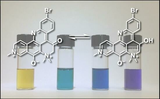
2.2. pKa of Petrosamine (1), Does the Structure of 1 Exhibit Substantial Enol Content?

To estimate the pKa of petrosamine, the UV-vis spectra of 1 were measured in buffered D2O at different pH. Across the pH 2–10 (Figure S1), the UV-vis spectrum of 1 is unchanged. From the Henderson-Hasselbach relationship (Equation (1)) [18] for Brønsted acid HA, the condition pH = pKa is met when concentrations of conjugate species are equal ([HA] = [A]). We surmise the pKa of 1 lies outside this pH range (pKa > 10). Indeed, a bathochromic shift in the UV-vis spectrum of 1 is only observed when a methanolic solution is treated with strong alkali NaOH (2 M, pH > 13). Conversely, only when a sample of 1 is dissolved in a very strong Brønsted acid (neat CF3COOH) are conditions met for a reasonable expectation of diprotonated petrosamine ([1•2H]2+). In the event when 1 was dissolved in neat TFA, the blue-purple color changed to bright yellow. The latter observation contrasts with the supposition drawn by Quinn and coworkers of doubly-protonated petrosamine B ([2•2H]2+) from their observation that 2 remains “bright blue…when dissolved in methanol”, under the relatively benign conditions used in C18-reversed phase isolation of the alkaloid (5% TFA-MeOH) [6]. In other words, N-10 in the monocationic molecules 13 is ‘pyridinium-like’ and non-basic (resembling 1d), and the C-8 C=O is insufficiently Brønsted-basic to be substantially protonated under ordinary isolation conditions.
pH = pKa + log10 [A]/[HA]
It is evident from the 1H and 13C NMR (DMSO-d6) that C-5 in petrosamine (1) is in the C8 keto form, a conclusion also reached by both the Suwanborirux and coworkers [5], and independently by Quinn and coworkers for petrosamine B (2) [6]. The single crystal X-ray crystallography of 1 is concordant with the C8 keto form. Solid state 1 is best represented by the di-keto tautomer, e.g., interatomic distances C8-O2, 1.256(17) Å; C5-O1, 1.203(15), and C5-C6, 1.495(21) [1].

2.3. Kinetic Measurements of Hydrogen-Deuterium Exchange of 1

None of the resonance forms of 1ad (Figure 2)—or more precisely, pathways of electron delocalization—explain the complete exchange of the H-6 protons by deuterium when 1 was dissolved in deuteric solvents (D2O, CD3OD) [1]. The latter can only be rationalized by consideration of the possibility of enolization (Figure 6), either through the positively charged 8a in neutral to weakly acidic pH or the charge-neutral (zwitterionic) enolate 8b under highly basic pH. The kinetic parameters for successive H replacement by D in 1, defined by rate constants k1 and k2, are intrinsic properties as opposed to thermodynamic properties that relate to the equilibrium constants Keq1 and Keq2; the latter are largely dependent upon the strength and concentration of added base, B (Figure 6).
In hydroxylic solvents, 1 also resides largely in the di-keto form. However, we found that in both D2O and CD3OD, the C-6 methylene protons undergo rapid exchange to give the C-6 CD2-isotopomer (1-d2) [1]; a rate so fast that we were unable to detect the C-6 CH2 or the intermediate forms within the time frame between sample preparation and measurement of the 1H NMR spectra. This observation was supported by 1H NMR, HSQC and HMBC measurements of 1 in protic solvent (CD3OH) where the C-6-CH2 group is still observable (HSQC correlation: H-6 to C-6, δH 4.64, s; δC 70.4 ppm). The simplest explanation for both phenomena is catalytic H-D exchange α-to the C-5 C=O group favored by an intermediate, the extensively-conjugated enol tautomer 8a (Figure 6).
To examine the kinetics of H-D exchange in 1 and place an upper bound on the rate of H-D exchange, the time-dependent appearance of the CD2-isotopmer by ESI mass spectrometry upon rapid dissolution of 1 in CD3OD (Figure 7). Under controlled conditions (23 °C, in situ measurement of the ESIMS of petrosamine with rapid sampling (sampled every t = 15 s) measurement—see Supplementary Materials (S6–S9 and Experimental (S14)), 1-d0 (C21H1789BrN3O2, calculated m/z 422.0499 [M+]) quickly disappeared and was replaced by an ephemeral isotopologue 1-d1 (m/z 423.0561), followed by convergence upon 1-d2 (m/z 424.0624). Complete exchange (>90%) was observed within 90 s of dissolution of 1. From triplicate measurements and rapid sampling, we could fit the kinetic deuterium exchange data to a first-order rate law. We estimated the apparent first and second H-D exchange rate constants to be (Table 2), k1 = 0.131(68) s−1 and k2 = 0.0755(26) s−1, respectively (best R2; see S6–S10). As expected, rate constant k2 is about half of k1, consistent with the expected rate law (k1/k2 = 1.74(94)), negating the involvement of a substantial primary kinetic isotope effect.
The exchange of 1 to 1-d2, in the absence of added acid, appears to be much faster than H-D exchange rates of other phenones, e.g., propiophenone (pKa 24.4, DMSO [19] α-tetralone (pKa 24.7 [20,21]. The rates of acid-promoted keto-enol equilibration of acetophenone (pKa 18.4 ± 0.03) have been measured. For example, the rate of ketonization of acetophenone enol (1-phenylethen-1-ol) is linearly dependent upon [H3O+] with a catalytic efficiency determined to be kH+ = (1.25 ± 0.02) × 103 M−1s−1 [22]. We conclude that tautomerism of 1, too, must be very fast, even in the absence of added acid. Either the enol 8a or enolate 8b, although undetectable as a discrete species in the time frame of 1H NMR, present a pathway to the acid-base equilibria of 1 and 1-d2, but enolization of the keto form is likely dominant.
Some amount of discussion has been given on the keto-enol state of 1. The NMR data for 1 reported by Suwanborirux and coworkers support the C5 keto form in DMSO-d6 [5] and—in agreement with Molinski and Faulkner, the enol form in D2O or CD3OD [1]—but Quinn and coworkers find, “no evidence for this keto-enol isomerism” in 2. Likely, the Keq of keto-enol tautomerism lies on some continuum, dependent upon solvent dielectric and H-bond donor ability.
We find it unlikely that the position of the Br in ring A—the only difference between structures 1 and 2—would exert a profound difference in physicochemical behavior. Quinn’s argument—“no involvement for this keto-enol isomerism” in 2—is confounded by two uncertainties: the composition of their NMR solvent (TFA, D2O, DMSO-d6) is not specified quantitatively, and facile interpretation of the 13C NMR signals for the C8 signal: “in contrast C-8 resonated at 155.8 ppm, supporting it as a phenolic resonance. It was, therefore, more likely that petrosamine also exists in the C-8 enol form and C-8 [Molinski and Faulkner [1] was the carbon at 161 ppm”, contrasts with those of Suwanborirux (δ 174.7 ppm, DMSO-d6) and their observation of, “the broad methylene carbon at δC 69.2” (DMSO-d6, “100 µmol NaOD” [5]). For comparative purposes, we synthesized a model quinoline S1 (see Supplementary Material)—a merocyanine of class (n = 3, see labeling of 1a,b) and found the 13C NMR chemical shift of C8 in CD3OD to be δC 172.2 ppm. Aside from these points, none of the three different 13C NMR values for C8 in 1 and 2 are incompatible with the structures, both of which are not strictly aryl ketones but vinylogous amides, the dominant resonance form of which will be highly dependent upon solvent dielectric and H-bond donor properties.
The observed 1H and 13C NMR spectra of petrosamine (1), and the C6 exchange to CD2 in CD3OD/NaOD [5], support the enol form in hydroxylic solvents. It is likely fast exchange between the hybrid structure of 1 and 8a,b with an equilibrium constant Keq1 largely in favor of 1 in DMSO-d6, but moving to the dominant form 8a in hydroxylic solvents. The enolate 8b may be favored as the catalytically important intermediate under ‘neutral’ conditions for reasons of charge neutralization, but insufficient data preclude testing this hypothesis with more certainty. In either case—enol or enolate intermediate—we surmise the electron-withdrawing quaternized N+Me2 group in 1 and the related petrosamines, 2, 3, plays a significant role in accelerating the rates of H-D exchange and lowering the pKa of the C-6 CH2 group.
To the best of our knowledge, rapid H-D exchange of a substituted β-quaternary ammonium ketone within a natural product has been observed only in one other instance, coulteroberbinone (7, Figure 8 [23]), an N-quaternary ammonium isoquinoline alkaloid from the leaves of Romneya trichocalyx. The authors note the C-14 C-H in 7, assigned to the α-proton between the carbonyl and quaternized nitrogen (δH 5.64, s), underwent rapid proton-deuterium exchange in D2O or CD3OD to C-D (7-d1) under ambient conditions (supported by 1H NMR and ESIMS data).
Two major factors most likely explain the relatively low pKa of 1 and 7: the electron-withdrawing (inductive) effect of the -N+Me2 quaternary ammonium group (N-7) and stabilization of enol 5 (or enolate 6) through extensive conjugation of the heteroaromatic core, not unlike the stabilization of the enolates of alkyl phenones, e.g., acetophenone, determined by UV-vis (pKa = 18.4). Indirect determinations of the pKa of the enol of acetophenone have also been obtained from the kinetics of reactions of acetophenone: e.g., α-chlorination [24] and aminolysis of the corresponding enol acetate [25]. In contrast, 1 appears to undergo rapid enolization without added Brønsted acid, suggesting that this tautomerization may even be autocatalytic.
Is the enol 8a of petrosamine (1) present in substantial concentrations? From the 1H NMR spectrum of 1 (DMSO-d6), we detect no signals attributable to 8a. It can be ascertained from 1H NMR that Keq1 (Figure 7) is very small (estimated from the limits of integration, Keq1 < 0.05), and the equilibrium of tautomers lies well towards the keto form 1. As noted above, in highly-basic aqueous solutions of 1, the charge-minimal enolate 8b appears in substantial concentrations, placing a lower boundary of pKa ~ 15 for 1. In the absence of base, the mechanism of H-D exchange 1 to 1-d2 in CD3OD most likely engages substantial equilibrium concentrations 8a to allow rapid exchange of the CH2 to CD2 in deuteric solvents within less than 2 min at 23 °C. An accurate pKA of 1 is not accessible from measurements in aqueous solvents, and securing an estimate will likely require measures in a suitable aprotic solvent (e.g., titration in DMSO [20,26]). Bordwell values of pKa are conventionally obtained by titration of a solution of the weak Brønsted acid in DMSO with its non-nucleophilic conjugate base, ‘dimsyl sodium’ (CH3(S=O)CH2 Na+) with Ph3CH as an indicator [26] where a colored endpoint is presented by the deep-red Ph3C anion. In the case of 1, the high color of the substrate and its conjugate base should lend itself to ‘self-indicating’.

3. Materials and Methods

3.1. General Experimental Procedures

Inverse detected 2D NMR spectra were measured on a Jeol ECA (500 MHz) spectrometer equipped with a 5 mm 1H{13C} 5 mm probe or a Bruker Avance III (600 MHz) NMR spectrometer with a 1.7 mm 1H{13C} microcryoprobe. 13C NMR spectra were collected on a Varian NMR spectrometer (125 MHz) with a 5 mm Xsens 13C{1H} cryoprobe. NMR spectra were referenced to residual solvent signals, (CD3)2CO (δH 2.05, δC 29.8). High-resolution ESITOF analyses were conducted on an Agilent 1200 HPLC coupled to an Agilent 6230 TOFMS. Low-resolution MS measurements were made on a Thermoelectron Surveyor UHPLC coupled to an MSD single-quadrupole detector. HPLC was performed on an Agilent 1200 HPLC. UV-vis spectra were measured on a Jasco V-630 spectrometer in quartz cells (1.00 cm path length, Helma). FTIR spectra were collected on thin film samples using a Jasco FTIR-4100 fitted with an ATR accessory (ZnSe plate). Optical densities (OD, λ nm) in microplate wells were measured using a Molecular Devices Spectramax 384 Plus. Measurements of pH were made with a digital pH meter (Denver Instrument, Arvada, CO, USA, Model 220), calibrated against standard solutions (NaH2PO4-Na2HPO4).

3.2. UV-Vis Measurements

Standard solutions of accurately-weighed 1 and 6b were prepared in volumetric flasks and used for serial dilutions to the final working concentrations in specified media, either pure HPLC grade solvent (DCM, DMSO, acetone or DMF—see Supporting Information for 6b in acetone) or mixtures of aqueous HPLC solvents of defined H2O composition.
pH Dependence: Britton-Robinson buffer was prepared by dissolving 2.48 g of boric acid, 2.30 mL acetic acid and 2.72 mL phosphoric acid in 1 L of water and titrating in a 2.0 M solution of NaOH to the desired pH.
40 μL of a 2.37 mM of petrosamine (1) in MeOH was added to 960 μL of the desired buffer solution, and the UV-vis spectrum was measured from λ 190–700 nm.
Solvent-Dependence: Solutions of 1 in H2O (95.0 μM) and acetone (95.0 μM) were mixed in the desired ratio, and the UV-vis spectrum was measured from λ 320–700 nm.

3.3. DFT Calculations of 1

The X-ray coordinates of petrosamine (1) were used as a starting point for DFT calculations of the energy, geometry minimized structure, polar isosurfaces and LUMO-HOMO. Calculations were performed within the Spartan ’20 package [27]. DFT energy minimized structure of 1 was obtained using B3LYP with the 6-31G* basis set under a polarization continuum model (=H2O). Calculated minimized structure with overlaid frontier π-orbitals and electrostatic potential isosurfaces of petrosamine (1) are shown in Figure 4 and Figure 5.

3.4. Synthesis of Merocyanine Dye (6b) [16]

Iodomethane (3.11 mL, 50 mmol) was slowly added to a cold mixture of 4-methylpyridine (4.86 mL, 50 mmol) and 2-propanol (5 mL). The stirred mixture was heated at reflux overnight. Removal of the solvent gave the crude 4-methylpyridinium methiodide (10.23 g) as an off-white solid. A portion of the latter salt (3.00 g, 12.8 mmol), 4-hydroxybenzaldehyde and piperidine (1.06 mL, 10.7 mmol) were dissolved in anhydrous EtOH (16 mL) and heated at reflux overnight, with stirring. Upon cooling, a red precipitate was deposited. The red solid was filtered (Büchner funnel), dissolved in KOH solution (0.2 M, 17 mL, 15 mmol), and heated with stirring for 30 min. Blue-red shiny crystals were recovered by filtration, washed with cold water, and dried to give 6b (2.00 g, 65%). The 1H NMR of the sample was consistent with the expected product.

3.5. H-D Exchange Measurements of 1 by ESI-TOFMS

H-D exchange measurements were made by ESI-TOFMS using the following conditions: VCap: 3500V; fragmentor voltage: 160 V; nozzle voltage: 500 V; drying gas temperature: 325 C, sheath gas temperature: 325 C, drying gas flow rate: 7.0 L/Min; sheath gas flow rate: 10 L/min; nebulizer pressure: 40 psi. A petrosamine (1) sample was rapidly dissolved in CD3OD (23 °C). An aliquot of the solution was immediately introduced into the inlet of the TOF mass spectrometer, and the ratios of 1-d0, 1-d1 and 1-d2 were measured ‘on the fly’ from the corresponding molecular ion intensities [M]+ normalized to the [M]+ of a solution of 1 in CH3OH at the same concentration. Subsequent measurements were made at 15 s intervals over a total reaction time of 90 s. HRMS data were acquired and stored in centroided mode. See Figure 7 and S6–S9.

4. Conclusions

Evidence supports that the major tautomer of petrosamine is the C5 and C8 di-keto form 1, both in solution and solid states. While the enol form 8a was not detectable by 1H NMR in DMSO-d6, it is nevertheless likely responsible for the H-D exchange of 1 in deuteric solvents. The exceptional solvatochromism of 1 is best attributable to charge delocalized resonance forms 1a and 1d—partial structures within 1 that are analogous to Brooker merocyanine dyes [11]. As with the latter, the hyposochromic solvent effects mostly correlate with polar-solvent stabilization of the ground state (HOMO) and attribution to the color changes of 1. The complete isotopic exchange of the C6 CH2 group in 1 to CD2 in a deuterated solvent under ambient conditions is supported by 1H NMR and fast-sampled ESIMS measurements. The exchange mechanism likely proceeds through rapid acid-catalyzed keto-enol tautomerism.
Refinement of the molecular structure of 1 by QM methods maps the electron delocalization and accompanying charge distribution in 1. These and other refinements complement computational docking studies that can lend a more precise definition of host-guest interactions of 1 in its cognate enzyme active site. In turn, these observations may assist in the design and synthesis of new AChE inhibitors based on the pyridoacridine skeleton: a privileged alkaloid molecular framework produced exclusively by marine invertebrates.

Supplementary Materials

The following supporting information can be downloaded at: https://www.mdpi.com/article/10.3390/md21080446/s1, Figure S1: UV-vis spectra of 1. pH dependence in Britton-Robinson buffer (normalized); Figure S2: FTIR of 6b (film, ATR); Figure S3: UV-vis spectra of 6b in pure acetone; Figure S4: UV-vis spectra of 6b in acetone-H2O mixtures (normalized); Figure S5: In situ ESIMS measurements of the rate of H-D exchange of 1 in CD3OD; Figure S6: Derivation of kinetic parameters of H-D exchange of 1 in CD3OD and uncertainty analysis; Figure S7: HRMS. Time course of 1 in CD3OD (triplicate runs) and Fitted Curves; Figure S8: ESI-TOFMS of 1 collected in (a) in CH3OH (b) CD3OD at 30 s) (c) CD3OD at 90 s; Figure S9: Time course of ESI-TOF-MS for petrosamine (1); Figure S10: 1H NMR of 1 in a. CD3OH and b. CD3OD after 5 min; Figure S11: 1H NMR temperature study of petrosamine (1) (CD3OH, 500 MHz); Table S1: λmax and absorbance of merocyanine dye 6b in H2O-acetone mixtures. Table S2: 1H and 13C NMR of 1 in CD3OD and CD3OH; Table S3: 1H NMR data for compounds S1 and S6 (CD3OD, 500 MHz); Scheme S1: Synthesis of compound S1: Experimental: Synthesis of Model Compound S1. References [28,29] are cited in the Supplementary Materials.

Author Contributions

C.J.G. carried out the photophysical measurements with consultation from T.F.M., X.W. and Y.S. and conducted the kinetic analysis of H-D exchange in 1. The manuscript was co-written by C.J.G., X.W. and T.F.M. All authors have read and agreed to the published version of the manuscript.

Funding

The 500 MHz NMR spectrometer and the HPLC ESITOFMS instrument were purchased with funding from the NSF (Chemical Research Instrument Fund, CHE0741968) and the NIH Shared Instrument Grant (S10RR025636) programs, respectively. This research was supported by the Academic Senate of UC San Diego.

Institutional Review Board Statement

Not applicable.

Data Availability Statement

Original data will be made available upon reasonable request.

Acknowledgments

We are most grateful for a generous gift of petrosamine (1) from P. Stout (Sirenas Marine Discovery, LLC, San Diego, CA, USA) and to C. Perrin (UCSD) for helpful discussions and N. Goodyear for assistance with measurements

Conflicts of Interest

The authors declare no conflict of interest.

References and Notes

  1. Molinski, T.F.; Faulkner, D.J. Petrosamine, a Novel Pigment from the Marine Sponge Petrosia sp. J. Org. Chem. 1988, 53, 1341–1343. [Google Scholar] [CrossRef]
  2. Molinski, T.F. Marine Pyridoacridine Alkaloids: Structure, Synthesis, and Biological Chemistry. Chem. Rev. 1993, 93, 1825–1838. [Google Scholar] [CrossRef]
  3. Schmitz, F.J.; Agarwal, S.K.; Gunasekera, S.P.; Schmidt, P.G.; Shoolery, J.N. Amphimedine, New Aromatic Alkaloid from a Pacific Sponge, Amphimedon sp. Carbon Connectivity Determination from Natural Abundance 13C-13C Coupling Constants. J. Am. Chem. Soc. 1983, 105, 4835–4836. [Google Scholar] [CrossRef]
  4. Marshall, K.M.; Barrows, L.R. Biological activities of pyridoacridines. Nat. Prod. Rep. 2004, 21, 731–751. [Google Scholar] [CrossRef] [PubMed]
  5. Nukoolkarn, V.S.; Saen-oon, S.; Rungrotmongkol, T.; Hannongbua, S.; Ingkaninan, K.; Suwanborirux, K. Petrosamine, a potent anticholinesterase pyridoacridine alkaloid from a Thai marine sponge Petrosia n. sp. Bioorg. Med. Chem. 2008, 16, 6560–6567. [Google Scholar] [CrossRef] [PubMed]
  6. Carroll, A.R.; Ngo, A.; Quinn, R.J.; Redburn, J.; Hooper, J.N.A. Petrosamine B, an Inhibitor of the Helicobacterpylori Enzyme Aspartyl Semialdehyde Dehydrogenase from the Australian Sponge Oceanapia sp. J. Nat. Prod. 2005, 68, 804–806. [Google Scholar] [CrossRef] [PubMed]
  7. Wei, X.; Bugni, T.S.; Harper, M.K.; Sandoval, I.T.; Manos, E.J.; Swift, J.; Van Wagoner, R.M.; Jones, D.A.; Ireland, C.M. Evaluation of Pyridoacridine Alkaloids in a Zebrafish Phenotypic Assay. Mar. Drugs 2010, 8, 1769–1778. [Google Scholar] [CrossRef] [Green Version]
  8. Choi, D.-Y.; Choi, H. Natural products from marine organisms with neuroprotective activity in the experimental models of Alzheimer’s disease, Parkinson’s disease and ischemic brain stroke: Their molecular targets and action mechanisms. Arch. Pharm. Res. 2015, 38, 139–170. [Google Scholar] [CrossRef]
  9. Britton, H.T.K.; Robinson, R.A. CXCVIII.—Universal buffer solutions and the dissociation constant of veronal. J. Chem. Soc. 1931, 1456–1462. [Google Scholar] [CrossRef]
  10. Exchange of 1H-13C for 2H-13C results in dramatic attenuation of the 13C signal due to quadrupolar relaxation, and absence of the heteronuclear nOe, among other effects.
  11. Brooker, L.G.S.; Heyes, G.H.; Sprague, R.H.; Van Dyke, R.H.; Van Lare, E.; Van Zandt, G.; White, F.L. Studies in the Cyanine Dye Series. XI. The Merocyanines. J. Am. Chem. Soc. 1951, 73, 5326–5332. [Google Scholar] [CrossRef]
  12. Brooker, L.G.S.; Heyes, G.H.; Sprague, R.H.; Van Dyke, R.H.; Van Lare, E.; Van Zandt, G.; White, F.L.; Cressman, H.W.J.; Dent, S.G. Color and Constitution. X. Absorption of the Merocyanines. J. Am. Chem. Soc. 1951, 73, 5332–5350. [Google Scholar] [CrossRef]
  13. Other possible resonance forms, for example invoking electron donation from Br, are considered only minor contributors.
  14. Simpson, W.T. A Mathematical Treatment of the Color of the Merocyanine Dyes. J. Am. Chem. Soc. 1951, 73, 5359–5363. [Google Scholar] [CrossRef]
  15. Morley, J.O.; Morley, R.M.; Docherty, R.; Charlton, M.H. Fundamental Studies on Brooker’s Merocyanine. J. Am. Chem. Soc. 1997, 119, 10192–10202. [Google Scholar] [CrossRef]
  16. Minch, M.J.; Sadiq Shah, S. A Merocyanin Dye Preparation for the Introductory Organic Laboratory. J. Chem. Educ. 1977, 54, 709. [Google Scholar] [CrossRef]
  17. We confirm the solvatochromism of one example, i, by synthesis and its solvent-dependent UV-vis properties. See Supporting Information.
  18. Hasselbalch, K.A. Die Berechnung der Wasserstoffzahl des Blutes aus der freien und gebundenen Kohlensäure desselben, und die Sauerstoffbindung des Blutes als Funktion der Wasserstoffzahl. Biochem. Z. 1917, 78, 112–114. [Google Scholar]
  19. Bordwell, F.K.; Harrelson, J.A., Jr. Acidities and homolytic bond dissociation energies of the αC—H bonds in ketones in DMSO. Can. J. Chem. 1990, 68, 1714–1718. [Google Scholar] [CrossRef]
  20. Bordwell, F.G. Equilibrium acidities in dimethyl sulfoxide solution. Acc. Chem. Res. 1988, 21, 456–463. [Google Scholar] [CrossRef]
  21. Available online: https://organicchemistrydata.org/hansreich/resources/pka/pka_data/pka-compilation-reich-bordwell.pdf (accessed on 8 July 2023).
  22. Chiang, Y.; Kresge, J.; Wirz, J. Flash-Photolytic Generation of Acetophenone Enol. The Keto-Enol Equilibrium Constant and pKa of Acetophenone in Aqueous Solution. J. Am. Chem. Soc. 1984, 106, 6392–6395. [Google Scholar] [CrossRef]
  23. Valpuesta, M.; Díaz, A.; Suau, R. Coulteroberbinone, a quaternary isoquinoline alkaloid from Romneya coulteri. Phytochemistry 1999, 51, 1157–1160. [Google Scholar] [CrossRef]
  24. Guthrie, J.P.; Cossar, J.; Klym, A. pKa values for substituted acetophenones: Values determined by rates of halogenation. Can. J. Chem. 1987, 65, 2154–2159. [Google Scholar] [CrossRef]
  25. Novak, M.; Marc Loudon, G. Aminolysis of Acetoxystyrenes. The pKa of Acetophenones in Aqueous Solution. J. Am. Chem. Soc. 1976, 98, 3591–3597. [Google Scholar] [CrossRef]
  26. Steiner, E.C.; Gilbert, J.M. The Acidities of Weak Acids in Dimethyl Sulfoxide. J. Am. Chem. Soc. 1963, 85, 3054–3055. [Google Scholar] [CrossRef]
  27. Shao, Y.; Gan, Z.; Epifanovsky, E.; Gilbert, T.B.; Wormit, M.; Kussmann, J.; Lange, A.W.; Behn, A.; Deng, J.; Feng, X.; et al. Advances in molecular quantum chemistry contained in the Q-Chem 4 program package. Mol. Phys. 2015, 113, 184–215. [Google Scholar] [CrossRef] [Green Version]
  28. Graulich, A.; Scuvée-Moreau, J.; Seutin, V.; Liégeois, J.-F. Synthesis and radioligand binding studies of C-5-and C-8-substituted 1-(3, 4-dimethoxybenzyl)-2, 2-dimethyl-1, 2, 3, 4-tetrahydroisoquinoliniums as SK channel blockers related to N-methyl-laudanosine and N-methyl-noscapine. J. Med. Chem. 2005, 48, 4972–4982. [Google Scholar] [CrossRef]
  29. Molinski, T.F.; Faulkner, D.J. An antibacterial pigment from the sponge Dendrilla membranosa. Tetrahedron Lett. 1988, 29, 2137–2138. [Google Scholar] [CrossRef]
Figure 1. Structures of petrosamine (1), analogs 2, 3 and galanthamine (4).
Marinedrugs 21 00446 g001
Figure 2. Canonical resonance forms—neutral (a) and dipolar (b)—for merocyanines defined by bond path, n (see [11]). (b) Petrosamine ‘neutral’ and dipolar resonance forms: 1a (n = 3), 1b (n = 2), 1c (n = 4) and 1d (n = 4). (c) Brooker merocyanine resonance forms; neutral (5a, 6a) and dipolar (5b, 6b) forms [11].
Marinedrugs 21 00446 g002
Figure 3. Solvatochromism in normalized electronic UV-vis spectra of petrosamine (1) in H2O-DMSO solvents of variable composition (H2O v/v = 0%, 20%, 40%, 60%, 80% and 100%).
Marinedrugs 21 00446 g003
Figure 4. Calculated minimized structure and overlaid frontier π-orbitals of petrosamine (1) (DFT B3LYP 6-31G*, polarization continuum model = H2O) (a) HOMO and (b) LUMO.
Marinedrugs 21 00446 g004
Figure 5. Minimized energy structure of petrosamine (1, DFT, Spartan’20, EDF2 6-31G, dipole moment µ = 19.2 D). (a) electrostatic potential surface and (b) the corresponding molecular framework of 1 (dipolar 1d and ‘charge-minimal’ 1 resonance forms).
Marinedrugs 21 00446 g005
Figure 6. Keto-enol tautomerism of 1. Enol 8a (acid-catalyzed) and enolate 8b (base-promoted).
Marinedrugs 21 00446 g006
Figure 7. Representative ESIMS measurements of the rate of H-D exchange of petrosamine (1 in CD3OD, 23 °C) and fitted curves (non-linear regression). See S6–S10 for rate law and k determinations.
Marinedrugs 21 00446 g007
Figure 8. Rapid H-D exchange of coulterberbinone (7) in deuteric solvent under ambient conditions.
Marinedrugs 21 00446 g008
Table 1. UV-vis properties of λmax1 1 and λmax2 1 in petrosamine (1) in DMSO-H2O 1.
% H2O (v/v)λmax1 (nm)ε1 1λmax2 (nm)ε2 1
0296184,00064824,000
20290172,00062220,600
40288161,00060419,400
60287141,70058916,500
80282122,25258112,900
10028168,5005704700
1 See text for definitions. ε1,2 values normalized from the literature value of λmax2 (ε 4700) [1].
Table 2. Rate constants k1 and k2 for H-D exchange in 1 in CD3OD (23 °C, see Figure 7) 1.
Reactionk1 (s−1)k2 (s−1)95% Confidence Interval
1 –> 1-d10.1311(68){0.1176, 0.1447}
1-d1 –> 1-d20.0755(26){0.0702, 0.0807}
1 See S6–S10 for plotted raw ESIMS data and detailed kinetic analysis.
Disclaimer/Publisher’s Note: The statements, opinions and data contained in all publications are solely those of the individual author(s) and contributor(s) and not of MDPI and/or the editor(s). MDPI and/or the editor(s) disclaim responsibility for any injury to people or property resulting from any ideas, methods, instructions or products referred to in the content.

