Article

New Naphthalene Derivatives from the Mangrove Endophytic Fungus Daldinia eschscholzii MCZ-18

1
Collaborative Innovation Center of Ecological Civilization, School of Chemistry and Chemical Engineering, Hainan University, Haikou 570228, China
2
Institute of Applied Chemistry, Jiangxi Academy of Sciences, Nanchang 330096, China
3
Institute Research and Utilization on Seaweed Biological Resources, Key Laboratory of Haikou, Hainan University, Haikou 570228, China
*
Author to whom correspondence should be addressed.
Mar. Drugs 2024, 22(6), 242; https://doi.org/10.3390/md22060242
Submission received: 26 April 2024 / Revised: 20 May 2024 / Accepted: 24 May 2024 / Published: 27 May 2024
(This article belongs to the Special Issue Bio-Active Products from Mangrove Ecosystems 2.0)

Abstract

:
Five new naphthalene derivatives dalesconosides A–D, F (14, 6), a known synthetic analogue named dalesconoside E (5), and eighteen known compounds (724) were isolated from Daldinia eschscholzii MCZ-18, which is an endophytic fungus obtained from the Chinese mangrove plant Ceriops tagal. Differing from previously reported naphthalenes, compounds 1 and 2 were bearing a rare ribofuranoside substituted at C-1 and the 5-methyltetrahydrofuran-2,3-diol moiety, respectively. Their structures were determined by detailed nuclear magnetic resonance (NMR) and mass spectroscopic (MS) analyses, while the absolute configurations were established by theoretical electronic circular dichroism (ECD) calculation. Compounds 1, 3, 1317 and 19 showed broad ranges of antimicrobial spectrum against five indicator test microorganisms (Enterococcus faecalis, Methicillin-resistant Staphylococcus aureus, Escherichia coli, Pseudomonas aeruginosa and Candida albicans); especially, 1, 16 and 17 were most potent. The variations in structure and attendant biological activities provided fresh insights concerning structure−activity relationships for the naphthalene derivatives.

1. Introduction

Microorganisms from special ecological niches such as the mangrove endosymbionts have been demonstrated to be a reliable source of structurally novel and pharmacologically potent natural products (NPs) and have likewise drawn the attention of NP researchers [1,2]. Mangrove-associated fungi produce a larger number of bioactive secondary metabolites compared to any other mangrove-derived microbes; more than 70% were isolated from endophyte fungi [3,4]. Filamentous fungi of the species Daldinia eschscholtzii is known as an endophyte commonly found in multiple hosts such as dead trees [5], insects [6], alga [7], broad-leaved forest [8] and mangroves [9,10]. It has been demonstrated to be a prolific source of bioactive polyketides including immunosuppressive dalesconols A and B, acetodalmanols A and B, (+)-daeschol A, dalmanol A, 2,16-dihydroxyl-benzo[j]fluoranthene [6,11], free radical scavengers (±)-daeschol A [11], cytotoxic (±)-acetodalmanol B, 2,16-dihydroxyl-benzo[j]fluoranthene [11], fungistatic helicascolide C [7], stem cell differentiation inducer selesconol [12], NLRP3 inflammasome activation inhibitors spirodalesol [13] and α-glucosidase inhibitors chromones, naphthoquinones, and naphthofurans [14,15]. As part of our ongoing investigations on mangrove endophytic fungi [16,17,18], Daldinia eschscholtzii-Mcz18 was isolated from a fresh branch of the mangrove plant Ceriops tagal. Five new aromatic polyketide dalesconosides A–D, F (14, 6), a known synthetic analogue named dalesconoside E (5) (this is the first isolation from a natural source), as well as 18 known compounds (724) (Figure 1) were isolated and identified from the mangrove endophytic fungus Daldinia eschscholzii MCZ-18. These compounds were examined for antimicrobial and cytotoxic activities. Details of the isolation, structure elucidation, and antimicrobial and cytotoxic activities of these compounds are reported herein.

2. Results and Discussion

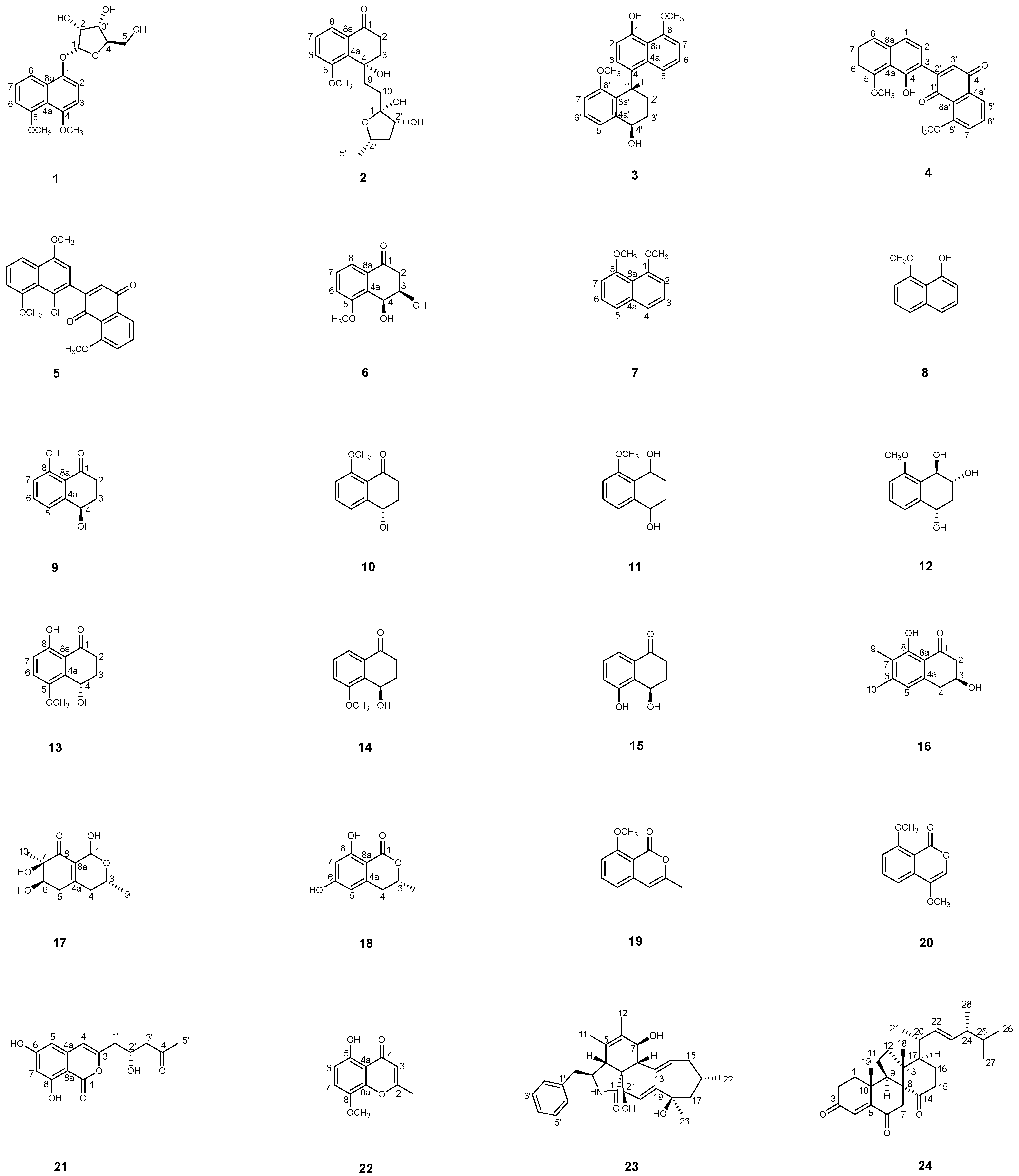
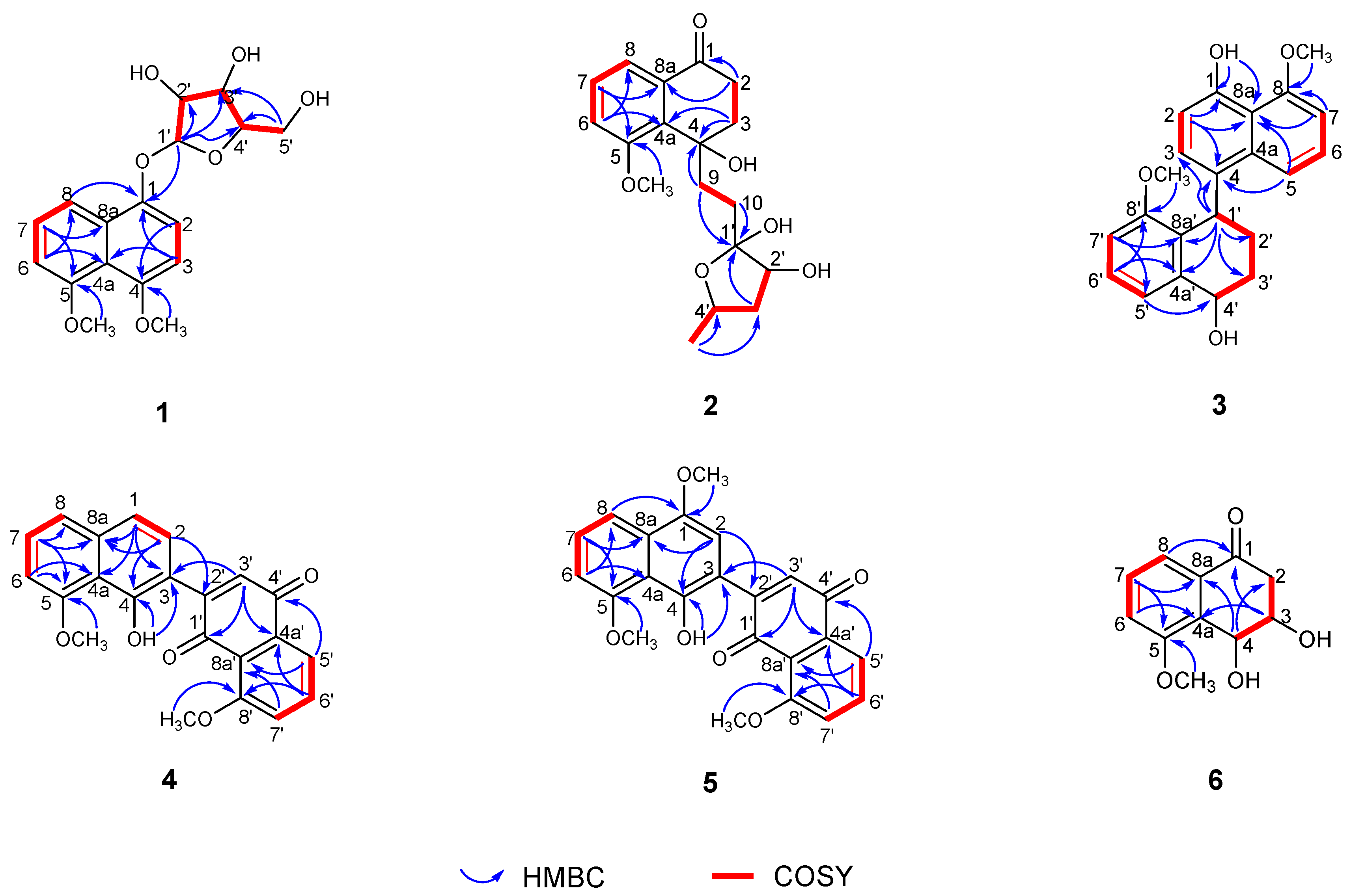
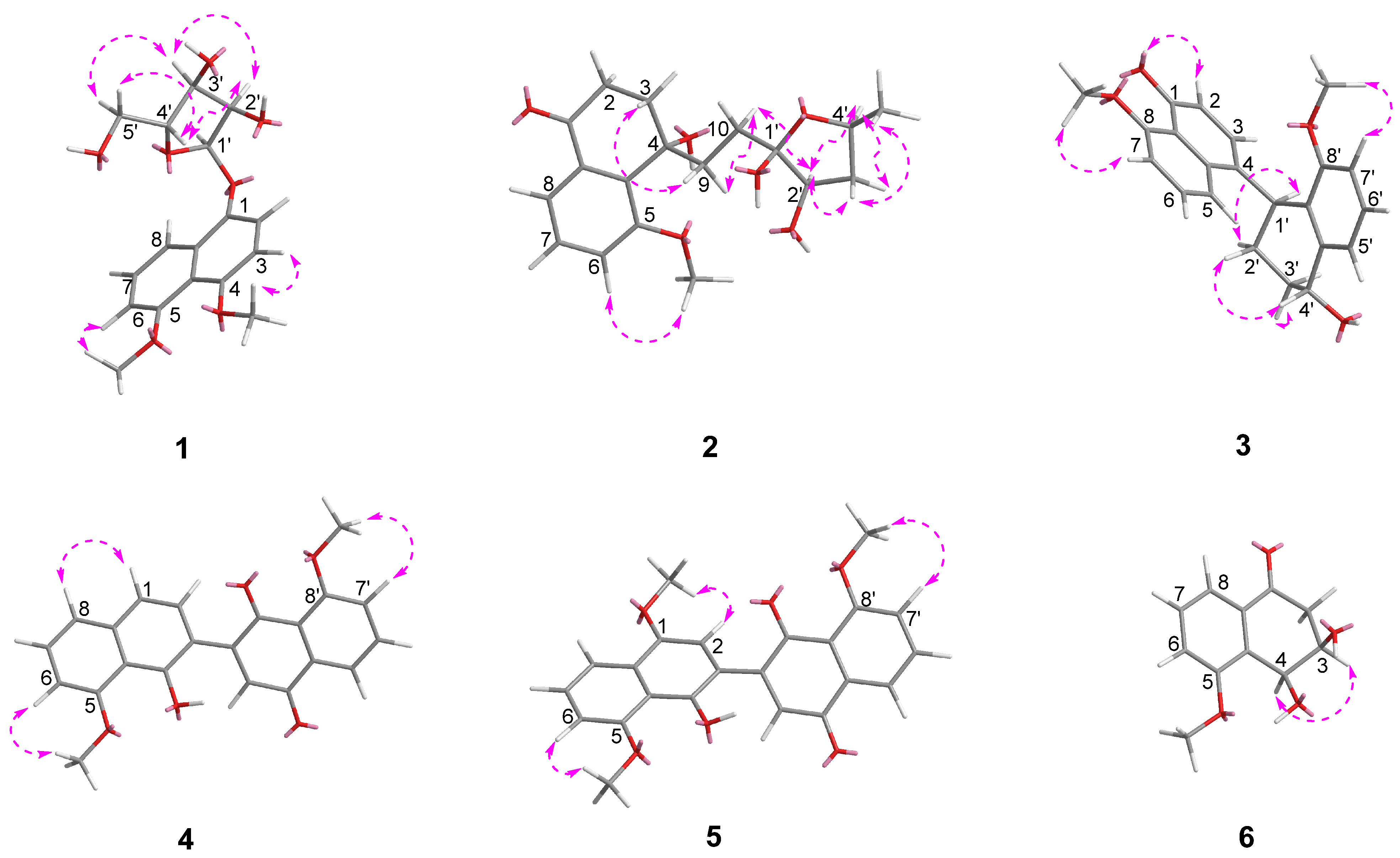
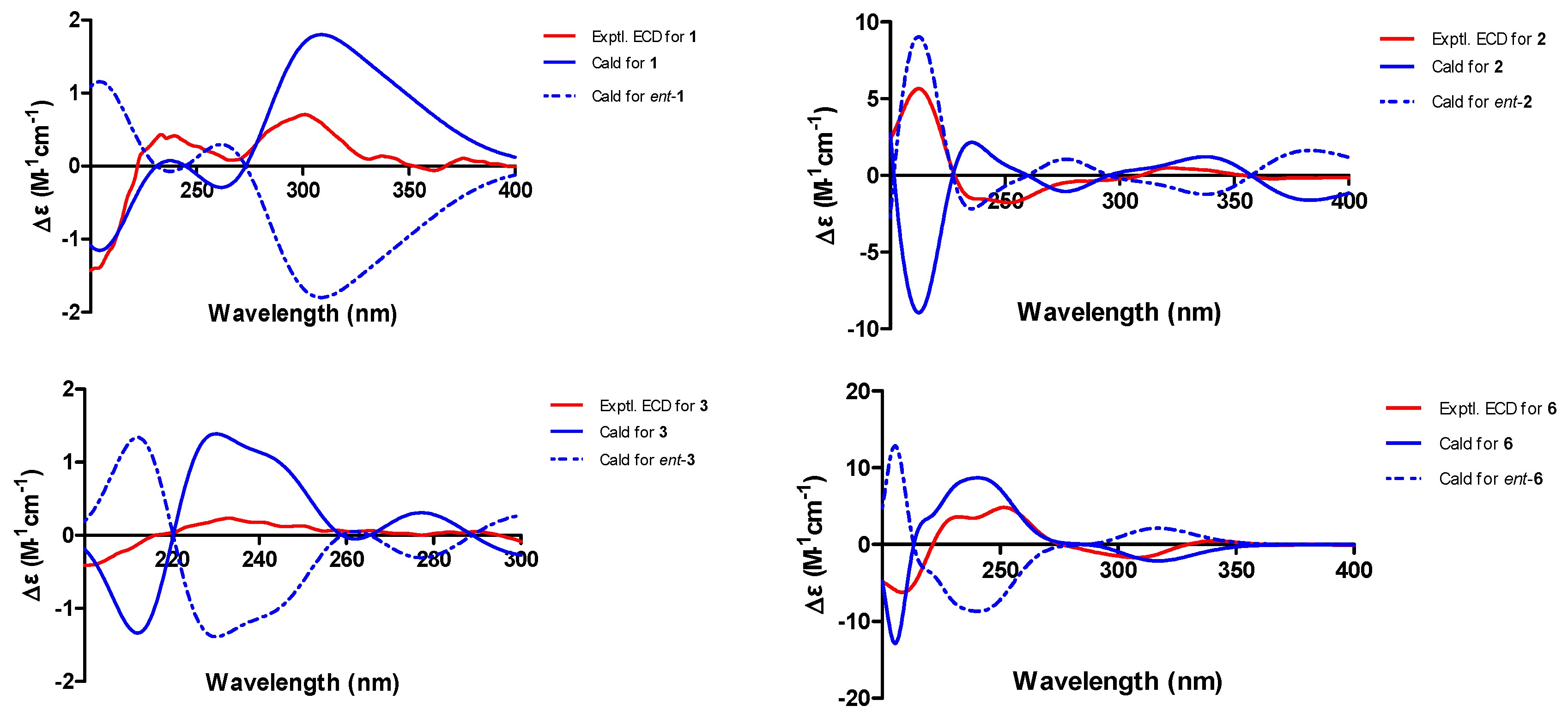
2.1. Structure Elucidation

Dalesconoside A (1) was isolated as yellow needles with the molecular formula C17H20O7 established by HR-ESIMS (m/z 337.1289, calcd for [M+H]+ 337.1282). Consequently, 1 had eight degrees of unsaturation. The 1H and 13C NMR data (Table 1 and Table 2) of 1 indicated that seven out of eight degrees of unsaturation are derived from a naphthalene moiety. Therefore, the last unsaturation degree was attributed to an additional ring system. The 1H and 13C NMR in combination with the 1H-1H COSY and HSQC spectra showed that the compound had five signals of aromatic protons at [δH 8.00 (d, J = 6.4 Hz), δC 116.5, d, CH-8; δH 7.39 (t, J = 6.5, 6.4 Hz), δC 127.2, d, CH-7; 7.18 (d, J = 6.8 Hz), δC 113.1, d, CH-3; δH 6.96 (d, J = 6.1 Hz), δC 108.4, d, CH-6; 6.84 (d, J = 6.8 Hz), δC 108.2, d, CH-2], an anomeric proton [δH 5.63 (d, J = 3.6 Hz), δC 103.9, d, CH-1′], two methoxy groups [δH 3.92, s, δC 56.9, q, 5-OCH3; δH 3.87, s, δC 57.7, q, 4-OCH3], and signals of a furanoside moiety at δH 3.65–4.24. Comparison of the 1H and 13C NMR data with those of 1,8-dimethoxynaphthalene (7) co-isolated in the culture medium and isotorachrysone-6-O-α-D-ribofuranoside [19] revealed that 1 was a 1,8-dimethoxynaphthalene ribofuranoside. The HMBC correlations (Figure 2) observed from H-1′ (δH 5.63) to C-1 (δC 156.6) established that the connection of the furanoside and naphthalene moieties were through the ether bridge. The relative configuration of 1 was interpreted by the diagnostic NOE interactionsH-1′/H-2′, H-2′/H-3′, H-1′/H2-5′, and H-3′/ H2-5, which revealed their co-facial relationship (Figure 3). The α configuration of ribose was confirmed by the chemical shifts (δH 5.63, δC 103.9, CH-1′) and coupling constant of the anomeric proton J1′,2′ value 3.6 Hz. The sample limitations precluded further acid hydrolysis to study the absolute configuration of the α-ribofuranoside in 1, whereas the calculated ECD spectrum method can be used to predict the absolute configuration of C-1′ (Figure 4, Tables S1 and S2). Consequently, the absolute configuration of C-1′ was assigned to be R, and the sugar moiety was assigned as α-D-ribofuranoside. Thus, compound 1 was elucidated as 4,5-dimethoxynaphthalen-1-O-α-D-ribofuranoside and named dalesconoside A (Figures S1–S8).
Dalesconoside B (2) was obtained as a colorless oil, and it possessed the same molecular formula of C18H24O6 as 1 with seven degrees of unsaturation as determined by HRESIMS (m/z 359.1464, calcd for [M+Na] +359.1465). The 1H NMR spectrum of 1 (Table 1) exhibited resonances for three aromatic protons [δH 7.60 (dd, J = 7.8, 1.0 Hz), H-8; δH 7.42 (dd, J = 8.1, 7.8 Hz), H-7; δH 7.32 (dd, J = 8.1, 1.0 Hz), H-6], which indicated the presence of a trisubstituted benzene ring. The 1H NMR spectrum further showed signals for 12 aliphatic protons, six oxygenated protons, including two oxygenated methines (δH 4.15 (t, J = 7.5 Hz), H-2′; δH 4.29, m, H-4′), one methoxy group (δH 3.89, s, 5-OCH3) and one secondary methyl group [δH 1.21 (d, J = 6.3 Hz), H3-5′]. The 13C NMR spectrum disclosed 18 carbon resonances, including two sp3 methyls, five sp3 methylenes, two sp3 methines, two sp3 quaternary carbons (including a dioxysubstituted sp3 carbon at δC 115.8 (C-1′)), three sp2 olefinic methines, and four sp2 nonprotonated carbons (including one ketone carbonyl), as supported by the DEPT and HSQC spectra. 1H–1H COSY spectrum (Figure 2) suggested the presence of fragments of –CH2(2)–CH2(3)–, –CH(5)–CH(6)–CH(7)–, –CH2(9)–CH2(10)–, and –CH(2′)–CH2(3′)–CH(4′)–CH3(5′), suggesting that 2 contains a tetralone skeleton and a 5-methyltetrahydrofuran-2,3-diol moiety. This was confirmed by the HMBC correlations from H2-2 (δH 2.46, m) and H-7 (δH 7.42) to C-8a (δC 134.7); H2-3 (δH 2.76, m; δH 2.63, m) and H-6 (δH 7.32) to C-4a (δC 134.6); and H2-3 and H2-9 (δH 2.54, m; δH 2.33, m) to C-4 (δC 84.8). Other correlations in the HMBC spectrum from H2-9, H2-10 (δH 2.56, m; δH 2.23, m) and H2-3′ (δH 2.00, m; δH 1.83, m) to C-1′ (δC 115.8); 5-OCH3 (δH 3.89) to C-5 (δC 159.3) further supported the atom connectivity in compound 2. The relative configuration of 2 was assigned by analysis of the NOESY spectrum (Figure 3). There were observed correlations between H-2′/ H-4′, H-2′/ Hb-3′, Ha-3′/ H3-5′, H-2′/ Ha-10, Ha-10/ Hb-9, and Ha-9/ Hb-3, indicating that H-2′, Hb-3′, H-4′, Hb-9 and Ha-10 are on the same face and Ha-3′, H3-5′, Ha-9 and Hb-3 are on the other face, which is consistent with a 4S*,1′R*,2′S*,4′R* relative configuration. The absolute configuration of 2 was then determined by comparing experimental and calculated ECD spectra using time-dependent density-functional theory (TDDFT) as shown in Figure 4, Tables S3 and S4. However, theoretical computations with the expected structure provided an ECD spectrum that was an excellent fit with the mirror image of the measured spectrum of 2. Finally, the structure of 2 was assigned to be (R)-4-(2-((1′S,2′R,4′S)-1′,2′-dihydroxy-4′-methyltetrahydrofuran-1′-yl)ethyl)-4-hydroxy-5-methoxy-3,4-dihydronaphthalen-1(2H)-one (Figures S9–S16).
Dalesconoside C (3) was obtained as yellow needles, which has the molecular formula C22H22O4 established by HR-ESIMS (m/z 351.1597, calcd for [M+H]+ 351.1591). Consequently, 3 had 12 degrees of unsaturation. The 1H and 13C NMR data of 3 (Table 1 and Table 2) indicated that 11 of the 12 units of unsaturation come from a naphthalene moiety and an aromatic ring. Thus, the last remaining degree of unsaturation must be attributed to an additional ring system. The 1D NMR spectroscopic data indicated the presence of a structure with two units, one of which is identical with those of 8-methoxy-1-naphthol (8), which was isolated from the same source. The second part showed signals corresponding to 1,2,3,4-tetrahydro-5-methoxynaphthalene with the substitution at C-4. The 1H-1H COSY and HSQC spectra of 3 allowed the assignments of the fragments –CH(2)–CH(3)–, –CH(5)–CH(6)–CH(7)–, –CH(1′)–CH2(2′)–CH2(3′)–CH(4′)– and –CH(5′)–CH(6′)–CH(7′)–. The HMBC correlations from H-1′ (δH 5.04) to C-3 (δC 126.6), C-4 (δC 131.5), C-2′ (δC 23.5), C-3′ (δC 26.7), C-4a′ (δC 140.4) and C-8a′ (δC 128.2); from 1-OH (δH 9.37) to C-1 (δC 152.8), C-2 (δC 109.4) and C-8a (δC 115.6); from 8-OCH3 (δH 4.08) to C-8 (δC 156.9); and from 8′-OCH3 (δH 3.49) to C-8′ (δC 157.2) enable the establishment of the planer structure of 3. The relative configuration of 3 was based on the NOESY correlations as indicated in Figure 3. The NOESY correlations observed of H-1′/Hb-2′(δH 1.86), H-1′/H-5 (δH 7.91), Hb-2′/H-5 and Hb-2′/H-4′ (δH 4.87) indicated that H-1′ and H-4′ were on the same side of the 1,2,3,4-tetrahydronaphthalene moiety. The absolute configuration of 1 were also determined by comparing experimental and calculated electronic circular dichroism (ECD) spectra for the truncated model (1′S, 4′S) -3a and the truncated model (1′R, 4′R) -3b using time-dependent density-functional theory (TDDFT) (Tables S5 and S6). The calculated electronic circular dichroism (ECD) curve for 1a was in good agreement with the experimental ECD spectrum of 3 (Figure 4). Therefore, the absolute configuration of 3 was firmly assigned as 1′S, 4′S. Thus, the complete structure of 3 was established (Figures S17–S24).
Dalesconoside D (4) was obtained as a red amorphous powder. Its molecular formula, C22H16O5 (15 degrees of unsaturation), was established by HR-ESI-MS (m/z 361.1071, calcd for [M+H]+ 361.1071). Using the 1H and 13C NMR (Table 1 and Table 2) combined with the HSQC spectra of 4, a phenolic hydroxyl signal at δH 9.82 (s, 4-OH), the signals for nine aromatic signals at [δH 7.78 (dd, J = 7.6, 2.9 Hz), δC 118.8, d, CH-5′; δH 7.68 (t, J = 8.2, 7.8 Hz), δC 134.6, d, CH-6′; 7.43 (d, J = 7.6 Hz), δC 121.9, d, CH-8; δH 7.37 (t, J = 7.8 Hz), δC 126.8, d, CH-7; δH 7.35 (d, J = 7.6 Hz), δC 118.7, d, CH-1; δH 7.33, s, δC 128.8, d, CH-2; δH 7.31 (d, J = 8.2 Hz), δC 117.8, d, CH-7′; δH 7.06, s, δC 134.5, d, CH-3′; 6.82 (d, J = 7.6 Hz), δC 104.6, d, CH-6], and signals of two methoxy groups [δH 4.05, s, δC 56.2, q, 5-OCH3; δH 4.01, s, δC 56.5, q, 8′-OCH3] were observed. The coupling constants and 1H-1H COSY spectrum showed the presence of three 1H-1H spin systems from –CH(1)–CH(2)–, –CH(6)–CH(7)–CH(8)– and –CH(5′)–CH(6′)–CH(7′)–. The NMR spectroscopic data indicated that 4 comprised one 7-hydroxy-1-methoxyanthracene-9,10-dione subunit and one 8-methoxy-1-naphthol (8) subunit [20]. The HMBC spectrum (Figure 2) and NOESY correlations (Figure 3) supported the assignments of the methoxy groups 5-OCH3 at C-5 (δC 156.7) and 8′-OCH3 at C-8′ (δC 159.7). Moreover, HMBC correlations of 4-OH with C-4 (δC 152.8) and C-3(δC 116.2), H-2 with C-3(δC 116.2) and C-2 (δC 150.4), H-2 with C-2′(δC 150.4), and H-3′ with C-3 (δC 116.2) provided evidence for the C-3-C-2′ linkage of 4. On the basis of the above results, the structure of dalesconoside D (4) was identified as 1′-hydroxy-8,8′-dimethoxy-[2,2′-binaphthalene]-1,4-dione (Figures S25–S32).
Dalesconoside E (5) was obtained as a red amorphous powder. Its molecular formula C23H18O6 (i.e., differing from that of 4 by an additional OCH3 group) was established from HRESIMS at m/z 391.1177 [M+H]+. The 1H and 13C NMR data (Table 1 and Table 2) of 5 were similar to those of 4 except for the significant downfield shift of C-1(δC 147.9) and the presence of a methoxy signal (δH 3.96, s, δC 56.0, q, 1-OCH3) indicative of 5 was a methoxylated analogue of 4. The HMBC correlations (Figure 2) of 1-OCH3 with C-1 revealed that the extra methoxy group is bound to C-1. Hence, 5 was 1-methoxydalesconoside D (Figures S33–S40). Compound 5 is here reported for the first time as a natural product. It was previously prepared via a two-step sequence involving the oxidative dimerization of hydroquinone monomethyl ethers [21].
Dalesconoside F (6) was isolated as a colorless amorphous powder. Its molecular formula C11H12O4 was deduced from HRESIMS at m/z 209.0806 [M+H]+, indicating six degrees of unsaturation. The planer structure of 6 was determined as 3,4-dihydroxy-5-methoxy-3,4-dihydronaphthalen-1(2H)-one [22], which was chemically synthesized from 3,4,5-trihydroxy-1-tetralone with diazomethane, on the basis of 1H and 13C NMR observations, including 2D 1H-1H COSY and HSQC and HMBC spectral data (Figure 2). However, the observed NOESY correlations (Figure 3) of H-3 with H-4 suggested that these protons were on the same spatial orientation. Furthermore, the theoretical ECD spectrum (Figure 4, Tables S7 and S8) was also calculated by a quantum chemical method at the [B3LYP/ 6-311+G(2d,p)] level, and the predicted ECD curve of (3R,4S)-6 was in good agreement with that of the experimental one. Finally, the absolute configuration of 6, named dalesconoside F, was unambiguously determined to be 3R,4S (Figures S41–S48).
By comparing physical and spectroscopic data with those reported in the literature, the known compounds were elucidated as 1,8-dimethoxynaphthalene (7), 8-methoxy-1-naphthol (8) [23], regiolone (9) [24], nodulisporone (10) [25], nodulisporol (11) [25], xylariol A (12) [26], (4R)-4,8-dihydroxy- 3-hydro-5-methoxy-1-naphthalenone (13) [27], (4R)-O-methylsclerone (14) [28], (4R)-3,4-dihydro-4,5-dihydroxynaphthalen-1(2H)-one (15) [29], (3S)-3,8-dihydroxy-6,7-dimethyl-a-tetralone (16) [9], fusaraisochromenone (17) [30], 3R-3,4-dihydro-6,8-dihydroxy-3-methylisocoumarin (18) [31], 2-acetyl-7-methoxybenzofuran (19) [32], 4,8-dimethoxy-1H-isochromen-1-one (20) [22], (+)-citreoisocoumarin (21) [33], 5-hydroxy-8-methoxy-2-methyl-4H-1-benzopyran-4-one(22) [34], cytochalasin O (23) [35], and dankasterone A (24) [36].

2.2. Biological Activity

Compounds 124 were tested for antimicrobial activity against Pseudomonas aeruginosa, Enterococcus faecalis, methicillin-resistant Staphylococcus aureus and Escherichia coli as well as antifungal activity against Candida albicans. As shown in Table 3, all metabolites showed antimicrobial activity against at least one of the five tested indicator pathogens at the selected concentration of 50 μg/mL, but all of the antimicrobial activities were weaker than that of the positive control. Compounds 1, 3, 1317 and 19 exhibited a wide range of antimicrobial activities; especially, 1 (against P. aeruginosa and E. coli), 16 (against E. coli) and 17 (against MRSA) were most potent, and their resistant strain with MIC values reached 6.25 µg/mL. The comparison between compounds 1 and 7 approved that the substitution of additional furanose was the critical structural component for its antimicrobial activity. The antibacterial results of the comparison of 216 suggested that the dihydronaphthalen-1-one nucleus may not be sensitive to the tested microbes in some cases, but the extra auxochromes, e.g., -OCH3, -OH or CH3 enhanced the antibacterial effect of the compound against the tested bacteria and fungi. Meanwhile, when the hydroxyl group is oxidized to a carbonyl group to a certain extent, the antibacterial activity of the metabolite against some indicator bacteria is weakened. Comparing the antimicrobial activities of compounds 1722 indicated that the O-containing heterocyclic substructure may increase the compound’s effect on P. aeruginosa, E. faecalis, MRSA and E. coli. When the -OH and -OCH3 groups are differently substituted on the benzene ring or the lactone/quionone of metabolites’ isocoumarin backbone, different indicator bacteria have different promotion and inhibition. In general, the broad antimicrobial activities of these naphthalene derivatives support their continued investigation so as to develop a deeper understanding of structure−activity relationships and mechanism of antimicrobial action.

3. Materials and Methods

3.1. General Experimental Procedure

Optical rotation was recorded at 20 °C using a WYA-2S digital Abbe polarimeter (Shanghai Physico-optical Instrument Factory, Shanghai, China). UV spectral data were obtained from online UV spectra acquired on a Shimadzu UV-2401 PC spectrophotometer. NMR spectra were recorded on a Bruker AV-400 and AV-500 (Bruker Corporation, Fällanden, Switzerland) instrument with TMS as an internal standard. ESI-MS spectra were recorded on a VG Auto Spec-3000 mass spectrometer (VG, Manchester, UK). High-resolution ESI-MS were recorded on an Agilent 6210 mass spectrometer (Agilent Technologies, Waldbronn, Germany) employing peak matching. The ECD spectra were measured on a JASCO J-715 spectra polarimeter (Japan Spectroscopic, Tokyo, Japan). SephadexLH-20 (Beijing Biotopped Science & Technology Co., Ltd., Beijing, China), SiliaSphere C18 (SiliCycle, Quebec, Canada) and Silica gel (200–300 mesh, Qingdao Marine Chemical Factory, Qingdao, China) were used for column chromatography (CC). Thin-layer chromatography (TLC) was performed on precoated silica gel GF254 plates (Qingdao Marine Chemical Factory, Qingdao, China).

3.2. Fungal Material

The strain MCZ-18 was isolated from a fresh, healthy branch of Ceriops tagal collected from the Dong Zhai Gang-Mangrove Garden on Hainan Island, China. The fungus was isolated under sterile conditions from the inner tissue of the flower following an isolation protocol described previously [37] and identified as Daldinia eschscholzii (GenBank accession no. MH712260) by morphologic traits and molecular identification. A voucher strain was deposited in the School of Chemical Engineering and Technology, Hainan University, Haikou, China.

3.3. Fermentation, Extraction and Isolation

The Daldinia eschscholzii MCZ-18 strain was cultivated on an autoclaved rice solid-substrate medium, consisting of 80 1 L Erlenmeyer flasks. Each flask included 80 g of rice, 0.24 g of sea salt and 80 mL of water. The culture was maintained at a temperature of 25 °C. Following a fermentation period of 29 days, the mycelia and rice were subjected to three extractions using EtOAc. The extracts underwent filtration and evaporation under decreased pressure to yield a crude extract of 75 g. The crude extracts were then submitted to vacuum liquid chromatography (VLC) on silica gel employing a step gradient of petroleum ether/ethyl acetate (1:0–0:1, v/v) to obtain five fractions (Fr.1–Fr.5). Fr.1 was eluted equivalently with petroleum ether to give 8 (200 mg). Fr.2 was eluted equivalently with petroleum ether / ethyl acetate (50:1, v/v) to obtain 7 (150 mg). Fr.3 was eluted with a step gradient of petroleum ether/ethyl acetate (20:1, 10:1, 5:1, 3:1, 1:1, v/v) to give 3 fractions (Fr.3.1–Fr.3.3). Subsequently, Fr.3.1 was further separated by Sephadex LH-20 chromatography (CH2Cl2/MeOH, 1:1, v/v) and finally by an ODS column eluting with MeOH-H2O (70:30, 80:20, 90:10, 100:0, v/v) giving 24 (4.5 mg) and 22 (20 mg). Fr.3.2 was eluted equivalently with petroleum ether/ethyl acetate (5:1, v/v) to give 3 subfractions (Fr.3.2.1–Fr.3.2.3). Fr.3.2.2 was further purified by preparative TLC (developing solvents: CH2Cl2/MeOH, 100: 3, v/v) to obtain 14 (12 mg) and 16 (24 mg). Fr.3.2.3 was purified by Sephadex LH-20 CC (CH2Cl2/MeOH, 1:1, v/v) and finally by an ODS column eluting with MeOH-H2O (60:40, 70:30, v/v) to obtain 3 (2 mg), 19 (6 mg), 11 (1.0 mg), 5 (2.6 mg), 4 (1.2 mg) and 9 (2 mg). 2 (1.8 mg) and 6 (2.2 mg) was acquired by Sephadex LH-20 chromatography (CH2Cl2/MeOH, 1:1, v/v) and C-18 ODS column (MeOH/H2O, 60:40, 70:30, v/v). Fr.3.3 was eluted with a step gradient of petroleum ether/ethyl acetate (3:1, 2:1, 1:1, v/v) to give 3 fractions (Fr.3.3.1–Fr.3.3.3). Fr.3.3.2 was purified by Sephadex LH-20 CC (CH2Cl2/MeOH, 1:1, v/v) and finally by an ODS column eluting with MeOH-H2O (60:40, v/v) to obtain 13 (3 mg), 18 (3.2 mg) and 12 (2 mg). Fr.3.3.3 was purified by Sephadex LH-20 CC (MeOH) and finally by an ODS column eluting with MeOH-H2O (45:55, v/v) to obtain 21 (3.3 mg), 10 (3.6 mg) and 17 (4.4 mg). Fr.4 was purified by an ODS column eluting with MeOH-H2O (40:60, 50:50, 60:40, 70:30, 80:20, 90:10, 100:0, v/v) to obtain 3 subfractions (Fr 4.1–Fr 4.3) and 23 (3.8 mg). Fr.4.2 was purified by Sephadex LH-20 CC (MeOH) and finally by an ODS column eluting with MeOH-H2O (45:55, v/v) to obtain 15 (3 mg), 20 (2.6 mg) and 1 (5.5 mg).
Dalesconoside A (1): yellow needles; [α]20D +100 (c 0.0001, MeOH); UV (MeOH) λmax 226 nm; 1H and 13C NMR data, see Table 1 and Table 2; (+)-HRESIMS at m/z 337.1289 [M+H]+ (calcd for C17H21O7 337.1282).
Dalesconoside B (2): colorless oil; [α]20D +10 (c 0.0001, MeOH); UV (MeOH) λmax 205 nm; 1H and 13C NMR data, see Table 1 and Table 2; (+)-HRESIMS at m/z 359.1464 [M+Na] + (calcd for C18H24O6Na 359.1465).
Alesconoside C (3): yellow needles; [α]20D +80 (c 0.0001, MeOH); UV (MeOH) λmax 202 nm; 1H and 13C NMR data, see Table 1 and Table 2; (+)-HRESIMS at m/z 351.1597 [M+H]+ (calcd for C22H23O4 351.1591).
Dalesconoside D (4): red amorphous solid; UV (MeOH) λmax 201 nm; 1H and 13C NMR data, see Table 1 and Table 2; (+)-HRESIMS at m/z 361.1071 [M+H]+ (calcd for C22H17O5 361.1071).
Dalesconoside E (5): red amorphous solid; UV (MeOH) λmax 202 nm; [α]20D +70 (c 0.0001, MeOH); UV (MeOH) λmax 201 nm; 1H and 13C NMR data, see Table 1 and Table 2; (+)-HRESIMS at m/z 391.1177 [M+H]+ (calcd for C23H19O6 391.1176).
Dalesconoside F (6): colorless solid; [α]20D +80 (c 0.0001, MeOH); UV (MeOH) λmax 221 nm; 1H and 13C NMR data, see Table 1 and Table 2; (+)-HRESIMS at m/z 209.0806 [M+H]+ (calcd for C11H13O4 209.0808).

3.4. ECD Calculations

The Monte Carlo conformational searches were carried out by means of the Spartan’s 14 software (v1.1.4) using a Merck Molecular Force Field (MMFF). The conformers of 13 and 6 with a Boltzmann population of over 5% were chosen for ECD calculations, and then the conformers were initially optimized at B3LYP/6-31g (d, p) in gas. The theoretical calculation of ECD was conducted in MeOH using time-dependent density functional theory (TD-DFT) at the B3LYP/6-31+g (d, p) level for all conformers of compounds. Rotatory strengths for a total of 30 excited states were calculated. ECD spectra were generated using the program SpecDis 1.6 (University of Würzburg, Würzburg, Germany) and GraphPad Prism 5 (University of California San Diego, CA, USA) from dipole-length rotational strengths by applying Gaussian band shapes with sigma = 0.3 eV.

3.5. Antimicrobial Activity Assay

The microplate assay method was used to assess the antimicrobial activities of compounds 124 against four terrestrial pathogenic bacteria (Pseudomonas aeruginosa, Methicillin-resistant Staphylococcus aureus, Bacillus subtilis and Escherichia coli) and one pathogenic fungus (Candida albicans) (Guangdong Microbial Culture Collection Center, China, CDMCC) according to previously reported methods [38]. Ciprofloxacin and amphotericin B were used as a positive control.

4. Conclusions

In conclusion, five new naphthalene derivatives (14, 6), a new natural product (5) and eighteen known compounds (724) were isolated from mangrove endophytic fungus Daldinia eschscholzii MCZ-18. Compounds 1 and 2 are naphthalenes bearing a rare ribofuranoside substituted at C-1 and the 5-methyltetrahydrofuran-2,3-diol moiety, respectively. This is the first report of these naphthalene subtypes being isolated from mangrove-derived fungal sources. All the isolated metabolites showed more or less antimicrobial activities. Several naphthalene derivatives (1, 3, 1317 and 19) showed broad ranges of antimicrobial spectrum. Based on the structure–activity relationship analysis, oxygen heterocyclic rings such as ribofuranoside and tetrahydropyran moieties, auxochromes, e.g., -OCH3, -OH or CH3 were considered as the pharmacophores. The results presented herein reinforce the importance of the mangrove microbial environment as a source of antimicrobial compounds with remarkably varied applications.

Supplementary Materials

The following supporting information can be downloaded at https://www.mdpi.com/article/10.3390/md22060242/s1, Figures S1–S48; Tables S1–S8. The 1D, 2D NMR and HRESIMS spectra for 124 are available online at Figures S1–S48.

Author Contributions

J.X. designed and supervised this research, structured the elucidation, and wrote the draft and final revision of the manuscript. Z.X. performed the isolation. T.F. and B.C. helped organize and test compound data. Z.W. measured the NMR spectra. Q.L. and P.L. carried out the biological activity. The final revision of the manuscript was revised by all the authors. All authors have read and agreed to the published version of the manuscript.

Funding

The National Natural Science Foundation of China (No. 82160675/81973229), the Key Research Program of Hainan Province (ZDYF2021SHFZ108), the Collaborative Innovation Center Foundation of Hainan University (XTCX2022STB01) and the Guangdong Key Laboratory of Marine Materia Medica Open Fund (LMM2021-4) are gratefully acknowledged for the grants that co-financed this project.

Institutional Review Board Statement

Not applicable.

Data Availability Statement

The original data presented in the study are included in the article/Supplementary Material; further inquiries can be directed to the corresponding author.

Conflicts of Interest

The authors declare no conflicts of interest.

References

  1. Lin, W.; Li, G.; Xu, J. Bio-Active Products from Mangrove Ecosystems. Mar. Drugs 2023, 21, 239. [Google Scholar] [CrossRef] [PubMed]
  2. Wu, M.J.; Xu, B.; Guo, Y.W. Unusual Secondary Metabolites from the Mangrove Ecosystems: Structures, Bioactivities, Chemical, and Bio-Syntheses. Mar. Drugs 2022, 20, 535. [Google Scholar] [CrossRef] [PubMed]
  3. Xu, J. Biomolecules Produced by Mangrove-Associated Microbes. Curr. Med. Chem. 2011, 18, 5224–5266. [Google Scholar] [CrossRef] [PubMed]
  4. Xu, J. Bioactive Natural Products Derived from Mangrove-Associated Microbes. RSC Adv. 2015, 5, 841–892. [Google Scholar] [CrossRef]
  5. Karnchanatat, A.; Petsom, A.; Sangvanich, P.; Piapukiew, J.; Whalley, A.J.S.; Reynolds, C.D.; Gadd, G.M.; Sihanonth, P. A Novel Thermostable Endoglucanase from the Wood-Decaying Fungus Daldinia eschscholzii (Ehrenb.:Fr.) Rehm. Enzym. Microb. Technol. 2008, 42, 404–413. [Google Scholar] [CrossRef]
  6. Zhang, Y.L.; Ge, H.M.; Zhao, W.; Dong, H.; Xu, Q.; Li, S.H.; Li, J.; Zhang, J.; Song, Y.C.; Tan, R.X. Unprecedented Immunosuppressive Polyketides from Daldinia eschscholzii, a Mantis-Associated Fungus. Angew. Chem. Int. Ed. 2008, 47, 5823–5826. [Google Scholar] [CrossRef] [PubMed]
  7. Tarman, K.; Palm, G.J.; Porzel, A.; Merzweiler, K.; Arnold, N.; Wessjohann, L.A.; Unterseher, M.; Lindequist, U. Helicascolide C, a New Lactone from an Indonesian Marine Algicolous Strain of Daldinia eschscholzii (Xylariaceae, Ascomycota). Phytochem. Lett. 2012, 5, 83–86. [Google Scholar] [CrossRef]
  8. Luo, Y.; Qiu, L.; Deng, Y.; Yuan, X.-H.; Gao, P. A New Chromone and a New Aliphatic Ester Isolated from Daldinia eschscholtzii. J. Asian Nat. Prod. Res. 2018, 20, 883–888. [Google Scholar] [CrossRef] [PubMed]
  9. Kongyen, W.; Rukachaisirikul, V.; Phongpaichit, S.; Sakayaroj, J. A New Hydronaphthalenone from the Mangrove-Derived Daldinia eschscholtzii PSU-STD57. Nat. Prod. Res. 2015, 29, 1995–1999. [Google Scholar] [CrossRef]
  10. Yang, L.-J.; Liao, H.-X.; Bai, M.; Huang, G.-L.; Luo, Y.-P.; Niu, Y.-Y.; Zheng, C.-J.; Wang, C.-Y. One New Cytochalasin Metabolite Isolated from a Mangrove-Derived Fungus Daldinia eschscholtzii HJ001. Nat. Prod. Res. 2018, 32, 208–213. [Google Scholar] [CrossRef]
  11. Zhang, Y.L.; Zhang, J.; Jiang, N.; Lu, Y.H.; Wang, L.; Xu, S.H.; Wang, W.; Zhang, G.F.; Xu, Q.; Ge, H.M.; et al. Immunosuppressive Polyketides from Mantis-Associated Daldinia eschscholzii. J. Am. Chem. Soc. 2011, 133, 5931–5940. [Google Scholar] [CrossRef] [PubMed]
  12. Zhang, A.H.; Tan, R.; Jiang, N.; Yusupu, K.; Wang, G.; Wang, X.L.; Tan, R.X. Selesconol, a Fungal Polyketide That Induces Stem Cell Differentiation. Org. Lett. 2016, 18, 5488–5491. [Google Scholar] [CrossRef] [PubMed]
  13. Zhang, A.H.; Liu, W.; Jiang, N.; Wang, X.L.; Wang, G.; Xu, Q.; Tan, R.X. Sequestration of Guest Intermediates by Dalesconol Bioassembly Lines in Daldinia eschscholzii. Org. Lett. 2017, 19, 2142–2145. [Google Scholar] [CrossRef] [PubMed]
  14. Liao, H.-X.; Shao, T.-M.; Mei, R.-Q.; Huang, G.-L.; Zhou, X.-M.; Zheng, C.-J.; Wang, C.-Y. Bioactive Secondary Metabolites from the Culture of the Mangrove-Derived Fungus Daldinia eschscholtzii HJ004. Mar. Drugs 2019, 17, 710. [Google Scholar] [CrossRef] [PubMed]
  15. Liao, H.-X.; Zheng, C.-J.; Huang, G.-L.; Mei, R.-Q.; Nong, X.-H.; Shao, T.-M.; Chen, G.-Y.; Wang, C.-Y. Bioactive Polyketide Derivatives from the Mangrove-Derived Fungus Daldinia eschscholtzii HJ004. J. Nat. Prod. 2019, 82, 2211–2219. [Google Scholar] [CrossRef]
  16. Feng, Z.; Zhang, X.; Wu, J.; Wei, C.; Feng, T.; Zhou, D.; Wen, Z.; Xu, J. Immunosuppressive cytochalasins from the mangrove endophytic fungus Phomopsis asparagi DHS-48. Mar. Drugs 2022, 20, 526. [Google Scholar] [CrossRef] [PubMed]
  17. Feng, T.; Wei, C.; Deng, X.; Chen, D.; Wen, Z.; Xu, J. Epigenetic manipulation induced production of immunosuppressive chromones and cytochalasins from the mangrove endophytic fungus Phomopsis asparagi DHS-48. Mar. Drugs 2022, 20, 616. [Google Scholar] [CrossRef]
  18. Wu, J.; Chen, D.; Li, Q.; Feng, T.; Xu, J. Metabolomics-Guided Discovery of New Dimeric Xanthones from Co-Cultures of Mangrove Endophytic Fungi Phomopsis asparagi DHS-48 and Phomopsis sp. DHS-11. Mar. Drugs 2024, 22, 102. [Google Scholar] [CrossRef] [PubMed]
  19. Huang, H.; Lin, Y.; Zhou, S.; Vrijmoed, L.L.P. Metabolites of Mangrove Endophytic Fungus 3920 from the South China Sea. Acta Sci. Nat. Univ. Sunyatseni 2005, 6, 137–138. [Google Scholar]
  20. Lu, M.M.; Chen, Y.C.; Liu, H.X.; Li, S.N.; Li, H.H.; Tao, M.H.; Hao, Z.B.; Zhang, W.M. Secondary Metabolites of Endophytic Daldinia eschscholtzii A630 from Pogostemon cablin and Their Cytotoxic Activities. Nat. Prod. Res. Dev. 2018, 30, 1176–1180. [Google Scholar] [CrossRef]
  21. Takeya, T.; Kondo, H.; Otsuka, T.; Tomita, K.; Okamoto, I.; Tamura, O. A Novel Construction of Dibenzofuran-1,4-Diones by Oxidative Cyclization of Quinone-Arenols. Org. Lett. 2007, 9, 2807–2810. [Google Scholar] [CrossRef] [PubMed]
  22. Fujimoto, Y.; Yokoyama, E.; Takahashi, T.; Uzawa, J.; Morooka, N.; Tsunoda, H.; Tatsuno, T. Studies on the Metabolites of Penicillium Diversum Var. Aureum. I. Chem. Pharm. Bull. 1986, 34, 1497–1500. [Google Scholar] [CrossRef] [PubMed]
  23. Nadeau, A.K.; Sorensen, J.L. Polyketides Produced by Daldinia loculata Cultured from Northern Manitoba. Tetrahedron Lett. 2011, 52, 1697–1699. [Google Scholar] [CrossRef]
  24. Deng, Q.; Li, G.; Sun, M.; Yang, X.; Xu, J. A New Antimicrobial Sesquiterpene Isolated from Endophytic Fungus Cytospora sp. from the Chinese Mangrove Plant Ceriops tagal. Nat. Prod. Res. 2020, 34, 1404–1408. [Google Scholar] [CrossRef]
  25. Kamisuki, S.; Ishimaru, C.; Onoda, K.; Kuriyama, I.; Ida, N.; Sugawara, F.; Yoshida, H.; Mizushina, Y. Nodulisporol and Nodulisporone, Novel Specific Inhibitors of Human DNA Polymerase λ from a Fungus, Nodulisporium sp. Bioorganic Med. Chem. 2007, 15, 3109–3114. [Google Scholar] [CrossRef] [PubMed]
  26. Guo, Z.-L.; Zheng, J.-J.; Cao, F.; Wang, C.; Wang, C.-Y. Chemical Constituents of the Gorgonian-Derived Fungus Chaetomium globosum. Chem. Nat. Compd. 2017, 53, 199–202. [Google Scholar] [CrossRef]
  27. Buchanan, M.S.; Hashimoto, T.; Takaoka, S.; Kan, Y.; Asakawa, Y. A 10-Phenyl-[11]-Cytochalasan from a Species of Daldinia. Phytochemistry 1996, 42, 173–176. [Google Scholar] [CrossRef]
  28. Chang, C.; Chang, H.; Cheng, M.; Liu, T.; Hsieh, S.; Yuan, G.; Chen, I. Inhibitory Effects of Constituents of an Endophytic Fungus Hypoxylon investiens on Nitric Oxide and Interleukin-6 Production in RAW264.7 Macrophages. Chem. Biodivers. 2014, 11, 949–961. [Google Scholar] [CrossRef] [PubMed]
  29. Couché, E.; Fkyerat, A.; Tabacchi, R. Stereoselective Synthesis of cis- and trans-3,4-Dihydro-3,4,8-trihydroxynaphthalen-1(2H)-one. Helv. Chim. Acta 2009, 92, 903–917. [Google Scholar] [CrossRef]
  30. Boonyaketgoson, S.; Trisuwan, K.; Bussaban, B.; Rukachaisirikul, V.; Phongpaichit, S. Isochromanone Derivatives from the Endophytic Fungus Fusarium sp. PDB51F5. Tetrahedron Lett. 2015, 56, 5076–5078. [Google Scholar] [CrossRef]
  31. Krohn, K.; Bahramsari, R.; Flörke, U.; Ludewig, K.; Kliche-Spory, C.; Michel, A.; Aust, H.-J.; Draeger, S.; Schulz, B.; Antus, S. Dihydroisocoumarins from Fungi: Isolation, Structure Elucidation, Circular Dichroism and Biological Activity. Phytochemistry 1997, 45, 313–320. [Google Scholar] [CrossRef] [PubMed]
  32. Wen, L.; Guo, Z.; Li, Q.; Zhang, D.; She, Z.; Vrijmoed, L.L.P. A New Griseofulvin Derivative from the Mangrove Endophytic Fungus Sporothrix Sp. Chem. Nat. Compd. 2010, 46, 363–365. [Google Scholar] [CrossRef]
  33. Youssef, D.T.A.; Shaala, L.A.; Genta-Jouve, G. Asperopiperazines A and B: Antimicrobial and Cytotoxic Dipeptides from a Tunicate-Derived Fungus Aspergillus sp. DY001. Mar. Drugs 2022, 20, 451. [Google Scholar] [CrossRef] [PubMed]
  34. Sun, Y.; Liu, G.; Huang, H.; Yu, P. Chromone Derivatives from Halenia elliptica and Their Anti-HBV Activities. Phytochemistry 2012, 75, 169–176. [Google Scholar] [CrossRef] [PubMed]
  35. Zhang, S.; Liao, H.; Liang, D. Chemical constitutes from Clerodendrum japonicum. China J. Chin. Mater. Medica 2018, 43, 2732–2739. [Google Scholar] [CrossRef] [PubMed]
  36. Amagata, T.; Tanaka, M.; Yamada, T.; Doi, M.; Minoura, K.; Ohishi, H.; Yamori, T.; Numata, A. Variation in Cytostatic Constituents of a Sponge-Derived Gymnascella dankaliensis by Manipulating the Carbon Source. J. Nat. Prod. 2007, 70, 1731–1740. [Google Scholar] [CrossRef] [PubMed]
  37. Zhou, J.; Li, G.; Deng, Q.; Zheng, D.; Yang, X.; Xu, J. Cytotoxic Constituents from the Mangrove Endophytic Pestalotiopsis sp. Induce G0/G1 Cell Cycle Arrest and Apoptosis in Human Cancer Cells. Nat. Prod. Res. 2018, 32, 2968–2972. [Google Scholar] [CrossRef]
  38. Wei, C.W.; Sun, C.X.; Feng, Z.; Zhang, X.X.; Xu, J. Four New Chromones from the Endophytic Fungus Phomopsis asparagi DHS-48 Isolated from the Chinese Mangrove Plant Rhizophora mangle. Mar. Drugs 2021, 19, 348. [Google Scholar] [CrossRef]
Figure 1. Chemical structures of compounds 124.
Marinedrugs 22 00242 g001
Figure 2. Key COSY and HMBC correlations of compounds 16.
Marinedrugs 22 00242 g002
Figure 3. Key NOESY correlations of compounds 16.
Marinedrugs 22 00242 g003
Figure 4. Experimental and calculated electronic circular dichroism (ECD) spectra of 13 and 6.
Marinedrugs 22 00242 g004
Table 1. 1HNMR data of 16.
Position1 a,d2 a,c3 b,c4 b,c5 b,c6 a,c
1 7.33, d (7.6)
26.84, d (6.8)2.46, m6.59, d (8.0)7.35, d (7.6)6.72, sHa 3.18, dd (16.9, 2.8)
Hb 2.64, dd (16.9, 3.2)
37.18, d (6.8)Ha 2.76, m6.37, d (8.0) 4.37, dd (3.2, 2.8)
Hb 2.63, m
4 5.13, d (2.8)
5 7.91, d (8.7)
66.96, d (6.1)7.32, dd (8.1, 1.0)7.45, t (8.4, 7.7)6.82, d (7.6)6.87, d (7.7)7.29, d (8.1)
77.39, t (6.5, 6.4)7.42, t (8.1, 7.8)6.86, d (7.7)7.37, t (7.8)7.39, t (8.3, 8.0)7.42, t (8.1, 7.8)
88.00, d (6.4)7.60, dd (7.8, 1.0) 7.43, d (7.6)7.87, d (7.9)7.56, d (7.8)
9 Ha 2.54, m
Hb 2.33, m
10 Ha 2.56, m
Hb 2.23, m
1′5.63, d (3.6)
2′4.24, m (overlap)4.15, t (7.5)Ha 2.38, m; Hb 1.86, m
3′4.15, dd (6.6, 3.1)Ha 2.00,mHa 1.83, m; Hb 1.78, m7.06, s7.05, s
Hb 1.83, m
4′4.23, m (overlap)4.29, m4.87, t (2.7)
5′Ha 3.71, dd (9.7, 3.8)
Hb 3.65, dd (9.7, 3.2)
1.21, d (6.3)7.11, d (7.6)
6′ 7.32, t (7.9)7.78, dd (7.6, 2.9)7.77, dd (7.6, 1.0)
7′ 6.79, d (8.0)7.68, t (8.2, 7.8)7.68, t (8.3, 7.7)
8′ 7.31, d (8.2)7.32, d (8.1)
1-OCH3 3.96, s
4-OCH33.87, s
5-OCH33.92, s3.89, s 4.05, s4.03, s3.93, s
8-OCH3 4.08, s
8′-OCH3 3.49, s4.01, s4.01, s
4-OH 9.82, s9.42, s
a In CD3OD. b In CDCl3.c 1H (400 MHz). d 1H (500 MHz).
Table 2. 13C NMR data of 16.
Position1 a,d2 a,c3 b,c4 b,c5 b,c6 a,c
1148.6, C199.8, C152.8, C118.7, CH147.9, C198.9, C
2108.2, CH38.6, CH2109.4, CH128.8, CH107.2, CH41.8, CH2
3113.1, CH 36.6, CH2126.6, CH116.2, C114.9, C71.4, CH
4153.6, C84.8, C131.5, C152.8, C146.4, C65.1, CH
4a131.7, C 134.6, C134.1, C114.8, C115.4, C131.2, C
5158.0, C159.3, C117.9, CH156.7, C156.5, C160.1, C
6108.4, CH118.6, CH125.5, CH104.6, CH105.6, CH117.3, CH
7127.2, CH130.3, CH103.8, CH126.8, CH126.4, CH130.4, CH
8116.5, CH120.1, CH156.9, C121.9, CH116.0, CH118.7, CH
8a119.7, C134.7, C115.6, C137.4, C129.0, C133.8, C
9 36.1, CH2
10 35.1, CH2
1′103.9, CH115.8, C33.7, CH 183.3, C183.3, C
2′73.7, CH75.0, CH23.5, CH2150.4, C151.0, C
3′71.2, CH40.4, CH226.7, CH2134.5, CH134.5, CH
4′87.6, CH73.3, CH67.3, CH185.4, C 185.4, C
4a′ 140.4, C134.5, C 134.4, C
5′63.3, CH222.4, CH3121.6, CH118.7, CH 118.7, CH
6′ 127.2, CH134.6, CH134.5, CH
7′ 110.2, CH117.8, CH 117.8, CH
8′ 157.2, C159.7, C159.7, C
8a′ 128.2, C121.0, C121.2, C
1-OCH3 56.0, CH3
4-OCH357.7, CH3
5-OCH356.9, CH356.1, CH3 56.2, CH356.2, CH356.5, CH3
8-OCH3 56.2, CH3
8′-OCH3 55.6, CH356.5, CH356.5, CH3
a In CD3OD. b In CDCl3.c 13C (100 MHz). d 13C (125 MHz).
Table 3. Antimicrobial activities of isolated compounds 124.
MIC(μg/mL)
CompoundP. aeruginosaE. faecalisMRSAE.coliC. albicans
16.252512.56.2525
250>5050>50>50
325252512.5>50
4255050>50>50
512.55025>5050
6>50>505050>50
712.550252550
825>50255012.5
9>5025>50>5025
1050>505050>50
1150255050>50
12252525>5025
132512.512.52525
142525252525
155025502525
1612.52512.56.2550
1712.512.56.2512.525
18>50>5012.525>50
1912.5252512.5>50
20>50>505050>50
2150>505050>50
22>50>502550>50
23>50>502525>50
2450>50>50>50>50
Ciprofloxacin0.780.6250.31250.625
Amphotericin B 0.78
Disclaimer/Publisher’s Note: The statements, opinions and data contained in all publications are solely those of the individual author(s) and contributor(s) and not of MDPI and/or the editor(s). MDPI and/or the editor(s) disclaim responsibility for any injury to people or property resulting from any ideas, methods, instructions or products referred to in the content.

