Article

Two C23-Steroids and a New Isocoumarin Metabolite from Mangrove Sediment-Derived Fungus Penicillium sp. SCSIO 41429

1
Guangdong Provincial Key Laboratory of Chinese Medicine Pharmaceutics, School of Traditional Chinese Medicine, Southern Medical University, Guangzhou 510515, China
2
CAS Key Laboratory of Tropical Marine Bio-Resources and Ecology, Guangdong Key Laboratory of Marine Materia Medica, South China Sea Institute of Oceanology, Chinese Academy of Sciences, Guangzhou 510301, China
*
Author to whom correspondence should be addressed.
These authors contributed equally to this work.
Mar. Drugs 2024, 22(9), 393; https://doi.org/10.3390/md22090393
Submission received: 5 August 2024 / Revised: 22 August 2024 / Accepted: 28 August 2024 / Published: 30 August 2024
(This article belongs to the Section Marine Pharmacology)

Abstract

:
Two new C23-steroids derivatives, cyclocitrinoic acid A (1) and cyclocitrinoic acid B (2), and a new isocoumarin metabolite, (3R,4S)-6,8-dihydroxy-3,4,5-trimethyl-7-carboxamidelisocoumarin (10), together with 12 known compounds (39, 1115) were isolated from the mangrove-sediment fungus Penicillium sp. SCSIO 41429. The structures of the new compounds were comprehensively characterized by 1D and 2D NMR, HRESIMS and ECD calculation. All isolates were evaluated for pancreatic lipase (PL) inhibitory and antioxidant activities. The biological evaluation results revealed that compounds 2, 14 and 15 displayed weak or moderate inhibition against PL, with IC50 values of 32.77, 5.15 and 2.42 µM, respectively. In addition, compounds 7, 12 and 13 showed radical scavenging activities against DPPH, with IC50 values of 64.70, 48.13, and 75.54 µM, respectively. In addition, molecular docking results indicated that these compounds had potential for PL inhibitory and antioxidant activities, which provided screening candidates for antioxidants and a reduction in obesity.

1. Introduction

In recent decades, the prevalence of obesity has demonstrated a significant global escalation and has become a major public health challenge that needs to be addressed [1]. In this context, the biological function of pancreatic lipase (PL), a key enzyme in the regulation of fat metabolism, has received increasing attention from the scientific community. PL, which is mainly synthesized by human pancreatic cells and secreted into the duodenum, plays a central role in the hydrolysis of dietary fats, and is an important target for the control of fat absorption and metabolism [2]. Currently, PL inhibitors, such as orlistat, have become commonly used in the clinical treatment of obesity to reduce fat absorption by effectively inhibiting PL activity, thereby achieving weight loss. However, the widespread use of these drugs is also accompanied by a series of side effects, especially gastrointestinal discomfort, which limits the safety of their long-term use and patient compliance [3]. Therefore, it is important to explore and develop efficient and safe novel weight loss drugs to replace or optimize the existing therapeutic regimens for the management of obesity and its related metabolic diseases.
A variety of human diseases occur due to the excessive production of reactive oxygen species (ROS) in the body during oxidative stress states, capable of attacking and damaging biomolecules [4]. Moreover, antioxidants, as a class of compounds capable of slowing down or blocking oxidative processes, show great potential in the prevention and treatment of oxidative stress-related diseases. They protect cells from damage by providing electrons or hydrogen atoms to oxidative substrates and neutralizing ROS [5]. The wide application of antioxidants is not only limited to the pharmaceutical field but is also commonly found in food preservation and cosmetic industries, as exemplified by using ascorbic acid (vitamin C) in the preservation of fruits and vegetables. Because of the above background, this study aimed to screen potential antioxidant candidate molecules using the DPPH radical scavenging assay as a rapid and effective means of assessing antioxidant activity, which provides a scientific basis for subsequent in-depth research and application development.
Marine fungi have proven to be a very promising source of bioactive compounds [6,7,8]. In particular, mangrove fungi, a unique and biodiversity-rich group of marine fungi, have demonstrated a strong secondary metabolite production capacity, providing a wealth of candidate molecules for drug discovery [9]. Penicillium spp. fungi are common microbial taxa in mangrove ecosystems. From 2007 to 2020, 276 new secondary metabolites isolated and identified from mangrove Penicillium spp. of mangrove origin were reported, of which 140 compounds exhibited biological activity [10]. During our continuous study exploring new bioactive natural products from mangrove fungi [11,12,13,14], two unusual C23-steroids (Figures S1–S22), cyclocitrinoic acid A (1) and cyclocitrinoic acid B (2), one new isocoumarin derivative, (3R,4S)-6,8-dihydroxy-3,4,5-trimethyl-7-carboxamidelisocoumarin (10), and 12 known natural products (39, 1115) (Figure 1) were obtained from the fungus Penicillium sp. SCSIO 41429, which was isolated from mangrove sediment. Herein, we described the fermentation, isolation, structural determination and biological activities of these compounds.

2. Results and Discussion

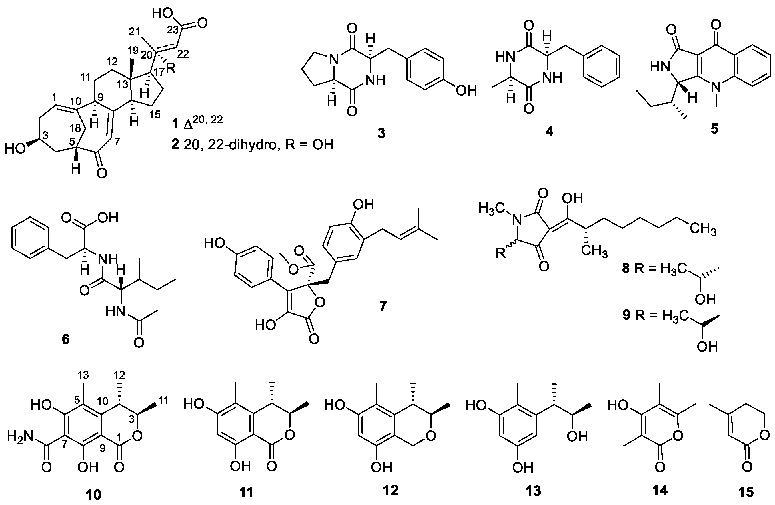
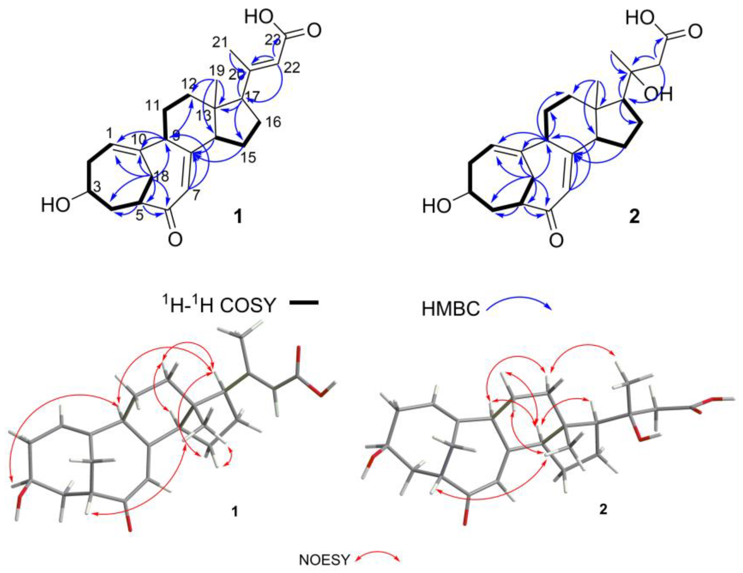
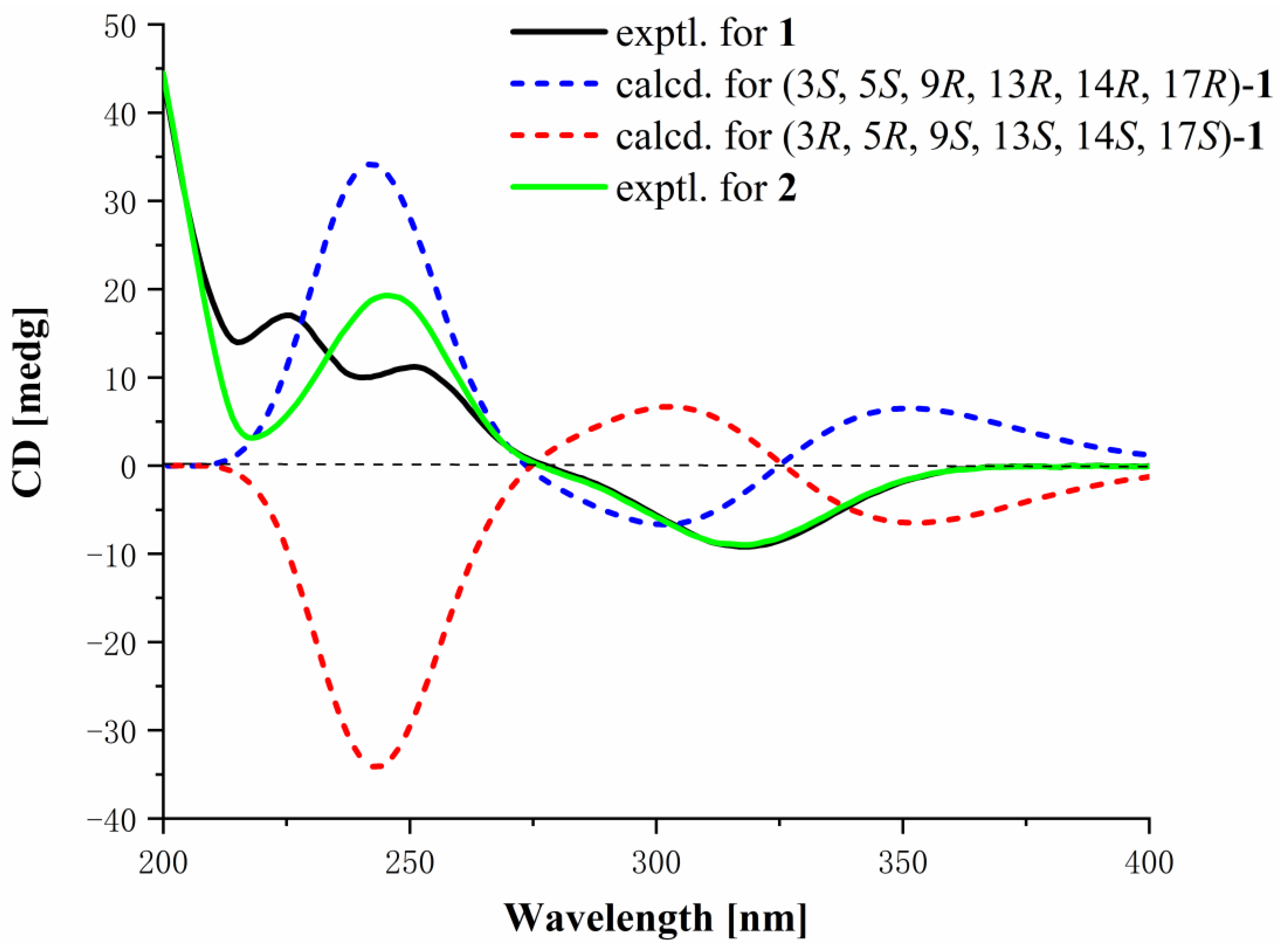
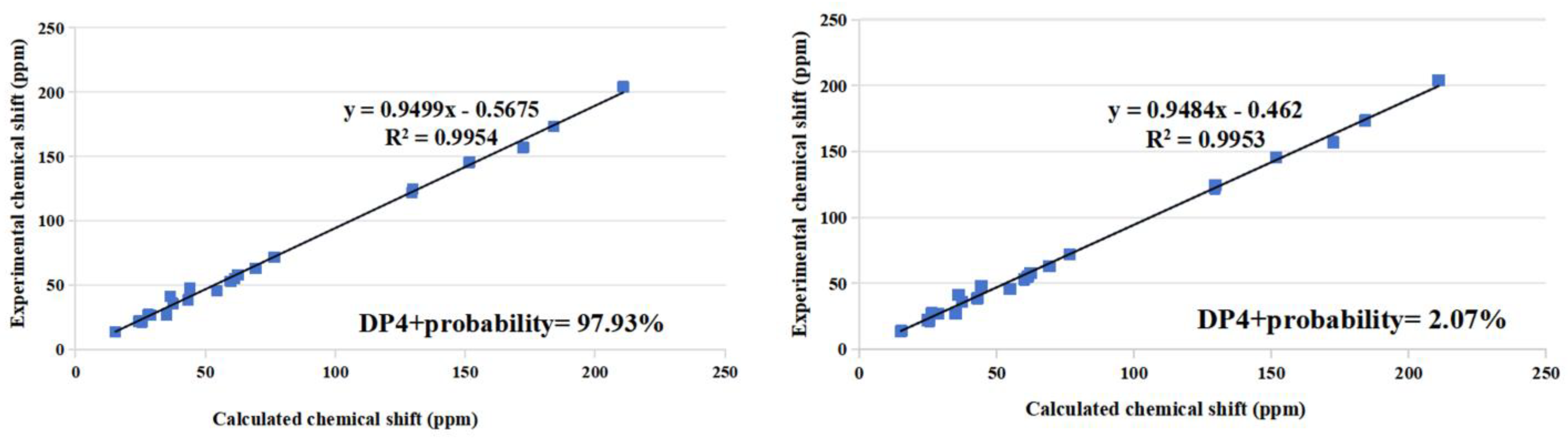
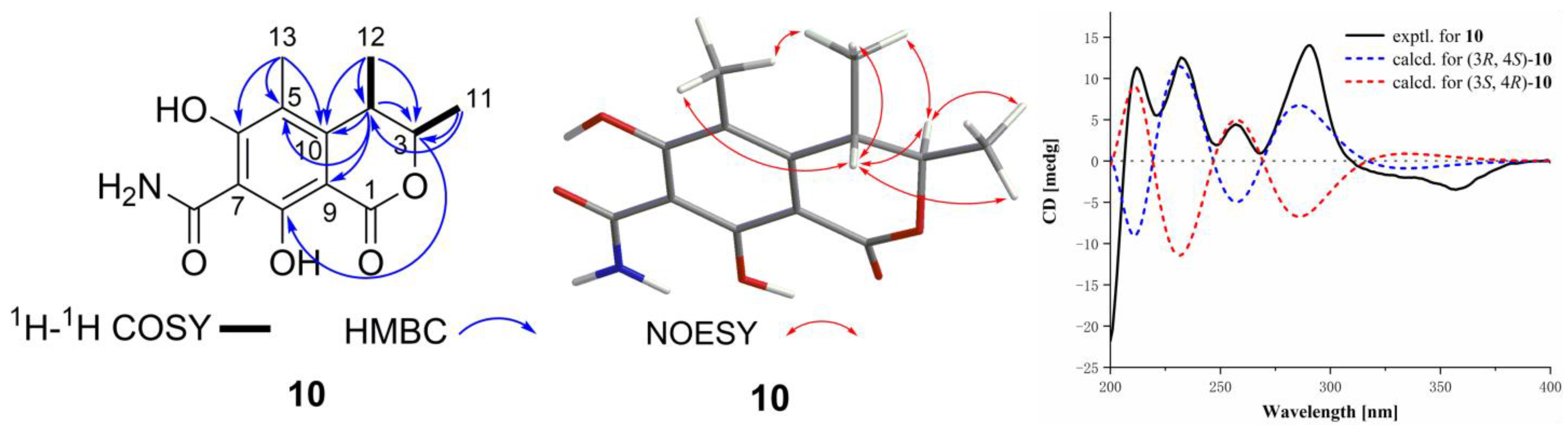
Structural Identification of New Compounds

Compound 1 was obtained as a yellow solid, and its molecular formula was established as C23H30O4 by HERESIMS ion peak at m/z 369.2081 [M − H] (calcd for C23H29O4, 369.2071), indicating nine degrees of unsaturation. Its IR spectrum exhibited absorption bands at 3392.79 and 1672.28 cm−1, indicative of hydroxy and carbonyl groups, respectively. The 1H NMR showed signals for three alkene hydrogens at δH 5.66 (1H, s, H-22), 5.56 (1H, dd, J = 8.5, 6.0 Hz, H-1), and 5.43 (1H, s, H-7), and two methyls at δH 2.10 (3H, s, H3-21) and 0.53 (3H, s, H3-19). The 13C NMR and DE PT spectra (Table 1) revealed 23 carbon-atom resonances including one carbonyl at δC 204.1 (C-6); one carboxyl at δC 167.6 (C-23); four quaternary carbons at δC 156.5 (C-20), 156.5 (C-8), 145.4 (C-10), and 47.1 (C-13); eight methine carbons at δC 48.1 (CH-5), 53.1 (CH-9), 54.5 (CH-14), 59.9 (CH-17), 63.0 (CH-3), 117.4 (CH-22), 122.2 (CH-1), and 124.5 (CH-7); seven methylene carbons at δC 22.3 (CH2-16), 23.9 (CH2-15), 27.2 (CH2-18), 27.4 (CH2-11), 35.9 (CH2-2), 36.9 (CH2-12), and 41.4 (CH2-4); and two methyl carbons at δC 13.3 (CH3-19) and 19.8 (CH3-21). The details of 1D-NMR data and 2D-NMR spectra (Figure 2) showed that 1 has a similar tetracyclic steroid skeleton with a bicyclo [4.4.1] A/B ring system to the reported compound, neocyclocitrinol A [15]. On the side chain, compound 1 and neocyclocitrinol A both exhibited signals for a methyl carbon at δC 19.8 (CH3-21), and double-bond carbons at δC 156.5 (C-20) and 117.4 (CH-22). However, they differed in that compound 1 lacked signals for C-24 and C-25, and exhibited a carboxylate signal at δC 167.6 (C-23) instead of the tertiary alcohol carbon. Moreover, the fragment of C20–C23 of compound 1 was supported by the HMBC correlations (Figure 2) of 2.42 (H-17)/156.5 (C-20), 2.10 (H3-21)/156.5 (C-20), 5.66 (H-22)/59.9 (CH-17), 5.66 (H-22)/156.5 (C-20) and 5.66 (H-22)/167.6 (C-23).
The NOEs between pairs of the protons (Figure 2), H-3/H-9, H-9/H-17, H-17/Hα-12 and Hα-12/H-14, indicated that orientations of H-3, H-9, H-14, and H-17 were on the same side of the A–D ring system, and H-17/Hα-15, H3-19/Hβ-15, H-5/H3-19, indicated that H-5 and H3-19 were on the other side of the ring system in 1, so the relative configuration of 1 was Rel-(3S, 5S, 9R, 13R, 14R, 17R). Then, the ECD calculation (Figure 3) showed that the absolute configuration of compound 1 was 3S, 5S, 9R, 13R, 14R, 17R. Finally, the structure of 1 was determined as shown in Figure 1 and named cyclocitrinoic acid A.
Compound 2 was obtained as white solid, and its molecular formula was established as C23H32O5 by HERESIMS ion peak at m/z 389.2320 [M + H]+ (calcd for C23H33O5+, 389.2323), indicating eight degrees of unsaturation. Its IR spectrum exhibited absorptions bands at 3392.79 and 1697.36 cm−1, indicative of hydroxy and carbonyl groups, respectively. Comparison of the 1H and 13C NMR (Table 1) data revealed that 2 was an analogue of 1. The obvious differences were the changed chemical shifts at C-20 (−84.5 ppm), CH2-22 (−69.5 ppm), CH3-21 (+7.6 ppm), and C-23 (+6.0 ppm) and the replacement of a methine group (δH 5.66 (H-22)/δC 117.4 (CH-22) in 1 by a methylene group (δH 2.24 (H2-22)/δC 72.0 (CH2-22)) in 2. Thus, it was implied that compound 2 differed in the absence of double-bond Δ20,22 and the hydroxylation at C-20. The above deduction was supported by the HMBC correlations of 1.64~1.80 (H-17)/72.0 (C-20), 1.30 (H3-21)/72.0 (C-20), 2.24 (H2-22)/72.0 (C-20), and 2.24 (H2-22)/173.5 (C-23).
The NOEs between pairs of the protons (Figure 2), H-5/H3-19, indicated orientations of H-5 and H3-19 on the same side of the A–D ring system, and H-9/H-14, H-9/Hα-12, H-14/H-17, Hα-12/H-21, Hα-11/H-14, and Hβ-11/H3-19, indicated H-9, H-14, H-17 and H-21 on the other side of the ring system in 2. It was postulated that the A–D rings of compounds 1 and 2 were in the same absolute configurations because they were analogues that had similar ECD curves, despite the absence of NOESY signals of H-3. It was possible to confirm that the absolute configuration of C-3 in compound 2 is 3S. To determine the absolute configuration of the chiral center C-20, the 13C NMR chemical shift calculation of two possible stereoisomers, (3S, 5S, 9R, 13S, 14R, 17S, 20R*)-2 and (3S, 5S, 9R, 13S, 14R, 17S, 20S*)-2, was performed using the gauge including atomic orbital (GIAO) method. The calculation results showed that the experimental NMR data for 2 most closely match those of (3S, 5S, 9R, 13S, 14R, 17S, 20R)-2 based on DP4+ probability analysis of carbon chemical shift (Figure 4), suggesting the absolute configuration of 2 was established as 3S, 5S, 9R, 13S, 14R, 17S, 20R and named as cyclocitrinoic acid B.
Compound 10 was obtained as yellow solid, and its molecular formula was established as C13H15NO5 by HRESIMS ion peak at m/z 266.1025 [M + H]+ (calcd for C13H16NO5, 266.1023). The unsaturation degree of seven and IR spectrum exhibited bands at 3402.16 cm−1 (hydroxyl group) and 1638.37 cm−1 (carbonyl group). A detailed analysis of 1H NMR data (Table 2) of 10 exhibited the presence of two methines at δH 4.86 (q, J = 6.6 Hz, H-3) and 3.18 (q, J = 7.1 Hz, H-4); and three methyls at δH 2.01 (s, H3-13), 1.22 (d, J = 6.6 Hz, H3-12), and 1.17 (d, J = 7.1 Hz, H3-11). The 13C NMR data and HSQC spectrum displayed 14 carbon signals including two ester carbons at δC 175.1 (C-7-CONH2) and 170.2 (C-1); six olefinic tertiary carbons at δC 168.8 (C-6), 166.0 (C-8), 144.8 (C-10), 111.9 (C-5), 100.4 (C-7), and 94.20 (C-9); two methine carbons at δC 80.2 (CH-3) and 33.9 (CH-4); and three methyl carbons at δC 18.6 (CH3-11, 12) and 9.1 (CH3-13). The 1H-1H COSY correlations of H3-11/H-3 and H3-12/H-4 revealed partial structures of CH-3/CH3-11 and CH-4/CH3-12. The above NMR data indicated that 10 had the same isocoumarin skeleton as the co-isolated 11. The main distinction was the presence of an amide group at C-7 of 10 instead of the alkene hydrogen at C-7 of 11, which was supported by HSQC correlations of 14.86 (H2-7-CONH2)/175.1 (C-7-CONH2) and 100.4 (C-7) of 10 instead of 6.29 (H-7)/100.2 (C-7). Thus, the planar structure of 10 was defined as shown in Figure 5, and the other HMBC correlations supported the deduction. To determine the absolute configurations of C-3/C-4, the NOESY analysis and ECD calculation methods were used. The NOESY correlations of H-3/H3-12 and H-4/H3-11 supported the different orientations of CH3-11 and CH3-12. As shown in Figure 5, the experimental ECD was matched to the calculated ECD spectrum of (3R,4S)-10. The 1H coupling constant data of H-3 (4.86, q, J = 6.6 Hz) and H-4 (3.18, q, J = 7.1 Hz) of compound 10 were similar to H-3 (4.68, q, J = 6.6 Hz) and H-4 (3.06, q, J = 7.1 Hz) of the known compound 11. The data provided further evidence of the differing orientations of CH3-11 and CH3-12. Consequently, the absolute configuration of 10 was determined as 3R,4S and named (3R,4S)-6,8-dihydroxy-3,4,5-trimethyl-7-carboxamidelisocoumarin.
In addition to the isolation of the above new compounds 1, 2, and 10, 12 known compounds were isolated, including cyclo-(l-Pro-l-Tyr) (3) [16], cyclo-(l-Phe-l-Ala) (4) [17], guinolactacin A1 (5) [18], N-(N-acetyl-valyl)-phenylalanine (6) [19], butyrolactone I (7) [20], penicillenol A1 (8) [21], penicillenol A2 (9) [21], stoloniferol B (11) [22], decarboxydihydrocitrinin (12) [23], phenol A (13) [24], 4-hydroxy-3,5,6-trimethyl-2H-pyran-2-one (14) [25], and 4-methyl-5,6-dihydropyren-2-one (15) [26]. These structures were elucidated through a comparison of their NMR and MS data with reported literature.
All compounds (115) were evaluated for pancreatic lipase inhibitory and antioxidant activity in vitro, according to the reported methods [27,28,29]. Compounds 2, 14, and 15 displayed weak or moderate inhibition against PL, with IC50 values of 32.77, 5.15, and 2.42 µM, respectively. Orlistat was used as a positive control, with IC50 value of 0.079 µM. Meanwhile, compounds 7, 12, and 13 showed radical scavenging activities against DPPH, with IC50 values of 64.70, 48.13, and 75.54 µM, respectively. Ascorbic acid was used as a positive control, with a IC50 value of 42.61 µM.
To further understand the interaction between the compounds and proteins, docking studies were carried out for compounds in the active site of PL (PDB ID: 1ETH) and superoxide dismutase (PDB ID: 7wx0) to gain insights into their molecular interactions.
As a result, these ligands were favorably accommodated within the binding cleft with analogous anchoring conformations. Compounds 2 and 14 exhibited the binding free energy of −5.84 and −5.23 kcal/mol. Their grid box size was 78 × 58 × 126, centered at x: 72.007, y: 31.282, z: 145.92. As shown in Figure 6, Compound 2 formed one hydrogen-bonding interaction with residue ARG-65, and four hydrophobic-bonding interactions with residues GLU-64, LEU-357, TYR-370 and GLU-371, and displayed two salt bridges with residue LYS-42 and ARG-65. Compound 14 revealed one hydrogen bond with residue SER-78, and five hydrophobic-bonding interactions with residues PHE-78, TYR-115, PRO-181 and PHE-216, and exhibited a salt bridge with residue with HIS-264, and a π-stacking with residue PHE-78. The results of this molecular docking study of compound 15 were by those reported previously [30]. The result as reported showed that compound 15 exhibited the binding free energy of −5.82 kcal/mol, and the dimensions of the grid box size used was 78 × 58 × 126, centered at x: 72.007, y: 31.282, z: 145.92. Compound 15 interacted with residues ASN-329 and AGR-340 via two hydrogen bonds, and three hydrophobic bonds were formed with residues ALA-282, PHE-284 and ARG-340. Compounds 2, 14 and 15 could inhibit PL by tightly binding to catalytic amino acid residues through diverse types of interactions.
Compound 2 was a derivative of compound 1 after hydrolysis of the double-bond at C-20 and C-22, but compound 1 did not show biological activity, suggesting that the hydroxyl group at C-20 of compound 2 was the key for PL inhibitory activity. Compounds 14 and 15 showed similar PL inhibitory activities, which suggested that the lactone group may be the key acting group. Compounds 10 and 11 were both isocoumarins. Despite the presence of lactone groups, they did not exhibit PL inhibitory activity. It was postulated that the benzene ring adjacent to the lactone group may prevent their interaction with PL enzyme active sites, thereby resulting in their inactivity.
Furthermore, molecular docking between the active compounds 7, 12, and 13 with superoxide dismutase (PDB ID: 7wx0) was performed to gain functional and structural insight (Figure 6). The results showed that compounds 7, 12, and 13 displayed the binding free energy of −6.06, −5.59, and −5.49 kcal/mol, respectively. The size of compounds 7, 12, and 13 was 60 × 58 × 96, centered at x: 14.313, y: 6.955, z: 67,949. Compound 7 interacted with the residues ASN-86, THR-88, ILE-96 and GLU-100 via five hydrogen bonds, and a salt bridge interaction was formed with residue LYS-75, and displayed three hydrophobic interactions with residues with PRO-74 and ILE-99. Compound 12 revealed five hydrogen-bonding interactions with residues VAL-8, LYS-10, CYS-146 and VAL-148, and one hydrophobic-bonding interaction with residue VAL-8. Compound 13 formed six hydrogen-bonding interactions with residues VAL-8, LYS-10, CYS-146 and VAL-148, and four hydrophobic interactions with residues LYS-10, ASN-53 and VAL-148. The docking studies suggested that compounds 7, 12, and 13 could inhibit superoxide dismutase by tightly binding to catalytic amino acid residues through diverse types of interactions.

3. Materials and Methods

3.1. General Experimental Procedures

Optical rotations were measured in MeOH using a PerkinElmer MPC 500 (Hertford, UK). IR spectra were recorded on an IR Affinity-1 (Shimadzu, Beijing, China) and Bruker Tensor II spectrometer (Ettlingen, Germany). UV spectra were acquired on a UV-2600 spectrophotometer (Shimadzu, Kyoto, Japan). NMR spectra were obtained on a Bruker AVANCE spectrometer (500 and 700 MHz for 1H NMR; 125 and 175 MHz for 13C NMR (Broker, Fallanden, Switzerland)) using TMS as an internal standard. HRESIMS spectra were generated on a Bruker miXis TOF-QII mass spectrometer (Bruker, Fallanden, Switzerland). ECD data were measured by use of a Chirascan circular dichroism spectrometer (Applied Photophysics, Surrey, UK). Semi-preparative HPLC was performed on the Hitachi Primide using an ODS column (YMC-pack ODS-A (Kyoto, Japan), 10 × 250 mm, 5 µm).

3.2. Fungal Material

The fungus Penicillium sp. SCSIO 41429 used for this study was isolated from mangrove sediment collected in Zhangjiang, Guangdong province, China (21.235° N, 110.451° E). Currently, the strain was stored on MB agar (malt extract of 15 g, sea salt of 10 g, agar of 16 g, H2O of 1 L, and pH 7.4–7.8) slants at 4 °C and deposited at the Key Laboratory of Tropical Marine Bio-resources and Ecology, Chinese Academy of Sciences. The strain was identified as a Penicillium sp. (GenBank No. PP940097) by the ITS sequence of its rDNA. Thus, the strain was identified as Penicillium sp. SCSIO 41429.

3.3. Fermentation, Extraction, and Isolation

The fungus Penicillium sp. SCSIO 41429 was cultivated on the plate of MB agar for 5 days. The mycelia of the strain were cut into small pieces (1 × 1 × 0.5 cm3) and inoculated into 110 × 1000 mL Erlenmeyer flasks each containing Sabouraud’s dextrose broth (3 g of peptone, 12 g of glucose, 0.03 g of chloramphenicol and 1 L of water, pH = 5.6 ± 0.2) for 45 days at 25 °C. The fermented whole broth (33 L) was filtered through cheesecloth to separate into filtrate and mycelia. The filtrate was extracted three times with EtOAc, while mycelia were extracted four times with CH3OH. The EtOAc and CH3OH solutions were concentrated under reduced pressure to get dark brown gum, respectively, and then were combined to obtain crude extract (30 g).
The crude extract was subjected to silica gel CC using step gradient elution with petroleum ether/EtOAc (0–100%, v/v) and EtOAc/CH3OH (0–100%, v/v) to obtain four subfractions (Frs.1–4) based on TLC properties.
Fr.2 was subjected by MPLC with ODS column (MeOH/H2O, 0% to 100% MeOH) to obtain 11 subfractions (Fr.2.1–Fr.2.11). Fr.2.2 was separated by semipreparative reverse-phase HPLC (38% MeOH/H2O, 3 mL/min) to afford 14 (6.6 mg, tR = 19.1 min). Fr.2.4 was purified by semipreparative reverse-phase HPLC (28%MeOH/H2O, 3 mL/min) to yield 13 (12.9 mg, tR = 11.5 min), 10 (6.3 mg, tR = 18.5 min), and 12 (16.6 mg, tR = 32.5 min). Fr.2.7 was purified by semipreparative reverse-phase HPLC (61% MeOH/H2O, 3 mL/min) to give 1 (4.2 mg, tR = 9.5 min), 7 (8.6 mg, tR = 11.5 min), 8 (13.8 mg, tR = 17.8 min), and 9 (13.3 mg, tR = 19.5 min). Fr.3 was subjected by further MPLC with ODS column (MeOH/H2O, 0% to 100% MeOH) to obtain 10 subfractions (Fr.3.1–Fr.3.10). Fr.3.2 was purified by reverse-phase HPLC (20% MeOH/H2O, 3 mL/min) to give 4 (2.6 mg, tR = 10.5 min), 5 (14.7 mg, tR = 12.5 min) and 3 (14.7 mg, tR = 17.6 min). Fr.3.10 was purified by reverse-phase HPLC (53% MeOH/H2O, 3 mL/min) to give 6 (10.3 mg, tR = 20.0 min), 2 (3.3 mg, tR = 21.5 min), and 11 (2.0 mg, tR = 24.0 min). Fr.4 was separated by MPLC with ODS column (MeOH/H2O, 0% to 100% MeOH) to obtain six subfractions (Fr.4.1–Fr.4.6). Fr.4.6 was purified by semipreparative reverse-phase HPLC (8% MeCN/H2O, 3 mL/min) to yield 15 (4.5 mg, tR = 30.5 min).

3.4. Structural Elucidation of the New Compounds 1, 2, and 10

Cyclocitrinoic acid A (1): white solid; [ α ] D 25 = + 89.5°, (c 0.1, MeOH); UV (MeOH) λmax (log ε) 240 (0.9) nm; IR (film) vmax 3392.79, 1672.28, 1644,71, 1298.09, 1246.02, 1174.65, 1033.85, 867.97 cm−1; HRESIMS at m/z 369.2081 [M − H] (calcd for C23H29O4, 369.2071); ECD (0.25 mg/mL, MeOH) λmaxε) 225 (17.05), 251 (11.20), 319 (−9.22) nm. 1H and 13C NMR: see Table 1.
Cyclocitrinoic acid B (2): yellow solid; [ α ] D 25 = +95.2°, (c 0.1, MeOH); UV (MeOH) λmax (log ε) 245 (0.5) nm; IR (film) vmax 3392.79, 1697.36, 1672,21, 1456.26, 1396.46, 1205.51, 1024.20, 896.90, 829.39 cm−1; HRESIMS at m/z 389.2320 [M + H]+ (calcd for C23H33O5+, 389.2323); ECD (0.25 mg/mL, MeOH) λmaxε) 218 (3.13), 246 (19.26), 318 (−9.00) nm. 1H and 13C NMR: see Table 1.
(3R,4S)-6,8-Dihydroxy-3,4,5-trimethyl-7-carboxamidelisocoumarin (10): yellow solid; [ α ] D 25 = +23.9°, (c 0.1, MeOH); UV (MeOH) λmax (log ε) 240 (1.90), 290 (0.98), 340 (0.88) nm; IR (film) vmax 3402.16, 3212.06, 1638.37 cm−1; HRESIMS at m/z 266.1025 [M + H]+ (calcd for C13H16NO5+, 266.1023); ECD (0.25 mg/mL, MeOH) λmaxε) 212 (11.32), 232 (12.53), 257 (4.44), 290 (14.00) nm. 1H and 13C NMR: see Table 2.

3.5. Pancreatic Lipase Inhibition Activity In Vitro Assay

The in vitro PL inhibitory activity of compounds 115 was evaluated with the established protocol [27,28]. A 2.5 mg/mL solution of pancreatic lipase was prepared by dissolving it in Tris–HCl buffer (pH = 8.4). The test sample was mixed with enzyme buffer and incubated at 37 °C for 10 min. Subsequently, p-nitrophenyl palmitate was added, and the enzyme reaction was permitted to continue at 37 °C for a further 10 min. PL activity was ascertained through measurement of the hydrolysis of p-nitro-phenyl palmitate to p-nitrophenol at 405 nm in a microplate reader. All compounds were subjected to a primary screening for enzyme inhibition activity at a concentration of 50 µg/mL, with orlistat as the positive control.

3.6. DPPH Free Radical Scavenging Assay

2,2-Diphenyl-1-picrylhydrazyl (DPPH) assay was employed to assess the free radical scavenging activities of all compounds. A specific experimental procedure was employed, based on the methodology proposed by Yen and Chen [29]. Then, 100 µL of 0.2 mM DPPH prepared in MeOH was mixed with 100 µL of each test sample ranging from 10 to 100 µg/mL in 96-well plates. After incubation for 30 min at room temperature in the dark, the absorbance of the resulting solution was measured spectrophotometrically at 517 nm. Ascorbic acid was used as the positive control, with MeOH serving as the blank. Decreased absorbance in the reaction mixture indicates heightened free radical scavenging efficacy. The scavenging ability of the compounds was calculated using the standard equation. IC50 values were calculated using the Origin 2018 software via a non-linear curve-fitting approach.

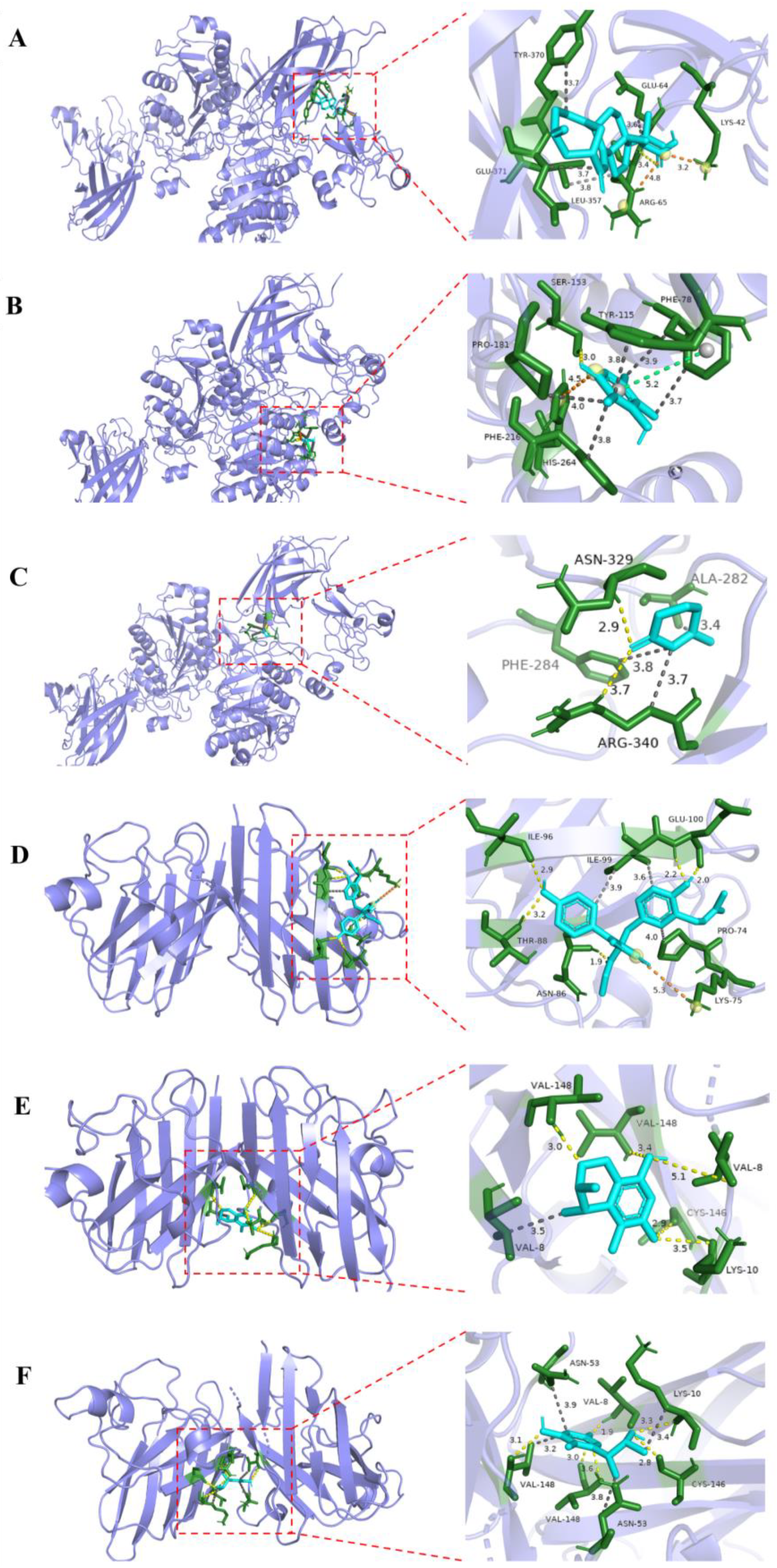
3.7. Molecular Docking Analysis

The molecular docking simulation was implemented by utilizing software AutoDock tools (ADT 1.5.6). The crystal structures of PL (PDB ID: 1ETH) and superoxide dismutase (PDB ID: 7wx0) were obtained from the Protein DataBank. The structures of ligands were generated in ChemBioOffice 18.0 (ChemBioOffice version 14.0), followed by an MM2 calculation to minimize the conformation energy. The other docking parameters, settings, and calculations were defaulted, and the docking results were analyzed using the software PyMOL 2.4.0.

3.8. ECD Calculations

Conformational searches were conducted by Spartan’14 software with the Merck molecular force field (MMFF). The stable conformers were subsequently optimized using Gaussian09 software at the B3LYP/6-31G(d) level in the gas phase. The optimized stable conformers were selected for further ECD calculations at the B3LYP/6-311G(d,p) level in methanol. The overall ECD data were weighted by Boltzmann distribution with a half-bandwidth of 0.3 eV using GuassView6.0 software, and ECD curves were produced by Origin 2018 after UV correction. Meanwhile, the best optimized conformer of 2 was chosen for NMR chemical shift calculations performed by GIAO at the PCM/mPW1PW91/6-311+G(d,p) level in dimethylsulfoxide.

4. Conclusions

Two C23-steroids, cyclocitrinoic acid A (1) and cyclocitrinoic B acid (2), and a new isocoumarin metabolite, (3R,4S)-6,8-dihydroxy-3,4,5-trimethyl-7-carboxamidelisocoumarin (10), together with 12 known metabolites were isolated from the mangrove-sediment fungus Penicillium sp. SCSIO 41429, which had been fermented using Sabouraud’s dextrose broth. The new structures, including absolute configurations, were identified through the application of spectroscopic methods coupled with the calculated ECD. During the screening for pancreatic lipase inhibitory and antioxidant activity, compounds 2, 14, and 15 displayed weak or moderate inhibition against PL, with IC50 values of 32.77, 5.15, and 2.42 µM, respectively. It was suggested that these compounds might be of further research value as potential pancreatic lipase inhibitor candidates in the management of obesity and the treatment of related metabolic diseases. Meanwhile, compounds 7, 12, and 13 showed radical scavenging activities against DPPH, with IC50 values of 64.70, 48.13, and 75.54 µM, respectively, which suggested their potential application as antioxidants in healthy food or drug development. The present study not only augmented the chemical diversity of secondary metabolites of mangrove fungi but also illuminated the prospective applications of some of these compounds in the domain of PL inhibitory and antioxidant activity through bioactivity screening. This provided a valuable material foundation and scientific basis for subsequent drug discovery and development studies.

Supplementary Materials

The following supporting information can be downloaded at: https://www.mdpi.com/article/10.3390/md22090393/s1, Figures S1–S32: NMR, HRESIMS, UV, IR, CD spectra of 1, 2 and 10; Tables S1 and S3: Energies of 1 and 10 at B3LYP/6-311G(d,p) level; Table S2: Energies of 2 at PCM/mPW1PW91/6-311g+(d,p) level The physicochemical and spectroscopic data of 39, 1115.

Author Contributions

Funding acquisition, L.H. and H.T.; Investigation, L.H., C.C., J.C., Y.C., Y.Z. and B.Y.; Methodology, L.H. and C.C.; resources, H.T., Y.L. and X.Z.; Supervision, L.H. and H.T.; Writing—original draft, L.H. and C.C. All authors have read and agreed to the published version of the manuscript.

Funding

This research was funded by Key-Area Research and Development Program of Guangdong Province (2023B1111050008), Natural Science Foundation of Guangdong Province (2024A1515011051), and Postdoctoral Fellowship Program of CPSF (GZC20232777).

Data Availability Statement

The data presented in this study are available on request from the corresponding author.

Acknowledgments

We are grateful to Aijun Sun, Yun Zhang, and Xuan Ma in the analytical facility at SCSIO and Xin Li in the central laboratory at SMU for recording spectroscopic data.

Conflicts of Interest

The authors declare no conflicts of interest.

References

  1. Wilding, J. The future of obesity treatment. EXS 2000, 89, 181–191. [Google Scholar] [PubMed]
  2. Rahul, B.B.; Kamlesh, B.K. Pancreatic lipase inhibitors from natural sources: Unexplored potential. Drug Discov. Today 2007, 12, 879–889. [Google Scholar]
  3. Drent, M.L.; Veen, E.A.d.v. First clinical studies with orlistat: A short review. Obes. Res. 1995, 4, 623S–625S. [Google Scholar] [CrossRef] [PubMed]
  4. Gulcin, İ.; Gulcin, A. Antioxidants and antioxidant methods: An updated overview. Arch. Toxicol. 2020, 94, 651–715. [Google Scholar] [CrossRef]
  5. Apak, R.; Gorinstein, S. Methods of measurement and evaluation of natural antioxidant capacity/activity (IUPAC Technical Report). Pure Appl. Chem. 2013, 85, 957–998. [Google Scholar] [CrossRef]
  6. Fisher, F.J.; Shahriar, M. β-Lactams from the ocean. Mar. Drugs 2023, 21, 86. [Google Scholar] [CrossRef] [PubMed]
  7. Deepika, N.P.; Rahman, M.H.; Duraiswamy, B. The emerging role of marine natural products for the treatment of parkinson’s disease. CNS Neurol. Disord. 2023, 22, 801–816. [Google Scholar] [CrossRef]
  8. Manoharan, S.; Perumal, E. Potential role of marine bioactive compounds in cancer signaling pathways: A review. Eur. J. Pharmacol. 2022, 936, 175330. [Google Scholar] [CrossRef]
  9. Li, K.; Chen, S.; Pang, X.; Cai, J.; Zhang, X.; Liu, Y.; Zhu, Y.; Zhou, X. Natural products from mangrove sediments-derived microbes: Structural diversity, bioactivities, biosynthesis, and total synthesis. Eur. J. Med. Chem. 2022, 230, 114117. [Google Scholar] [CrossRef]
  10. Zeng, W.; Huang, G.; Wang, B.; Cai, J.; Zheng, C. Secondary metabolites and biological activities of Penicillium spp. fungi of mangrove origin (2007–2020). Chin. J. Org. Chem. 2021, 41, 4255–4278. [Google Scholar] [CrossRef]
  11. Chen, C.; Chen, W.; Pang, X.; Liao, S.; Wang, J.; Lin, X.; Yang, B.; Zhou, X.; Luo, X.; Liu, Y. Pyrrolyl 4-quinolone alkaloids from the mangrove endophytic fungus Penicillium steckii SCSIO 41025: Chiral resolution, configurational assignment, and enzyme inhibitory activities. Phytochemisty 2021, 186, 112730. [Google Scholar] [CrossRef] [PubMed]
  12. Cai, J.; Li, M.; Chen, C.; Yang, B.; Gao, C.; Liu, Y.; Luo, X.; Tan, Y.; Zhou, X. Peniditerpenoids A and B: Oxidized indole diterpenoids with osteoclast differentiation inhibitory activity from a mangrove-sediment-derived Penicillium sp. J. Nat. Prod. 2024, 87, 1401–1406. [Google Scholar] [CrossRef] [PubMed]
  13. Xiao, Z.; Cai, J.; Chen, Y.; Zhu, Y.; Chen, C.; Yang, B.; Zhou, X.; Tao, H. Two new sesquiterpenoids and a new shikimic acid metabolite from mangrove sediment-derived fungus Roussoella sp. SCSIO 41427. Mar. Drugs 2024, 22, 103. [Google Scholar] [CrossRef]
  14. Chen, C.; Chen, W.; Tao, H.; Yang, B.; Zhou, X.; Luo, X.; Liu, Y. Diversified polyketides and nitrogenous compounds from the mangrove endophytic fungus Penicillium steckii SCSIO 41025. Chin. J. Chem. 2021, 39, 2132–2140. [Google Scholar] [CrossRef]
  15. Du, L.; Zhu, T.; Fang, Y.; Gu, Q.; Zhu, W. Unusual C25 steroid isomers with bicyclo [4.4.1]A/B rings from a volcano ash-derived fungus Penicillium citrinum. J. Nat. Prod. 2008, 71, 1343–1351. [Google Scholar] [CrossRef]
  16. Jayatilake, S.G.; Thornton, P.M.; Leonard, C.A.; Grimwade, E.J.; Baker, J.B. Metabolites from an antarctic sponge-associated bacterium, Pseudomonas aeruginosa. J. Nat. Prod. 1996, 59, 293–296. [Google Scholar] [CrossRef]
  17. Yang, H.; Wei, N.; Jiang, Q.; Chen, G.; Wen, L. Otoprotective compounds from the metabolites derived from mangrove symbiotic actinomycete Streptomyces sp. 1624105. Chem. Nat. Compd. 2021, 57, 792–794. [Google Scholar] [CrossRef]
  18. Song, T.; Tang, M.; Ge, H.; Chen, M.; Lian, X.; Zhang, Z. Novel bioactive penicipyrroether A and pyrrospirone J from the marine-derived Penicillium sp. ZZ380. Mar. Drugs 2019, 17, 292. [Google Scholar] [CrossRef]
  19. Tang, X.; Liu, S.; Yan, X.; Tang, B.; Fang, M.; Wang, X.; Wu, Z.; Qiu, Y. Two new cytotoxic compounds from a deep-sea Penicillum citreonigrum XT20-134. Mar. Drugs 2019, 17, 509. [Google Scholar] [CrossRef]
  20. Triana, R.D.; Sanro, T.; Sofa, F.; Muhammad, H. α-Glucosidase inhibitor compounds from Aspergillus terreus RCC1 and their antioxidant activity. Med. Chem. Res. 2014, 24, 737–743. [Google Scholar]
  21. Li, S.; Mou, Q.; Xu, X.; Qi, S.; Leung, P.H.M. Synergistic antibacterial activity between penicillenols and antibiotics against methicillin-resistant Staphylococcus aureus. R. Soc. Open Sci. 2018, 5, 172466. [Google Scholar] [CrossRef] [PubMed]
  22. Xin, Z.; Tian, L.; Zhu, T.; Wang, W.; Du, L.; Fang, Y.; Gu, Q.; Zhu, W. Isocoumarin derivatives from the sea squirt-derived fungus Penicillium stoloniferum QY2-10 and the halotolerant fungus Penicillium notatum B-52. Arch. Pharm. Res. 2007, 30, 816–819. [Google Scholar] [CrossRef] [PubMed]
  23. Wei, J.; Chen, X.; Ge, Y.; Yin, Q.; Wu, X.; Tang, J.; Zhang, Z.; Wu, B. Citrinin monomer, trimer, and tetracyclic alkaloid derivatives from the hydrothermal vent-associated fungus Penicillium citrinum TW132-59. J. Org. Chem. 2022, 87, 13270–13279. [Google Scholar] [CrossRef]
  24. Song, T.; Chen, M.; Ge, Z.; Chai, W.; Li, X.; Zhang, Z.; Lian, X. Bioactive penicipyrrodiether A, an adduct of GKK1032 analogue and phenol A derivative, from a marine-sourced fungus Penicillium sp. ZZ380. J. Org. Chem. 2018, 83, 13395–13401. [Google Scholar] [CrossRef]
  25. Wang, D.; Zhou, L.; Zhou, H.; Hu, H.; Hou, G. Chemical composition and protective effect of guava (Psidium guajava L.) leaf extract on piglet intestines. J. Sci. Food Agric. 2021, 101, 2767–2778. [Google Scholar] [CrossRef]
  26. Shintani, R.; Hayashi, T. Rhodium-catalyzed asymmetric 1, 4-addition of sodium tetraarylborates to β, β-disubstituted α, β-unsaturated esters. Org. Lett. 2011, 13, 350–352. [Google Scholar] [CrossRef]
  27. Jo, Y.H.; Kim, S.B.; Ahn, J.H.; Turk, A.; Kwon, E.B.; Kim, M.O.; Hwang, B.Y.; Lee, M.K. Xanthones from the stems of Cudrania tricuspidata and their inhibitory effects on pancreatic lipase and fat accumulation. Bioorg. Chem. 2019, 92, 103234. [Google Scholar] [CrossRef] [PubMed]
  28. Peng, Q.; Cai, J.; Long, J.; Yang, B.; Lin, X.; Wang, J.; Xiao, J.; Liu, Y.; Zhou, X. New azaphthalide and phthalide derivatives from the marine coral-derived fungus Aspergillus sp. SCSIO 41405. Phytochem. Lett. 2021, 43, 94–97. [Google Scholar] [CrossRef]
  29. Yen, G.; Chen, H. Antionxidant activity of various tea extracts in relation to their antimutagenicity. J. Agric. Food Chem. 1995, 43, 27–32. [Google Scholar] [CrossRef]
  30. Huang, L.; Chen, C.; Cai, J.; Chen, Y.; Zhu, Y.; Yang, B.; Zhou, X.; Liu, Y.; Tao, H. Discovery of enzyme inhibitors from mangrove sediment derived fungus Trichoderma harzianum SCSIO 41051. Chem. Biodivers. 2024, 21, e202400070. [Google Scholar] [CrossRef]
Figure 1. Structures of compounds 115.
Marinedrugs 22 00393 g001
Figure 2. The key 1H-1H COSY, HMBC, and NOESY correlations of 1 and 2.
Marinedrugs 22 00393 g002
Figure 3. The experimental and calculated ECD spectra of 1, and experimental of ECD spectra of 2.
Marinedrugs 22 00393 g003
Figure 4. Linear regression analysis of calculated 13C NMR shifts of (3S, 5S, 9R, 13S, 14R, 17S, 20R)-2 (left) and (3S, 5S, 9R, 13S, 14R, 17S, 20S)-2 (right) against the experimental shifts of 2 and the DP4+ probability for assignment of 2 to the candidate stereoisomers.
Marinedrugs 22 00393 g004
Figure 5. The key 1H-1H COSY, HMBC, and NOESY correlations of 10 and experimental and calculated ECD spectra of 10.
Marinedrugs 22 00393 g005
Figure 6. Molecular docking proposed binding interaction of compounds 2 (A), 14 (B) and 15 (C) with the active site residues of PL (PDB ID: 1ETH), and compounds 7 (D), 12 (E), and 13 (F) with the active site residues of superoxide dismutase (PDB ID: 7wx0). Yellow dotted line: hydrogen bond; gray dotted line: hydrophobic interaction; orange dotted line: salt bridge; green dotted line: ππ stacking interaction.
Marinedrugs 22 00393 g006
Table 1. The 1H (500 MHz) and 13C (125 MHz) NMR data of 1 and 2 in DMSO-d6.
Pos.12
δC, TypeδH (J in Hz)δC, TypeδH (J in Hz)
1122.2, CH5.56 (dd, 8.5, 6.0)122.0, CH5.53 (dd, 8.5, 6.0)
35.9, CH22.04~2.08 (overlapped)35.9, CH22.04~2.13 (overlapped)
2.28~2.37 (overlapped)2.34 (m)
363.0, CH3.12 (m)63.0, CH3.12 (m)
41.4, CH21.50~1.62 (overlapped)41.3, CH21.41~1.53 (overlapped)
2.64 (m)2.63 (m)
548.1, CH2.69 (m)48.1, CH2.68 (m)
6204.1, C 204.1, C
7124.5, CH5.43 (s)124.6, CH5.39 (s)
8156.5, C 156.9, C
953.1, CH2.86 (dd, 12.0, 5.5)53.1, CH2.81 (dd, 12.0, 5.5)
10145.4, C 145.6, C
11α27.4, CH21.50~1.62 (overlapped)27.2, CH21.41~1.53 (overlapped)
11β1.71~1.80 (overlapped)1.64~1.80 (overlapped)
12α36.9, CH21.71~1.80 (overlapped)38.8, CH22.04~2.13 (overlapped)
12β1.50~1.62 (overlapped)1.41~1.53 (overlapped)
1347.1, C 45.7, C
1454.5, CH2.28~2.37 (overlapped)55.3, CH2.04~2.13 (overlapped)
15α23.9, CH21.71~1.80 (overlapped)22.1, CH21.41~1.53 (overlapped)
15β1.85 (m)1.41~1.53 (overlapped)
16α22.29, CH21.50~1.62 (overlapped)21.6, CH21.64~1.80 (overlapped)
16β1.50~1.62 (overlapped)1.87 (m)
1759.9, CH2.42 (t, 9.5)58.0, CH1.64~1.80 (overlapped)
1827.2, CH22.48 (overlapped)27.1, CH22.47 (overlapped)
1913.3, CH30.53 (s)13.8, CH30.76 (s)
20156.5, C 72.0, C
2119.8, CH32.10 (s)27.4, CH31.30 (s)
22117.4, CH5.66 (s)47.9, CH22.24 (d, 5.0)
23167.6, C 173.5, C
Table 2. The 1H (500 MHz) and 13C (125 MHz) NMR data of 10 in DMSO-d6.
Pos.δC, TypeδH (J in Hz)
1170.2, C
380.2, CH4.86 (q, 6.6)
433.9, CH3.18 (q, 7.1)
5111.9, C
6168.8, C
7100.4, C
8166.0, C
994.2, C
10144.8, C
1118.6, CH31.22 (d, 6.6)
1218.6, CH31.17 (d, 7.1)
139.1, CH32.01 (s)
7-CONH2175.1, C14.86 (s)
Disclaimer/Publisher’s Note: The statements, opinions and data contained in all publications are solely those of the individual author(s) and contributor(s) and not of MDPI and/or the editor(s). MDPI and/or the editor(s) disclaim responsibility for any injury to people or property resulting from any ideas, methods, instructions or products referred to in the content.

