Article

Morphological, Toxicological, and Biochemical Characterization of Two Species of Gambierdiscus from Bahía de La Paz, Gulf of California

by
Leyberth José Fernández-Herrera
1,
Erick Julián Núñez-Vázquez
1,*,
Francisco E. Hernández-Sandoval
2,
Daniel Octavio Ceseña-Ojeda
1,
Sara García-Davis
3,
Andressa Teles
4,
Marte Virgen-Félix
5 and
Dariel Tovar-Ramírez
4
1
Laboratorio de Toxinas Marinas y Aminoácidos, Centro de Investigaciones Biológicas del Noroeste, Av. Instituto Politécnico Nacional 195, Playa Palo de Santa Rita, La Paz CP 23096, Mexico
2
Laboratorio de Microalgas Nocivas, Centro de Investigaciones Biológicas del Noroeste, Av. Instituto Politécnico Nacional 195, Playa Palo de Santa Rita, La Paz CP 23096, Mexico
3
Instituto Universitario de Bio-Orgánica Antonio González (IUBO AG), Universidad de La Laguna (ULL), Avenida Astrofísico Francisco Sánchez 2, 38206 La Laguna, Tenerife, Spain
4
Laboratorio de Fisiología Comparada y Genómica Funcional, Centro de Investigaciones Biológicas del Noroeste, Av. Instituto Politécnico Nacional 195, Playa Palo de Santa Rita, La Paz CP 23096, Mexico
5
Laboratorio de Colección de Microalgas, Centro de Investigaciones Biológicas del Noroeste, Av. Instituto Politécnico Nacional 195, Playa Palo de Santa Rita, La Paz CP 23096, Mexico
*
Author to whom correspondence should be addressed.
Mar. Drugs 2024, 22(9), 422; https://doi.org/10.3390/md22090422
Submission received: 31 July 2024 / Revised: 4 September 2024 / Accepted: 11 September 2024 / Published: 16 September 2024
(This article belongs to the Special Issue Commemorating the Launch of the Section "Marine Toxins")

Abstract

:
We describe five new isolates of two Gambierdiscus species from Bahía de La Paz in the southern Gulf of California. Batch cultures of Gambierdiscus were established for morphological characterization using light microscopy (LM) and scanning electron microscopy (SEM). Pigment and amino acid profiles were also analyzed using high-performance liquid chromatography (HPLC-UV and HPLC-DAD). Finally, toxicity (CTX-like and MTX-like activity) was evaluated using the Artemia salina assay (ARTOX), mouse assay (MBA), marine fish assay (MFA), and fluorescent receptor binding assay (fRBA). These strains were identified as Gambierdiscus cf. caribaeus and Gambierdiscus cf. carpenteri. Toxicity for CTX-like and MTX-like activity was confirmed in all evaluated clones. Seven pigments were detected, with chlorophyll a, pyridine, Chl2, and diadinoxanthin being particularly noteworthy. For the first time, a screening of the amino acid profile of Gambierdiscus from the Pacific Ocean was conducted, which showed 14 amino acids for all strains except histidine, which was only present in G. cf. caribeaus. We report the presence of Gambierdiscus and Fukuyoa species in the Mexican Pacific, where ciguatera fish poisoning (CFP) cases have occurred.

1. Introduction

Ciguatera fish poisoning (CFP) is the most prevalent marine toxin-related seafood poisoning worldwide, affecting up to 50,000 people annually. This illness is caused by the consumption of seafood contaminated with the lipophilic toxins ciguatoxins (CTXs) and, possibly, maitotoxins (MTXs), which are produced by benthic dinoflagellates of the genera Gambierdiscus and Fukuyoa [1,2]. Numerous fish species that are typically associated with coral reefs worldwide accumulate CTXs from these dinoflagellates through the consumption of contaminated prey, storing the toxins in their flesh and viscera [1]. In tropical and subtropical regions, CFP is a serious problem that impacts the fishing industry and human health. The true incidence rate of CFP is challenging to determine, given that CFP-related symptoms are underreported (2–10%) [1,2]. The symptoms of CFP are characterized by gastrointestinal, neurological, and cardiovascular disorders and have been described as a polymorphic syndrome, with more than 175 reported symptoms and clinical signs. Symptoms can persist for days, months, or even years, and in severe cases, CFP can lead to paralysis, coma, and death [2].
Recent reviews have described an increase in the frequency and intensity of harmful algal blooms in some regions of the world [3,4,5,6]. These trends can be partly attributed to improved monitoring and coastal eutrophication [3,7,8]. Despite being significantly underreported, cases of CFP are increasing worldwide, with reports indicating a 60% rise in the Pacific Islands over recent decades and in other regions [1,9]. Climate variability may be related to the incidence of CFP [1,9,10] and Gingold et al. [10], with some research indicating that climate change could increase the burden of this poisoning.
In Mexico, CFP primarily occurs in the Yucatan Peninsula (PYUC) in the Mexican Caribbean, the Gulf of Mexico, and the waters of the southern Baja California Peninsula (PBC) in the Eastern Tropical Pacific and Gulf of California [11]. In the PBC, CFP cases are rare and sporadic; however, at least 240 cases (some severe) have been reported in the southern region due to the consumption of carnivorous fish, such as snappers (Lutjanus spp.) and cabrillas and groupers (Epinephelus and Mycteroperca), from islands and islets [11,12,13,14,15,16,17].
Gambierdiscus is a benthic dinoflagellate that grows on a variety of seagrasses, macroalgae, corals, artificial substrates, and sediments. Until 1999, Gambierdiscus was considered a monotypic genus (G. toxicus); however, in recent decades, 19 species have been described in this genus (G. toxicus, G. australes, G. balechii, G. belizeanus, G. caribaeus, G. carolinianus, G. carpenteri, G. cheloniae, G. excentricus, G. holmesii, G. honu, G. jejuelnsis, G. lapillus, G. lewissi, G. pacificus, G. polynensiensis, G. scabrosus, G. silvae, and G. vietnamensis) [18]. Fukuyoa is closely related to Gambierdiscus and was originally included in this genus [18,19,20] before being reclassified by Gomez et al. [20]. Four species of Fukuyoa have been described: F. yasumutoi, F. ruetzleri, F. paulensis, and F. koreensis [20,21].
Gambierdiscus is widely distributed in coastal zones at tropical and subtropical latitudes; however, its presence has also been reported in temperate waters, including those near Australia, Japan, and the Mediterranean [22]. The breadth of species within this genus remains poorly studied, and the ability to discriminate between different Gambierdiscus species has only recently been developed [23]. Gambierdiscus produces multiple secondary metabolites, including bioactive compounds that can be either lipophilic or hydrophilic and vary in their modes of action and toxicity. These include CTXs, MTXs, gambierones, gambieroles, gambieroxide, and gambieric acids [18,22]. To date, more than 30 CTX congeners have been identified.
Ciguatoxins are lipophilic, ladder-shaped polyethers with 13–14 cyclic consecutively connected rings [22] and are classified as either CTX3C, Caribbean Sea CTXs (C-CTXs), Pacific Ocean CTXs (P-CTXs/CTX4A), or Indian Ocean CTXs (I-CTXs) based on the structural backbone of each molecule [22]. Recently, a new ciguatoxin was isolated from G. silvae and G. caribeus in the Caribbean; its structure was determined, and it was named C-CTX-5 [24,25]. Although some MTXs exhibit greater toxicity than CTXs, their role in CFP remains unclear. Six MTXs have been identified, and most of their structures have been elucidated. Like CTXs, MTXs are also polyether compounds, but their molecular masses vary significantly. Most MTXs are larger than CTXs, although they share structural similarities [22]. In addition to CTXs and MTXs, some Gambierdiscus species produce gambieric acids (GA), gambieroxide, gambierol, and gambierons. Some of these compounds have antifungal properties and participate in dinoflagellate defense mechanisms while also possibly regulating their growth. They also exhibit substantially lower toxicity than CTXs, opening the door for potential therapeutic and biomedical applications [22,26].
Evaluating the pigment profile, amino acids, and other metabolites in dinoflagellate species provides an opportunity to explore the chemical diversity of secondary metabolites. These metabolites can serve as biomarkers for identifying Gambierdiscus species (chemotaxonomy); they can also be used to evaluate possible ecotypes and to study toxin metabolism and biosynthesis, which may potentially involve allelochemical compounds [27,28].
Gambierdiscus toxicus, Gambierdiscus sp., F. yasumotoi, G. cf. carolinianus, G. cf. carpenteri, Gambierdiscus spp., and Fukuyoa sp. have been documented in the Eastern Tropical Pacific and Gulf of California around the southern PBC [29,30,31,32,33,34,35,36,37,38] (Table S1). In this work, we describe five new strains of G. cf. caribaeus and G. cf. carpenteri from Bahía de La Paz in the southern Gulf of California. We propose a morphological characterization using light microscopy (LM) and scanning electron microscopy (SEM) and report pigment and amino acid profiles using high-performance liquid chromatography and ultraviolet-visible spectrophotometry (HPLC-UV/VIS) and high-performance liquid chromatography-diode array detection (HPLC-DAD). Toxicity (CTXs-like and MTXs-like activity) was evaluated by four different assays: Artemia salina assay (ARTOX), mouse bioassay (MBA), marine fish assay (MFA), and fluorescent receptor binding assay (fRBA).

2. Results

2.1. Isolation and Algal Cultures

Gambierdiscus was present at all sampling sites in Bahía de La Paz (Table S2). Seventy-five percent of isolated cells were established in culture (n = 39 well plate); the lenticular cells were isolated directly from the substrate without shaking the algae or sonicating the sediment.

2.2. Morphological Identification

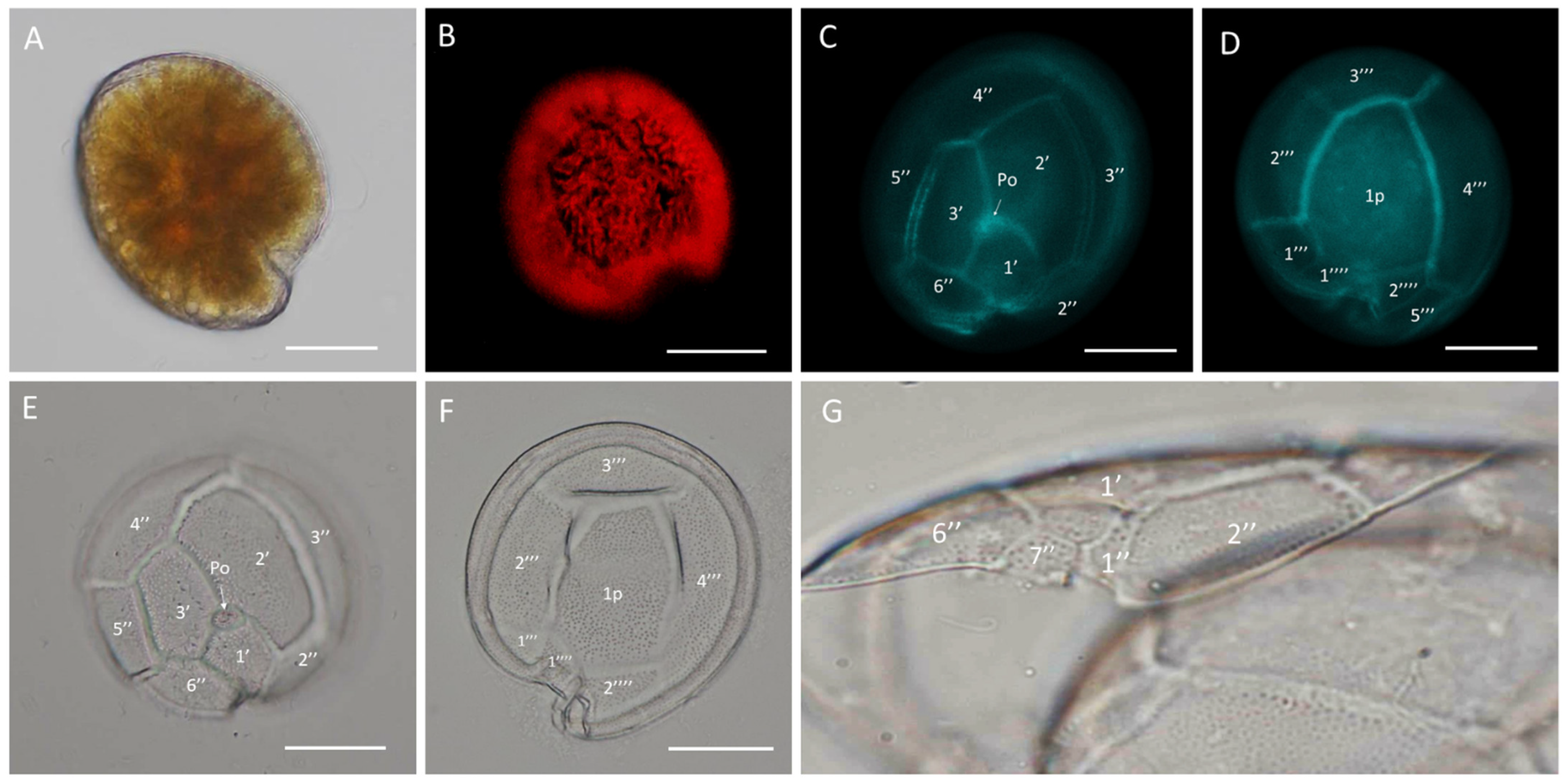
Gambierdiscus cf. caribaeus strains GbSa-2 and GbSa-7 were identified as photosynthetic, lens-shaped cells that were rounded in the apical view and compressed in the anterior-posterior axis, with golden granular chloroplasts (Figure 1A,B). The cells measured 70.31–91.61 ± 5.90 µm depth, 83.32–86.32 ± 7.90 µm width, and 61.12–64.57 ± 4.10 µm length (n = 30). The plate formula was Po, 3′, 7″, 6C, 6S, 5′′′, 1p, and 2′′′′ (Figure 1C–F). The apical pore plate Po was in contact with apical plate 1′ 2′, and 3′. The rectangular hatched-shaped 2′ plate was the largest apical plate, with plate 4″ being pentagonal and symmetrical (Figure 1C,E). In the antapical view, between 2′′′ and 4′′′, plate 1p was the largest hypothecal plate; it was broad and pentagonal in shape. In the ventral view, minuscule plate 1″ was rectangular, and trapezium-shape 7″ was also observed (Figure 1G).
SEM micrographs (n = 15) revealed a rounded apical shape with a smooth cell surface and multiple pores scattered across the thecal plates, including symmetrical plate 4″ (Figure 2A). The antapical view showed a broad 1p plate and detail of postcingular plates 1′′′, 2′′′, 3′′′, and 4′′′ (Figure 2B,D). The apical pore plate, Po, was elliptical with a fishhook-shaped opening (Figure 2C). Detailed images of the sulcal zone showed minuscule plates 1″ and 7″, with the cingulum gradually descending near the sulcus (Figure 2E). The dorsal view revealed symmetrical plate 4″ and a narrow, stylized cingulum edge (Figure 2F). Both LM and SEM micrographs of the GbSa-2 strain are shown in Figure S3.
The maximum cell density (1835 ± 117 cells mL−1) in G. cf. caribaeus was recorded during the exponential phase (day 35 for strain GbSa-2). For strain GbSa-7, the maximum cell density (2457 ± 394 cells mL−1) was recorded on day 49. The calculated growth rates were 0.25 ± 0.01 and 0.26 ± 0.05 division·day−1, respectively, with an average generation time of 2.71 ± 0.06 days for G. cf. caribaeus strains.
In G. cf. carpenteri strains GbSa-4, GbSa-5, and GbSa-7, the cells were photosynthetic and lens-shaped, predominantly oblong and irregular in apical view, and compressed in the anterior-posterior axis, with golden granular chloroplasts (Figure 3A,B). The cells measured 72.21–84.91 ± 5.90 µm depth, 71.72–77.62 ± 7.29 µm width, and 59.56–64.57 ± 6.15 µm length (n = 30). The plate formula was Po, 3′, 7″, 6C, 6S, 5′′′, 1p, and 2′′′′ (Figure 3C–F).
In the apical view, plates 1′, 2′, and 3′ were connected to the Po plate. Apical 2′ plate showed a hatched and large form, while plate 4″ was asymmetrically narrow toward plate 3″ and wider toward plate 5″. Plates 2″ and 6″ were the shortest in the precingular area (Figure 3C,E). Plate 1p was long, pentagonal, and broad, joining with small plates 1′′′′ and 2′′′′ in a ridge-like form. Plate 1p was also connected to plate 3″ in a quadrangular shape and to plates 2′′′ and 4′′′ to form the largest quadrangular plate (Figure 3D,F). In the ventral view, minuscule plate 1″ was rectangular and plate 7″ was trapezium-shaped (Figure 3G).
SEM micrographs (n = 15) of G. cf. carpenteri revealed a rounded apical shape with a smooth cell surface and multiple scattered pores. Asymmetrical plate 4″ protruded, forming an apparent rostrum where plate 3″ was articulated (Figure 4A). The antapical view revealed a long, pentagonal plate 1p (Figure 4B,D). The apical pore plate, Po, was elliptical with a fishhook-shaped opening (Figure 4C). The sulcal zone showed minuscule plate 1″ and small plate 7″, which formed ridges toward the adjacent part of the sulcus (Figure 4E). The cingulum was narrow, and the dorsal view highlighted asymmetrical 4″ plate and the apparent rostrum formed by the union of precingular 3″ and 4″ plates and postcingular 3′′′ and 4′′′ plates (Figure 4F). The LM and SEM micrographs of GbSa-5 and GbSa-6 strains are shown in Figures S4 and S5.
Gambierdiscus cf. carpenteri strains GbSa-4 and GbSa-6 presented higher cellular densities (2779 ± 424 and 1815 ± 329 cells mL−1, respectively) compared to that of strain GbSa-5 (892 ± 163 cells mL−1). The growth rates, based on cell counts for the strains with the highest abundance, were 0.25–0.27 ± 0.01 division·day−1, with an average generation time of 2.64 ± 0.09 per day. Strain GbSa-5 showed the lowest growth rate among all strains (0.21–0 ± 0.01 division·day−1, 3.36 ± 0.20 generation time). The growth curves are shown in Figure S6.
Morphological measurements revealed differences in the length-to-width ratios of G. cf. caribaeus cell size (1.05–1.13 ± 0.59 and 1.00–1.12 ± 1.29) compared to that of G. cf. carpenteri. The measurements of the apical pore plate were similar between the strains of both species, although the number of pores differed. Gambierdiscus cf. caribaeus and G. cf. carpenteri exhibited an average of 37.33 ± 3.77 pores and 40.66 ± 2.43 pores, respectively. The length-to-width ratio of plate 1p further distinguished the two species (G. cf. caribaeus: ratio of 1.28 ± 0.11; G. cf. carpenteri: 1.59 ± 0.24; Table 1).
Regarding the ratio of the 2′/4″ suture length and 2′/2″ suture length between the two species, no differences were observed in the measurements (n = 20). In G. cf. caribaeus, this ratio was 0.92 ± 0.01, while in G. cf. carpenteri, the ratio was 0.92 ± 0.04. After the dissociation of the apical and antapical plates, marked morphological differences were only observed in plates 1′, 3″, and 4″.
In addition to the dinoflagellates characterized in this study, we also detected the presence of the potentially harmful dinoflagellates G. cf. carolineanus, G. cf. toxicus, and F. cf. yasumotoi (Figures S7–S9).

2.3. Biochemical Characterization

2.3.1. Pigments Composition

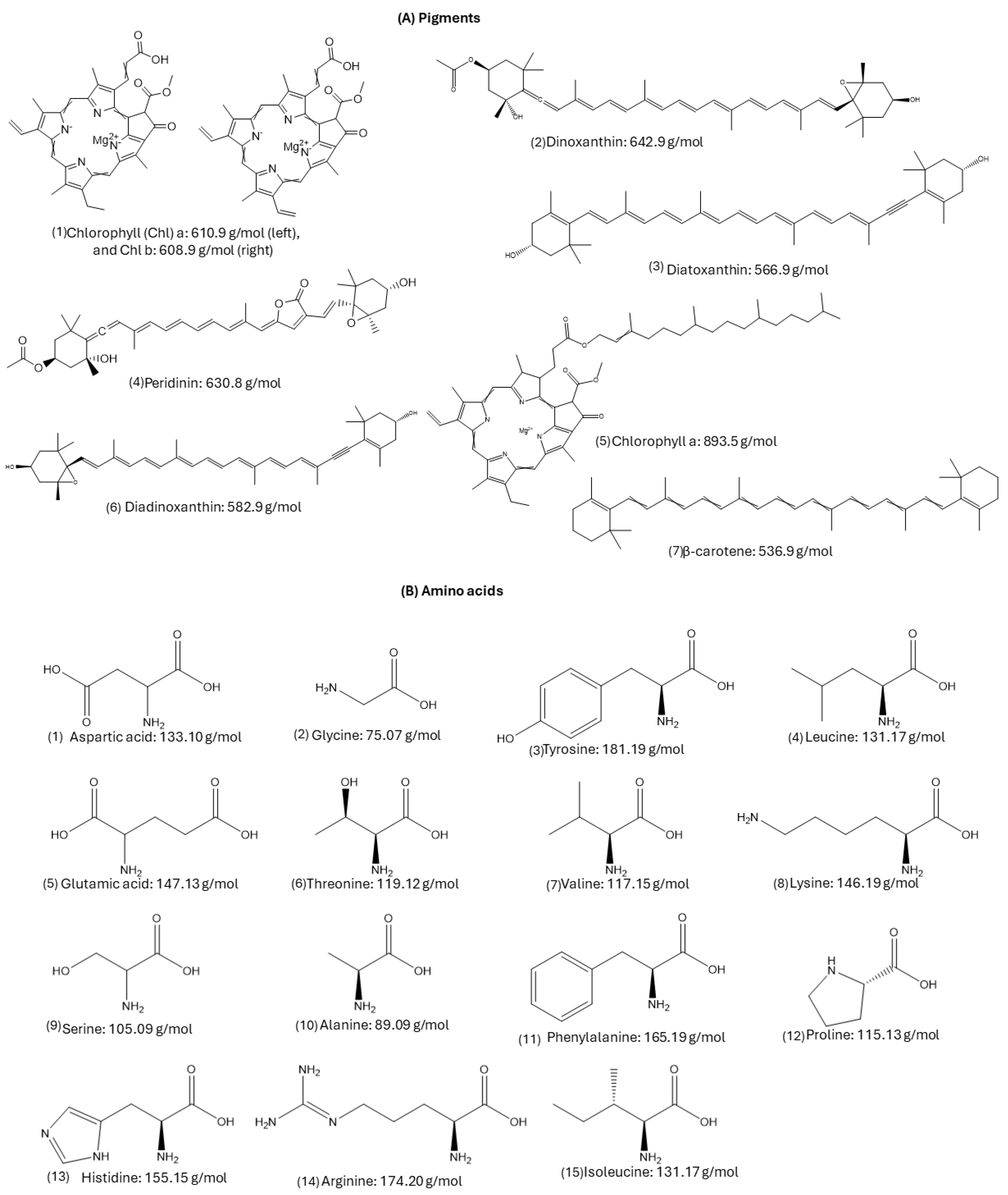
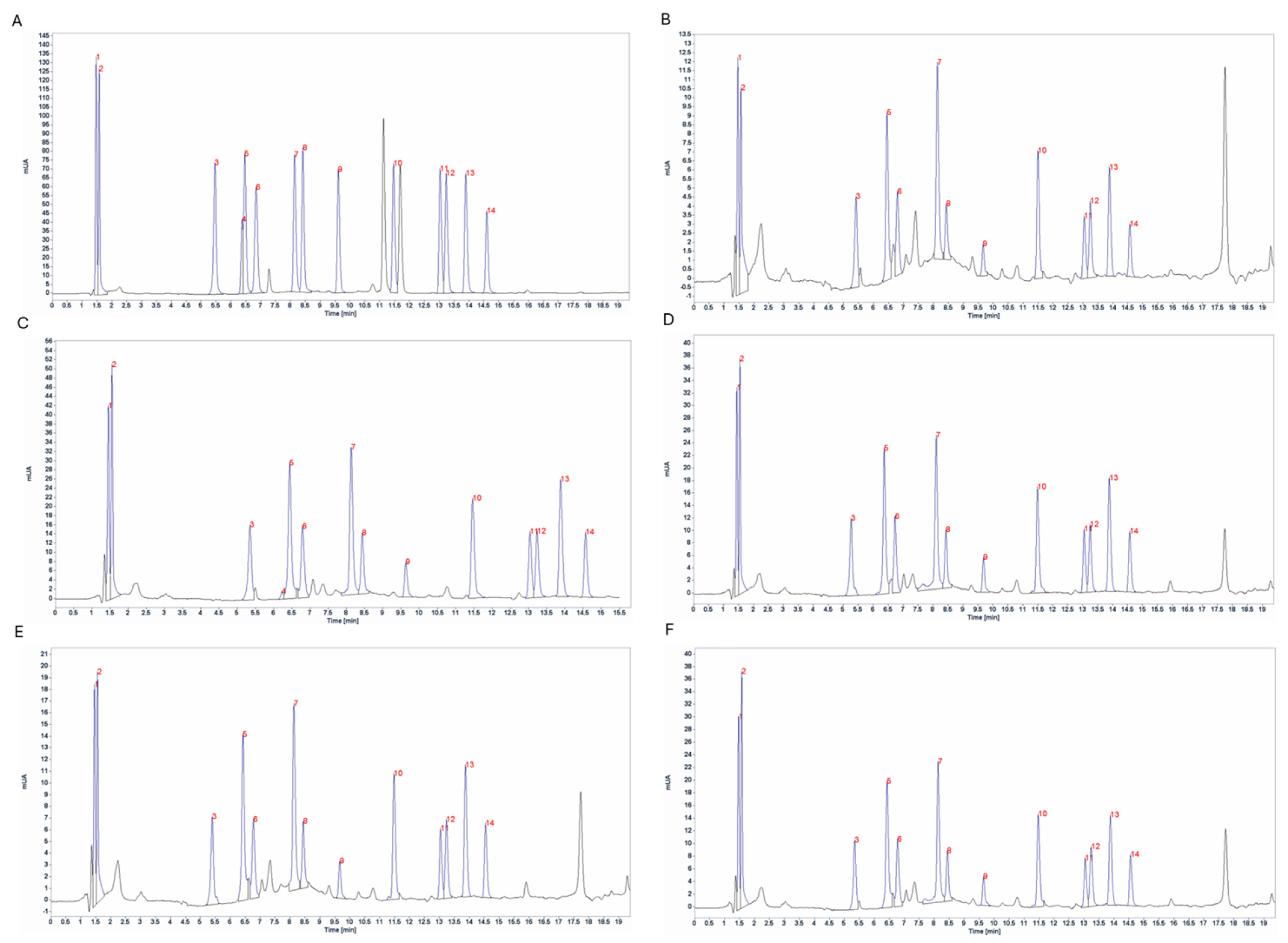
To investigate the composition and concentration of pigments, samples were collected between days 24–25 of culture during the early exponential growth phase. As a result of this, nine pigments were identified in Gambierdiscus strains (Figure 5 and Figure 6).
Pigment production varied between the strains (Table 2). Peridinine was the main carotenoid pigment present in the Gambierdiscus strains (23.89–81.26 pg cell−1). Chlorophyll a was the major pigment in G. cf. carpenteri GbSa-6 (278.9 pg·cell−1), followed by chlorophyll c1, c2 (57.59 pg·cell−1) in G. cf. caribaeus GbSa-2.

2.3.2. Amino Acid Composition

The amino acid profile of Gambierdiscus was determined using HPLC-DAD. Fourteen amino acids were detected, including glutamic acid, aspartic acid, serine, glycine, threonine, alanine, arginine, tyrosine, valine, phenylalanine, isoleucine, leucine, lysine, and proline. Histidine was detected only in G. cf. caribaeus Gbsa-7 (Figure 7).
The amino acid content varied between the strains (Table 3). Glutamic acid had the highest content (14.80–36.39 pg·cell−1), whereas glycine had the lowest (7.72–18.66 pg·cell−1). Strain GbSa-7 contained proline (<52.30 pg·cell−1) and was the only strain to contain histidine.

2.4. Toxicity Assay

2.4.1. Mouse Bioassay (MBA)

All Gambierdiscus strains tested by MBA were positive for CTX-like activity. The toxicity in G. cf. caribaeus GbSa-2 was 18.57 × 10⁻⁴ µg·kg−1·1000 cells−1, which was comparable to the toxicity of strain GbSa-7 (17.08 × 10⁻⁴ µg·kg−1·1000 cells−1). For G. cf. carpenteri strains, significant variations were observed. Strain GbSa-4 showed the highest toxicity (29.07 × 10⁻⁴ µg·kg−1·1000 cells−1), with CTXs-like activity three times higher compared to that of strain GbSa-5 (8.46 × 10⁻⁴ µg·kg−1·1000 cells−1) and more than twice that of strain GbSa-6 (12.54 × 10⁻⁴ µg·kg−1·1000 cells−1).
The main clinical signs related to CTXs-like activity were neurological: paralysis of the rear and front quarters, dyspnea, piloerection, spasms, loss of locomotion, body arching, arched and zig-zag tail movements, tail wriggling, blinking, and ataxia. Gastric signs included polyuria, lethargy due to peritoneal inflammation, diarrhea, and abundant defecation. Cardiovascular signs included labored breathing and vasoconstriction of the ear and other clinical signs like continuous itching and scratching, hypersalivation, closed eyes, and eventual death due to respiratory failure (Figure 8).
All strains of G. cf. caribaeus and G. cf. carpenteri showed toxicity related to MTX-like activity in the MBA at sublethal doses. After 24 h of observation, the LD50 was not recorded in any of the groups of injected mice, but several clinical signs related to MTX-like activity were observed.
The clinical signs associated with MTXs-like activity included neurological signs such as paralysis of the rear and front quarters, dyspnea, piloerection, spasms, body arching, arched tail and tail wriggling, blinking, ataxia, and convulsions. Gastric signs included polyuria, lethargy due to peritoneal inflammation, and abundant defecation. Cardiovascular signs included vasoconstriction of the ears. Other clinical signs included hypersalivation, closed eyes, and tear secretion (Figure 8).

2.4.2. Artemia Assay (ARTOX)

Toxicity was determined using the brine shrimp Artemia salina. The control group did not exhibit any mortality, whereas the organisms exposed to CTXs-like activity of Gambierdiscus strains showed mortality (Figure 9) and clinical signs of toxicity.
At 6 h, the mortality of the brine shrimp exposed to the CTXs-like activity of G. cf. caribaeus strains GbSa-2 and GbSa-7 was 9 ± 1% and 5 ± 0.5%, respectively. The mortality caused by G. cf. carpenteri were 5 ± 1% for strain GbSa-4, 30 ± 3% for strain GbSa-5, and 8 ± 1% for strain GbSa-6.
After 12 h, G. cf. caribaeus GbSa-2 caused 18 ± 1% mortality, while strain GbSa-7 caused 14 ± 1%. The mortalities caused by G. cf. carpenteri in brine shrimp were 19 ± 1% for strain GbSa-4, 40 ± 3% for strain GbSa-5, and 39 ± 2% for strain GbSa-6.
The highest mortality in A. salina was recorded at 18 h of exposure to the CTXs-like activity of G. cf. caribaeus GbSa-2 (45 ± 3%) and GbSa-7 (38 ± 2%). For the G. cf. carpenteri extract, mortality was 38 ± 1% for strain GbSa-4, 90 ± 6% for strain GbSa-5, and 78 ± 5% for strain GbSa-6. This trend remained consistent until 24 h when the mortality of A. salina was 45 ± 3% for GbSa-2 and 43 ± 2% for GbSa-7. The highest mortality was observed for G. cf. carpenteri GbSa-5 (90 ± 12%), followed by GbSa-6 (78 ± 5%) and GbSa-4 (54 ± 3%) (Figure 9A).
Clinical signs in A. salina exposed to CTX-like activity were characterized by changes in swimming behavior. Starting at 6 h, A. salina exposed to G. cf. caribaeus and G. cf. carpenteri showed a loss of orientation while swimming, characterized by swimming in circles and rising and falling from the surface to the bottom. These clinical signs intensified between 6 and 24 h, leading to gasping, lethargy, and paralysis of the thoracic appendages.
For A. salina exposed to MTX-like activity, mortality was recorded at 6 h: 5 ± 0.5% and 5 ± 1% for strains GbSa-2 and GbSa-7 of G. cf. caribaeus and 5 ± 0.5%, 10 ± 1%, and 8 ± 5% for strains GbSa-4, GbSa-5, and GbSa-6 of G. cf. carpenteri, respectively. After 12 h of exposure to the MTX-like activity of G. cf. caribaeus, the mortality of A. salina was 9 ± 1% (GbSa-2) and 11 ± 1% (GbSa-7). At this time, the highest mortality was recorded with GbSa-5 (30 ± 4%) and GbSa-6 (23 ± 2%) from G. cf. carpenteri, while GbSa-4 showed 10 ± 2% mortality. At 18 h, A. salina mortality did not increase further in any of the treatments. At 24 h, A. salina mortality increased by 10% for those exposed to the MTXs-like activity of G. cf. caribaeus GbSa-7 (<22 ± 2%) (Figure 9B).
Although mortality due to MTX-like activity in A. salina was lower compared to that of CTX-like activity, several clinical signs were present from the first hour of exposure. Brine shrimp exhibited erratic swimming, lethargy, a head-up posture, reduced response to stimuli, and gasping within the first 12 h. By the end of the exposure, 80% of brine shrimp had recovered.

2.4.3. Marine Fish Assay

A toxicity test was conducted using longfin yellowtail larvae (Seriola rivoliana). During the first 6 h, only larvae exposed to the CTX-like activity of G. cf. carpenteri GbSa-4 presented 10 ± 1% mortality. After 12 h of treatment, G. cf. caribaeus GbSa-7 caused 10 ± 0.5% mortality. At 18 h, there was no further increase in mortality in any of the treatments. The maximum mortality was recorded at 24 h, with 20 ± 2% mortality in S. rivoliana larvae exposed to the CTXs-like activity of G. cf. carpenteri GbSa-4. No mortality was observed in the control group (Figure 10A).
Clinical signs recorded in S. rivoliana larvae exposed to CTXs-like activity included a loss of balance with abnormal horizontal orientation, abnormal swimming behavior, loss of buoyancy control leading to sinking to the bottom, reduced response to stimuli, erratic swimming, shaking, body arching, irregular ventilation (e.g., hyperventilation), and notable skin darkening.
Larvae exposed to the MTXs-like activity of G. cf. caribaeus GbSa-2 exhibited 10 ± 1% mortality at 6 h, which increased to 20 ± 2% at 12 h. Larvae exposed to the MTX-like activity of G. cf. caribaeus GbSa-7 showed 10 ± 1% mortality at 6 h. At 18 h, mortality due to GbSa-2 and GbSa-7 increased to 30 ± 3%. In contrast, larvae treated with the MTXs-like activity of G. cf. carpenteri GbSa-4 showed 10 ± 0.5% mortality. After 24 h, the maximum mortality of S. rivoliana larvae was 60 ± 4% for those exposed to the MTXs-like activity of G. cf. caribaeus GbSa-2 and GbSa-7 and G. cf. carpenteri GbSa-4. The lowest mortality was observed in larvae exposed to the MTXs-like activity of G. cf. carpenteri GbSa-5 and GbSa-6, with 20 ± 1% mortality (Figure 10B).
The exposure of larvae to dissolved MTX-like activity led to abnormal horizontal orientation, loss of balance, and loss of buoyancy control, causing the larvae to sink to the bottom. Abnormal vertical orientation with head-down posture was also observed, along with behavioral changes such as erratic swimming and pulses of hypoactivity and hyperactivity. Additional signs included shaking, body arching, abnormal respiration (e.g., hyperventilation), irregular ventilation, head shaking, and skin darkening before death.

2.5. Fluorescent Receptor Binding Assay (fRBA)

To measure the binding affinity of the CTX analogs for voltage-gated sodium channel (VGSC) receptors, we employed the fRBA assay. This assay is based on the competition for VGSC site 5 between CTXs and a brevetoxin (PbTx) labeled with fluorescent compounds. The relative fluorescence unit (RFU), defined as the fluorescence relative to the culture biomass, was adjusted to the control (ethanol 100% binding value, representing maximum fluorescence). For the highest concentration of the standard curve, the PbTx standard at 0.1 µg·mL−1 showed 29.8 ± 4.95% RFU. The Karenia brevis (Kb-3) extract (positive for PbTx-2 and Pbtx-3) resulted in 95.15 ± 2.10% RFU, with a concentration of 1.3 ± 0.07 pg eq PbTx per cell. For G. cf. caribaeus GbSa-2, the blinding value was 95.6 ± 0.89% RFU with a maximum concentration of 2.59 ± 0.25 pg eq PbTx per cell. Strain GbSa-7 showed a binding value of 92.8 ± 1.01% RFU with a concentration of 2.19 ± 0.23 pg eq PbTx per cell.
The affinity for VGCS in G. cf. carpenteri GbSa-4 was 93.9 ± 1.27% RFU, with a maximum concentration of 2.51 ± 0.30 pg eq PbTx per cell. The highest competition for VGCS was recorded for strains GbSa-5 (89.8 ± 2.50% RFU and 1.15 ± 0.31 pg eq PbTx per cell) and GbSa-6 (95.8 ± 3.49% RFU and 1.57 ± 0.05 pg eq PbTx per cell).

3. Discussion

3.1. Morphological Identification

Two out of the five isolated strains were identified as G. cf. caribaeus, and three of them were identified as G. cf. carpenteri, as both species share similarities in the plate formula Po, 3′, 7″, 6C, 6S, 5′′′, 1p, and 2′′′′ (Figure 1, Figure 2, Figure 3 and Figure 4). However, they can be distinguished by the shape of plates 1p and 4″, as suggested by the dichotomous key of Litaker et al. [39] and the description of Diaz-Ascencio et al. [40]. Gambierdiscus cf. caribaeus (GbSa-2 and GbSa-7) is characterized by a rounded and lenticular-shaped cell, short and wide plate 1p, and symmetrical plate 4″, which is consistent with the original descriptions by Litaker et al. [39] and Soler-Onis et al. [41] in Atlantic waters and Diaz-Ascencio et al. [42] in the Caribbean region (Figure 2). Although G. caribaeus shares morphological features with its sister species, G. carpenteri and G. jejuensis, the presence of a symmetrical plate 4” allows for satisfactory differentiation. However, under culture conditions, this is not always reliable, as cellular deformation occurs during the initial generations, leading to aberrant cell shapes that complicate the identification of the typical plates characterizing the genus Gambierdiscus [40,42,43]. In this study, the number of deformed cells in G. cf. caribaeus strains was low, and the morphological features aligned with previous descriptions, suggesting that measurements were accurate.
Strains of G. cf. carpenteri (GbSa-4, GbSa-5, and GbSa-6) from the Gulf of California exhibited clear morphological characteristics similar to those described by Litaker et al. [39]. These included oval cells, a narrower and elongated plate 1p extending toward plate 3′′′′, and an asymmetric plate 4″ (Figure 3C–F). Cell size was similar to those of the strains reported for G. carpenteri isolated from Australia, Belize, the Mexican Caribbean, and the Philippines [39,44,45]. However, variations were observed in the shape of plate 2′, which ranged from rectangular to hatchet-shaped. Additionally, the G. cf. carpenteri strains described in this study presented several morphological characteristics similar to those described by Ramos-Santiago et al. [38] for G. carpenteri in the Gulf of California, with plates 2′ and 4″ corresponding to those described by Litaker et al. [39,44]. Furthermore, in the description by Litaker [39], G. carpenteri cells presented a rostrum resulting from the fusion of plates 3′ and 4″ and postcingular plates 3′′′ and 4′′′, which was similarly observed in G. cf. carpenteri (SEM GbSa-4 and GbSa-6, n = 14) (Figure 4A–F and Figure S6). However, the cellular morphology of G. cf. carpenteri (GbSa-4 and GbSa-6) was more consistent with the descriptions by Kohli et al. [44] and Ramos-Santiago et al. [38]. The variety of forms among the strains isolated from the same area supports the diversity and morphological plasticity of G. carpenteri, suggesting that the diverse coastal environments of the Gulf of California may harbor new ecotypes of this species.
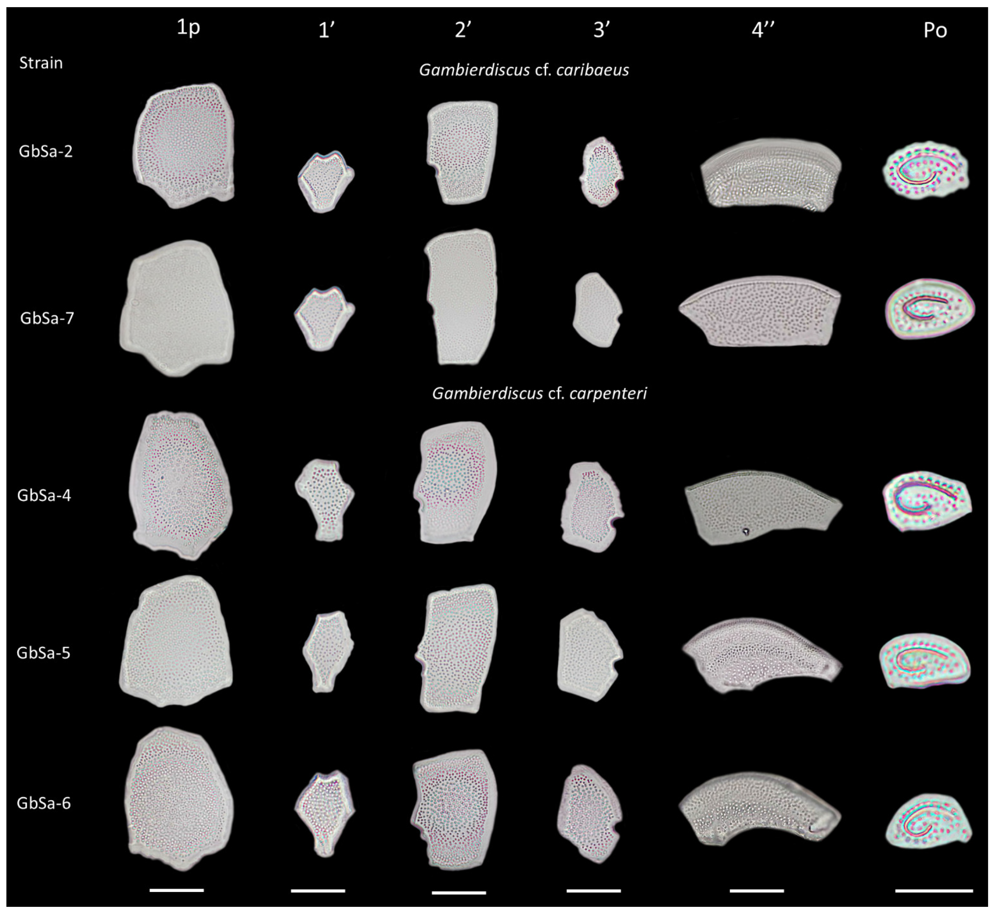
Plate dissociation allowed for precise measurements and detailed observations using LM, which confirmed the formula tabulation [39]. Through plate dissociation, it was possible to support the morphological distinction between G. cf. caribaeus and G. cf. carpenteri and to perform more precise measurements, particularly in plate 1p. Differences in the ratio were observed between G. cf. caribaeus and G. cf. carpenteri (Table 1), although the diversity in the shape of plate 2′ did not correspond to the ratio of the 2′/4″ suture length and 2′/2″ suture length between the two species, as suggested by Diaz-Asencio et al. [40], Vacarizas et al. [45], and Litaker et al. [39]. Plate dissociation was performed to compare the shape and size of plates 1′ and 3′, leading to the suggestion of adding the shape of plate 4″ to the tabulation to distinguish G. cf. caribaeus from G. cf. carpenteri (Figure 11). Plate 1′ was more compact and robust in G. cf. caribaeus, while plate 3′ was wider in G. cf. carpenteri than in G. cf. caribaeus, including in deformed cells (Figure 11). The asymmetric plate 4″ was similar in all G. cf. carpenteri strains, and upon dissociation, it was compared with fixed cells of G. autrales (VGO1250), G. excentricus (VGO911), and G. carolineanus (GbEs-4), which also exhibited an asymmetrical plate 4′. In G. cf. carpenteri, the width contrast in the 3″/4″ suture was twice as small as that in the 4″/5″ suture, whereas in G. autrales, G. excentricus, and G. carolineanus, the proportion was lower (1.5). Regarding the forms of plate Po, only the number of pores showed differences between G. cf. caribaeus and G. cf. carpenteri. Therefore, we recommend performing plate dissociation to morphologically distinguish G. caribaeus, G. carpenteri, and G. carolineanus (Figure 11 and Figure S7).
Gambierdiscus caribeaus and G. carpenteri are two species within the genus that present a cosmopolitan distribution [46]. They exhibit a wide range of tolerance to variations in temperature, salinity, and irradiance [44,46], which contributes to their broad distribution and ability to adapt to diverse habitats, as well as their efficient growth rates [47,48,49]. In this study, G. cf. caribaeus and G. cf. carpenteri showed the highest cell growth under culture conditions, with average growth rates of 0.25 div/day, similar to those reported in previous studies under optimal salinity (35) and temperature (25 °C) conditions [45,50]. However, G. cf. carpenteri strain GbSa-5 exhibited a slower growth rate and lower cell abundance compared with the GbSa-4 and GbSa-6 strains.
Although the toxicity levels of G. carpenteri are considered medium-to-low and their contribution to CFP is uncertain [46], it is important to consider the potential implications of the presence of this species in the Gulf of California. Various factors have been identified that could increase the risk of CFP cases [1,46,51], including climate change, coastal area modifications, and increased nutrient and pollution loads. Recently, Bahía de La Paz has been impacted by tropical storms and hurricanes, leaving numerous boats at the bottom of the sea in coastal areas and nearby islands [52,53]. Some studies have suggested that the abundance of Gambierdiscus spp. does not depend on the presence of macroalgae and that certain species are associated with artificial substrates [49,51,54,55,56]. Consequently, the remains of these vessels may represent substrates that promote the potential settlement and proliferation of Gambierdiscus and Fukuyoa species associated with CFP.
At sampling points near the coast, G. cf. carpenteri was the dominant species, while the highest diversity was recorded at Fang Ming, a sunken ship converted into an artificial reef near Espiritu Santo Island (Table S2, Figure 12), in addition to G. cf. carpenteri, G. cf. carolineanus, and F. cf. yasumotoi (but not G. cf. caribaeus). The Gulf of California has 244 islands, islets, and coastal areas [57]. Species of the genera Gambierdiscus and Fukuyoa are closely associated with the insular areas of the Gulf of California. Morquecho-Escamilla et al. [37] reported the presence of Gambierdiscus and Fukuyoa on San Jose Island, and Ramos-Santiago et al. [38] reported their presence on La Gaviota Island. Including this study, reports of Gambierdiscus and Fukuyoa cover only 1.2% of Gulf of California islands, suggesting that these islands are potential habitats for a great diversity of Gambierdiscus and Fukuyoa species, including novel species.

3.2. Biochemical Characterization

3.2.1. Pigments Composition by HPLC-DAD

Pigment analysis has been proposed not only for physiological studies of dinoflagellates [58,59,60,61] but also as a pigment fingerprint for monitoring harmful microalgae species during harmful algal blooms (HABs) [62,63,64,65,66,67]. Using thin-layer chromatography (TLC), Durand and Berkaloff [68] and Indelicato and Watson [69] conducted the first analyses of pigments produced by the genus Gambierdiscus, identifying the presence of Chl a, Chl c1, Chl c2, Diadino, Dino, Peri, and ββ-car. Since then, at least ten studies have been conducted to further understand pigment production and composition [63,70,71,72,73] (Table 4). Recently, Malto et al. [27] and Yon et al. [28] described the presence of these compounds using liquid chromatography coupled with mass spectrometry as part of a study on the metabolome of three strains belonging to three species of Gambierdiscus isolated from the Philippines and 15 strains of five species from the Atlantic Ocean. The analysis and evaluation of the pigment profile, along with other metabolites (e.g., fatty acids, sterols, carbohydrates, amino acids, and toxins), in various species of dinoflagellates allows for the chemical diversity of secondary metabolites to be explored. Biomarkers can then be identified to differentiate between Gambierdiscus species (chemotaxonomy), evaluate the possible presence of ecotypes, and study the metabolism and biosynthesis of toxins. In this study, we detected the presence of seven pigments in G. cf. caribaeus and G. cf. carpenteri, and the profiles obtained were compared globally with those from other isolates of species within this genus (Table 4; Figure 6).
The predominant pigment profile for these two species included Chl a, Chl c2, Diadino, Dino, Diato, Peri, and ββ-car. Notably, we observed that strain GbSa-7 was distinct from strain GbSa-2; both corresponded to G. cf. caribaeus, as it produced a higher concentration of Chl a (203.79 pg·cell−1) and Dino (4.71 pg·cell−1). This effect was also observed in the amino acids profile, with histidine (13.04 pg·cell−1) detected only in this strain, along with the highest concentration of proline (52.30 pg·cell−1; Table 3 and Figure 6). Therefore, we consider that this strain may represent an ecotype. This hypothesis will be further supported by an analysis of the toxin profile and other metabolites (e.g., fatty acids, sterols, and carbohydrates) using analytical methods.

3.2.2. Amino acid Composition

Analyzing the amino acid profiles of dinoflagellates is useful for exploring the chemical diversity of secondary metabolites; these profiles can serve as biomarkers for species identification in chemotaxonomy. They also facilitate the study of ecotypes, metabolism, and toxin biosynthesis. This analysis also allows for assessments of intracellular changes in response to diurnal variations in light, nitrogen, and nutrients, as well as the physiological status of natural dinoflagellate populations [74,75].
The amino acids profile of G. cf. caribaeus and G. cf. carpenteri strains revealed the presence of 14 amino acids: glutamic acid, aspartic acid, serine, glycine, threonine, alanine, arginine, tyrosine, valine, phenylalanine, isoleucine, leucine, lysine, and proline. Gambierdiscus. cf. caribaeus GbSa-7 was the only strain that presented histidine (13.04 pg·cell−1), and it also had the highest concentration of proline (52.30 pg·cell-1; Table 4 and Figure 6). Therefore, we considered that this strain may be a potential ecotype. Figure 6 provides a comparative analysis of the amino acid profile of Gambierdiscus detected in this study with those of other dinoflagellate species.
Studies by Okaichi et al. [76] and Hayashi [77], who used high-speed amino acid analyzers (HSAAA), in addition to the more recent analyses using high-performance liquid chromatography with fluorescence detection (HPLC-FLD) and HPLC-DAD have elucidated the amino acid profiles of various dinoflagellate species from the genera Prorocentrum, Noctiluca, Aureodinium, Glenodinium, Heterocapsa, Scrippsiella, Alexandrium, Gymnodinium, Ansanella, and Takayama [74,78]. Recently, Yon et al. [28] also described the presence of these compounds using high-performance liquid chromatography-mass spectrometry (HPLC-MS) as part of the metabolome of 15 strains from five species of Gambierdiscus originating from the Atlantic Ocean. Furthermore, for the first time, this study presents a screening of the amino acid profile of Gambierdiscus from the Pacific Ocean (Table 5).

3.3. Toxicity Assay

CTX-like and MTX-like activity was evaluated using four different assays: Artemia assay (ARTOX), mouse bioassay (MBA), marine fish assay (MFA) and fluorescent receptor binding assay (fRBA). Toxicity related to CTX-like and MTX-like activity was confirmed in all five clones, although the effects varied across the different toxicological assays.

3.3.1. Mouse Bioassay (MBA)

CTXs-like toxicity was evident in the MBA (18.57 × 10⁻⁴ µg·kg−1·1000 cell−1 and 17.08 × 10⁻⁴ µg·kg−1·1000 cell−1 in G. cf. caribaeus GbSa-2 and GbSa-7, respectively, and 29.07 × 10⁻⁴ µg·kg−1·1000 cell−1, 8.46 × 10⁻⁴ µg·kg−1·1000 cell−1, and 12.54 × 10⁻⁴ µg·kg−1·1000 cell−1 in G. cf. carpenteri GbSa-4, GbSa-5, and GbSa-6, respectively). Clinical signs were also observed for MTX-like activity; however, lethality was only associated with CTX-like activity. The predominant clinical signs were neurological (50–62%), followed by others (10–22%), cardiovascular (7–22%), and gastric (8–16%) across all analyzed strains. In the case of MTX-like activity, neurotoxicity signs predominated (42–70%), followed by gastric (10–25%), others (8–25%), and cardiovascular (8–14%) (Figure 8). These clinical signs were consistent with those described for these types of toxins [79,80]. When comparing the effects of these two types of toxins detected in strains of G. cf. caribaeus and G. cf. carpenteri, a higher percentage of neurotoxic clinical signs was observed for MTX-like activity, although it was not lethal in this assay.

3.3.2. Artemia Assay (ARTOX)

In ARTOX, toxicity was also higher for CTX-like activity compared to that of MTX-like activity (Figure 9A,B). The highest mortality was recorded at 18 h of exposure to the CTXs-like activity of G. cf. caribaeus (GbSa-2: 45 ± 3% and GbSa-7: 38 ± 2%) compared to those of the three strains of G. cf. carpenteri (GbSa-4: 38 ± 1%; GbSa-5: 90 ± 6%; GbSa-6: 78 ± 5%). Clinical signs in adult brine shrimp exposed to CTX-like activity were characterized by changes in swimming behavior, starting at 6 h in G. cf. caribaeus and G. cf. carpenteri. These changes included loss of swimming orientation characterized by circular swimming and rising and falling from the surface to the bottom, which intensified between 6 and 24 h, with gasping, lethargy, and paralysis of the thoracic appendages. These clinical signs were consistent with those described for this type of toxin in fish extracts and Gambierdiscus and Fukuyoa extracts [81,82]. Although mortality associated with MTX-like activity was lower, several clinical signs were observed, such as erratic swimming, lethargy, head-up posture, under-reactive response to stimuli, and gasping within the first 12 h. By the end of the exposure, 80% of the brine shrimp had recovered. The toxic effects of some species of benthic dinoflagellate species, such as Prorocentrum, Ostreopsis, Gambierdiscus, and Fukuyoa, have been previously demonstrated by Neves et al. [83], Heredia-Tapia et al. [84], Ajuzie [85], Faimali et al. [86], and Gong et al. [87]. Additionally, Gambierdiscus and Fukuyoa have been used as CTXs-like and MTXs-like bio-vehicles for metabolic studies of these polyether toxins in marine fish [88,89].

3.3.3. Marine Fish Assay

The MFA elucidated an opposite effect to that of the MBA, as MTX-like activity was highly ichthyotoxic for S. rivoliana larvae. The toxicological effects of CTXs-like activity produced by Gambierdiscus in fish (larvae, juveniles, and adults) have been evaluated under controlled conditions in various freshwater and marine species using different methods, such as feeding with contaminated fish pellets, dinoflagellates that produce these toxins, or a bio-vehicle like brine shrimp, as well as injections, and in some cases, dissolved toxins in seawater [89,90,91,92]. Lewis [93] described CTXs as potent ichthyotoxins. Pure ciguatoxin-1 (CTX-1) and CTX-2 added to water containing Gambusia affinis induced similar clinical signs, including pronounced opercular movement and uncoordinated swimming, preceding death.
Previous studies have reported the negative effects of CTXs-like activity (ranging from behavioral changes to toxicogenomic effects) on fish embryos, larvae, juveniles, and adults of marine and freshwater species (e.g., Thalassoma bifasciatum, Epinephelus fulvus, Lutjanus apodus, L. mahogoni, Micropterus salmoiedes, Oryzias melastigma, Oryzias latipes, Gambusia affinis, Serranus cabrilla, and Pterois volitans). These effects include skin color variations, hypoactivity, loss of equilibrium, erratic swimming, jerky feeding movements, loss of orientation, and death, as well as induced hyperkinetic twitching, severe spinal deformities, and an inability to feed [90,91,92,93,94,95,96,97,98,99,100,101,102,103,104,105,106].
Feeding cichlid fishes (Oreochromis niloticus and O. mossambicus) with Gambierdiscus causes balance abnormalities and erratic spiral swimming behavior [99]. Other studies under controlled laboratory conditions have also shown that feeding with different concentrations of Gambierdiscus causes changes in coloration, inactivity, adverse effects on swimming and orientation, an inability to eat, and, in some cases, death in reef fish species such as Thalassoma bifasciatum, Acanthurus bahianus, and A. chirurgis [91,92,94,98]. Gambierdiscus also causes color modifications and behavioral changes in Chromis chromis and a decrease in liver size and coloration of this organ in Serranus cabrilla [101]. Purified extracts of toxins from this dinoflagellate caused changes in Na+ content in the muscle of Chelon labrosus [95], epithelial degeneration of the intestine, dilation of the gill filaments in Pomacentrus wardi and Chromis nitida [96], and mortality in Gambusia affinis [93]. Galarza et al. [107] described the effect of CTXs (obtained from barracuda) on the aggregation of pigment granules in the melanophores of teleost fish. A detailed pathological study using transmission electron microscopy [101] demonstrated structural alterations in the liver of the cabrilla Serranus cabrilla related to the ingestion of Gambierdiscus in the diet. Marked intercellular fibrosis, a decrease in the number and length of microvilli in the sinusoidal and canalicular areas of the hepatocytes, lipid accumulation, a high number of binucleated cells, nucleolar disorganization, loss of condensed heterochromatin in the periphery of the nucleus, nuclear pyknosis, dilated rough endoplasmic reticulum, and dilated mitochondria were observed.
Although MTXs-like toxins are among the most potent ichthyotoxins [108] and some of the largest and most powerful toxins known [109,110], their accumulation in fish and their ichthyotoxicity has not been extensively evaluated [108,111,112]. MTX was first found in the viscera of the surgeon fish Ctenochaeus straiatus, which bears the Tahitian name “maito” and has been occasionally associated with CFP [110]. The amphiphilic nature of MTX, derived from the uneven distribution of polar functional groups in the molecule, is thought to play an important role in its interaction with cell membranes, contributing to its potent activity [108,110,113,114].
MTXs, which are also produced by Gambierdiscus, have been described for their negative effects on the gills of the freshwater white cloud mountain fish Tanichthys albonubes. MTX caused marked vacuolization of the cytoplasm of chloride cells in the epithelium. Additionally, marked apoptosis was observed not only in the gill epithelial cells but also in all mesenchymal cells of the gill filaments, except in the cartilage in the terminal state [115].
Igarashi et al. [108] evaluated the ichthyotoxic activity of MTX added to water in T. albonubes. The LC50 of MTX in these fishes in Ca2+-free media (pH 8) was 5 nM, but this value decreased significantly to 3 pMa after the pH increased to 8 and the Ca2+ concentration increased to 2 mM. Therefore, the authors suggested that in a marine environment, MTX is 2000 times more toxic to fish than brevetoxin-3 (PbTx-3), highlighting the need for special attention to be paid to the extraordinarily high ichthyotoxicity of MTX.
Kholi et al. [111] described the accumulation of MTXs under controlled conditions in carnivorous marine fish, using Gambierdiscus as a source of these toxins. Juvenile mullet (Aldrichetta forsteri) were fed pellets containing G. australes; these were also used as food for the snapper Pagrus auratus. The transfer of MTX-1 was confirmed by LC-MS/MS detection in viscera, liver, and muscle samples.
In this study, S. rivoliana larvae exposed to dissolved MTX-like activity presented clinical signs after 6 h after exposure. MTX-like activity was highly ichthyotoxic for S. rivoliana larvae, which exhibited changes such as abnormal horizontal position, loss of balance, loss of buoyancy, control leading to sinking to the bottom, abnormal vertical orientation with a head-down posture, behavioral changes in swimming, constant pulses of hypoactivity and hyperactivity, erratic movements, shaking, body arching, abnormal respiration activity, hyperventilation, irregular ventilation, head shaking, and notable skin darkening before death. After 24 h of exposure, the maximum mortality of S. rivoliana larvae was 60 ± 4% in those exposed to G. cf. caribaeus (GbSa-2 and GbSa-7) and G. cf. carpenteri (GbSa-4). The lowest mortality (20 ± 1%) was recorded in the groups of larvae exposed to MTX-like activity from G. cf. carpenteri (GbSa-5 and GbSa-6) (Figure 9B). However, after 24 h of the acute test, the surviving larvae were moribund, with most of them found at the bottom.
During epizootics and mass mortality events of reef fish (Pomacanthus paru, P. arcuatus, Holocanthus tricolor, Centropyge argi, Scarus taeniopterus, Chromis cyaneus, Diodon holocanthus, Cantherhines macroceros, Acanthurus chirurgis, Chaetodon sedentarius, and C. capistratus) observed in Florida and the Caribbean, Ladsberg [116] suggested a possible association with benthic HABs caused by dinoflagellates of the genera Gambierdiscus, Prorocentrum, and Ostreopsis and cyanobacteria of the genus Lyngbya as potential etiologies of these events. In Cabo Pulmo in the Gulf of California, Nuñez-Vazquez et al. [34] described epizootics and mass mortality of reef fish (tons) potentially associated with benthic HABs, highlighting the presence of Gambierdiscus, Ostreopsis, Prorocentrum, and Lyngbya species. Recently, massive die-offs of reef fish in Florida have been described, with individuals showing signs of acute poisoning similar to those observed in some trials of acute exposure to toxins from Gambierdiscus and other benthic dinoflagellates. Therefore, it is important to study whether Gambierdiscus toxins are associated with these epizootics, as has been recently suggested.
Based on existing knowledge and the results observed in the present study, new studies should be conducted that evaluate the ecotoxicological role of toxins and other related compounds from Gambierdiscus and other benthic species in the health of fish during different life stages in the wild and under aquaculture conditions. In the latter case, their importance as future animal models to study the metabolism and biosynthesis of CFP toxins and other syndromes of benthic origin should also be explored.

3.4. Fluorescent Receptor Binding Assay (fRBA)

Although fRBA is not specific in detecting the presence of toxins, the sensitivity of this test has demonstrated its efficiency in fish tissues, Gambierdiscus, and Fukuyoa for detecting compounds related to VGSC site 5, such as CTX and PbTx [42,117,118]. In this study, G. cf. caribaeus showed concentrations based on the VGSC competitor of 2.19–2.59 ± 0.24 pg eq PbTx cell−1, a medium-to-low concentration similar to that reported for G. caribeaus from Cuba [42]. The estimated concentration for G. cf. carpenteri samples ranged between 1.15–2.51 ± 0.31 pg eq PbTX·cell−1, which is three times lower than that reported by Vacarizas et al. [45] (7.48 ± 0.49 pg PbTx eq·cell−1). Although PbTx was used as a standard reference to quantify the activity of toxins related to the sodium channel, previous studies have reported better sensitivity during quantification when using CTX competition as a reference standard [41,117] compared to standardized tests such as CBA-N2a and ELA [47,118]. In the present study, strains of G. cf. caribaeus and G. cf. carpenteri (Table 6) support the conclusion that these species present medium-to-low toxicity compared to G. exentricus and G. polynesiensis, which are representatives of the genus with higher CTXs production per cell. Future studies should compare the binding percentage with a CTX standard due to variations in the capacity to bind VGSC, as the extracts tested are likely to contain a mixture of analogs that could affect the sensitivity and binding signal, which could differ when quantifying using the brevetoxin standard.
Table 6 describes and compares the main toxicity assays performed at the global level and the marine toxins detected in the G. caribaeus and G. carpenteri strains. Various assays and analytical techniques have been used to determine the toxicity and chemical composition of the toxins produced by these two species in different parts of the world. Toxicity varies from nontoxic strains to those that present high toxicity. Some reports indicate 1.4 ± 0.6 fg CTX3C eq cell−1 in strains from Hawaii [126]), 0.3–1.4 fg CTX3C eq cell−1 in strains isolated from the Caribbean [39], and 7.48 ± 0.49 pg PbTx-3 eq cell−1 and 12.36 ± 4.38 pg PbTx-3 eq cell−1 in strains from the Philippines (all G. carpenteri) [27,44,45]. In addition, G. carpenteri strains from Australia have been reported to have a toxicity of 2.4 mg kg−1 per MBA, and Ramos-Santiago et al. [38] reported a CTX-like toxicity of 5.9 mg kg−1 by MBA in Isla Gaviota, Gulf of California.
In G. caribaeus, a toxicity of 0.66 ± 0.34 fg CTX3C eq cell−1 was described in strains from the Caribbean [42], 1.0–1.6 fg CTX3C eq cell−1 in strains from Hawaii [118,126], 15.89 fg CTX1B eq cell−1 in strains from the Canary Islands (127), 0.54 fg CTX3C eq cell−1 in strains from the Gulf of China [123], and 0.6 ± 0.2 pg cell−1 and 9.8 ± 0.6 pg cell−1 (Gambierone) and 7.1 ± 4.2 pg cell−1 (44-methylgambierone) in strains from St. Thomas, US Virgin Islands [24]. For the strains of G. caribaeus and G. silvae, a new ciguatoxin was isolated in the Caribbean and named C-CTX-5 [24,25]. This finding represents a significant advance in research on the origin of CFP in the Caribbean, as the precursor toxin from its algal source and its relationship with CTXs in fish that cause the disease was previously unknown, hindering the development of detection methods, diagnostics, and monitoring programs. In the same study, various in vitro and chemical techniques were applied to identify the new analog (C-CTX-5), and it was verified that the treatment of this analog using fish liver microsomes converted it to C-CTX1/2, the dominant C-CTX in ciguatoxic fish from the Caribbean. Recently, C-CTX-5 was also identified for the first time in an “amberjack” (Seriola sp.) involved in a CFP case in the Canary Islands, Spain [127]. Based on this, it is necessary to investigate whether G. caribaeus strains from other latitudes, such as those in the present study, can also produce this new ciguatoxin.
The genetic identification of the Gambierdiscus and Fukuyoa species evaluated in this study and chemical analysis of the toxins will be carried out in a subsequent study using analytical tools, given the complexity of obtaining standards and other inputs necessary to detect and analyze CTXs, MTXs, and other related compounds. The toxicity and composition of CTXs, MTXs, and other related compounds in these two species are also described in Table 6.
Over the past decades, the occurrence and geographic distribution of CFP have notably expanded due to anthropogenic activities and climate change, leading to an increase in human illness, greater public health impacts, and larger economic losses. The global spread of CFP has made Gambierdiscus and its toxins a concern for environmental and human health worldwide [22].
In the PBC, cases of CFP are rare and sporadic. However, at least 240 human cases have been recorded in the southern region due to the consumption of the meat, liver, and heads of carnivorous fish, such as snappers (Lutjanus spp.), cabrillas, and groupers (Epinephelus and Mycteroperca), caught near islands and islets [11,12,13,14,15,16,17]. Additionally, epizootics and the massive mortality of reef fish potentially associated with benthic HABs have been reported, with Gambierdiscus, Ostreopsis, Prorocentrum, and Lyngbya species being primarily detected [34].
The PBC is also a region of notable fishery importance, supporting artisanal fisheries that exploit numerous species of teleost fish, elasmobranchs, mollusks, and crustaceans. In the Gulf of California, coastal fisheries target around 70 species, with an annual catch of nearly 200,000 tons. Sport fishing is also prominent, targeting species such as yellowfin tuna (Thunnus albacares), skipjack (Katsuwonus pelamis), billfish striped marlin, blue marlin (Makaira nigricans), black marlin (Makaira indica), sailfish (Istiophorus platypterus), swordfish (Xiphias gladius), dorado (Coryphaena hippurus), yellowtail amberjack (Seriola lalandi), longfin yellowtail (Seriola rivoliana), snappers (Lutjanus spp.), “groupers” (Ephinpehelus spp. and Mycteroperca spp.), and around 40 species of sharks, including Mustelus, Carcharhinus, Alopias, Sphyrna, and Squatina [128]. The region is also important for activities such as whale watching (e.g., Eschrichtius robustus, Megaptera novaeangliae, Balaenoptera musculus, and Balaenoptera physalus) and observing dolphins (e.g., Tursiops truncatus and Delphinus spp.), sea lions (Zalophus californianus), sea turtles, whale sharks (Rhincodon typus), reef fish, a great diversity of invertebrates, and algae [128,129,130]. In recent decades, the southern PBC has experienced increased tourism and urban growth, increased demand for services, and the development of marine fish farming, bivalve aquaculture, and shrimp production in Bahía de La Paz.
The annual economic contribution of sport fishing in the southern PBC was estimated to be $80,801,119 USD between 2008 and 2010, equivalent to 1.4% of the Gross Domestic Product (GDP) of Mexico during that period, according to the National Institute of Geography and Statistics (INEGI) [131,132].
In this study, in addition to G. caribaeus and G. carpenteri, we report the presence Gambierdiscus (G. cf. carolinianus and G. cf. toxicus) and Fukuyoa (Fukuyoa cf. yasumotoi) species in the PBC, where cases of CFP have occurred. The Gulf of California is a potential hotspot for the biodiversity and endemism of benthic dinoflagellate species, which provides an important opportunity to research CTXs, MTXs, CFP-related compounds, other polyether toxins, and secondary metabolites. Studies must be conducted that aim to monitor and evaluate the toxicity, toxin composition, and secondary metabolites of different species of Gambierdiscus and Fukuyoa in this region. The results of these studies are essential for protecting human and animal health. These studies also represent an opportunity to advance research on chemical diversity (metabolomics), which could contribute to characterizing the species and ecotypes present and their potential biotechnological or therapeutic applications.

4. Materials and Methods

4.1. Isolated and Algal Cultures
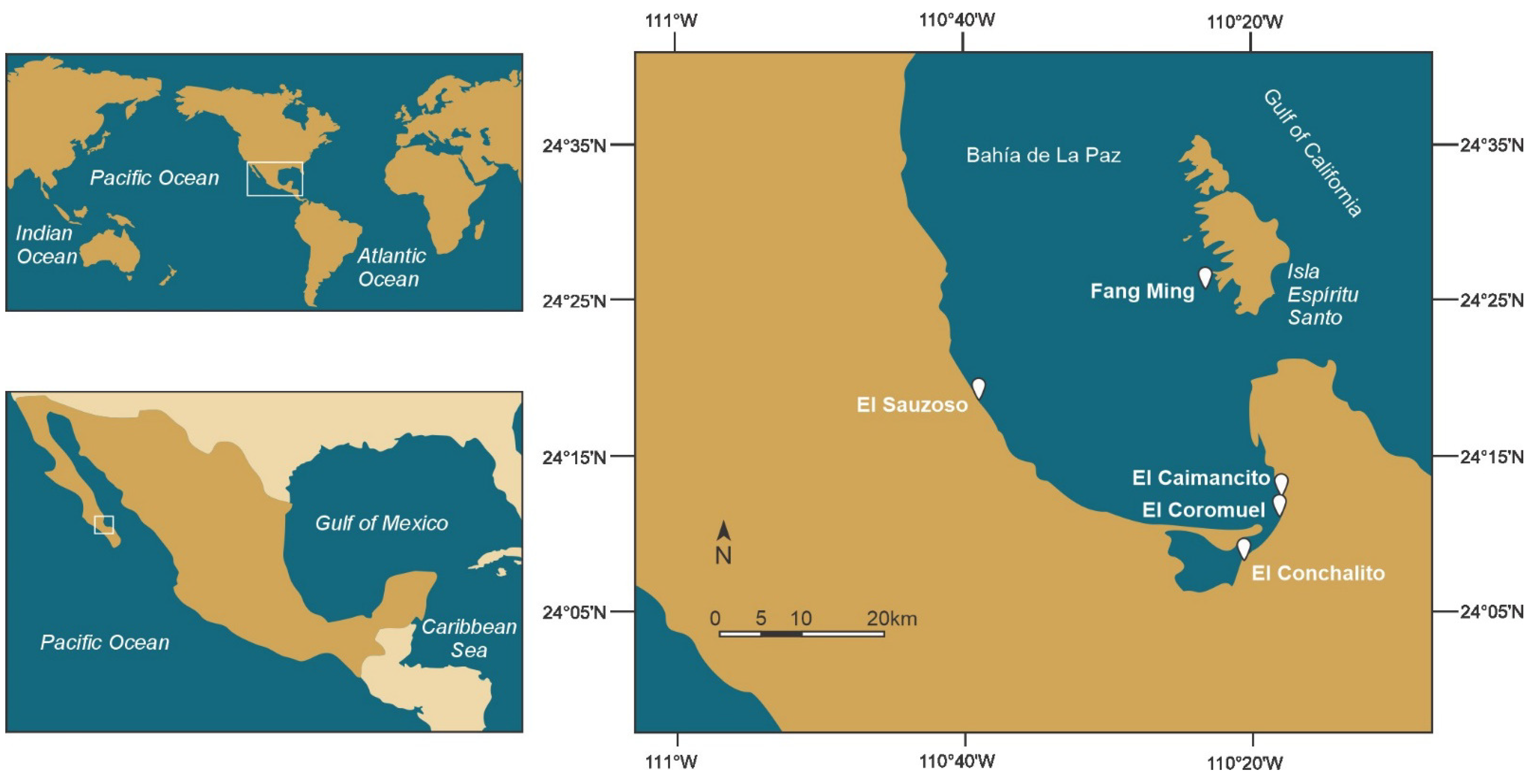
During August 2022, samples of the macroalgae Dyctiota spp. and Padina spp. (associated with corals) were collected by snorkeling at a depth of less than 4 m in the coral reef of El Sauzoso, from artificial substrates at Playa Coromuel, from the rocky reef at El Caimancito, and from the sandy intertidal plane of El Conchalito. Additional samples were collected at the Fang Ming scuba diving site in Espiritu Santo Island (Figure 12).
Figure 12. Sampling sites in Bahía de La Paz used to collect Gambierdiscus species.
Marinedrugs 22 00422 g012
Samples were carefully incubated in Nalgene 1-L bottles and immediately transported to the Marine Toxins and Amino Acids laboratory of CIBNOR. Gambierdiscus cells were isolated individually from macroalgae, rocks, and sandy sediments using a capillary micropipette under an inverted microscope (Labomed, TCM 400, Hicksville, NY, USA). To establish the culture, the isolated cells were transferred into 24-well flat bottom plates (Costar, Boston, MA, USA) with 1 mL of modified K medium [133] containing 1 mg/L of germanium oxide. After 45 days, the wells containing more than 20 cells were transferred to flat bottom culture tubes (Pyrex®, Sunderland, UK) containing 20 mL of medium. These cultures were maintained in the culture room of the CIBNOR microalgae collection (25 ± 2 °C, 35 salinity, a 12:12 light-dark cycle, and irradiance of 35 mmol photon m−2 s−1 supplied by LED light (MAGGA T8 LED STD 18 W, CDMX, Mexico)). All cultures were carried out under these conditions.
Growth rates and generations per day were calculated according to the methods described by Guillard and Ryther [134] and Guillard [135]. Briefly, triplicate cultures of each species were maintained in stock cultures in 300-mL Erlenmeyer flasks with 100 mL of medium at an initial density of 50 cells·mL−1. Every seven days, 2-mL samples were fixed with Lugol’s solution Utermöhl for cell counts on Sedgwick-Rafter slides under a compound light microscope (Olympus BX41, Tokyo, Japan). Serial culture batches were established in 2.8-L Fernbach flasks (Pyrex®) with 1 L of culture medium to increase biomass to evaluate morphological taxonomy, pigments, and amino acids, as well as the mouse bioassay, fluorescent receptor binding assay (fRBA), artemia assay (ARTOX), and larvae fish assay.

4.2. Morphological Identification

4.2.1. Light Microscopy LM

Observations, measurements, and photomicrographs were conducted on 30 randomly selected live Gambierdiscus cells. Cell size, including depth (ventral/dorsal distance), width (transdiameter), and length (apical/antapical axis), was recorded under a microscope (Olympus BX41, Japan) equipped with a camera (SC-MOUNT-YW500 Sony sensor IMX335, ImageView software 2.0), and the system was calibrated using a micrometric slide (1–1000 µm) with achromatic (40×/0.65 Ph2) and 100×/1.25 Oil Ph3 ∞/−) Olympus objectives. Ten milliliters of the culture were collected in 15-mL conical tubes, and cells were fixed with 4% glutaraldehyde. To identify cell plates, a 5-mL aliquot was centrifuged at 1200× g for 2 min, the fixative was removed, and cells were washed three times with distilled water before being fixed with 4% sodium hypochlorite to dissociated cell plates. Plate tabulation nomenclature followed the description of Ramos-Santiago et al. [38]. Another 2 mL of fixed sample was washed again with distilled water by centrifugation and treated with Fluorescent Bright 28 (Sigma Aldrich, St. Lois, MO, USA) [136]. Fluorescent Bright 28 (blue emission) and natural autofluorescence of chloroplasts were documented using a scanning laser confocal microscope (Leica® DMi8, Wetzlar, Germany).

4.2.2. Scanning Electron Microscopy SEM

Ten milliliters of culture were fixed with osmium in 15-mL sterile conical tubes (Corning®, Corning, NY, USA). After 24 h, the cell pellet was filtered through membrane filters with a 0.2-µm pore size and 25-mm diameter (Gelman Science, Bloomfield Hills, MI, USA). Cells were washed four times with filtered distilled water and sequentially dehydrated with 30, 50, 75, 95, and 100% EtOH. After critical-point-drying, the cells were transferred and mounted on 12-mm carbon tabs PK/10 (West Chester, PA, USA), coated with gold, and examined under a scanning electron microscope (Hitachi® S300N, Tokyo, Japan) using image capture software (Quartz PCI 200).

4.3. Biochemical Characterization

4.3.1. High-Performance Liquid Chromatography (HPLC)

The pigment composition of five strains of two species of Gambierdiscus species was analyzed by HPLC-UV/VIS according to the methods [66,137], with some modifications. Briefly, 30 mL of Gambierdiscus cells were collected on a glass microfiber filter GF/F (Ahlstrom—Munksjö, Sweden). The filters were extracted with 2 mL of 100% acetone in an ice bath, hand-ground with a glass rod, and stored overnight at −40 °C for full extraction. The extract was recovered after centrifugation (3000× g for 5 min). A 200-µL aliquot of the extract was mixed with 100 µL of 0.5 N ammonium acetate and injected through a 100-µL loop into an HPLC system (Hewlett Packard series 1260) equipped with a photodiode array detector (DAD, 1.2 nm optical resolution). Pigments were separated and quantified using an isocratic HPLC in reverse-phase mode, as described by Vidussi et al. [137] and Bustillos et al. [66]. The mobile phase consisted of MeOH (0.5 N aqueous ammonium acetate, 70:30% v/v (solvent A), and MeOH [solvent B]), with the following gradient (minute (percent of solvent A-percent of solvent B)): 0 (75–25), 1 (50–50), 15 (0–100), and 19 (75–25). Quantification was based on the absorbance at 440 nm, and the response factor (peak area/pigment concentration) for each pigment was determined as described by Mantoura and Repeta [138]. Pigment identification was based on retention time, spectral characteristics, and chromatography using certified commercial standards (International Agency for 14C determinations, Hørsholm, Denmark).

4.3.2. High-Performance Liquid Chromatography-Diode Array Detection (HPLC-DAD) for Amino Acid Composition

The amino acid composition of five isolates of two species of Gambierdiscus was analyzed by HPLC-DAD according to Gratzfeld-Huesgen [139] and AOAC [140], with some modifications. A 150-mL volume of Gambierdiscus cultures was collected on a GF/F filter (Sartoriuos, Goettingen, Germany), and the filter was dried in an oven (Symphony™ VWR®, Radnor, PA, USA) at 60 °C. Dried samples were hydrolyzed in a mixture of 6 N hydrochloric acids/phenol at 110 °C for 24 h. After acid hydrolysis, samples were washed with deionized water and dried in a rotary evaporator (IKA®-WERKE RV05, Staufen im Breisgau, Germany) before undergoing oxidation with performic acid and alkaline hydrolysis with 4.2 M sodium hydroxide (NaOH). Finally, samples were filtered through syringe filters with 0.2–0.45 µm pore sizes before analysis on the HPLC system. An L-amino acid kit from SIGMA Chemicals Corp. (Saint Louis, MO, USA) was used. O-phthalaldehyde (OPA) solution: o-phthalaldehyde and 3-mercaptopropionic acid (Agilent Technologies Part no. 5061-3335, Santa Clara, CA, USA); 9-fluorenylmethylchloformate (FMOC) solution: 9-fluorenylmethylchloformate (Agilent Technologies Part No. 5061-3337) and Borate Buffer 0.4 M at PH 10.2 (Part Number: 5061-3339). For calibration, the following profile of L-amino acids (SIGMA Chemicals Corp., Saint Louis, MO, USA) was used: aspartic acid (Asp), glutamic acid (Glu), serine (Ser), histidine (His), glycine (Gly), threonine (Thr), alanine (Ala), arginine (Arg), tyrosine (Tyr), valine (Val), phenylalanine (Phe), Isoleucine (Iso) leucine (Leu), lysine (Lys), and Proline (Prol) at a concentration of 0.5 µmol/mL in 0.1 N HCl.
Sample derivatization method: derivatization with OPA was used for primary amino acids and FMOC for secondary amino acids. Automatic mixing of 1 µL of the sample was performed in the injector, with 5 µL borate buffer at pH 10.4, 1 µL OPA, and 1 µL FMOC. An Agilent Technologies 1100 series HPLC with a UV-VIS diode array detector was used. A Hypersil 5 AA-ODS (Agilent Technologies) reversed-phase column (200 mm length, 2.1 mm diameter, and 5 μm particle size [Part No. 79916AA-572]) was used. A C18 4 × 3.0 mm Phenomenex column guard (Part No. AJ0-4287) was used to protect the column. Detection was performed by UV at 338 nm for primary amino acids and at 262 nm for secondary amino acids at 40 °C with a flow rate of 0.450 mL/min and an injection volume of 8 µL using a binary gradient program. The mobile phase was prepared based on a binary gradient (A, B), with phase A of 20 mM sodium acetate buffer with 0.018% (v/v) triethylamine and 0.03% tetrahydrofuran, and phase B consisting of 20% of 100 mM sodium acetate buffer, 40% acetonitrile, and 40% MeOH.

4.4. Toxicity Assay

4.4.1. Toxins Extraction

Batch cultures were harvested during the early exponential phase, and the number of cells was recorded. Harvesting was carried out by concentrating cells on glass microfiber filters with a pore size of 0.7 μm and a diameter of 47 mm (Whatman™, Maidstone, UK). Toxin extraction from Gambierdiscus cells was performed following the method described by Chinain et al. [141]. Methanol (50 mL) was added, and the filters were macerated with a glass rod for 5 min, followed by sonication for 20 min (Branson® 1210, Branson, MO, USA) three times. Methanolic extracts were dried at 40 °C using a Bücher Rotary evaporator. The extract was resuspended and partitioned in a separating funnel by adding dichloromethane (DCM) (50 mL per million cells) and MeOH:H2O (6:4. v/v) (25 mL per million cells) [142,143]. Two fractions were obtained: the methanol:water phase containing MTX and the DCM fraction containing CTX.

4.4.2. Mouse Bioassay (MBA)

The toxicity of Gambierdiscus was assessed using MBA for CTX-like and MTX-like toxins. The mouse model was maintained under comfortable conditions (temperature of 22 ± 1 °C, a 12:12 light:dark cycle, and a maximum humidity of 60%) in compliance with the Official Mexican Standard NOM-062-ZOO-1999. The mice were provided with bedding made of Aspen shavings from Nepco and were fed sterile formulated pellets (Zeigler, Gardners, PA, USA) and sterile acidified water provided ad libitum. The DCM and MeOH:H2O phases were evaporated and resuspended in 1% Tween 60 and 0.9% sterile saline solution. A 1 mL dose was intraperitoneally (i.p.) injected into male mice weighing 18–20 g each (strain CD-1 Harlan Laboratories, CDMX, Mexico) divided in groups of two animals, with a control group injected with Tween solution. Biological activity and toxicity were determined following the recommendations described by Satake et al. [79,142,143]. The mouse unit (MU), defined as the i.p. The LD50 dose for a 20-g mouse was calculated according to Lewis [77]. To determine the toxicity of a mixture of ciguatoxins, the following formula was used:
l o g   M U = 2.3 l o g 1 + 1 T
To determine the toxicity of a mixture of maitotoxins, the following formula was used:
l o g   M U = 6.7 l o g 1 + 1 T
Continuous monitoring was conducted every hour for 24 h, during which the clinical signs of poisoning in mice were recorded and reported as a percentage according to the affected body system.

4.4.3. Artemia salina Assay (ARTOX)

The A. salina toxicity assay was performed to evaluate the acute toxicity of the CTXs-like and MTX-like extracts, following the procedure described by Heredia-Tapia et al. and Neves et al. [82,83]. Adult brine shrimp (A. salina) were cultured in plastic containers and exposed to the extracts (1 mL in 1% Tween 60 and 0. 9% sterile saline solution) of G. cf. caribeaus and G. cf. carpenteri, equivalent to 450,000 cel mL−1 in a glass container of brine shrimp (n = 10). Clinical signs were observed over a period of 24 h, with observation at 6, 12, 18, and 24 h, to determine the mortality rate associated with each extract.

4.4.4. Marine Fish Assay

Groups of S. rivoliana larvae were conditioned for 24 h in cylindrical glass tanks containing 400 mL of seawater with aeration at 25 °C, after which the water was changed. To evaluate the acute toxicity of the extracts, 3 mL of CTXs-like and MTXs-like extracts, equivalent to 450,000 cel mL−1 of G. cf. caribaeus and G. cf. carpenteri, were added to 0. 9% sterile saline solution. Two control groups were established, one with saline solution and the other with seawater. Mortality was recorded for each treatment at 6, 12, 18, and 24 h, and clinical signs were documented following the Organisation for Economic Co-operation and Development OECD guidelines for testing chemicals (Test No. 203: Fish, Acute Toxicity Test). Mortality was reported as a percentage, comparing the number of survivors to dead individuals in the control groups with the number of survivors to dead S. rivoliana larvae in the treatments with the dissolved CTXs-like and MTXs-like extracts.

4.5. Fluorescent Receptor Binding Assay (fRBA)

The SeaTox Research Inc. (Wilmington, NC, USA) Brevetoxin/Ciguatoxin RBA test kit was used to detect the presence of compounds with binding affinity to VGSC site 5, which is specific for the receptor but not for toxins (e.g., PbTx-1, PbTx-2, and PbTx-3 and ciguatoxin P-CTX-1, P-CTX-3C, and C-CTX-1) [117]. In this assay, the receptor was mixed with a fluorescent ligand and an unlabeled ligand. In the presence of a low competitor (e.g., ethanol control or 0.1 ng/mL−10 log in the standard brevetoxin assay), the receptor site was predominantly bound by the fluorescent ligand, resulting in high fluorescence levels in the samples. When the concentration of a competitor was high (e.g., 1 or 0.1 µg/mL standard brevetoxin; -6 and -7 log in the assay), most receptor sites were occupied by the unlabeled ligands, and low fluorescence levels were measured from the sample.
The reagents were prepared in 1× buffer (10 mL of buffer from the kit with 90 mL of sterile distilled water) and stored at 4 °C until use. Fluorescent ligands were resuspended in 60 µL of HPLC-grade ethanol to create a 100× solution in an amber glass vial, sonicated, and vortexed to ensure resuspension. A 1× working solution was prepared by diluting 55 µL of fluorescent ligand (100×) in 5.5 mL of assay buffer (1×) and kept at –20 °C until use. The brevetoxin standard (100 µg/mL) was prepared by adding 35 µL of ethanol directly to the vial, followed by sonication and vortexing to achieve a final concentration of 1 µg/mL. A series of five 1:10 dilutions (0.1–0.00001 µg/mL standard brevetoxin) were prepared in HPLC vials, starting with 10 µL of brevetoxin standard stock 1, adding 90 µL of solvent for standard 2, and continuing this process to standard 5. The samples were stored at −20 °C until use. Synaptosome receptors were refrigerated until use and were completely and gently resuspended in 5 mL of sterile distillate water. During the test, the resuspended aliquot was always kept on ice. Samples were prepared by evaporating a 3-mL aliquot at a concentration of 400,000 cell·mL−1 for fRBA, following the procedure described by Darius et al. [118]. The Gambierdiscus DCM phase, corresponding to the ciguatoxin mixture, was resuspended in 1× assay buffer. An extract of Karenia brevis with a known profile (PbTx-2 and PbTx-3 by LC-MS/MS) was tested, and ethanol was used as a control.
The fRBA reaction assay plate was carried out in qualitative mode: single concentration (triplicate), 5 µL added to each series of the standard dilution, 5 µL of Gambierdiscus extract, 5 µL of K. brevis extract, and 6 wells with 5 µL of ethanol control. Then, 195 µL of assay buffer and 50 µL of cold synaptosome solution were added to each well, and the plate was incubated and covered on ice for 30 min with agitation and in darkness. Afterwards, 50 µL of fluorescent ligand (1×) working solution and 200 µL of assay buffer (1×) were added to each well and incubated in darkness for 60 min on ice, with gentle mixing on a table shaker. During incubation, 250 µL of 1× assay buffer was passed through the wells of the filter plate provided in the kit assay and dried using a vacuum system until no liquid remained on the paper towel. After incubation, the reaction mixtures were transferred from the plate to the filter plate and dried in a vacuum manifold system. The filter wells were rinsed with 500 µL of assay buffer, and air was removed from the plate by vacuum for at least 30 s after the final rinse. The plate filter was read at 490–10 nm (excitation filter) and 520–10 nm (emission filter), with wavelengths determined from the extra fluorescent ligand provided and the empty filter plate to minimize background fluorescence, using a fluorescent plate reader (Thermo Scientific Varioskan Flash 3001-2158, Waltham, MA, USA).
Data were analyzed as the relative fluorescence unit (RFU) of total binding (%), using an ethanol control (100% fluorescent ligand). Background RFU values were subtracted from the final RFU data for each well, and the values were extrapolated to picograms equivalent brevetoxin per cell (pg eq PbTX) from the standard curve generated by the brevetoxin standard.

5. Conclusions

  • Gambierdiscus cf. carpenteri is the most abundant species in Bahía de La Paz and is associated with the production of toxins related to CFP. The presence of G. cf. caribeaus, G. cf. carolineanus, G. cf. toxicus, and F. cf. yasumotoi was also recorded.
  • There are differences in pigment production and in the amino acid profiles between the strains G. cf. caribaeus and G. cf. carpenteri. These results suggest the presence of ecotypes, even among strains of the same species. The biochemical profiles could potentially establish chemotaxonomic criteria for the identification of Gambierdiscus species.
  • Toxicity for CTX-like and MTX-like activity was confirmed in all strains of G. cf. caribeaus and G. cf. carpenteri. CTX-like and MTX-like toxicity produced a positive result in the MBA and caused the mortality of A. salina. In the marine fish assay, the CTXs-like extracts presented a low-to-medium toxicity through competition for VGSC site 5, as quantified by the fluorescent receptor binding assay.
  • The differences in the responses across the various assays suggest that G. cf. caribeaus and G. cf. carpenteri from Bahía de La Paz may produce different metabolites associated with the genus Gambierdiscus.
  • The Gulf of California is a potential hotspot of biodiversity and endemism for benthic dinoflagellate species and a key area for research on CTXs and MTXs, CFP-related compounds, other polyether toxins, and secondary metabolites.

Supplementary Materials

The following supporting information can be downloaded at https://www.mdpi.com/article/10.3390/md22090422/s1, Table S1: Species of dinoflagellates of the genera Gambierdiscus and Fukuyoa in the Mexican Pacific and Gulf of California; Table S2. Gambierdiscus and Fukuyoa species recorded in the site collected; Figure S3. LM plate tabulation Gambierdiscus cf. caribaeus (GbSa-2); Figure S4. LM plate tabulation Gambierdiscus cf. carpenteri (GbSa-5); Figure S5. LM plate tabulation Gambierdiscus cf. carpenteri (GbSa-6); Figure S6. Growth curve of Gambierdiscus cf. caribaeus and Gambierdiscus cf. carpenteri; Figure S7. LM Gambierdiscus cf. carolineanus (GbEs-4); Figure S8. LM Gambierdiscus cf. toxicus (GbEs-3); Figure S9. LM Fukuyoa cf. yasumotoi (FyEs-1); Figure S10. Chromatogram of the amino acid proline; Figure S11. Sigmoid dose response curve as a percentage of total binding.

Author Contributions

All authors contributed significantly to this study. Conceptualization, methodology, software, formal analysis, investigation, data curation, writing—original draft preparation, visualization, review and editing: L.J.F.-H.; Conceptualization, methodology, supervision, formal analysis, investigation, data curation, writing—original draft preparation, visualization, resources, funding acquisition, review and editing: E.J.N.-V.; methodology, software, formal analysis, investigation, data curation, writing—original draft preparation, visualization, review and editing: F.E.H.-S.; methodology, software, formal analysis, investigation, data curation, writing—original draft preparation, visualization, review and editing: D.O.C.-O.; software, visualization, writing, review and editing: S.G.-D.; methodology, review and editing: M.V.-F.; methodology, writing, review and editing: A.T.; investigation, resources, funding acquisition, writing, review and editing: D.T.-R. All authors have read and agreed to the published version of the manuscript.

Funding

This research was financed by CONAHCYT (project 319865), Ciencia Básica y/o Ciencia de Frontera Modalidad Paradigmas y Controversias de la Ciencia 2022: “Toxicogenómica en peces de importancia socio-económica: establecimiento de dos nuevos modelos acuícolas para el estudio del efecto de toxinas marinas y de agua dulce” and PRONAII-CONAHCYT (Project 319104) “Atención de la problemática asociada florecimientos algales nocivos en Baja California: integración del conocimiento a necesidades socioambientales y económicas”.

Institutional Review Board Statement

The experiment complied with the Mexican Government (NOM-062-ZOO-1999) for the production, care, and use of experimental animals, the ARRIVE guidelines, and the Guidelines of the European Union Council (2010/63/EU). The acute toxicity test in the mouse model was carried out based on the Mexican Standard NOM-242-SSA1-2009 (https://dof.gob.mx/normasOficiales/4295/salud2a/salud2a.htm (accessed on 11 July 2024)) of the Ministry of Health (official method in Mexico for analysis of CTXs) and in accordance with the Official Mexican Standard NOM-062-ZOO-1999 (https://www.gob.mx/senasica/documentos/nom-062-zoo-1999 (accessed on 11 July 2024)) of the Ministry of Agriculture, Livestock, Rural Development, Fisheries and Food and the technical specifications for production, care and use of laboratory animals and followed the recommendations of Heldrich, H. (2012). The laboratory mouse. 2ed. Academic Press, Elsevier, Oxford, UK.

Data Availability Statement

Data is contained within the article or Supplementary Materials.

Acknowledgments

E. J. N.-V dedicates this work to the memory of Maasaki Kodama (1943–2020) of the University of Tokyo. This study received support from Centro de Investigaciones Biológicas del Noroeste (CIBNOR) and Consejo Nacional de Humanidades, Ciencia y Tecnología (CONAHCYT). We thank A. Cruz-Villacorta (CIBNOR) for the scanning electronic microscopy service and Gabriel González-Gomez, Julian Garzon-Favela, and Atilano Ramirez Octavio (Aquaculture Genetic Laboratory and Laboratory of Microalgae of the CIBNOR). We thank Amaury Cordero-Tapia and Guadalupe Sánchez (Bioterium of the CIBNOR) for technical support related to organism maintenance, Patricia Hinojosa (Laboratory of Comparative Physiology and Functional Genomics of the CIBNOR) for their assistance, and Adriana Landa from CIBNOR for editing the pigment chromatogram figure, and Andrea Lievana MacTavish for English editing. Leyberth J. Fernández-Herrera received a postdoctoral fellowship (CONAHCYT; project 319865 and Postdoctoral residence for Mexico 2022 (3) CVU 337101) and Andressa Teles received a postdoctoral fellowship (CONAHCYT) project 319865 and Postdoctoral residence for Mexico 2022 (3). We thank (ENI) Exploration and Diving for their help during the sampling campaign and Victor Henandez (IUBO-ULL) for the fixed samples of G. exentricus and G. autrales.

Conflicts of Interest

The authors declare no conflicts of interest.

References

  1. Chinain, M.; Gatti, C.M.I.; Darius, H.T.; Quod, J.P.; Tester, P.A. Ciguatera poisonings: A global review of occurrences and trends. Harmful Algae 2021, 102, 101873. [Google Scholar] [CrossRef] [PubMed]
  2. FAO; WHO. Report of the Expert Meeting on Ciguatera Poisoning: Rome, 19–23 November 2018; Food Safety and Quality No. 9; World Health Organization: Rome, Italy, 2020. [Google Scholar] [CrossRef]
  3. Anderson, D.M.; Glibert, P.M.; Burkholder, J.M. Harmful algal blooms and eutrophication: Nutrient sources, composition, and consequences. Estuaries 2002, 25, 704–726. [Google Scholar] [CrossRef]
  4. Glibert, P.M.; Anderson, D.A.; Gentien, P.; Granéli, E.; Sellner, K.G. The global, complex phenomena of harmful algal blooms. Oceanography 2005, 18, 136–147. [Google Scholar] [CrossRef]
  5. Hallegraeff, G.M. Ocean climate change, phytoplankton community responses, and harmful algal blooms: A formidable predictive challenge. J. Phycol. 2010, 46, 220–235. [Google Scholar] [CrossRef]
  6. Gobler, C.J.; Doherty, O.M.; Hattenrath-Lehmann, T.K.; Griffith, A.W.; Kang, Y.; Litaker, R.W. Ocean warming since 1982 has expanded the niche of toxic algal blooms in the North Atlantic and North Pacific oceans. Proc. Natl. Acad. Sci. USA 2017, 114, 4975–4980. [Google Scholar] [CrossRef]
  7. Heisler, J.; Glibert, P.M.; Burkholder, J.M.; Anderson, D.M.; Cochlan, W.; Dennison, W.C.; Dortch, Q.; Gobler, C.J.; Heil, C.A.; Humphries, E.; et al. Eutrophication and harmful algal blooms: A scientific consensus. Harmful Algae 2008, 8, 3–13. [Google Scholar] [CrossRef]
  8. Griffith, A.W.; Gobler, C.J. Harmful algal blooms: A climate change co-stressor in marine and freshwater ecosystems. Harmful Algae 2020, 91, 101590. [Google Scholar] [CrossRef]
  9. Skinner, M.P.; Brewer, T.D.; Johnstone, R.; Fleming, L.E.; Lewis, R.J. Ciguatera fish poisoning in the Pacific Islands (1998 to 2008). PLoS Negl. Trop. Dis. 2011, 5, e1416. [Google Scholar] [CrossRef]
  10. Gingold, D.B.; Strickland, M.J.; Hess, J.J. Ciguatera fish poisoning and climate change: Analysis of National Poison Center data in the United States, 2001–2011. Environ. Health Perspect. 2014, 122, 580–586. [Google Scholar] [CrossRef]
  11. Núñez-Vázquez, E.J.; Almazán-Becerril, A.; López-Cortés, D.J.; Heredia-Tapia, A.; Hernández-Sandoval, F.E.; Band-Schmidt, C.J.; Bustillos-Guzmán, J.J.; Gárate-Lizárraga, I.; García-Mendoza, E.; Salinas-Zavala, C.A.; et al. Ciguatera in Mexico (1984–2013). Mar. Drugs 2019, 17, 13. [Google Scholar] [CrossRef]
  12. Parrilla-Cerrillo, M.C.; Vázquez-Castellanos, J.L.; Sáldate-Castañeda, E.O.; Nava-Fernández, L.M. Brotes de toxiinfecciones alimentarias de origen microbiano y parasitario. Salud Pública Mex. 1993, 35, 456–463. [Google Scholar] [PubMed]
  13. Barton, E.D.; Tanner, P.; Turchen, S.G.; Tungen, C.L.; Manoguerra, A.; Clarck, R.F. Ciguatera fish poisoning: A Southern California epidemic. West. J. Med. 1995, 163, 31–35. [Google Scholar] [PubMed]
  14. Lechuga-Devéze, C.; Sierra-Beltrán, A. Documented case of ciguatera on the Mexican Pacific Coast. Nat. Toxins 1995, 3, 415–418. [Google Scholar] [CrossRef] [PubMed]
  15. Núñez-Vázquez, E.; Sierra-Beltrán, A.P.; Ochoa, J.L. Presencia de Biotoxinas Tipo Ciguatoxinas en Peces Carnívoros de Baja California Sur. In Proceedings of the XI Simposium Internacional de Biología Marina, La Paz, Mexico, 18–22 November 1996. [Google Scholar]
  16. Núñez-Vázquez, E.J.; Sierra-Beltrán, A.P.; and Ochoa, J.L. Fish Poisoning in Mexico. In Proceedings of the Abstracts of the VIII Conferencia Internacional sobre Algas Nocivas, Vigo, Spain, 25–29 June 1997. [Google Scholar]
  17. Núñez-Vázquez, E.J.; Bustillos-Guzmán, J.; Heredia-Tapia, A.; Yasumoto, T.; Cruz-Villacorta, A.; Band-Schmidt, C.J.; Gárate-Lizárraga, I.; López-Cortés, D.; Hernández-Sandoval, F.E.; Ochoa, J.L. Múltiples toxinas marinas en el hígado de Mycteroperca prionura, M. rosacea y Lutjanus colorado asociados a la ciguatera en la isla El Pardito, B.C.S. México. In Resúmenes del III Taller sobre Florecimientos Algales Nocivos: Integración del Conocimiento Sobre Eventos de FAN en México; Laboratorio Estatal de Salud Pública Dr. Galo Soberón y Parra; Secretaría de Salud México: Acapulco, Mexico, 2009; p. 52. [Google Scholar]
  18. Murray, J.S.; Passfield, E.M.F.; Rhodes, L.L.; Puddick, J.; Finch, S.C.; Smith, K.F.; van Ginkel, R.; Mudge, E.M.; Nishimura, T.; Funaki, H.; et al. Targeted Metabolite Fingerprints of Thirteen Gambierdiscus, Five Coolia and Two Fukuyoa Species. Mar. Drugs 2024, 22, 119. [Google Scholar] [CrossRef]
  19. Holmes, M.J. Gambierdiscus yasumotoi, sp. nov. (Dinophyceae), a toxic benthic dinoflagellate from Southeastern Asia. J. Phycol. 1998, 34, 661–668. [Google Scholar] [CrossRef]
  20. Gómez, F.; Qiu, D.; Lopes, R.M.; Lin, S. Fukuyoa paulensis gen. et sp. nov., a New Genus for the Globular Species of the Dinoflagellate Gambierdiscus (Dinophyceae). PLoS ONE 2015, 10, e0119676. [Google Scholar] [CrossRef]
  21. Li, Z.; Park, J.S.; Kang, N.S.; Chomérat, N.; Mertens, K.N.; Gu, H.; Lee, K.-W.; Kim, K.H.; Baek, S.H.; Shin, K.; et al. A new potentially toxic dinoflagellate Fukuyoa koreansis sp. nov. (Gonyaulacales, Dinophyceae) from Korean coastal waters: Morphology, phylogeny, and effects of temperature and salinity on growth. Harmful Algae 2020, 109, 102107. [Google Scholar] [CrossRef]
  22. Wang, D.Z.; Xin, Y.H.; Wang, M.H. Gambierdiscus and Its Associated Toxins: A Minireview. Toxins 2022, 14, 485. [Google Scholar] [CrossRef] [PubMed] [PubMed Central]
  23. Kohli, G.S.; Farrell, H.; Murray, S.A. Gambierdiscus, the cause of ciguatera fish poisoning: An increased human health threat influenced by climate change. In Climate Change and Marine and Freshwater Toxins; Botana, L.M., Louzao, C.M., Vilariño, N., Eds.; De Gruyter: Berlin, Germany, 2015; pp. 271–312. [Google Scholar]
  24. Mudge, E.M.; Miles, C.O.; Ivanova, L.; Uhlig, S.; James, K.S.; Erdner, D.L.; Fæste, C.K.; McCarron, P.; Robertson, A. Algal ciguatoxin identified as source of ciguatera poisoning in the Caribbean. Chemosphere 2023, 330, 138659. [Google Scholar] [CrossRef]
  25. Miles, C.O.; Burton, I.W.; Lewis, N.I.; Robertson, A.; Giddings, S.D.; McCarron, P.; Mudge, E.M. Isolation of Caribbean Ciguatoxin-5 (C-CTX5) and confirmation of its structure by NMR spectroscopy. Tetrahedron 2024, 162, 134115. [Google Scholar] [CrossRef]
  26. Flores-Holguín, N.; Salas-Leiva, J.S.; Núñez-Vázquez, E.J.; Tovar-Ramírez, D.; Glossman-Mitnik, D. Exploring marine toxins: Comparative analysis of chemical reactivity properties and potential for drug discovery. Front. Chem. 2023, 11, 1286804. [Google Scholar] [CrossRef] [PubMed]
  27. Malto, Z.B.L.; Benico, G.A.; Batucan, J.D.; De la Cruz, J.; Romero, M.L.J.; Azanza, R.V.; Salvador-Reyes, L.A. Global mass spectrometric analysis reveals chemical diversity of secondary metabolites and 44-Methylgambierone production in Philippine Gambierdiscus strains. Front. Mar. Sci. 2022, 8, 767024. [Google Scholar] [CrossRef]
  28. Yon, T.; Réveillon, D.; Sibat, M.; Holland, C.R.; Litaker, W.; Nascimento, S.M.; Rossignoli, A.E.; Riobó, P.; Hess, P.; Bertrand, S. Targeted and non-targeted mass spectrometry to explore the chemical diversity of the genus Gambierdiscus in the Atlantic Ocean. Phytochemistry 2024, 222, 114095. [Google Scholar] [CrossRef] [PubMed]
  29. Sierra, B.A.; Cruz, A.; Núñez-Vázquez, E.J.; Del Villar, L.M.; Cerecero, J.; Ochoa, J.L. An overview of the marine food poisoning in Mexico. Toxicon 1998, 36, 1493–1502. [Google Scholar] [CrossRef]
  30. Núñez-Vázquez, E.J.; Almazán-Becerril, A.; Heredia-Tapia, A.; Hernández-Becerril, D.U.; Troccoli-Ghinaglia, L.; Arredondo-Vega, B.O.; Herrera-Silveira, J.A.; Vázquez-Castellanos, J.L.; Ochoa, J.L. Incidencia del envenenamiento por Ciguatera en Mexico. In Proceedings of the 4th Reunión de Expertos en Envenenamientos por Animales Ponzoñosos, Cuernavaca, Mexico, 30–31 March 2000; pp. 56–57. [Google Scholar]
  31. Okolodkov, Y.B.; Gárate-Lizárraga, I. An annotated checklist of dinoflagellates (Dinophyceae) from the Mexican Pacific. Acta Botánica Mex. 2006, 74, 1–154. [Google Scholar]
  32. Gárate-Lizárraga, I.; Band-Schmidt, C.J.; Verdugo-Díaz, G.; Muñetón-Gómez, M.J.; Félix-Pico, E. Dinoflagelados (Dinophyceae) del sistema lagunar Magdalena-Almejas. In Estudios Ecológicos en Bahía Magdalena; Funes-Rodríguez, R., Gómez-Gutiérrez, J., Palomares-García, R., Eds.; Gobierno del Estado de B. C. S.: La Paz, Mexico; Secretaria de Turismo de B. C. S.: La Paz, Mexico; Fondo para la Protección de los Recursos Marinos de B. C. S.: La Paz, Mexico; Instituto Politécnico Nacional, Centro Interdisciplinario de Ciencias Marinas: La Paz, Mexico, 2007; pp. 145–174. [Google Scholar]
  33. Cortés-Altamirano, R. Two new localities for Gambierdiscus toxicus in the Mexican Pacific. Harmful Algae News 2012, 45, 10–11. [Google Scholar]
  34. Núñez-Vázquez, E.J.; Hernández-Sandoval, F.E.; Cordero-Tapia, A.; Almazán-Becerril, A.; Band-Schmidt, C.J.; Bustillos-Guzmán, J.; López-Cortés, D.; Salinas-Zavala, C.A.; Morales-Zárate, M.V.; Mejía-Rebollo, A.; et al. Presencia de microalgas bénticas potencialmente nocivas y mortandad de peces asociadas al Parque Marino de Cabo Pulmo, B.C.S. In Proceedings of the II Congreso Nacional de Sociedad Mexicana para el estudio de los Florecimientos Algales Nocivos, Manzanillo, Mexico, 30–31 October 2013; p. 41. [Google Scholar]
  35. Garate-Lizárraga, I.; Okolodkov, Y.B.; Cortés-Altamirano, R. Microalgas formadoras de florecimientos algales en el Golfo de California. In Florecimientos Algales Nocivos en Mexico; García-Mendoza, E., Quijano-Scheggia, S.I., Olivos-Ortíz, A., Núñez-Vázquez, E.J., Eds.; CICESE: Ensenada, Mexico, 2016; pp. 130–145. ISBN 978-607-95688-5-6. [Google Scholar]
  36. Okolodkov, Y.B.; Gárate-Lizárraga, I.; Martínez-Cruz, M.E.; Galicia-García, C. Epibenthic dinoflagellates of the southern Gulf of California: Species composition and abundance (2015–2019). In Proceedings of the 19th International Conference on Harmful Algae (ICHA 2021), La Paz, Mexico, 10–15 October 2021. [Google Scholar]
  37. Morquecho-Escamilla, L.; Reyes-Salinas, A.; Okolodkov, Y.B.; Gárate-Lizárraga, I.; Mazariegos-Villarreal, A.; Galicia-García, C. Aislamiento y caracterización taxonómica de dinoflagelados epífitos en Bahía de La Paz e Isla San José, Golfo de California. In Proceedings of the Memorias del IV Congreso Somefan y II Reunión Alen, Cancun, Mexico, 23–27 October 2017. [Google Scholar]
  38. Ramos-Santiago, A.E.; Band-Schmidt, C.J.; Leyva-Valencia, I.; Fernández-Herrera, L.J.; Núñez-Vázquez, E.J.; Okolodkov, Y.B. Gambierdiscus carpenteri (Dinophyceae) from Bahía de La Paz, Gulf of California: Morphology, genetic affinities, and mouse toxicity. Bot. Mar. 2024, 67, 309–324. [Google Scholar] [CrossRef]
  39. Litaker, R.W.; Vandersea, M.W.; Faust, M.A.; Kibler, S.R.; Chinain, M.; Holmes, M.J.; Holland, W.C.; Tester, P.A. Taxonomy of Gambierdiscus including four new species, Gambierdiscus caribaeus, Gambierdiscus carolinianus, Gambierdiscus carpenteri and Gambierdiscus ruetzleri (Gonyaulacales, Dinophyceae). Phycologia 2009, 48, 344–390. [Google Scholar] [CrossRef]
  40. Díaz-Asencio, L.; Rojas Abrahantes, G.; Pérez Avilleira, G.; Chamero, D.; Moreira González, A. Clave dicotómica para la identificación preliminar de las especies de Gambierdiscus y Fukuyoa reportadas en la región del Caribe. Rev. De Investig. Mar. 2023, 43, 25–35. Available online: https://hdl.handle.net/1834/43015 (accessed on 11 July 2024).
  41. Soler-Onís, E.; Fernández-Zabala, J.; Ojeda-Rodriguez, A.; Amorim, A. Bloom of Gambierdiscus caribaeus in the temperate-subtropical waters of El Hierro, Canary Islands (north East Atlantic). Harmful Algae News 2016, 55, 14–17. [Google Scholar]
  42. Díaz-Asencio, L.; Vandersea, M.; Chomérat, N.; Fraga, S.; Clausing, R.J.; Litaker, R.W.; Chamero-Lago, D.; Gómez-Batista, M.; Moreira-González, A.; Tester, P.; et al. Morphology, toxicity and molecular characterization of Gambierdiscus spp. towards risk assessment of ciguatera in south central Cuba. Harmful Algae 2019, 86, 119–127. [Google Scholar] [CrossRef] [PubMed]
  43. Smith, K.F.; Rhodes, L.; Verma, A.; Curley, B.G.; Harwood, D.; Kohli, G.S.; Solomona, D.; Rongo, T.; Munday, R.; Murray, S.A. A new Gambierdiscus species (Dinophyceae) from Rarotonga, Cook Islands: Gambierdiscus cheloniae sp. nov. Harmful Algae 2016, 60, 45–56. [Google Scholar] [CrossRef]
  44. Kohli, G.S.; Murray, S.A.; Neilan, B.A.; Rhodes, L.L.; Harwood, D.T.; Smith, K.F.; Meyer, L.; Capper, A.; Brett, S.; Hallegraeff, G.M. High abundance of the potentially maitotoxic dinoflagellate Gambierdiscus carpenteri in temperate waters of New South Wales, Australia. Harmful Algae 2014, 39, 134–145. [Google Scholar] [CrossRef]
  45. Vacarizas, J.; Benico, G.; Austero, N.; Azanza, R. Taxonomy and toxin production of Gambierdiscus carpenteri (Dinophyceae) in a tropical marine ecosystem: The first record from the Philippines. Mar. Pollut. Bull. 2018, 137, 430–443. [Google Scholar] [CrossRef]
  46. Chinain, M.; Gatti, C.M.; Roue, M.; Darius, H.T. Ciguatera-causing dinoflagellates in the genera Gambierdiscus and Fukuyoa: Distribution, ecophysiology and toxicology. In Dinoflagellates: Classification, Evolution, Physiology and Ecological Significance; Subba Rao, D.V., Ed.; Nova Science Publishers: Hauppauge, NY, USA, 2020; pp. 405–457. [Google Scholar]
  47. Litaker, R.W.; Holland, W.C.; Hardison, D.R.; Pisapia, F.; Hess, P.; Kibler, S.R.; Tester, P.A. Ciguatoxicity of Gambierdiscus and Fukuyoa species from the Caribbean and Gulf of Mexico. PLoS ONE 2017, 12, e0185776. [Google Scholar] [CrossRef]
  48. Tester, P.; Wickliffe, L.; Jossart, J.; Rhodes, L.; Enevoldsen, H.; Adachi, M.; Litaker, W. Global distribution of the genera Gambierdiscus and Fukuyoa. Harmful Algae 2018, 138, 138–143. [Google Scholar]
  49. Richlen, M.L.; Horn, K.; Uva, V.; Fachon, E.; Heidmann, S.L.; Smith, T.B.; Parsons, M.L.; Anderson, D.M. Gambierdiscus species diversity and community structure in St. Thomas, USVI and the Florida Keys, USA. Harmful Algae 2024, 131, 102562. [Google Scholar] [CrossRef]
  50. Xu, Y.; Richlen, M.L.; Liefer, J.D.; Robertson, A.; Kulis, D.; Smith, T.B.; Parsons, M.L.; Anderson, D.M. Influence of environmental variables on Gambierdiscus spp. (Dinophyceae) growth and distribution. PLoS ONE 2016, 11, e0153197. [Google Scholar] [CrossRef]
  51. Loeffler, C.R.; Tartaglione, L.; Friedemann, M.; Spielmeyer, A.; Kappenstein, O.; Bodi, D. Ciguatera Mini Review: 21st Century Environmental Challenges and the Interdisciplinary Research Efforts Rising to Meet Them. Int. J. Environ. Res. Public Health 2021, 18, 3027. [Google Scholar] [CrossRef]
  52. Cementerio de Yates: Negligencia y Omisiones en la Bahía de La Paz Tras Paso de Norma. Available online: https://causanaturamedia.com/periodismo-cn/Cementerio-de-yates-negligencia-y-omisiones-en-la-bahia-de-La-Paz-tras-paso-de-Norma (accessed on 1 April 2024).
  53. Gobierno de BCS Sin Capacidad de Retirar Embarcaciones Varadas en La Paz: VCC. Available online: https://tribunademexico.com/gobierno-de-bcs-sin-capacidad/ (accessed on 11 July 2024).
  54. Parsons, M.L.; Settlemier, C.J.; Ballauer, J.M. An examination of the epiphytic nature of Gambierdiscus toxicus, a dinoflagellate involved in ciguatera fish poisoning. Harmful Algae 2011, 10, 598–605. [Google Scholar] [CrossRef]
  55. Parsons, M.L.; Brandt, A.L.; Ellsworth, A.; Leynse, A.K.; Rains, L.K.; Anderson, D.M. Assessing the use of artificial substrates to monitor Gambierdiscus populations in the Florida Keys. Harmful Algae 2017, 68, 52–66. [Google Scholar] [CrossRef] [PubMed]
  56. Tester, P.A.; Kibler, S.R.; Holland, W.C.; Usup, G.; Vandersea, M.W.; Leaw, C.P.; Teen, L.P.; Larsen, J.; Mohammad-Noor, N.; Faust, M.A.; et al. Sampling harmful benthic dinoflagellates: Comparison of artificial and natural substrate methods. Harmful Algae 2014, 39, 8–25. [Google Scholar] [CrossRef]
  57. Islas y Áreas Protegidas del Golfo de California. Available online: https://www.gob.mx/semarnat/articulos/islas-y-areas-protegidas-del-golfo-de-california-269050 (accessed on 12 April 2021).
  58. Heredia-Tapia, A.; Arredondo-Vega, B.O.; NuñezVázquez, E.J.; Ochoa, J.L. Partial biochemical characterization and toxicological evaluation of the dinoflagellate Prorocentrum lima (=Exuviaella lima) isolated from El Pardito Island in Baja California Sur, México. In Proceedings of the Memorias 9th International Conference on Algal Blooms, Hobart, Australia, 7–11 February 2000. [Google Scholar]
  59. Nascimento, S.M.; Purdie, D.A.; and Morris, S. Morphology, toxin composition and pigment content of Prorocentrum lima strains isolated from a coastal lagoon in southern UK. Toxicon 2005, 45, 633–649. [Google Scholar] [CrossRef] [PubMed]
  60. David, H.; Laza-Martínez, A.; Kromkamp, J.C.; Orive, E. Physiological response of Prorocentrum lima (Dinophyceae) to varying light intensities. FEMS Microbiol. Ecol. 2018, 94, 1–12. [Google Scholar] [CrossRef]
  61. Wu, H.; Zhang, H.; Peng, J.; Zheng, G.; Lu, S.; Tan, Z. Adaptive responses of geographically distinct strains of the benthic dinoflagellate, Prorocentrum lima (Dinophyceae), to varying light intensity and photoperiod. Harmful Algae 2023, 127, 102479. [Google Scholar] [CrossRef]
  62. Zapata, M.; Freire, J.; Garrido, J.L. Pigment composition of several harmful algae as determined by HPLC using pyridine-containing mobile phase polymeric octadecylsilica column. In Harmful Algae; Reguera, B., Blanco, J., Fernández, M.L., Wyatt, T., Eds.; UNESCO—Xunta de Galicia: Santiago de Compostela, Spain, 1998; pp. 304–307. [Google Scholar]
  63. Zapata, M.; Fraga, S.; Rodríguez, F.; Garrido, J.L. Pigment-based chloroplast types in dinoflagellates. Mar. Ecol. Prog. Ser. 2012, 465, 33–52. [Google Scholar] [CrossRef]
  64. Carreto, J.I.; Carignan, M.O.; Montoya, N.G. Comparative studies on mycosporine-like amino acids, paralytic shellfish toxins and pigment profiles of the toxic dinoflagellates Alexandrium tamarense, Acatenella and A. minutum. Mar. Ecol. Prog. Ser. 2001, 223, 49–60. [Google Scholar] [CrossRef]
  65. Carreto, J.I.; Seguel, M.; Montoya, N.G.; Clément, A.; Carignan, M.O. Pigment profile of the ichthyotoxic dinoflagellate Gymnodinium sp. from a massive bloom in southern Chile. J. Plank. Res. 2001, 23, 1171–1175. [Google Scholar] [CrossRef]
  66. Bustillos-Guzmán, J.; Garate-Lizarraga, I.; López-Cortés, D.; Hernández-Sandoval, F.E. The use of pigment “fingerprints” in the study of harmful algal blooms. Rev. Biol. Trop. 2004, 52 (Suppl. S1), 17–26. [Google Scholar]
  67. Liu, S.; Peng, Y.; Yu, Z.; Dong, L.I.; Deng, C.; Yu, Z. HPLC pigment Profiles of 31 Harmful Algal Bloom Species Isolated from the Coastal Sea Areas of China. Ocean. Coast. Sea Res. 2014, 13, 941–950. [Google Scholar] [CrossRef]
  68. Durand, M.; Berkaloff, C. Pigments composition and chloroplast organization of Gambierdiscus toxicus Adachi and Fukuyo (Dinophyceae). Phycologia 1985, 24, 217–223. [Google Scholar] [CrossRef]
  69. Indelicato, S.R.; Watson, D.A. Identification of the Photosynthetic Pigments of the Tropical Benthic Dinoflagellate Gambierdiscus toxicus. Mar. Fish. Rev. 1986, 48, 44–47. [Google Scholar]
  70. Bomber, J.W.; Tindall, D.R.; Venable, C.W.; Miller, D.M. Pigment composition and low-ligth response of fourteen clones of Gambierdiscus toxicus. In Toxic Marine Phytoplankton; Graneli, E., Ed.; Elsevier Science Publishing Co., Inc.: Amsterdam, The Netherlands, 1990; pp. 263–268. [Google Scholar]
  71. Miller, D.M.; Tindall, D.R.; Venable, C.W. NMR Spectroscopy of Clorophyll(s)-a isolated from Gambierdiscus toxicus. In Toxic Marine Phytoplankton; Graneli, E., Ed.; Elsevier: Amsterdam, The Netherlands, 1993; pp. 305–310. [Google Scholar]
  72. Bustillos-Guzmán, J.; Diogene, J. Pigment content and toxicity in three strains of Gambierdiscus toxicus: Implications of cell volume differences. In Harmful Algae; Reguera, B., Blanco, J., Fernández, M.L., Wyatt, T., Eds.; UNESCO—Xunta de Galicia: Santiago de Compostela, Spain, 1998; pp. 372–373. [Google Scholar]
  73. Fraga, S.; Rodríguez, F.; Caillaud, A.; Diogène, J.; Raho, N.; Zapata, M. Gambierdiscus excentricus sp. nov. (Dinophyceae), a benthic toxic dinoflagellate from the Canary Islands (NE Atlantic Ocean). Harmful Algae 2011, 11, 10–22. [Google Scholar] [CrossRef]
  74. Flynn, K.; Flynn, K.J.; Jones, K.J. Changes in dinoflagellate intracellular amino acids in response to diurnal changes in light and N supply. Mar. Ecol. Prog. Ser. 1993, 100, 245–252. [Google Scholar] [CrossRef]
  75. Flynn, K.J.; Jones, K.J.; Raine, R.; Richard, J.; Flynn, K. Use of intracellular amino acid analysis as an indicator of the physiological status of natural dinoflagellate populations. Mar. Ecol. Prog. Ser. 1994, 103, 175–186. [Google Scholar] [CrossRef]
  76. Okaichi, T. Significance of Amino Acid Composition of Phytoplankton and suspensoid in Marine Biological Production. Bull. Jap. Soc. Sc. Fish. 1974, 40, 471–478. [Google Scholar] [CrossRef]
  77. Hayashi, T.; Suitani, Y.; Murakami, M.; Yamaguchi, K.; Konosu, S.; and Noda, H. Protein and Amino Acid Compositions of Five Species of Marine Phytoplankton. Bull. Japan Soc. Sc. Fish. 1986, 52, 337–343. [Google Scholar] [CrossRef]
  78. Lim, A.S.; Jeong, H.J.; Kim, S.J.; Ok, J.E. Amino acids profiles of six dinoflagellate species belonging to diverse families: Possible use as animal feeds in aquaculture. Algae 2018, 33, 279–290. [Google Scholar] [CrossRef]
  79. Lewis, R.L. Detection of ciguatoxins and related benthic dinoflagellate toxins: In vivo and in vitro methods. In Manual on Harmful Marine Microalgae; Hallegraeff, G.M., Anderson, D.M., Cembella, A.D., Enevoldsen, H.O., Eds.; UNESCO-IOC: Paris, France, 1995; pp. 135–161. [Google Scholar]
  80. FAO. Marine Biotoxins; Food and Nutrition Paper 80; Food and Agriculture Organozation of the United Nations: Rome, Italy, 2004; 278p. [Google Scholar]
  81. Bienfang, P.; Oben, B.; DeFelice, S.; Moeller, P.; Huncik, K.; Oben, P.; Bowen, B. Ciguatera: The detection of neurotoxins in carnivorous reef fish from the coast of Cameroon, West Africa. Afr. J. Mar. Sci. 2008, 30, 533–540. [Google Scholar] [CrossRef]
  82. Ley-Martínez, T.C.; Núñez-Vázquez, E.J.; Balart-Páez, E.F.; Poot-Delgado, C.A.; Band-Schmidt, C.J.; Almazán-Becerril, A. Identificación y cuantificación de ciguatoxinas en peces carnívoros de la Península de Yucatán. In Proceedings of the Memorias del IV Congreso Nacional de la SOMEFAN y II Congreso Latinoamericano de la ALEAN, Cancun, Mexico, 23–27 October 2017; p. 88. [Google Scholar]
  83. Neves, R.A.F.; Fernandes, T.; Santos, L.N.d.; Nascimento, S.M. Toxicity of benthic dinoflagellates on grazing, behavior and survival of the brine shrimp Artemia salina. PLoS ONE 2017, 12, e0175168. [Google Scholar] [CrossRef]
  84. Heredia-Tapia, A.; Arredondo-Vega, B.O.; Núñez-Vázquez, E.J.; Yasumoto, T.; Yasuda, M.; Ochoa, J.L. Isolation of Prorocentrum lima (Syn. Exuviaella lima) and diarrhetic shellfish poisoning (DSP) risk assessment in the Gulf of California, Mexico. Toxicon 2002, 40, 1121–1127. [Google Scholar] [PubMed]
  85. Ajuzie, C.C. Palatability and fatality of the dinoflagellate Prorocentrum lima to Artemia salina. J. Appl. Phycol. 2007, 19, 513–519. [Google Scholar] [CrossRef]
  86. Faimali, M.; Giussani, V.; Piazza, V.; Garaventa, F.; Corrà, C.; Asnaghi, V.; Privitera, D.; Gallus, L.; Cattaneo-Vietti, R.; Mangialajo, L.; et al. Toxic effects of harmful benthic dinoflagellate Ostreopsis ovata on invertebrate and vertebrate marine organisms. Mar. Environ. Res. 2012, 76, 97–107. [Google Scholar] [CrossRef] [PubMed]
  87. Gong, Y.; Zhang, K.; Geng, N.; Wu, M.; Yi, X.; Liu, R.; Challis, J.K.; Codling, G.; Xu, E.G.; Giesy, J.P. Molecular mechanisms of zooplanktonic toxicity in the okadaic acid-producing dinoflagellate Prorocentrum lima. Environ. Pollut. 2021, 279, 116942. [Google Scholar] [CrossRef] [PubMed]
  88. Yan, M.; Mak, M.Y.L.; Cheng, J.; Li, J.; Gu, J.R.; Leung, P.T.Y.; Lam, P.K.S. Effects of dietary exposure to ciguatoxin P-CTX-1 on the reproductive performance in marine medaka (Oryzias melastigma). Mar. Pollut. Bull. 2020, 152, 110837. [Google Scholar] [CrossRef]
  89. Li, L.; Chen, S.; Xu, S.Y.; Li, D.W.; Li, H.Y.; Yang, W.D. Toxicity and underlying mechanism of the toxic dinoflagellate Gambierdiscus caribaeus to the fish Oryzias melastigma. Ecotoxicol. Environ. Saf. 2022, 247, 114223. [Google Scholar] [CrossRef]
  90. Helfrich, P.; Bannerg, A. Experimental Induction of Ciguatera Toxicity in Fish through Diet. Nature 1963, 197, 1025–1026. [Google Scholar] [CrossRef]
  91. Davin, W.T., Jr.; Kohler, C.C.; Tindall, D.R. Effects of ciguatoxins on the bluehead. Trans. Am. Fish. Soc. 1986, 115, 908–912. [Google Scholar] [CrossRef]
  92. Davin, W.T., Jr.; Kohler, C.C. Ciguatera toxins adversely affect piscivorous fishes. Trans. Am. Fish. Soc. 1988, 117, 374–384. [Google Scholar] [CrossRef]
  93. Lewis, R.J. Ciguatoxins are potent ichthyotoxins. Toxicon 1992, 30, 207–211. [Google Scholar] [CrossRef]
  94. Magnelia, S.J.; Kohler, C.C. Acanthurids Do Not Avoid Consuming Cultured Toxic Dinoflagellates yet Do Not Become Ciguatoxic. Trans. Am. Fish. Soc. 1992, 121, 737–745. [Google Scholar] [CrossRef]
  95. Quod, J. Action de la Ciguatoxine sur Léquilibrie de Distribution Hydroionique et sur la Perméabilité au Na+ du Muscle Blanc Dún Téléostéen, Chelon Labrosus; University of Bordeaux: Bordeaux, France, 1983. [Google Scholar]
  96. Capra, M.F.; Cameron, J.; Flowers, A.E.; Coombe, I.F.; Blanton, C.G.; Hahn, S.T. The Effects of Ciguatoxin on Teleosts. In Proceedings of the 6th International Coral Reef Congress, Townsville, Australia, 8–12 August 1988; pp. 37–41. [Google Scholar]
  97. Durand-Clement, M.; Amade, P.; Puiseux-Dao, S. Induction of toxicity in fishes fed with cultures of the ciguateric dinoflagellate Gambierdiscus toxicus. In Proceedings of the 4th International Meeting of the SAA, Villeneuve d’Ascq, France, 15–17 September 1987; Stadier, T., Karamanos, Y., Verdus, M.C., Mollion, J., Christiaen, D., Eds.; Elsevier: Amsterdam, The Netherlands, 1988; p. 19. [Google Scholar]
  98. Kohler, C.C.; Paleudis, G.A.; Tindall, D.R. Behavior abnormalities displayed by ocean surgeon following consumption of ciguatoxigenic dinoflagellates. Proc. Assn. Island Mar. Lab. Carib. 1989, 22, 34. [Google Scholar]
  99. Kelly, A.M.; Kohler, C.C.; Tindall, D.R. Are crustaceans linked to the ciguatera food chain? Environ. Biol. Fish. 1992, 33, 275–286. [Google Scholar] [CrossRef]
  100. Kohler, C.C.; Kholer, S. Ciguatera tropical fish poisoning: What’s happening in the food chain. In Proceedings of the 26th Meeting of the Association of Marine Laboratories of the Caribbean, San Salvador, The Bahamas, 11–16 June 1994; Gerace, D.T., Ed.; UNESCO; Paris, France, 1998; pp. 112–125. [Google Scholar]
  101. González, G.; Brusle, J.; Crespo, S. Ultrastructural alterations of cabrilla sea bass Serranus cabrilla liver related to experimental Gambierdiscus toxicus (dinoflagellate) ingestion. Dis. Aqua. Org. 1994, 18, 187–193. [Google Scholar] [CrossRef]
  102. Edmunds, J.S.; McCarthy, R.A.; Ramsdell, J.S. Ciguatoxin reduces larval survivability in finfish. Toxicon 1999, 37, 1827–1832. [Google Scholar] [CrossRef]
  103. Mak, L.Y.; Li, J.; Liu, C.N.; Cheng, S.H.; Lam, P.K.S.; Cheng, J.; Chan, L.L. Physiological and behavioural impacts of Pacific ciguatoxin-1 (P-CTX-1) on marine medaka (Oryzias melastigma). J. Hazard. Mater. 2017, 321, 782–790. [Google Scholar] [CrossRef]
  104. Clausing, R.J.; Ben Gharbia, H.; Sdiri, K.; Sibat, M.; Rañada-Mestizo, M.L.; Lavenu, L.; Hess, P.; Chinain, M.; Bottein, M.-Y.D. Tissue Distribution and Metabolization of Ciguatoxins in an Herbivorous Fish following Experimental Dietary Exposure to Gambierdiscus polynesiensis. Mar. Drugs 2024, 22, 14. [Google Scholar] [CrossRef]
  105. Yan, M.; Leung, P.T.; Ip, J.C.; Cheng, J.P.; Wu, J.J.; Gu, J.R.; Lam, P.K. Developmental toxicity and molecular responses of marine medaka (Oryzias melastigma) embryos to ciguatoxin P-CTX-1 exposure. Aquat. Toxicol. 2017, 185, 149–159. [Google Scholar] [CrossRef]
  106. Leite, I.D.P.; Sdiri, K.; Taylor, A.; Viallon, J.; Gharbia, H.B.; Mafra Júnior, L.L.; Swarzenski, P.; Oberhaensli, F.; Darius, H.T.; Chinain, M.; et al. Experimental Evidence of Ciguatoxin Accumulation and Depuration in Carnivorous Lionfish. Toxins 2021, 13, 564. [Google Scholar] [CrossRef]
  107. Galarza, J.R.; Edwards, R.A.; Baden, D.G.; Tosteson, T.R. Effect of Barracuda Ciguatoxins on Pigment Granule Aggregation in Teleost Melanophores. Bull. De La Soc. De Pathol. Exot. 1993, 85, 528. [Google Scholar]
  108. Igarashi, T.; Aritake, S.; Yasumoto, T. Mechanisms underlying the hemolytic and ichthyotoxic activities of maitotoxin. Nat. Toxins 1999, 7, 71–79. [Google Scholar] [CrossRef]
  109. Murata, M.; Naoki, H.; Iwashita, T.; Matsunaga, S.; Sasaki, M.; Yokoyama, A.; Takeshi, Y. Structure of Maitotoxin. J. Am. Chem. Soc. 1993, 115, 2060–2062. [Google Scholar] [CrossRef]
  110. Nicolaou, K.C.; Frederick, M.O.; Aversa, R.J. The continuing saga of the marine polyether biotoxins. Angew. Chem. Int. Ed. Engl. 2008, 47, 7182–7225. [Google Scholar] [CrossRef]
  111. Kohli, G.S.; Papiol, G.G.; Rhodes, L.L.; Harwood, D.T.; Selwood, A.; Jerrett, A.; Murray, S.A.; Neilan, B.A. A feeding study to probe the uptake of Maitotoxin by snapper (Pagrus auratus). Harmful Algae 2014, 37, 125–132. [Google Scholar] [CrossRef]
  112. Yasumoto, T.; Bagnis, R.; Vernoux, J.P. Toxicity of the surgeonfishes. II. Properties of the principal water-soluble toxin. Bull. Jpn. Soc. Fish. 1976, 42, 359–365. [Google Scholar] [CrossRef]
  113. Takahishi, M.; Tatsumi, M.; Ohizumi, Y.; Yasumoto, T. Ca2+ channel activation function of maitotoxin the most potent marine toxin known, in clonal rat pheochromocytoma cells. J. Biol. Chem. 1983, 258, 10944–10949. [Google Scholar] [CrossRef]
  114. Murata, M.; Yasumoto, T. The structure elucidation and biological activities of high molecular weight algal toxins: Maitotoxin, prymnesins and zooxanthellatoxins. Nat. Prod. Rep. 2000, 17, 293–314. [Google Scholar] [CrossRef]
  115. Terao, K.; Ito, E.; Igarashi, T.; Aritake, S.; Seki, T.; Satake, M.; Yasumoto, T. Effects of Prymnesin, Maitotoxin and Gymnomidine on the structure of Gills of small fish Akahire, Tanichtys albonubes. Lin. In Harmful and Toxic Algal Blooms; Yasumoto, T., Oshima, Y., Fukuyo, Y., Eds.; IOC-UNESCO: Paris, France, 1996; pp. 479–481. [Google Scholar]
  116. Landsberg, J.H. Tropical reef fish disease outbreaks and mass mortalities in Florida: What is the role of dietary biological toxins? Dis. Aquat. Org. 1995, 22, 83–100. [Google Scholar] [CrossRef]
  117. McCall, J.R.; Jacocks, H.M.; Niven, S.C.; Poli, M.A.; Baden, D.G.; Bourdelais, A.J. Development and utilization of a fluorescence-based receptor-binding assay for the site 5 voltage-sensitive sodium channel ligands brevetoxin and ciguatoxin. J. AOAC Int. 2014, 97, 307–315. [Google Scholar] [CrossRef]
  118. Pisapia, F.; Holland, W.C.; Hardison, D.R.; Litaker, R.W.; Fraga, S.; Nishimura, T.; Adachi, M.; Nguyen-Ngoc, L.; Séchet, V.; Amzil, Z.; et al. Toxicity screening of 13 Gambierdiscus strains using neuro-2a and erythrocyte lysis bioassays. Harmful Algae 2017, 63, 173–183. [Google Scholar] [CrossRef]
  119. Holland, W.C.; Litaker, R.W.; Tomas, C.R.; Kibler, S.R.; Place, A.R.; Davenport, E.D.; Tester, P.A. Differences in the toxicity of six Gambierdiscus (Dinophyceae) species measured using an in vitro human erythrocyte lysis assay. Toxicon 2013, 65, 15–33. [Google Scholar] [CrossRef] [PubMed]
  120. Rossignoli, A.E.; Tudó, A.; Bravo, I.; Díaz, P.A.; Diogène, J.; Riobó, P. Toxicity Characterisation of Gambierdiscus Species from the Canary Islands. Toxins 2020, 12, 134. [Google Scholar] [CrossRef] [PubMed]
  121. Murray, J.S.; Nishimura, T.; Finch, S.C.; Rhodes, L.L.; Puddick, J.; Harwood, D.T.; Larsson, M.E.; Doblin, M.A.; Leung, P.; Yan, M.; et al. The role of 44-methylgambierone in ciguatera fish poisoning: Acute toxicity, production by marine microalgae and its potential as a biomarker for Gambierdiscus spp. Harmful Algae 2020, 97, 101853. [Google Scholar] [CrossRef] [PubMed]
  122. Xu, Y.; He, X.; Lee, W.H.; Chan, L.L.; Lu, D.; Wang, P.; Tao, X.; Li, H.; Yu, K. Ciguatoxin-Producing Dinoflagellate Gambierdiscus in the Beibu Gulf: First Report of Toxic Gambierdiscus in Chinese Waters. Toxins 2021, 13, 643. [Google Scholar] [CrossRef]
  123. Munday, R.; Murray, S.; Rhodes, L.; Larsson, M.; Harwood, D. Ciguatoxins and Maitotoxins in extracts of sixteen Gambierdiscus isolates and one Fukuyoa isolate from the South Pacific and their toxicity to mice by intraperitoneal and oral administration. Mar. Drugs 2017, 15, 208. [Google Scholar] [CrossRef]
  124. Larsson, M.; Laczka, O.; Harwood, D.; Lewis, R.; Himaya, S.; Murray, S.; Doblin, M. Toxicology of Gambierdiscus spp. (Dinophyceae) from tropical and temperate Australian waters. Mar. Drugs 2018, 16, 7. [Google Scholar] [CrossRef]
  125. Darius, H.T.; Roué, M.; Sibat, M.; Viallon, J.; Gatti, C.M., II; Vandersea, M.W.; Tester, P.A.; Litaker, R.W.; Amzil, Z.; Hess, P.; et al. Toxicological Investigations on the Sea Urchin Tripneustes gratilla (Toxopneustidae, Echinoid) from Anaho Bay (Nuku Hiva, French Polynesia): Evidence for the Presence of Pacific Ciguatoxins. Mar. Drugs 2018, 16, 122. [Google Scholar] [CrossRef]
  126. Darius, H.T.; Revel, T.; Viallon, J.; Sibat, M.; Cruchet, P.; Longo, S.; Hardison, D.R.; Holland, W.C.; Tester, P.A.; Litaker, R.W.; et al. Comparative Study on the Performance of Three Detection Methods for the Quantification of Pacific Ciguatoxins in French Polynesian Strains of Gambierdiscus polynesiensis. Mar. Drugs 2022, 20, 348. [Google Scholar] [CrossRef] [PubMed] [PubMed Central]
  127. Estevez, P.; Oses-Prieto, J.; Castro, D.; Penin, A.; Burlingame, A.; Gago-Martinez, A. First Detection of Algal Caribbean Ciguatoxin in Amberjack Causing Ciguatera Poisoning in the Canary Islands (Spain). Toxins 2024, 16, 189. [Google Scholar] [CrossRef]
  128. Lluch-Cota, S.; Aragón-Noriega, E.A.; Arreguín-Sánchez, F.; Aurioles-Gamboa, D.; Bautista-Romero, J.J.; Brusca, R.C.; Cervantes-Duarte, R.; Cortés-Altamirano, R.; Del-Monte-Luna, P.; Esquivel-Herrera, A.; et al. The Gulf of California: Review of ecosystem status and sustainability challenges. Prog. Oceanogr. 2007, 73, 1–26. [Google Scholar] [CrossRef]
  129. Brusca, R.C.; Findley, L.T.; Hastings, P.A.; Hendrickx, M.E.; Torre, J.; van der Heiden, A.M. Macrofaunal diversity in the Gulf of California. In Biodiversity, Ecosystems, and Conservation in Northern Mexico; Cartron, J.L.E., Ceballos, G., Felger, R.S., Eds.; Oxford University Press: Oxford, UK, 2005; pp. 179–203. [Google Scholar]
  130. Brusca, R.C. The Gulf of California: Biodiversity and Conservation. Arizona-Sonora Desert Museum Studies in Natural History; University of Arizona Press: Tucson, AZ, USA, 2012. [Google Scholar]
  131. Gómez-Cabrera, I. Pesca Deportiva y Pesca Ribereña en Baja California Sur, México: Comparación del Valor Económico. Ph.D. Thesis, Universidad Autónoma de Baja California Sur, La Paz, Mexico, 2012. [Google Scholar]
  132. Gómez-Cabrera, I.D.; Ivanova-Boncheva, A. Valor económico de la pesca deportiva como fuente principal de atracción turística en Los Cabos, Baja California Sur, México. TURYDES Tur. Y Desarro. Local Sosten. 2013, 6, 25. [Google Scholar]
  133. Keller, M.D.; Selvin, R.C.; Claus, W.; Guillard, R.R.L. Media for the culture of oceanic ultraphytoplankton. J. Phycol. 1987, 23, 633–638. [Google Scholar] [CrossRef]
  134. Guillard, R.R.L.; Ryther, J.H. Studies of marine planktonic diatoms. I. Cyclotella nana Hustedt, and Detonula confervacea (Cleve) Gran. Can. J. Microbiol. 1962, 8, 229–239. [Google Scholar] [CrossRef] [PubMed]
  135. Guillard, R.R. Culture of phytoplankton for feeding marine invertebrates. In Culture of Marine Invertebrate Animals; Springer: Boston, MA, USA, 1975; pp. 29–60. [Google Scholar]
  136. Fritz, L.; Triemer, R.E. A rapid simple technique utilizing Calcofluor White M28 for the visualization of dinoflagellate thecal plates. J. Phycol. 1985, 21, 662–664. [Google Scholar] [CrossRef]
  137. Vidussi, F.; Claustre, H.; Bustillos-Guzmán, J.; Cailleau, C.; Marty, J.C. Rapid HPLC method for determination of phytoplankton chemotaxinomic pigments: Separation of chlorophyll a from divinyl-chlorophyll a and zeaxanthin from lutein. J. Plankton Res. 1996, 18, 2377–2382. [Google Scholar] [CrossRef]
  138. Montoura, R.F.C.; Repeta, D. Calibration methods for HPLC. In Phytoplankton Pigments in Oceanography: Guidelines to Modern Methods; Jeffrey, S.W., Mantoura, R.F.C., Wright, S.W., Eds.; UNESCO: Paris, France, 1997; pp. 407–428. [Google Scholar]
  139. Gratzfeld-Huesgen, A. Sensitive and Reliable Amino Acid Analysis in Protein Hydrolysates Using the Agilent 1100 Series HPLC. Agilent Technologies. Publication Number 5968–5658E. 1999. Available online: https://channel.gimitec.com/sites/default/files/59685658.pdf (accessed on 12 July 2024).
  140. AOAC. AOAC Official Method 994.12 Amino Acids in Feeds; Association of Official Analytical Collaboration (AOAC International): Rockville, ML, USA, 2000; 1p. [Google Scholar]
  141. Chinain, M.; Darius, H.T.; Ung, A.; Cruchet, P.; Wang, Z.; Ponton, D.; Laurent, D.; Pauillac, S. Growth and toxin production in the ciguatera-causing dinoflagellate Gambierdiscus polynesiensis (Dinophyceae) in culture. Toxicon 2010, 56, 739–750. [Google Scholar] [CrossRef]
  142. Satake, M.; Murata, M.; Yasumoto, T. The structure of CTX3C, a ciguatoxin congener isolated from cultured Gambierdiscus toxicus. Tetrahedr. Lett. 1993, 34, 1975–1978. [Google Scholar] [CrossRef]
  143. Holmes, M.J.; Lewis, R.J. Purification and characterisation of large and small maitotoxins from cultured Gambierdiscus toxicus. Nat. Toxins 1994, 2, 64–72. [Google Scholar] [CrossRef]
Figure 1. Light microscopy of Gambierdiscus cf. caribaeus (GbSa-7). (A) Live cell, (B) Epifluorescence of chloroplast, (C) Plate tabulation apical view stained with Fluorescent Brightener 28, (D) Antapical view, (E) Apical view, (F) Antapical view, and (G) Ventral view. Scale bars: (AF) 50 µm and (G) 30 µm.
Marinedrugs 22 00422 g001
Figure 2. Scanning electron micrographs of Gambierdiscus cf. caribaeus (GbSa-7). (A) Apical view, (B) Antapical view, (C) Po plate, (D) Sulcal area in antapical view, (E) Sulcal area in ventral view, and (F) Dorsal view. Scale bars: (A,B) 50 µm, (C) 10 µm, (D) 20 µm, and (E,F) 30 µm.
Marinedrugs 22 00422 g002
Figure 3. Light microscopy of Gambierdiscus cf. carpenteri (GbSa-4). (A) Live cell, (B) Epifluorescence of chloroplast, (C) Plate tabulation apical view stained with Fluorescent Brightener 28, (D) Antapical view, (E) Apical view, (F) Antapical view, and (G) Ventral view. Scale bars: (AF) 50 µm and (G) 30 µm.
Marinedrugs 22 00422 g003
Figure 4. SEM micrographs of Gambierdiscus cf. carpenteri (GbSa-4). (A) Apical view arrow showing an apparent rostrum, (B) Antapical view, (C) Po plate, (D) Sulcal area in antapical view, (E) Sulcal area in ventral view, and (F) Dorsal view arrow points to apparent rostrum. Scale bars: (A,B) 50 µm, (C) 10 µm, (D) 20 µm, and (E,F) 30 µm.
Marinedrugs 22 00422 g004
Figure 5. To investigate the composition and concentration of pigments, samples were collected between days 24 and 25 of culture during the early exponential growth phase. As a result of this, nine pigments were identified in Gambierdiscus strains (Figure 5 and Figure 6).
Marinedrugs 22 00422 g005
Figure 6. Principal pigments and amino acids detected in five strains of Gambierdiscus cf. caribaeus and G. cf. carpenteri in this study. (A) Pigments: (1) chlorophyll c1-c2, (2) Dinoxanthin, (3) diatoxanthin, (4) peridinin, (5) Chlorophyll a, (6) diadinoxanthin, and (7) β, β-carotene. (B) Amino acids: (1) aspartic acid, (2) glycine, (3) tyrosine, (4) leucine, (5) glutamic acid, (6) threonine, (7) valine, (8) lysine, (9) serine, (10) alanine, (11) phenylalanine, (12) proline, (13) histidine, (14) arginine, and (15) isoleucine.
Marinedrugs 22 00422 g006
Figure 7. High-performance liquid chromatography (HPLC) chromatograms registered at 338 nm. (A) STD amino acids. Peak identification: (1) aspartic acid, (2) glutamic acid, (3) serine, (4) histidine, (5) glycine, (6) threonine, (7) alanine, (8) arginine, (9) tyrosine, (10) valine, (11) phenylalanine, (12) isoleucine, (13) leucine, and (14) lysine. G. cf. caribaeus strain (B) Gbsa-2, (C) Gbsa-7, G. cf. carpenteri strain (D) Gbsa-4, (E) Gbsa-5 and (F) Gbsa-6. The chromatograms for proline are shown in Figure S10.
Marinedrugs 22 00422 g007
Figure 8. Left ring charts showed clinical signs recorded during the mouse bioassay (MBA) using CTX-like activity for Gambierdiscus cf. caribaeus strains (A) GbSa-2 and (B) GbSa-7 and Gambierdiscus cf. carpenteri strains (C) GbSa-4, (D) GbSa-5, and (E) GbSa-6. Right ring chart clinical signs recorded during the MBA using MTX-like activity of Gambierdiscus cf. caribaeus strains (A) GbSa-2 and (B) GbSa-7) and Gambierdiscus cf. carpenteri strains (C) GbSa-4, (D) GbSa-5, and (E) GbSa-6.
Marinedrugs 22 00422 g008
Figure 9. Artemia salina lethality bioassay. (A) Brine shrimp exposed to CTX-like activity of Gambierdiscus cf. caribaeus (GbSa-2 and GbSa-7) and Gambierdiscus cf. carpenteri (GbSa-4, GbSa-5, and GbSa-6). (B) Brine shrimp exposed to MTX-like activity of Gambierdiscus cf. caribaeus (GbSa-2 and GbSa-7) and Gambierdiscus cf. carpenteri (GbSa-4, GbSa-5, and GbSa-6).
Marinedrugs 22 00422 g009
Figure 10. (A) Toxicity in Seriola rivoliana larvae exposed to the CTX-like activity of Gambierdiscus cf. caribaeus (GbSa-2 and GbSa-7) and Gambierdiscus cf. carpenteri (GbSa-4, GbSa-5, and GbSa-6). (B) Toxicity in larvae of Seriola rivoliana exposed to MTX-like activity of Gambierdiscus cf. caribaeus (GbSa-2 and GbSa-7) and Gambierdiscus cf. carpenteri (GbSa-4, GbSa-5, and GbSa-6).
Marinedrugs 22 00422 g010
Figure 11. Light microscopy of typical morphology plate 1p, 1′, 2′, 3′, 4″, Po plate under present in Gambierdiscus cf. caribaeus (GbSa-2, GbSa-7) and Gambierdiscus cf. carpenteri (GbSa-4, GbSa-5, GbSa-6). Scale bar: 20 µm.
Marinedrugs 22 00422 g011
Table 1. Morphological measurements of Gambierdiscus strains from El Sauzoso, Bahía de La Paz, obtained through light microscopy.
Cell Size (µm)Apical Pore Plate Po (µm)Po Apical Pore (µm)1p Plate (µm)
Specie/StrainDWD/WLWL/WNumberDiameterLWL/W
G. cf. caribaeus/
GbSa-2
87.15 ± 4.7383.32 ± 4.901.05 ± 0.5610.04 ± 0.356.21 ± 0.311.62 ± 0.0737.16 ± 2.320.48 ± 0.0549.88 ± 4.2437.55 ± 3.091.29 ± 0.13
G. cf. caribaeus/
GbSa-7
79.32 ± 8.7570.29 ± 5.981.13 ± 0.639.91 ± 1.226.23 ± 0.891.59 ± 1.3837.50 ± 5.230.41 ± 0.0545.55 ± 3.3136.19 ± 1.991.28 ± 0.10
G. cf. carpenteri/
GbSa-4
77.67 ± 7.1271.31 ± 5.741.09 ± 1.2410.35 ± 0.496.17 ± 0.371.68 ± 1.3240.50 ± 3.240.41 ± 0.0955.59 ± 2.9535.99 ± 3.721.62 ± 0.19
G. cf. carpenteri/
GbSa-5
79.44 ± 6.7371.32 ± 6.551.11 ± 1.0311.03± 0.957.28 ± 1.711.52 ± 0.5641.50 ± 1.580.42 ± 0.1046.99 ± 2.2630.90 ± 5.231.55 ± 0.29
G. cf. carpenteri/
GbSa-6
79.59 ± 8.6371.34 ± 9.251.12 ± 0.9310.47± 1.496.67 ± 1.291.57 ± 1.1640.00 ± 2.490.42 ± 0.0747.61 ± 4.3329.86 ± 4.151.61 ± 0.24
Depth (D), Width (W), Length (L), and ratio for measurement plate (L/W). Data expressed as means ± standard deviation. (n = 30).
Table 2. Pigment content (pg·cell−1) in Gambierdiscus species isolated from Bahía de La Paz, Gulf of California.
SpeciesStrain CodeChl aChl c2DiadinoDinoDiatoPeriββ-Car
Gambierdiscus cf. caribeusGbSa-2149.3857.7926.412.0526.4181.266.02
GbSa-7203.7937.0825.774.713.6755.736.45
Gambierdiscus cf. carpenteriGbSa-4152.4134.2421.100.003.9049.473.99
GbSa-5124.9522.8815.703.283.2634.424.10
GbSa-6278.6930.9615.994.745.6723.8912.85
Table 3. Concentrations of amino acids (pg·cell−1) in Gambierdiscus species isolated from Bahía de La Paz, Gulf of California.
SpeciesStrain CodeGluAspSerHisGlyThrAlaArgTyrValPheIsoLeuLysPro
Gambierdiscus cf.
caribeus
Gbsa-217.4115.8513.52Nd8.3114.9512.8417.7018.7115.2017.3616.5316.2415.2217.78
Gbsa-732.7428.1522.6813.0416.6924.7922.3325.6924.2624.4427.1123.0629.3323.2452.30
Gambierdiscus cf.
carpenteri
Gbsa-436.3930.9326.31Nd18.6628.9127.5330.6329.4728.4830.6827.2732.2026.5818.10
Gbsa-514.8012.7411.45Nd7.7212.5211.2614.5114.5212.7814.2013.0814.3512.8612.52
Gbsa-630.9427.1421.12Nd15.0423.8322.1325.2324.9323.2624.8623.1325.2621.9924.14
Aspartic acid (Asp), glutamic acid (Glu), serine (Ser), histidine (His), glycine (Gly), threonine (Thr), alanine (Ala), arginine (Arg), tyrosine (Tyr), valine (Val), phenylalanine (Phe), Isoleucine (Iso) leucine (Leu), lysine (Lys), and Proline (Pro).
Table 4. Pigment composition detected in isolates of the genus Gambierdiscus.
Specie/StrainPigmentsMethodReference
Gambierdiscus toxicus
Gambier Island
Chl c1, Chl c2, and PeriTLC[68]
Gambierdiscus toxicus
Southern coast Florida
Chl a, Chl c1, Chl c2, Diadino, Dino, Peri, and ββ-carTLC[69]
Gambierdiscus toxicus
Fourteen clones:
Bermuda
Great Issacs Light, Bahamas;
Drifth Algae, Gulf Stream;
Gingerbreads, Bahamas;
Hawaii;
Marathon key, Fl; Martnique, Caribbean; Virgin Gorda, V.I.
Chl a, Chl c2, Peri, Diadino, and DinoTLC, HPLC[70]
Gambierdiscus toxicus
Caribean clone
Chl aNMR[71]
Gambierdiscus toxicus
Three clones:
Chl aHPLC-DAD[72]
Gambierdiscus excentricus
Canary Islands (NE Atlantic Ocean)
Chl a, Chl a allomer, Chl c1, Chl c2, Diadchr, Diadino, Dino, Diato, (MgDVP), Peri, Perid-ol, Peri-like, Pyrrho, and ββ-carHPLC-DAD[73]
Gambierdiscus sp.Chl a, Chl c1, Chl c2, Diadino, Dino, and PeriHPLC-DAD[63]
Gambierdiscus cf. caribaeus

Gambierdiscus cf. carpenteri
Five clones: Bahía de La Paz,
Gulf of California
Chl a, Chl c2, Diadino, Dino, Diato, Peri; and ββ-car

Chl a, Chl c2, Diadino, Dino, Diato, Peri; and ββ-car
HPLC-DAD


HPLC-DAD
This study


This study
Chlorophyll a (Chl a), Chlorophyll c1 (Ch c1), Chlorophyll c2, Peridine (Peri), diadinoxantin (Diadino), dinoxantina (Dino), β, β-carotene (ββ-car), Chrophyll a allomer (Chl a allomer), diatoxanthin (Diato), diadinochrome (Diadchr), divinyl protochlorophyllide (MgDVP), peridinin-like Peridininol (Perid-ol), peridinin-like (Peri-like), and pyrrhoxanthin (Pyrrho).
Table 5. Amino acid composition detected in diverse species of dinoflagellates.
Dinoflagellate/StrainAmino AcidsMethodFamilyReference
Prorocentrum triestinumAsp, Ser, Thr, Glu, Pro, Gly, Ala, Cys, Val, Met, Iso, Leu, Tyr, Phe, Lys, His, Arg, Try Prorocentraceae[76]
Prorocentrum minimum, Noctiluca scintillansHis, Thr, Arg, Val, Phe, Iso; Leu, Lys, Asp, Glu, Ser, Gly, Ala, Tyr, Cys, Met, Try, and ProHSAAProrocentraceae, Noctilucaceae[77]
Aureodinium pigmentosum, Prorocentrum micans, Glenodinium foliaceum, Heterocapsa triquetra, Scrippsiella trochoidea, Alexandrium tamarense y Gymnodinium catenatumGlu, Gln, Arg, Tau, Gin/GluHPLC [74]
Heterocapsa rotundata
Shiwa Bay, Korea
His, Thr, Arg, Val, Phe, Iso; Leu, Lys, Asp, Glu, Ser, Gly, Ala, Tau, GABA, Tyr, and ProHPLC-FLDHeterocapsaceae[78]
Ansanella granifera
Shiwa Bay, Korea
His, Thr, Arg, Val, Met, Phe, Iso; Leu, Lys, Asp, Glu, Ser, Gly, Ala, Tau, GABA, Tyr, and ProSuessiaceae
Alexandrium andersonii
Jinhae Bay, Korea
His, Thr, Arg, Val, Met, Phe, Iso; Leu, Lys, Asp, Glu, Ser, Gly, Ala, Tau, GABA, Tyr, and ProOstreopsidaceae
Takayama tasmanica
Glenhaven, New Zeland
His, Thr, Arg, Val, Met, Phe, Iso; Leu, Lys, Asp, Glu, Ser, Gly, Ala, Tyr, and ProBrachidiniaceae
Tanayama helix
Tasman Sea, Australia
His, Thr, Arg, Val, Met, Phe, Iso; Leu, Lys, Asp, Glu, Ser, Gly, Ala, Tyr, and ProBrachidiniaceae
Gymnodnium smaydae
Shiwa Bay, Korea
His, Thr, Arg, Val, Met, Phe, Iso; Leu, Lys, Asp, Glu, Ser, Gly, Ala, Tau, GABA, Tyr, and ProGymnodinaceae
Gambierdiscus cf. caribaeus

Gambierdiscus cf. carpenteri
Five clones:
Bahía de La Paz, Gulf of California
His, Thr, Arg, Val, Phe, Iso; Leu, Lys, Asp, Glu, Ser, Gly, Ala, Tyr, and Pro


His, Thr, Arg, Val, Phe, Iso; Leu, Lys, Asp, Glu, Ser, Gly, Ala, Tyr, and Pro
HPLC-DAD


HPLC-DAD
Ostreopsidaceae


Ostreopsidaceae
This study


This study
Cysthine (Cys), histidine (His), threonine (Thr), arginine (Arg), valine (Val), methionine (Met), phenylalanine (Phe), isoleucine (Iso), leucine (Leu), lysine (Lys), aspartic acid (Asp), glutamic acid (Glu), glutamine (Gln), serine (Ser), glycine (Gly), alanine (Ala), taurine (Tau), gamma-aminobutyric acid (GABA), tryptophan (Try), tyrosine (Tyr), and proline (Pro). HSAAA: High-speed amino acid analyzer; HPLC-FLD: high-performance liquid chromatography with fluorescence detection.
Table 6. Toxicity assays performed and principal marine toxins detected in Gambierdiscus caribaeus and G. carpenteri strains.
Specie/StrainOriginAssays
Toxicity
Toxins
Detected
MethodReference
Gambierdiscus caribaeus
Gam19, Jar 17 gam 20, Gam 4, SW gam 1, NCMA 1733, TC tow Gam 3, Norval Cay, SW gam 5, CBC gam1
NCMA 1651
Mexico Algae 1 gam 1
Jamaica Algae 1 gam 1
ETB Gam 6, Outfish 7-1, Outhfish 7-3
Jar 2 Tow 3, Keys jar 7 gam 7
ST1 C5
SJ3 07
BB gam 4
BRP gam 4, Coral cove gam 1
Dive 1 fa gam 1
WBHR 21 gam 2, WBHR 26 gam 1
Belize

Gran Cayman)
Cancun, Mexico
Ocho ríos, Jamaica
DryTortugas

(Long key, Florida);
St. Thomas
St. Jhons
Bathtub Beach, Florida
Jupiter, Florida
Ft. Pierce, Florida
Flower Garden
HA------[119]
CBCGam1
CCMP1651
Dive1TA
KeysJar7

MexicoAlgae1
SWGam5

Carrie Bow Cay Belize
Gran Cayman Island, Caribbean
Carrie Bow Cay, Belize
Florida Keys, USA
Cancún, Mexico
Southwater Cay, Belize
CBA-N2a
Average, within species toxicity (fg CTX3C eq cell−1 (0.66 ± 0.34, 51%))
------[47]
BillHiGam8Waikiki Beach, Honolulu, HawaiiCBA-N2a and ELA (DSF: 1.6–1.0 fg CTX3C eq cell−1)
MSF: 5.3–1.0 pg MTX eq cell−1
------[119]
CUB4A5Cienfuegos, CubaRBA------[42]
10 strains Culture Collection of Harmful Microalgae of the Spanish Institute of Oceanography (CCVIEO)Canary IslandsNeuro-2a Cell Assay, ELA
(<LOD to 90.37 ± 15.89 fg CTX1B eq cell−1)
------[120]
1 strain

GCBG01
Micronesia

Weizhou Island, Beibu Gulf of China
MBA

Neuro-2a cells (0.54 fg CTX3C eq cell−1)
44-methylgambierone

---
LC-MS/MS

---
[121]

[122]
BPAug08 and USVI-08St. Thomas, US Virgin IslandsN2a–MTT
Gambierone (0.6 ± 0.2 pg cell−1 and 9.8 ± 0.6 pg cell−1
44-methylgambierone: 7.1 ± 4.2 pg cell−1)
Gambierone, 44-methylgambierone
C-CTX-5
LC–HRMS
[24]
DiveIFM4 NOAA
STIC5 NOAA
Atlantic Ocean---Gambierone, 44-methylgambieroneLC-MS/MS[28]
CAWD301Pohnpei---Gambieric acid A, methylgambierone, MTX-7 traceLC-MS/MS[18]
BPAug08 and USVI-08St. Thomas, US Virgin Islands---C-CTX-5NMR spectroscopy[25]
G. cf. caribaeus/
GbSa-2 and GbSa-7
El Sauzoso, Gulf of California, MexicoMBA, ARTOX, MFA (CTXs and MTXs-like activity)
fRBA
------This study
Gambierdiscus carpenteri
Mixed PR gam 4NCMA 1654

GT4

PatH1jar 5 gam 3
WBHR21

ETB Exp 24 gam 1

Jamaica Algae 2 gam 1
Puerto Rico Guam
Belize
Oahu, Hawaii
Flower Gardens
Dry Tortugas
Ocho Rios
Hemolytic assay------[119]
4 isolatesCook islands, French Polynesia and AustraliaMBA (LD50 20–
38 mg kg−1)
44-methylgambierone in Cook island and French Polynesia strains; Australia island negative strainsLC-MS/MS[123]
CAWD237
CAWD364
Australia---MTX-6 trace
Gambierone, 44-methylgambierone, Gambieric acid A
LC-MS/MS[18]
Bill Aruba Gam15
GT4
Jamaica Algae2Gam1

Mexico Algae2Gam1
WBHR21

Aruba, Caribbean
Carrie Bow Cay Belize
Ocho Rios, Jamaica, Caribbean
Cancun, Mexico
Flower Garden Banks Nat. Mar. Sanctuary (West Bank) Gulf of Mexico, USA
CBA-N2a Average, within species toxicity (fg CTX3C eq cell−1) (0.89 ± 0.41, 47%)------[48]
AWD237New South Wales, AustraliaMBA (LD50 5.1–14.4 mg/Kg)CTXs and MTXs no detectedLC-MS/MS[123]
Tropical
UTSH12C4
UTSH16C3
UTSH16A1
UTSH16D2
Heron Island Lagoon, Australia
Ca2+ Influx SH-SY5Y Cell FLIPR Bioassay MTX-like activity
(3–4)
MTX-3LC-MS/MS[124]
Gam1BOL_080513Bolinao, Pangasinan, Luzon Island PhillipnesRBA
(7.48 ± 0.49 pg PbTx eq cell−1)
------[45]
NHA19 and NAH20Anaho Bay (Nuku Hiva Island, Marquesas archipelago)CBA-N2a
Were nontoxic
------[125]
Gam1BOL080513Bolinao, PangasinanRBA44-methylgambieroneUPLC-MS/MS[27]
GCARBAPAZ3Isla Gaviota, Gulf of California, MexicoMBA (CTXs and MTXs-like activity)
5.9 mg kg−1 CTXs-like and 0.06 mg kg−1 MTX-like
------[38]
G. c.f. carpenteri/
GbSa-4, GbSa-5, and GbSa-6
El Sauzoso, Gulf of California, MexicoMBA, ARTOX, MFA (CTXs and MTXs-like activity)
f -RBA
------This study
HA: Hemolytic assay; CBA-N2a: neuroblastoma cytotoxicity assay; RBA: radioligand-receptor binding assay; ELA: erythrocyte lysis assay; MTT: 3-(4,5-dimethyl-2-thiazol)-2,5-diphenyl-2H-tetrazolium bromide (methylthiazolyldiphenyl-tetrazolium bromide) colorimetric assay; MBA: mouse bioassay; FLIPR bioassay: fluorescent imaging plate reader; ARTOX: artemia assay; MFA: marine fish assay; fRBA: fluorescent receptor binding assay; LC–HRMS; high-performance liquid chromatography/H nuclear magnetic resonance; LC-MS/MS: liquid chromatography coupled to tandem low-resolution mass spectrometry; NMR spectroscopy: nuclear magnetic resonance spectroscopy; UPLC-MS/MS: ultra-performance liquid chromatography/mass spectrometry.
Disclaimer/Publisher’s Note: The statements, opinions and data contained in all publications are solely those of the individual author(s) and contributor(s) and not of MDPI and/or the editor(s). MDPI and/or the editor(s) disclaim responsibility for any injury to people or property resulting from any ideas, methods, instructions or products referred to in the content.