Article

An Orthogonal Protection Strategy for the Synthesis of Conotoxins Containing Three Disulfide Bonds

1
Key Laboratory of Marine Drugs, Chinese Ministry of Education, School of Medicine and Pharmacy, Ocean University of China, Qingdao 266003, China
2
Laboratory for Marine Drugs and Bioproducts, Qingdao National Laboratory for Marine Science and Technology, Qingdao 266237, China
3
Institute for Molecular Bioscience, Australian Research Council Centre of Excellence for Innovations in Peptide and Protein Science, The University of Queensland, Brisbane, QLD 4072, Australia
4
Shandong Academy of Pharmaceutical Sciences, Jinan 250100, China
*
Authors to whom correspondence should be addressed.
Mar. Drugs 2025, 23(4), 168; https://doi.org/10.3390/md23040168
Submission received: 22 March 2025 / Revised: 8 April 2025 / Accepted: 11 April 2025 / Published: 14 April 2025

Abstract

:
Disulfide bonds are crucial for stabilizing bioactive peptides such as conotoxins. We have developed a method for synthesizing conotoxins with three disulfide bonds using Mob, Trt, and Acm protection groups for regionally selective synthesis. This approach enabled the efficient synthesis of peptides with the desired disulfide bond connectivities independent of their sequences. Using our strategy, we synthesized five conotoxins, achieving yields of 20–30%. The results demonstrate the potential of our method for synthesizing complex peptides with multiple disulfide bonds.

1. Introduction

Conotoxins are small, disulfide-bond-rich neurotoxic peptides with diverse structures, potent biological activity, and high selectivity [1]. They specifically bind to a wide range of ion channels, transporters, receptors, and other molecular targets, making them promising leads for the development of drugs to treat neurological diseases [2,3,4]. The ω-conotoxin MVIIA (ziconotide), which contains three disulfide bonds, was the first conotoxin approved for the treatment of acute and chronic pain [5].
Disulfide bonds are crucial for the stability and activity of conotoxins [6,7]. They impose conformational constraints, enhancing the stability of these peptides and improving their metabolic and thermal resistance [8]. As disulfide-rich peptides, conotoxins typically exhibit high selectivity and affinity for their molecular targets. However, their high cysteine content complicates their chemical synthesis. Although strategies for synthesizing conotoxins with two disulfide bonds have been developed [4,9], the synthesis of conotoxins with three or more disulfide bonds remains challenging. Achieving isoform specificity in disulfide bond formation during chemical synthesis is particularly challenging for such peptides, with syntheses typically resulting in low oxidative folding yields.
Non-selective disulfide bond formation in REDOX buffers can be a viable approach. Typically, peptides are kept at low concentrations, and mild oxidants such as oxidized glutathione/glutathione (GSSG/GSH) are employed to mimic the intracellular disulfide bond formation environment [10], resulting in thermodynamically stable products. However, this method does not ensure the correct formation of disulfide bonds in the final product. By contrast, the orthogonal synthesis approach achieves directed folding by employing the orthogonal protection of pairs of cysteine residues and selectively removing protective groups under different reaction conditions. Various cysteine-protecting groups have been developed for the orthogonal synthesis of peptides containing three disulfide bonds (Figure 1). Acid-labile protective groups, such as trityl (Trt), are widely used alongside acetamidomethyl (Acm), which is unstable in the presence of iodine. Tert-Butyl (tBu) serves as a classic sulfhydryl protecting group, stable under TFA and REDOX conditions, and can be effectively removed using PhS(O)Ph/CH3SiCl3 in TFA [11]. 4,4′-Bis (dimethylsulfinyl) benzhydryl (Msbh) is highly stable under acidic and REDOX conditions and is often removed by NH4I/DMS/TFA [12]. However, both groups are usually incompatible with tryptophan, limiting their applicability. The reducing-agent-labile tert-butylsulphenyl (StBu) has several advantages, including reduced dimerization and broad orthogonality. However, it is often difficult, and sometimes impossible, to remove using reducing agents [13]. Often, deprotection on the resin before TFA treatment is necessary, leading to by-products and low yields. A light-sensitive cysteine protection strategy using 1,2-nitrobenzyl (oNB) is frequently employed but may encounter side reactions and sequence dependency during synthesis [14,15]. Overall, synthesizing peptides with three disulfide bonds using orthogonal protection remains challenging.
4-Methoxybenzyl (Mob) has long been used as a thiol protection group and is commonly employed in Boc-SPPS. It is unstable in strong acids and can be removed by HF or boiling TFA [16]. However, its use is limited by these harsh removal conditions. In 2012, a method for S-Mob removal was developed using TFA/thioanisole/DTNP [17]. However, the reaction products are required to be treated with a thiol, which can disrupt existing disulfide bonds. Additionally, the inclusion of DTNP results in enhanced reactivity towards other protecting groups, such as Acm and tBu groups, complicating its application in orthogonal protection synthesis. More recently, increasing the temperature and extending the reaction time in the presence of TIS has also proven effective for S-Mob removal [18]. In 2019, a single Mob protection group was employed to protect one cysteine residue in insulin chain A [19]. However, the application of a single Mob group has limitations in synthesizing a single peptide chain. To overcome the synthesis challenges of disulfide-rich peptides and develop a universal orthogonal oxidation strategy, in this study, we utilized two Mob protection groups for the orthogonal synthesis of single-chain peptides containing three disulfide bonds under mild TFA conditions, in conjunction with Trt and Acm groups, for the first time. The introduction of two Mob groups presents the challenge of optimizing the deprotection conditions for both; however, the method developed in this study significantly improves upon previous approaches by reducing sequence dependency and successfully synthesizing five conotoxins with three disulfide bonds (Figure 2). The symbol * in Figure 2 indicates C-terminal amidation.

2. Results and Discussion

2.1. S-Mob Deprotection Conditions

In order to explore the removal conditions of S-Mob and its compatibility with S-Trt and S-Acm in the orthogonal oxidation strategy, it is necessary to verify the reaction conditions using a template peptide. We used Cz1107, a conotoxin containing just a single disulfide bond, to explore the favorable S-Mob deprotection conditions, with HPLC employed as an analytical tool (Figure 3; Table 1). Thiols were protected with Mob and incubated in various TFA solutions. After 1 h at 40 °C, the linear peptide was converted into the product Cz1107(SH) with exposed thiol groups and the intermediate Cz1107(SH, S-Mob) generated by the partial removal of the S-Mob group (Figure 3A). S-Mob was unstable in pure TFA, yielding 4% product and 25% intermediate, and in 95% TFA/H2O, yielding 8% product and 20% intermediate. The reaction rate significantly increased with TIS (48% product, 36% intermediate) or thioanisole (45% product, 33% intermediate). After 3 h at 40 °C, S-Mob groups were fully removed with TIS or thioanisole, resulting in trace disulfide-bonded product (Figure 3(Bi–Bii)). The addition of iodine facilitated disulfide bond formation, achieving high yields of 86% with TIS (Figure 3(Biii)) and 82% with thioanisole (Figure 3(Biv)). The results of Cz1107 (S-Mob) deprotection under different conditions are summarized in Table 1. The MS characterization of Cz1107 is provided in the Supplementary Materials (Figure S1). All yields in this study were HPLC yields.
These results indicated that S-Mob can be completely removed using TFA in the presence of TIS or thioanisole. In contrast, S-Trt is sensitive to TFA (1–10%) as an acid-labile protecting group [20,21], which is eliminated usually during resin cleavage. S-Mob, however, is relatively stable during this process. Notably, S-Mob can also be partially removed under conventional cleavage conditions and, thus, it should be avoided for excessive temperature and high concentrations of TFA during cleavage. Here, we adopt reagent K [22] for the reaction at room temperature (Figure 3(Ci); 12%). Given the small amount of removal and the significant difference in hydrophobicity between the by-products and the desired product, there is a negligible impact on the overall yield.
Acm as an orthogonal pair of sulfhydryl protective groups is a suitable option and can be removed with iodine. Linear peptide with Mob groups remained stable in the presence of iodine (Figure 3(Cii)). In contrast, S-Acm was unstable in TFA solution at 40 °C (Figure 3(Civ), 37% deprotection), showing increased removal over time. The by-product that removes the S-Acm groups could be difficult to separate from the desired product due to their similar physicochemical properties. The partial lability of S-Acm under S-Mob deprotection conditions necessitates its removal prior to S-Mob deprotection.

2.2. Synthesis of Conotoxin reg3b

Following the trial with Cz1107, we next developed a synthetic scheme for the three-disulfide conotoxin reg3b (Scheme 1). The process began with the addition of 4,4′-dipyridyl disulfide (DTDP) to form the first disulfide bond, achieving a 71.4% yield (Figure 4(Aii)). Excess iodine was then used to remove the S-Acm groups, yielding a second disulfide bond with 90.2% (Figure 4(Aiii)). The intermediate containing two disulfide bonds was incubated in TFA/TIS/H2O (95:2.5:2.5) at 45 °C. The structural rigidity from disulfide bond formation decreased the S-Mob removal rate, reaching a maximum yield of 59% after 18 h, and the product can be used in the next reaction without purification (Figure 4(Aiv)). HPLC was used to monitor the reaction progress, showing that TIS was more effective for longer peptides than thioanisole, which produced more by-products. Finally, iodine oxidized the last pair of thiols, producing the target peptide with three disulfide bonds (Figure 4 (Av), 46.8% yield).
This strategy yielded reg3b with 96% purity (Figure 4B) and an overall yield of 30%. The synthetic reg3b was confirmed by ESI-MS, and the CD spectrum indicated random coil characteristics as reported in the NMR studies (Figure 4C,D) [23]. Additional mass spectra data are provided in the Supplementary Materials (Figure S2).

2.3. Synthesis of Conotoxin MVIIA

Among the known conotoxin sequences, those with three disulfide bonds constitute the largest proportion, predominantly featuring cysteine frameworks III (as exemplified by reg3b) and VI/VII [9]. The conotoxins within framework VI/VII, characterized by the stable inhibitory cysteine knot (ICK) motif, include ziconotide (MVIIA), which is currently employed as an analgesic medication. MVIIA exhibits a distinct backbone and disulfide bonding pattern compared to reg3b. To verify the generality of our synthetic method, we chose to synthesize MVIIA following the successful synthesis of reg3b (Scheme 2).
The synthesis of MVIIA also employed DTDP to facilitate the first disulfide bond formation (Figure 5(Aii), 60.1% yield). Iodine was then utilized to mediate the formation of the second disulfide bond (Figure 5(Aiii), 78.1% yield). Unlike other synthetic tricyclic peptides, this solvent system for iodine oxidation utilized a 90% AcOH/H2O mixture to enhance the yield, in contrast to the H2O/ACN system previously employed. During the oxidation of the third pair of thiols, the yield of S-Mob de-protection peaked at 51% after treatment with TFA/TIS/H2O (95:2.5:2.5) at 40 °C for 4 h (Figure 5(Aiv)). After the addition of iodine, the third disulfide bond was successfully formed with a yield of 44.7% (Figure 5(Av)). Overall MVIIA was obtained with 99% purity (Figure 5B) and a yield of 21.0%. The CD spectrum of synthetic MVIIA exhibited a weak positive peak at 192 nm and a distinct negative peak at 205 nm, suggesting a typical β-sheet structure (Figure 5D), as reported in the NMR studies and the previous CD results [24,25,26]. Additional mass spectra data can be found in the Supplementary Materials (Figure S3).

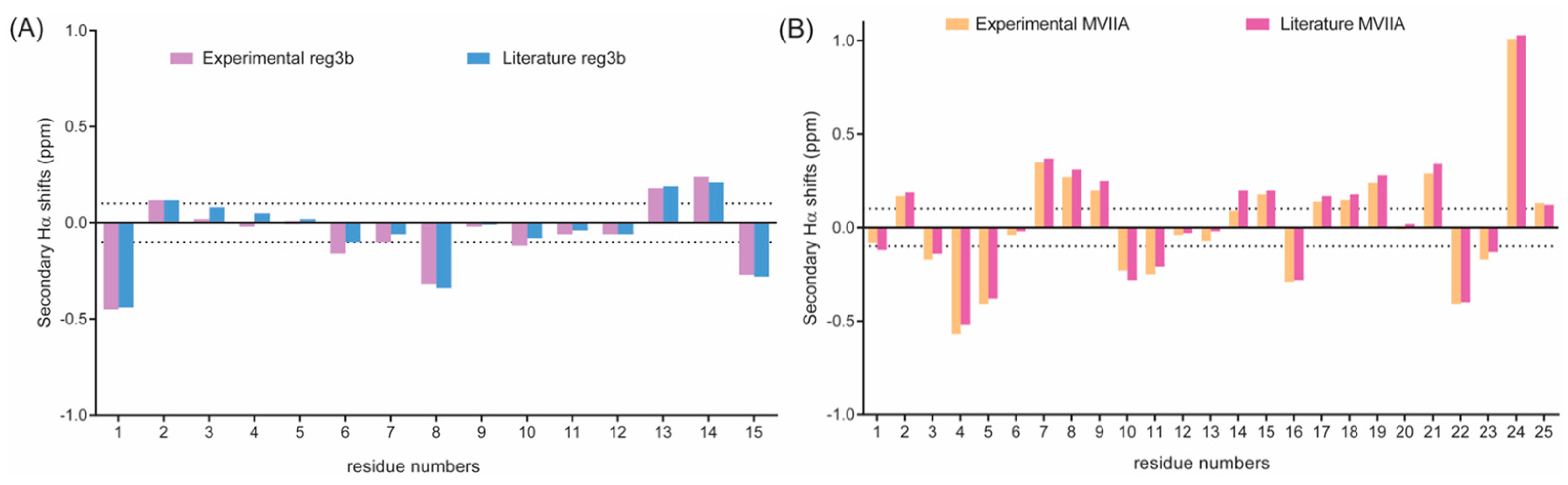
2.4. Comparisons of Secondary Hα Chemical Shifts

A detailed two-dimensional NMR analysis by NMR showed that the chemical shifts of reg3b and MVIIA in this work were identical within experimental error to the chemical shifts reported in the literature for these compounds [23,27], thus demonstrating that the molecules were correctly folded with the correct disulfide connectivity (Figure 6).

2.5. Application to the Other Three Conotoxins

We also applied the aforementioned synthesis strategy to three additional conotoxins with distinct structures and disulfide bond connection patterns. Following treatment with DTDP, iodine, a mixture of TFA/TIS/H2O (95:2.5:2.5), and iodine again, the final product was obtained with a purity exceeding 95%. The total yields were 26.1%, 23.4%, and 26.5%, respectively, with detailed synthetic schemes and results available in the Supplementary Materials (Figures S4–S12 and Figure S14). Notably, the rigid structures enhanced the resistance of S-Mob to TFA. During the synthesis of tricyclic peptides, the S-Mob removal became more efficient as the sequence length increased. MVIIA, BuIIIB, and gm9a, containing 25, 24, and 27 amino acid residues, respectively, achieved maximum yields after 4 h of reaction at 40 °C. In contrast, KIIIA and reg3b required 18 h at 45 °C to reach maximum yields. Furthermore, the reaction rate was positively correlated with temperature. For instance, KIIIA required over 24 h to reach its maximum yield at 40 °C, but this time decreased to 18 h at 45 °C and 12 h at 50 °C, with the structure remaining stable at all test temperatures (Figure S13).

3. Materials and Methods

3.1. General Information

All amino acids and resins were purchased from GL Biochem Ltd. (Shanghai, China), except for Fmoc-Cys(Mob)-OH (CAS 141892-41-3), which was obtained from Bidepharm (Shanghai, China). Preparative RP-HPLC was conducted using FL162040-H100-A0021 system, while analytical RP-HPLC was carried out on EasySep 1010 system. The peptide preparation utilized a 5C18-MS-I column (20 × 250 mm) with detection at 214 nm, whereas a 5C18-MS-II column (4.6 ID × 250 mm) was employed for analytical RP-HPLC. The HPLC solvents were prepared as follows: Solvent A was 0.05% TFA in H2O/ACN (v/v: 90:10), and solvent B was 0.05% TFA in ACN/H2O (v/v: 90:10). The flow rate for preparative RP-HPLC varied from 6 to 10 mL/min using a linear gradient from 10% to 50% Solvent B over 60 min. The analytical RP-HPLC was conducted with a linear gradient of 0.5–2% per minute, progressing from 10% to 70% Solvent B at a flow rate of 1 mL/min.

3.2. Synthesis of Linear Peptides

Linear peptides were synthesized using standard Fmoc solid-phase peptide synthesis (SPPS) protocols. Coupling reactions were performed with Fmoc-protected amino acids (4 eq.), HCTU (4 eq.), and DIEA (8 eq.) in DMF for 40 min, followed by deprotection of the Fmoc group with 20% piperidine in DMF for 20 min. After each reaction, the resin was thoroughly washed with DMF (2 × 10 mL), DCM (2 × 10 mL), and DMF (2 × 10 mL). The progress of SPPS reaction was monitored using the Kaiser test. Peptide synthesis utilized Rink-amide MBHA resin (0.42 mmol/g) and 2Cl-resin pre-loaded with the first amino acid (0.35 mmol/g), following previously described methods [28,29]. In the final step, the resin was washed with DCM (6 × 10 mL), and the crude peptide was cleaved from the resin while simultaneously deprotecting the side chains except for the S-Acm and S-Mob protection groups by treating it with a mixture of TFA/thioanisole/H2O/phenol/EDT (82.5:5:5:5:2.5) for 2 h at 25 °C. The peptide was then precipitated with cold diethyl ether twice and subsequently dissolved in H2O/ACN for preparative RP-HPLC purification.

3.3. Oxidative Folding of Peptides

All folding processes were conducted at a concentration of 2 mg/mL. DTDP was dissolved in a small amount of methanol and slowly added dropwise to the linear peptide in H2O/ACN under neutral conditions to form the first disulfide bond. The second disulfide bond was formed through I2-mediated S-Acm oxidation under acidic conditions. MVIIA was reacted in 90% acetic acid aqueous solution, while all other peptides were processed within the H2O/ACN system. After the reaction, ascorbic acid was added to quench the process, and the oxidized products were isolated via HPLC and lyophilized. The resulting oxidized peptides, containing two disulfides, were dissolved in TFA that also contained 2.5% v/v TIS and 2.5% v/v H2O. These solutions were kept in a shaker at 40 °C or 45 °C. After the reaction, the peptides were precipitated using cold diethyl ether and dissolved in H2O/ACN. A small amount of iodine was added to facilitate the formation of a third disulfide bond. Finally, the target product was purified by HPLC, and all reaction processes were monitored by MS.

3.4. Circular Dichroism (CD)

CD spectra were recorded at room temperature under a nitrogen atmosphere using a Jasco J-810 spectropolarimeter (JASCO, Tokyo, Japan). Measurements were taken over a wavelength range of 300–185 nm with a 1.0 mm path length cell, a bandwidth of 1.0 nm, and a response time of 2 s. Each spectrum values were averaged over three scans. Peptides were dissolved in H2O at a concentration of 0.2 mg/mL. The spectra are expressed as molar ellipticity ([θ]), calculated as follows: [θ] = 1000·mdeg/(l·c), where mdeg represents the raw CD data, c indicates the peptide’s molar concentration (mM), and l is the cell path length (mm).

3.5. NMR Spectroscopy

MVIIA and reg3b were analyzed in this study. Each peptide (~1 mg) was dissolved in 90% H2O/10% D2O, and 1 µL of a 1 mg/mL solution of 4,4-dimethyl-4-silapentane-1-sulfonic acid (DSS) was added as an internal reference. One-dimensional 1H spectra and two-dimensional spectra (TOCSY and NOESY) were acquired. All spectra were recorded on a Bruker Avance 600 MHz spectrometer at 298 K and were processed using TOPSIN 3.6.1 (Bruker) program. NMR data were analyzed using CCPNMR spectra assignment program (version 2.4.2) and all sequential assignments were performed according to the procedure described by Wishart et al. [30].

4. Conclusions

In summary, we developed a facile route for synthesizing conotoxins with three disulfide bonds through regionally selective synthesis. Peptides with three pairs of thiols protected by Trt, Acm, and Mob efficiently folded into desired disulfide connectivities in a sequence-independent manner, using conditions similar to conventional cleavage methods. This strategy enabled the synthesis of five conotoxins with different skeletons and three disulfide bonds, yielding target products of 20–30%. However, some shorter peptides required a longer reaction time, indicating future areas for improvement in deprotection conditions. Overall, this approach demonstrates versatility and simplicity, showcasing significant potential for the orthogonal protection synthesis of complex peptides with multiple disulfide bonds.

Supplementary Materials

The following supporting information can be downloaded at: https://www.mdpi.com/article/10.3390/md23040168/s1, Table S1: MS characterization of synthetic peptides; Figures S1–S6 are the MS characterization diagrams of Cz1107, reg3b, MVIIA, KIIIA, gm9a, and BuIIIB, respectively; Figures S7–S9 are the synthetic schemes of KIIIA, BuIIIB, and gm9a, respectively; Figures S10–S12 are the HPLC diagrams of the synthesis processes of conotoxins KIIIA, gm9a, and BuIIIB, respectively; Figure S13: HPLC analysis of KIIIA reactions for S-Mob removal at different temperatures; Figure S14: HPLC traces of crude peptides after treatment with reagent K; Figure S15: 1H NMR of MVIIA and reg3b. References [23,31,32,33,34,35] are cited in the Supplementary Materials.

Author Contributions

Conceptualization, R.Y. and H.Z. (Hengyu Zhang); writing—original draft preparation, H.Z. (Hengyu Zhang), L.Y.C. and H.Z. (Huanhuan Zhang); writing—review and editing, R.Y., W.C., T.J. and D.J.C. All authors have read and agreed to the published version of the manuscript.

Funding

This research was funded by the Jinan Innovation Team Project (202333022), National Natural Science Foundation of China (NSFC) (Grant Nos. 82122064 and 32101017), Qingdao National Laboratory for Marine Science and Technology (2022QNLM030003-4), Shandong Major Innovation Project (2022CXGC020414), and Shandong Supporting Funds for Talents (2022GJJLJRC02-046).

Institutional Review Board Statement

Not applicable.

Data Availability Statement

The data underlying this study are available in the published article and its Supplementary Materials.

Conflicts of Interest

The authors declare no conflicts of interest.

Abbreviations

SPPSSolid-phase peptide synthesis
HCTU5-Chloro-1-[bis(dimethylamino)methylene]-1H-benzotriazolium 3-oxide hexafluorophosphate
DIEAN,N-Diisopropylethylamine
DTDP4,4′-Dipyridyl disulfide
TrtTrityl
AcmAcetamidomethyl
Mob4-Methoxybenzyl

References

  1. Hone, A.J.; McIntosh, J.M. Nicotinic acetylcholine receptors: Therapeutic targets for novel ligands to treat pain and inflammation. Pharmacol. Res. 2023, 190, 106715. [Google Scholar] [CrossRef] [PubMed]
  2. Carstens, B.B.; Clark, R.J.; Daly, N.L.; Harvey, P.J.; Kaas, Q.; Craik, D.J. Engineering of conotoxins for the treatment of pain. Curr. Pharm. Des. 2011, 17, 4242–4253. [Google Scholar] [CrossRef]
  3. Li, X.; Tae, H.S.; Chu, Y.; Jiang, T.; Adams, D.J.; Yu, R. Medicinal chemistry, pharmacology, and therapeutic potential of α-conotoxins antagonizing the α9α10 nicotinic acetylcholine receptor. Pharmacol. Ther. 2021, 222, 107792. [Google Scholar] [CrossRef]
  4. Jin, A.H.; Muttenthaler, M.; Dutertre, S.; Himaya, S.W.A.; Kaas, Q.; Craik, D.J.; Lewis, R.J.; Alewood, P.F. Conotoxins: Chemistry and Biology. Chem. Rev. 2019, 119, 11510–11549. [Google Scholar] [CrossRef] [PubMed]
  5. Prommer, E. Ziconotide: A new option for refractory pain. Drugs Today 2006, 42, 369–378. [Google Scholar] [CrossRef]
  6. Yu, R.; Seymour, V.A.; Berecki, G.; Jia, X.; Akcan, M.; Adams, D.J.; Kaas, Q.; Craik, D.J. Less is More: Design of a Highly Stable Disulfide-Deleted Mutant of Analgesic Cyclic α-Conotoxin Vc1.1. Sci. Rep. 2015, 5, 13264. [Google Scholar] [CrossRef] [PubMed]
  7. Khoo, K.K.; Gupta, K.; Green, B.R.; Zhang, M.M.; Watkins, M.; Olivera, B.M.; Balaram, P.; Yoshikami, D.; Bulaj, G.; Norton, R.S. Distinct disulfide isomers of μ-conotoxins KIIIA and KIIIB block voltage-gated sodium channels. Biochemistry 2012, 51, 9826–9835. [Google Scholar] [CrossRef]
  8. Tabassum, N.; Tae, H.S.; Jia, X.; Kaas, Q.; Jiang, T.; Adams, D.J.; Yu, R. Role of CysI-CysIII Disulfide Bond on the Structure and Activity of α-Conotoxins at Human Neuronal Nicotinic Acetylcholine Receptors. ACS Omega 2017, 2, 4621–4631. [Google Scholar] [CrossRef]
  9. Cuthbertson, A.; Indrevoll, B. Regioselective formation, using orthogonal cysteine protection, of an alpha-conotoxin dimer peptide containing four disulfide bonds. Org. Lett. 2003, 5, 2955–2957. [Google Scholar] [CrossRef]
  10. Ramming, T.; Appenzeller-Herzog, C. The physiological functions of mammalian endoplasmic oxidoreductin 1: On disulfides and more. Antioxid Redox Signal 2012, 16, 1109–1118. [Google Scholar] [CrossRef]
  11. Akaji, K.; Tatsumi, T.; Yoshida, M.; Kimura, T.; Fujiwara, Y.; Kiso, Y. Disulfide bond formation using the silyl chloride-sulfoxide system for the synthesis of a cystine peptide. J. Am. Chem. Soc. 1992, 114, 4137–4143. [Google Scholar] [CrossRef]
  12. Dekan, Z.; Mobli, M.; Pennington, M.W.; Fung, E.; Nemeth, E.; Alewood, P.F. Total synthesis of human hepcidin through regioselective disulfide-bond formation by using the safety-catch cysteine protecting group 4,4′-dimethylsulfinylbenzhydryl. Angew. Chem. Int. Ed. Engl. 2014, 126, 2975–2978. [Google Scholar] [CrossRef]
  13. Góngora-Benítez, M.; Tulla-Puche, J.; Paradís-Bas, M.; Werbitzky, O.; Giraud, M.; Albericio, F. Optimized Fmoc solid-phase synthesis of the cysteine-rich peptide linaclotide. Biopolymers 2011, 96, 69–80. [Google Scholar] [CrossRef]
  14. Wang, S.; Zhou, Q.; Li, Y.; Wei, B.; Liu, X.; Zhao, J.; Ye, F.; Zhou, Z.; Ding, B.; Wang, P. Quinoline-Based Photolabile Protection Strategy Facilitates Efficient Protein Assembly. J Am Chem Soc. 2022, 144, 1232–1242. [Google Scholar] [CrossRef]
  15. Laps, S.; Atamleh, F.; Kamnesky, G.; Sun, H.; Brik, A. General synthetic strategy for regioselective ultrafast formation of disulfide bonds in peptides and proteins. Nat. Commun. 2021, 12, 870. [Google Scholar] [CrossRef]
  16. Akabori, S.; Sakakibara, S.; Shimonishi, Y.; Nobuhara, Y. A new method for the protection of the sulfhydryl group during peptide synthesis. Bull. Chem. Soc. Jpn. 1964, 37, 433–434. [Google Scholar] [CrossRef]
  17. Schroll, A.L.; Hondal, R.J.; Flemer, S., Jr. 2, 2′-Dithiobis (5-nitropyridine)(DTNP) as an effective and gentle deprotectant for common cysteine protecting groups. J. Pept. Sci. 2012, 18, 1–9. [Google Scholar] [CrossRef] [PubMed]
  18. Marie, E.J.S.; Hondal, R.J. Reduction of cysteine-S-protecting groups by triisopropylsilane. J. Pept. Sci. 2018, 24, e3130. [Google Scholar] [CrossRef] [PubMed]
  19. Zheng, N.; Karra, P.; VandenBerg, M.A.; Kim, J.H.; Webber, M.J.; Holland, W.L.; Chou, D.H.C. Synthesis and characterization of an A6-A11 methylene thioacetal human insulin analogue with enhanced stability. J. Med. Chem. 2019, 62, 11437–11443. [Google Scholar] [CrossRef]
  20. Góngora-Benítez, M.; Mendive-Tapia, L.; Ramos-Tomillero, I.; Breman, A.C.; Tulla-Puche, J.; Albericio, F. Acid-labile Cys-protecting groups for the Fmoc/tBu strategy: Filling the gap. Org. Lett. 2012, 14, 5472–5475. [Google Scholar] [CrossRef]
  21. Barlos, K.; Gatos, D.; Hatzi, O.; Koch, N.; Koutsogianni, S. Synthesis of the very acid-sensitive Fmoc-Cys(Mmt)-OH and its application in solid-phase peptide synthesis. Int. J. Pept. Protein Res. 1996, 47, 148–153. [Google Scholar] [CrossRef]
  22. King, D.S.; Fields, C.G.; Fields, G.B. A cleavage method which minimizes side reactions following Fmoc solid phase peptide synthesis. Int. J. Pept. Protein Res. 1990, 36, 255–266. [Google Scholar] [CrossRef]
  23. Franco, A.; Dovell, S.; Möller, C.; Grandal, M.; Clark, E.; Marí, F. Structural plasticity of mini-M conotoxins–expression of all mini-M subtypes by Conus regius. FEBS J. 2018, 285, 887–902. [Google Scholar] [CrossRef] [PubMed]
  24. Nielsen, K.J.; Thomas, L.; Lewis, R.J.; Alewood, P.F.; Craik, D.J. A consensus structure for omega-conotoxins with different selectivities for voltage-sensitive calcium channel subtypes: Comparison of MVIIA, SVIB and SNX-202. J. Mol. Biol. 1996, 263, 297–310. [Google Scholar] [CrossRef] [PubMed]
  25. Price-Carter, M.; Gray, W.R.; Goldenberg, D.P. Folding of ω-conotoxins. 2. Influence of precursor sequences and protein disulfide isomerase. Biochemistry 1996, 35, 15547–15557. [Google Scholar] [CrossRef] [PubMed]
  26. Kim, J.I.; Ohtake, A.; Sato, K. Circular dichroism spectra of calcium channel antagonist ω-conotoxins. Biochem. Biophys. Res. Commun. 1997, 230, 133–135. [Google Scholar] [CrossRef]
  27. Basus, V.J.; Nadasdi, L.; Ramachandran, J.; Miljanich, G.P. Solution structure of omega-conotoxin MVIIA using 2D NMR spectroscopy. FEBS Lett. 1995, 370, 163–169. [Google Scholar] [CrossRef]
  28. Amblard, M.; Fehrentz, J.A.; Martinez, J.; Subra, G. Methods and protocols of modern solid phase Peptide synthesis. Mol. Biotechnol. 2006, 33, 239–254. [Google Scholar] [CrossRef]
  29. Behrendt, R.; White, P.; Offer, J. Advances in Fmoc solid-phase peptide synthesis. J. Pept. Sci. 2016, 22, 4–27. [Google Scholar] [CrossRef]
  30. Wishart, D.S.; Bigam, C.G.; Yao, J.; Abildgaard, F.; Dyson, H.J.; Oldfield, E.; Markley, J.L.; Sykes, B.D. 1H, 13C and 15N chemical shift referencing in biomolecular NMR. J. Biomol. NMR 1995, 6, 135–140. [Google Scholar] [CrossRef]
  31. Mohan, M.K.; Abraham, N.; Jayaseelan, B.F.; Ragnarsson, L.; Lewis, R.J.; Sarma, S.P. Structure and allosteric activity of a single-disulfide conopeptide from Conus zonatus at human α3β4 and α7 nicotinic acetylcholine receptors. J. Biol. Chem. 2020, 295, 7096–7112. [Google Scholar] [CrossRef] [PubMed]
  32. Lin, J.; Chen, S.; Butt, U.D.; Yan, M.; Wu, B. A comprehensive review on ziconotide. Heliyon 2024, 10, e31105. [Google Scholar] [CrossRef] [PubMed]
  33. Zhao, Z.; Pan, T.; Chen, S.; Harvey, P.J.; Zhang, J.; Li, X.; Yu, R. Design, synthesis, and mechanism of action of novel μ-conotoxin KIIIA analogues for inhibition of the voltage-gated sodium channel Nav1.7. J. Biol. Chem. 2023, 299, 103068. [Google Scholar] [CrossRef] [PubMed]
  34. Holford, M.; Zhang, M.M.; Gowd, K.H.; Azam, L.; Green, B.R.; Watkins, M.; Olivera, B.M. Pruning nature: Biodiversity-derived discovery of novel sodium channel blocking conotoxins from Conus bullatus. Toxicon 2009, 53, 90–98. [Google Scholar] [CrossRef]
  35. Miles, L.A.; Dy, C.Y.; Nielsen, J.; Barnham, K.J.; Hinds, M.G.; Olivera, B.M.; Norton, R.S. Structure of a novel P-superfamily spasmodic conotoxin reveals an inhibitory cystine knot motif. J. Biol. Chem. 2002, 277, 43033–43040. [Google Scholar] [CrossRef]
Figure 1. Structures of the thiol protection groups.
Marinedrugs 23 00168 g001
Figure 2. Primary structures and 3D NMR structures of five exemplar conotoxins containing three disulfide bonds.
Marinedrugs 23 00168 g002
Figure 3. Exploration of S-Mob deprotection using conotoxin Cz1107 as a template. (A) Linear Cz1107 with two Mob groups was incubated under different conditions for 1 h at 40 °C: (i) untreated; (ii) TFA; (iii) TFA/H2O (95:5); (iv) TFA/TIS/H2O (95:2.5:2.5); (v) TFA/thioanisole/H2O (95:2.5:2.5). (B) Linear Cz1107 with Mob groups was incubated for 3 h at 40 °C with TIS or thioanisole added: (i) TFA/TIS/H2O (95:2.5:2.5); (ii) TFA/thioanisole/H2O (95:2.5:2.5); (iii) iodine added to the product of (i); (iv) iodine added to the product of (ii). (C) Orthogonality exploration of S-Mob, S-Acm, and S-Trt. (i) Treated Cz1107(S-Mob) with reagent K for 2 h at room temperature; (ii) treated Cz1107(S-Mob) with iodine at room temperature for 1 h; (iii) linear Cz1107 with two Acm groups; (iv) treated Cz1107(S-Acm) with TFA/TIS/H2O (95:2.5:2.5) for 1 h at 40 °C.
Marinedrugs 23 00168 g003
Scheme 1. Synthesis of conotoxin reg3b.
Marinedrugs 23 00168 sch001
Figure 4. Synthesis of conotoxin reg3b. (A) HPLC chromatograms of the reaction progression: (i) linear peptide with one pair of free thiols and two pairs protected by S-Mob and S-Acm groups, respectively; (ii) DTDP was added to facilitate the formation of the first disulfide bond; (iii) the second disulfide bond was formed through I2-mediated S-Acm oxidation; (iv) treated with TFA/TIS/H2O (95:2.5:2.5) at 45 °C for 18 h to remove the S-Mob groups, and the product was not purified; (v) a small amount of iodine was added to the reaction products in (iv). (B) HPLC chromatogram of synthetic reg3b. (C) MS characterization of synthetic reg3b. (D) CD spectrum of synthetic reg3b. The HPLC elution gradient from 10% ACN/H2O to 70% ACN/H2O took 30 min for samples (iiii) and 60 min for samples (iv,v).
Marinedrugs 23 00168 g004
Scheme 2. Synthesis of conotoxin MVIIA.
Marinedrugs 23 00168 sch002
Figure 5. Synthesis of conotoxin MVIIA. (A) HPLC chromatograms of the reaction progression: (i) linear peptide with one pair of free thiols and two pairs protected by S-Mob and S-Acm groups; (ii) DTDP was added to facilitate formation of the first disulfide bond; (iii) the second disulfide bond was formed through I2-mediated S-Acm oxidation; (iv) treated with TFA/TIS/H2O (95:2.5:2.5) at 40 °C for 4 h to remove the S-Mob groups, and the product was not purified; (v) a small amount of iodine was added to the reaction products in (iv). (B) HPLC chromatogram of synthetic MVIIA. (C) MS characterization of synthetic reg3b. (D) CD spectrum of synthetic MVIIA.
Marinedrugs 23 00168 g005
Figure 6. Secondary Hα chemical shifts of reg3b and MVIIA. (A) Comparison of the secondary Hα chemical shifts of experimental reg3b with literature values obtained from Franco et al., 2018 [23] (B) Comparison of the secondary Hα chemical shifts of experimental MVIIA with literature values obtained from Basus et al., 1995 [27]. Three or more consecutive residues with secondary chemical shifts greater than +0.1 ppm or less than –0.1 ppm (dotted lines) indicate potential structured regions within the peptide.
Marinedrugs 23 00168 g006
Table 1. Recovery yield of Cz1107(S-Mob) for different conditions.
EntryExperimental ConditionsRecovery Yield (%) a
1TFA, 1 h at 40 °C71
2TFA/H2O (95:5 v:v), 1 h at 40 °C72
3TFA/TIS/H2O (95:2.5:2.5 v:v:v), 1 h at 40 °C16
4TFA/thioanisole/H2O (95:2.5:2.5 v:v:v), 1 h at 40 °C22
5TFA/TIS/H2O (95:2.5:2.5 v:v:v), 3 h at 40 °C3
6TFA/thioanisole/H2O (95:2.5:2.5 v:v:v), 3 h at 40 °C4
7Reagent K, 2 h at RT88
8Iodine, 1 h at RT97
a HPLC proportion of Cz1107 (S-Mob) residual amount.
Disclaimer/Publisher’s Note: The statements, opinions and data contained in all publications are solely those of the individual author(s) and contributor(s) and not of MDPI and/or the editor(s). MDPI and/or the editor(s) disclaim responsibility for any injury to people or property resulting from any ideas, methods, instructions or products referred to in the content.

