Article

Purpuramine R, a New Bromotyrosine Isolated from Pseudoceratina cf. verrucosa Collected in the Kingdom of Tonga

by
Jennie L. Ramirez-Garcia
1,2,
Hannah Lee-Harwood
2,3,
David Ackerley
2,3,
Michelle Kelly
4,
S. Vailala Matoto
5,
Patricia Hunt
1,
A. Jonathan Singh
2,6,* and
Robert A. Keyzers
1,2,*
1
School of Chemical & Physical Sciences, and Centre for Biodiscovery, Victoria University of Wellington, P.O. Box 600, Wellington 6140, New Zealand
2
Maurice Wilkins Centre for Molecular Biodiscovery, University of Auckland, Private Bag 92019, Auckland 1142, New Zealand
3
School of Biological Sciences, and Centre for Biodiscovery, Victoria University of Wellington, P.O. Box 600, Wellington 6140, New Zealand
4
Oceans Centre, National Institute of Water & Atmospheric Research, P.O. Box 9940, Auckland 1010, New Zealand
5
Ministry of Fisheries, Sopu, Vuna Road, Tongatapu P.O. Box 871, Tonga
6
Ferrier Research Institute, and Centre for Biodiscovery, Victoria University of Wellington, P.O. Box 600, Wellington 6140, New Zealand
*
Authors to whom correspondence should be addressed.
Mar. Drugs 2025, 23(5), 186; https://doi.org/10.3390/md23050186
Submission received: 29 March 2025 / Revised: 17 April 2025 / Accepted: 22 April 2025 / Published: 27 April 2025
(This article belongs to the Special Issue Structural Diversity in Marine Natural Products)

Abstract

:
Sponges in the verongiid genus Pseudoceratina Carter are well-known producers of bioactive secondary metabolites. Chemical screening of a Tongan P. cf. verrucosa Bergquist using NMR highlighted the presence of aromatic natural products. Subsequent extraction and purification of P. cf. verrucosa yielded a new bromotyrosine, purpuramine R (1), that exhibits moderate (MIC 16 µg/mL) antibacterial activity against Gram-positive Staphylococcus aureus. The E-geometry of the oxime was confirmed using a combination of NMR and computational approaches. Additionally, computational conformational analysis indicates that purpuramine R adopts a hairpin orientation, stabilized by intramolecular hydrogen and halogen bonds. Knowledge of this stabilized conformation can inform synthetic approaches to make analogues of the purpuramines for future SAR studies.

1. Introduction

Marine sponges continue to be a rich resource for the discovery of new bioactive natural products [1]. Within phylum Porifera, the dictyoceratid, dendroceratid, and verongid demosponge orders have yielded numerous marine natural products (MNPs), including ianthelliformisamines [2], bastadins [3], and psammaplins [4]. Whilst the Verongiida are relatively easy to identify in the field due to their rapid oxidation to deep oak brown or deep royal purple upon exposure to air [5,6], the lack of spicules in all three orders makes species identification a challenge. The dense, fleshy, rubbery texture of the Verongiida presents a particular problem; accurate genus and species identification are reliant upon the composition and architecture of the fibers which can be difficult to access without histological examination. More specifically, species of the verongiid genus Pseudoceratina are well-known producers of bioactive secondary metabolites. For example, specimens identified in the literature as P. purpurea (Carter) (which probably includes the more common Indo-Pacific species, P. arabica (Keller)) are purported to be the source of many new compounds, including merosesquiterpenoids [7,8] and bromotyrosines [9,10].
Exploration of the MNPs from organisms collected in the South Pacific’s Kingdom of Tonga is in its infancy when compared to studies focused on other island groups such as Fiji or Samoa. However, a variety of unique MNPs have been sourced from organisms collected in Tonga [11]. As part of our group’s ongoing examination of Tongan marine organisms for new MNPs [12,13,14,15], we have carried out spectroscopic screening of an extract of Pseudoceratina cf. verrucosa Bergquist, 1995, a species originally described in New Caledonia [16]. NMR experiments highlighted the presence of aromatic metabolites. Subsequent extraction of a larger amount of P. cf. verrucosa followed by repeated reversed-phase chromatography resulted in the isolation of a new bromotyrosine metabolite, purpuramine R (1, Figure 1). In addition, the known compounds hexadellin A, purealidin B, and purpuramine M were also isolated (Figure 2 and Figure S1) [17,18,19]. The structure of 1 was determined using a combination of NMR and MS techniques, in conjunction with computational approaches to help assign the oxime geometry, and to investigate the conformational space adopted by purpuramine R. Here, we report the structural characterization and bioactivity of purpuramine R (1).

2. Results

2.1. Structure Elucidation of Purpuramine R (1)

Purpuramine R (1) was isolated as an optically inactive colorless film. Positive-mode high-resolution electrospray mass spectrometric (HRESI-MS) analysis detected a natively charged ion at m/z 755.8913, suitable for the molecular formula C24H3079Br4N3O5+ (calc. m/z 755.8913, Δ = 0.0 ppm, Figure S7) and therefore requiring ten degrees of unsaturation. The presence of the four bromine atoms in the molecular formula was indicated by the molecular cluster of ions at m/z 755, 757, 759, 761, and 763 in a 1:4:6:4:1 ratio. Interrogation of the HSQC, HMBC, 1H, and 13C NMR spectra identified 24 individual carbon resonances, of which 13 were protonated (CD3OD; Figures S2–S6). The resonances include aromatic methines, a methoxy group, a series of methylenes, and, of particular note, a singlet 1H signal integrating for nine protons, the chemical shifts of which indicated bonding to a heteroatom (δC 53.6–53.7; δH 3.20).
Only two spin systems of mutually coupled proton resonances were detected in the COSY spectrum of 1. The first spin system comprised a pair of methylenes, CH2-19 (δC 28.8, δH 3.09) and CH2-20 (δC 67.7, δH 3.54). The chemical shifts of CH2-19 and CH2-20 indicated attachment to an aryl ring and a nitrogen atom, respectively, supported by appropriate 1JC,H coupling constants. An HMBC correlation from CH2-20 to the deshielded singlet that integrated for nine relative protons indicated the positioning of a terminal trimethyl ammonium moiety. Methylene CH2-19 presented HMBC correlations to the aromatic carbon quaternary, C-16 (δC 136.0), and methine singlet, CH-15/17 (δC 134.6, δH 7.58), which integrated for two relative protons, suggesting a symmetrical 1,2,3,5-tetrasubstituted benzene element in 1 (ring B, Figure 3). HMBC correlations from CH-15/17 to a slightly shielded non-protonated aromatic carbon resonance (C-14/18: δC 119.5) indicated the placement of two of the four bromine atoms of 1.
The second spin system of 1 comprised three contiguous methylenes, CH2-10 (δC 37.9, δH 3.58), via CH2-11 (δC 30.5, δH 2.10), to CH2-12 (δC 72.2, δH 4.02), as established from COSY correlations. The chemical shifts of CH2-10 and CH2-12 suggest the attachment of nitrogen and oxygen, respectively, which is again supported by the 1JC,H values. Methylene CH2-10 correlated to carbonyl C-9 (δC 166.8), indicating an amide linkage. Additionally, methylene singlet CH2-7 also correlated with this carbonyl, along with correlations to a deshielded quaternary carbon, C-8 (δC 154.9), aromatic quaternary carbons, C-1 (δC 123.0), and oxygenated C-2 (δC 151.7), as well as to aromatic methine CH-6 (δC 134.5, δH 7.42).
The tyrosine-based moiety of purpuramine R (1) was deduced via HMBC correlations from methine H-6 to non-protonated carbons, C-3 (δC 108.8), C-4 (δC 154.7). and C-5 (δC 106.8). The chemical shifts of both C-3 and C-5 were shielded to lower frequencies and established connections to the last two bromines. Carbon C-4 was confirmed to be linked to the methoxy CH3-22 (δC 60.8, δH 3.80) by HMBC, positioned para in relation to C-1 and consistent with the 1,3-dibromo-2-O-methoxybenzene (ring A) moieties mostly reported from Verongid sponges. Finally, an HMBC correlation from H2-12 to C-13 established the link between the two spin systems via the 1,2,3,5-tetrasubstitutued benzene ring (ring B, Figure 3).
The analysis so far accounted for all but one N, O, and H atom of the molecular formula, indicating that carbon C-8 must be part of an oxime functionality. Such a motif is common in Verongid bromotyrosine metabolites. Assignment of the oxime geometry in bromotyrosines has largely been based upon an empirical observation noted in the 1987 study by Arabshahi and Schmitz, who reported a dimeric bromotyrosine that incorporated both possible geometries. In their example, the carbon chemical shift of the methylene linking the aromatic ring to the oxime was shielded to ~27 ppm for the E-isomer, while the Z-geometric isomer resonated at ~36 ppm [19]. It should be noted, however, that the substitution pattern around the aromatic rings of their compound were much simpler, being 1,2,4-trisubstituted benzenes rather than the penta-substituted case in 1. It could be envisaged that electronic shielding effects, particularly from the hydroxy group ortho and the methoxy group para to the chain attachment, could influence the chemical shift of the methylene group. We therefore sought to use density functional theory calculations of the methylene chemical shifts to help corroborate this empirical approach.
To lower the computational cost, truncated model 2 was used to predict the most stable conformations of the E (2i) and Z (2ii) oxime geometries (Figure 4), recognizing that the trimethylammonium portion should not play a significant role in influencing chemical shifts of the tyrosine moiety of 1. Within each possible geometry, hydrogen bonding between OH-6 and the amide carbonyl, and between the oxime and the amide NH, provides four possible stabilized conformers. Both possible E-geometry conformers were found to be lower in energy than the two corresponding Z-conformers (Figure 4). Prediction of the NMR chemical shifts of these four conformers (Table 1) showed that the CH2-7 methylene chemical shift of purpuramine R matched better to those of the two E geometric stereoisomers than the Z counterparts; gratifyingly, these also match closely to those observed empirically by Arabshahi and Schmitz [19]. The structure of purpuramine R was therefore established as 1. The NMR data of 1 are presented in Table 2.

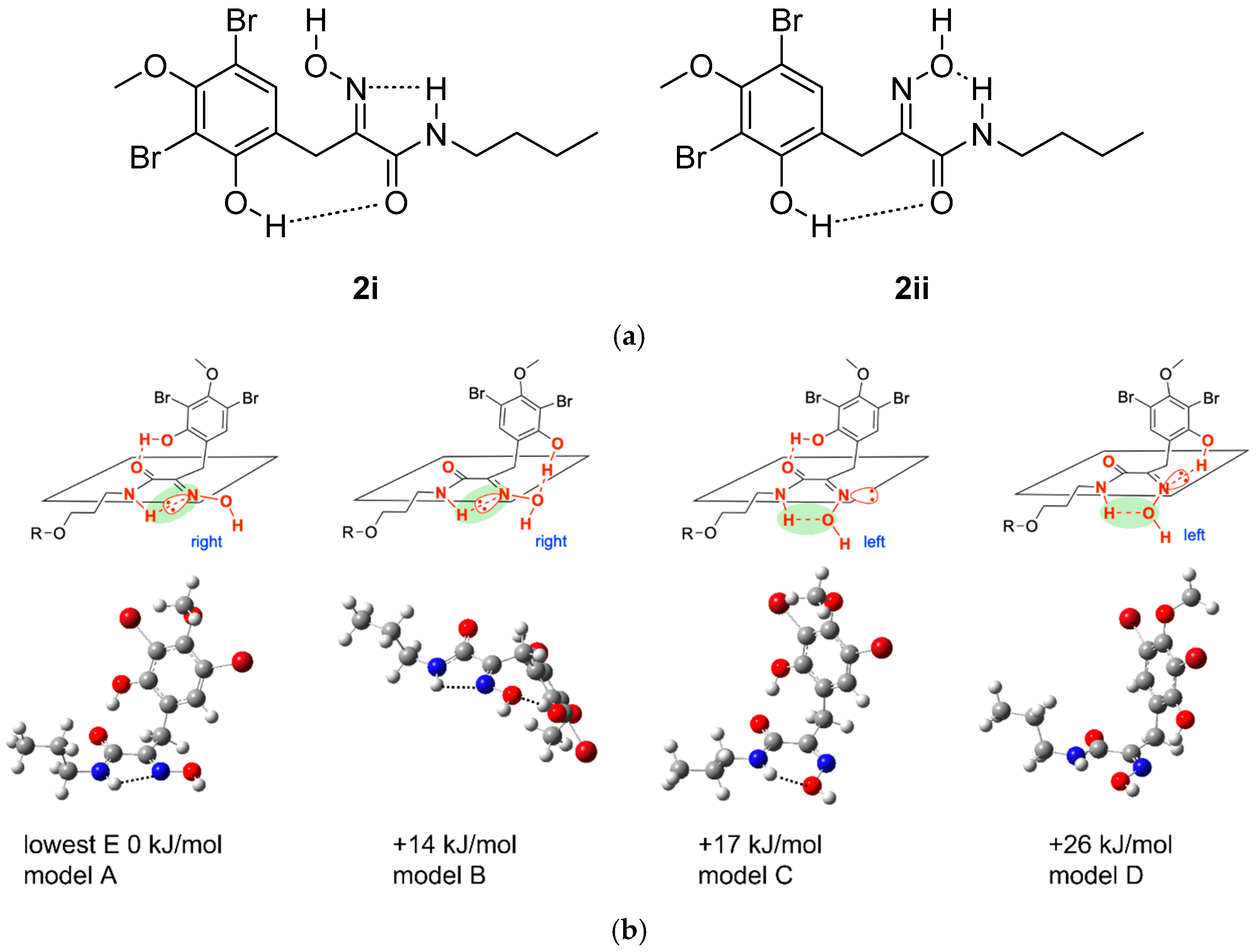
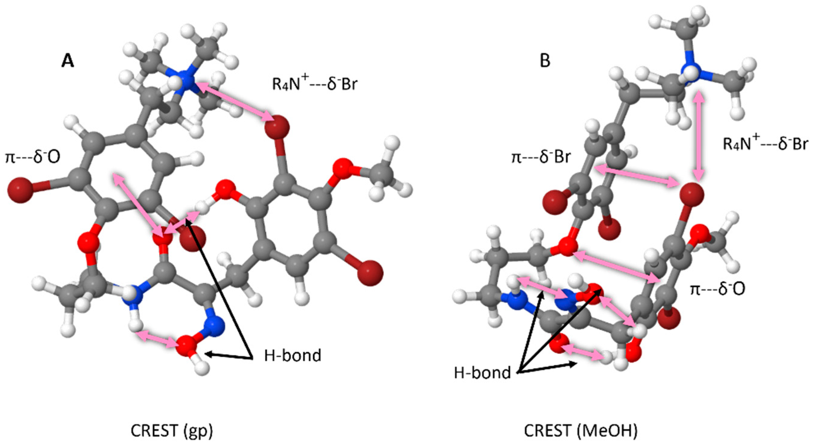
2.2. Conformational Analysis of Purpuramine R (1)

With the structure of purpuramine R (1) in hand, we next took advantage of the computational data already available to spearhead a study of its conformation in solution. We are not aware of any such conformational analyses of bromotyrosine metabolites to have been published to date. Moreover, knowledge of the likely conformation of 1 and related metabolites could inform the rational design of bioactive congeners to investigate structure–activity relationships (SARs) and assist with drug discovery efforts.
To this end, we employed CREST (Conformer—Rotamer Ensemble Sampling Tool) [20,21] to investigate the conformation space occupied by 1. Our initial assessment was carried out in the gas phase, which resulted in an ensemble of conformers, the lowest energy of which exhibited a “twisted” structure, stabilized by numerous noncovalent interactions, including hydrogen bonding, π-stacking, and ionic bridging (Figure 5A). To further assess the likely conformation of 1 in solution, CREST calculations were undertaken, employing a generalized solvent model and methanol (MeOH) parameters. The lowest-energy conformer adopts a folded, “hairpin”-like structure (Figure 5B). This solution conformer has a more obvious π-stacking arrangement, generating a pseudo-cyclophane-type arrangement which is “stitched” together by the ammonium moiety interacting with an aromatic bromine.
It is notable that this folded conformation is very different to the extended planar structure used as standard to depict bromotyrosine MNPs in the literature. It is entirely conceivable that many other related bromotyrosine metabolites will form similar compact folded conformations in solution. Knowledge of such conformational arrangements could also impact the bioactivity of such bromotyrosines. In particular, the more compact folded conformation could significantly impact the mobility of metabolites like purpuramine R through cell membranes or of binding to their receptor targets; therefore, synthetic efforts to make analogues of these metabolites could benefit from a deeper understanding of the three-dimensional structure they possess, enabling the exploitation of unusual functionalities. For example, the formation of a covalent bond between the terminal ammonium unit (to maintain charge carrying capacity) and the tyrosine ring would provide a chemically robust motif that permanently mimics the solution-phase conformation. Alternatively, knowledge of the key noncovalent interactions stabilizing the conformation of 1 could enable the prediction of synthetic modifications that would disrupt intramolecular bonding, providing access to a more flexible extended conformational space, akin to the way researchers normally draw these compounds. Similar conformation analyses of other bromotyrosine metabolites would be informative for future SAR studies of this MNP class.

2.3. Bioactivity of Purpuramine R (1)

Bromotyrosine metabolites are known to possess antibiotic activity [6,18,22,23,24,25]. Purpuramine R (1) was assessed for antibacterial activity against Gram-positive (Staphylococcus aureus) and Gram-negative (Escherichia coli, Klebsiella pneumoniae) strains (Table 3). A moderate effect was observed against S. aureus (MIC 16 µg/mL), but no activity was observed against the Gram-negative strains. Testing against a tolC deficient strain of E. coli (E. coli W3110 ΔtolC) was also attempted, resulting in moderate inhibition of the bacteria (MIC 16 µg/mL), suggesting that TolC plays an important role in facilitating purpuramine efflux to protect Gram-negative bacteria (Table 3).

3. Materials and Methods

3.1. General Experimental Procedures

Solvents for purification were obtained from Fisher Scientific (Hampton, NH, USA) and were HPLC or analytical grade. Water used for chromatography was glass distilled prior to use. All solvent mixtures are a reported as % v/v. Diaion HP-20® was obtained from Supelco (Spruce City, St Louis, MO, USA). Deuterated NMR solvents were obtained from Apollo Scientific (Bredbury, Stockport, United Kingdom). HPLC purifications were performed using an Agilent Technologies 1260 Infinity HPLC system (Santa Clara, CA, USA) featuring a temperature-controlled column compartment, a quaternary pump system, and both evaporative light scattering and diode array detectors. Semi-preparative purifications used a Phenomenex® Luna C18 column (10 mm W × 250 mm L, 5 µm particles; Torrance, CA, USA). UV/vis spectra were extracted from HPLC DAD outputs. High-resolution mass spectra were obtained using an Agilent Technologies 6530 QTOF mass spectrometer coupled to an Agilent Technologies 1260 Infinity HPLC. NMR data (Santa Clara, CA, USA) were acquired using a JEOL JNM-ECZ600R spectrometer (Akishima, Tokyo, Japan) with an N2-cooled 5 mm SuperCOOL cryogenic probe (operating at 600 MHz for 1H nuclei and 151 MHz for 13C nuclei). Chemical shifts (δ, ppm) were referenced to the residual solvent peak [26].

3.2. Sponge Material

Sponge specimens were collected from a submerged marine cave on the western coastline of ‘Eua Island in the Kingdom of Tonga (21.40° S, 174.97° W), on 7 June 2016, between 10 to 25 m depth. Specimens were transported frozen to New Zealand and stored at –20 °C until extraction. Identification was performed by author MK who considers the material to be most closely comparable to Pseudoceratina verrucosa Bergquist, 1995 (Order Verongiida, family Pseudoceratinidae), a species with a more restricted distribution than the pan Indo-Pacific and closely related species P. purpurea (Carter, 1880) and P. arabica (Keller, 1889). Pseudoceratina verrucosa has a more strongly warty surface than the former two species, sand inclusions in the fibers, and prominent oscules. The only other species in genus Pseudoceratina is P. durissima Carter, 1885, restricted to Australian waters. A Victoria University Wellington (VUW) voucher of the specimen (PTN4_36C) has been accessioned into the NIWA Invertebrate Collection (NIC), NIWA, Wellington: NIWA 143602.

3.3. Isolation of Purpuramine R (1)

Frozen P. cf. verrucosa (21.2 g) was macerated in MeOH twice overnight (2 × 50 mL). The second MeOH extract was passed through a HP-20® column (80 mL) that had been pre-equilibrated with Me2CO and MeOH (240 mL ea.). The first extract was then passed through the same column and combined with the second extract. The combined extract was diluted with H2O (100 mL) and passed back through the same column. Finally, the collected eluent was diluted once more with H2O (200 mL; final concentration 25% MeOH(aq)) and passed through the HP-20® column. The column was washed with water (240 mL, discarded), followed by 240 mL portions of 30% Me2CO(aq), 75% Me2CO(aq), and 100% Me2CO. The dried 30% Me2CO(aq) fraction (35.0 mg) was separated by semi-preparative HPLC (4 mL/min flow rate, 0–3 min: 30% ACN/H2O, 0.1% HCOOH; 3–11 min: 60% ACN/H2O, 0.1% HCOOH; 11–14 min: 100% ACN, 0.1% HCOOH) to give hexadellin A (tR = 6.8 min, 12.4 mg), purealidin B (tR = 7.7 min, 4.2 mg), purpuramine M (tR = 9.1 min, 4.6 mg), and purpuramine R (1; tR = 10.4 min, 1.4 mg).
Hexadellin A: yellow film; HRESIMS [M + H]+ observed m/z 713.8461, calculated for C21H2479Br4N3O5+ m/z 713.8444; 1H NMR (600 MHz, CD3OD) δ 7.55 (s, 2H), 6.42 (d, J = 0.9 Hz, 2H), 4.10–4.05 (m, 6H), 3.77 (s, 1H), 3.73 (s, 6H), 3.59 (t, J = 7.0 Hz, 4H), 3.16 (t, J = 7.5 Hz, 4H), 3.09 (s, 1H), 2.90 (t, J = 7.5 Hz, 4H), 2.12 (m, 2H); 13C NMR (151 MHz, CD3OD) δ 161.6, 155.3, 153.6, 149.3, 134.4, 132.2, 122.8, 119.5, 114.2, 92.4, 75.5, 72.2, 60.4, 41.4, 40.1, 37.9, 33.1, 30.6. Data concordant with those in the literature [17].
Purealidin B: colorless film; HRESIMS [M]+ observed m/z 755.8920, calculated for C24H3079Br4N3O5+ m/z 755.8913; 1H NMR (600 MHz, CD3OD) δ 7.62 (s, 3H), 6.42 (d, J = 0.9 Hz, 2H), 4.10–4.04 (m, 5H), 3.77 (s, 1H), 3.73 (s, 5H), 3.62–3.52 (m, 6H), 3.20 (s, 13H), 3.13–3.07 (m, 5H), 2.12 (m, 3H); 13C NMR (151 MHz, CD3OD) δ 161.6, 155.3, 153.7, 149.3, 136.1, 134.6, 132.1, 122.8, 119.4, 114.2, 92.4, 75.5, 72.2, 67.6, 60.3, 53.6, 40.1, 37.9, 30.6, 28.8. Data concordant with those in the literature [27].
Purpuramine M: white solid; HRESIMS [M + H]+ observed m/z 713.8431, calculated for C21H2479Br4N3O5+ m/z 713.8444; 1H NMR (600 MHz, CD3OD) δ 7.52 (s, 3H), 7.43 (s, 1H), 4.03 (t, J = 6.0 Hz, 3H), 3.81 (d, J = 6.0 Hz, 7H), 3.59 (t, J = 6.8 Hz, 3H), 3.15 (t, J = 7.6 Hz, 3H), 2.89 (t, J = 7.6 Hz, 3H), 2.10 (quin, J = 6.5 Hz, 3H); 13C NMR (151 MHz, CD3OD) δ 166.6, 154.7, 154.2, 153.3, 151.3, 136.9, 134.3, 134.1, 122.6, 119.3, 108.7, 107.1, 71.9, 60.4, 41.1, 37.7, 32.9, 30.2, 25.1. Data concordant with those in the literature [18].
Purpuramine R (1): white solid; UV/vis: λmax 207, 230, 290 nm (spectrum extracted from HPLC DAD); HRESIMS [M]+ observed m/z 755.8913, calculated for C24H3079Br4N3O5+ m/z 755.8913; HRESIMS/MS (30 eV) m/z (% relative intensity) 503.1067 (13), 429.0888 (21), 415.0365 (26), 355.0699 (29), 341.0179 (56), 337.9567 (29), 281.0510 (100), 221.0843 (37), 207.0321 (20), 147.0656 (42); for NMR data, see Table 2.

3.4. Computational Chemistry

Density functional theory (DFT) calculations were performed with the G16 suite of codes and visualized with GView6 [28,29]. For all calculations, (a) the density integration grid was set to a pruned (99,590) grid, i.e., 99 radial shells and 590 angular points per shell (int = ultrafine); (b) the scf convergence was set to 10−9 (scf = conver = 9) for optimization and 10−10 for frequency and NMR calculations. A value of 10−x corresponds to a maximum of 10−x on the RMS density matrix, with 10−(x−2) on the maximum value of the density matrix and the energy. Optimized conformers have been confirmed as the minima by the absence of imaginary modes.
A scaling method described on CHESHIRE CCAT (the Chemical Shift Repository for computed NMR scaling factors, with Coupling Constants Added Too) was employed [30,31]. Initial optimizations were carried out in the gas phase (GP) at the B3LYP(D3BJ)/6-311 + G(d,p) level. The lowest-energy conformers (A-D) were subsequently optimized in the GP at the B3LYP(D3BJ)/6-31 + G(d,p) level employing a Lanl2DZdp pseudo-potential (PP) and associated basis-set for the Br atoms. Single-point NMR evaluations were then carried out at the mPW1PW91-PCM(methanol)/6-311 + G(2d,p) level. Log files are available at DOI: 10.5281/zenodo.15098754
The NMR chemical shifts were scaled based on data given in CHESHIRE CCAT, as shown in eqn 1 (where σiso =the isotropic SCF GIAO Magnetic shielding tensor in ppm), and the RMSD given is for a test set of molecules [24,30]. We note that the D3BJ correction has been employed at the B3LYP level relative to the geometry optimization method defined in CHESHIRE CCAT B3LYP/6-31 + G(d,p). This change in method means that the fitting Equation (1) may not be as accurate as is indicated by the RMSD reported in CHESHIRE CCAT (2.0632). However, an error of ≈2 ppm in 13C NMR chemical shifts is expected.
13C s = −1.0399 σiso + 186.5993 RMSD = 2.0632
The initial B3LYP-optimized structures were first re-optimized using the XTB program (Semiempirical Extended Tight-Binding Program Package) employing the default options and using the Geometry, Frequency, Noncovalent, eXtended TB (GFN2-xTB) algorithm [32,33]. The CREST software (Conformer–Rotamer Ensemble Sampling Tool, v2.12) generates conformer/rotamer ensembles though meta-dynamic (MTD) simulations and sampling and employs a genetic z-matrix crossing algorithm (GC) [20,21]. CREST was run using the GFN2-xTB algorithm, first in the gas phase, and then including a generalized solvent description for methanol (ALPB). The iMTD-GC workflow was employed using default options: RMSD threshold 0.125, population threshold 0.05, conformer energy window 6 kcal/mol.
A “linear” bromotyrosine diagram (shown as 1) was constructed and optimized in the GP at the HF/3-21G level. The HF/3-21G structure was then optimized in the GP at the XTB level and a CREST conformer calculation carried out, 991 conformers/rotamers were obtained. The lowest-energy conformer was used to start a second CREST search which generated 1684 conformers/rotamers. The lowest-energy structure of the second search is very similar to that obtained in the first search. The HF/3-21G GP structure was also optimized within a generalized PCM solvent environment for MeOH at the XTB level and a CREST conformer calculation carried out, and 1470 conformers/rotamers were obtained and the lowest-energy conformer was retained for analysis. The lowest-energy XTB conformers for the GP and MeOH are given in Figure 5.

3.5. Bioassays

MIC (minimum inhibitory concentration, the lowest concentration at which visible growth is completely inhibited) assays were performed using a methodology adapted from Wiegand et al. [34]. Strains were freshly streaked onto Mueller Hinton (MH) agar plates and incubated for 16 h at 37 °C. Following colony formation, 5–10 colonies were resuspended in MH medium, and the OD600 of each culture was normalized to 0.002. Cells were seeded in wells containing MHB media supplemented with a final concentration of 3.2% DMSO or drug between 0.125 μg/mL to 32 μg/mL (in 2-fold dilutions). Plates were incubated at 37 °C for 16 h and then visually inspected for visible growth. Assays were performed in biological triplicates.

4. Conclusions

Verongid sponges such as Pseudoceratina cf. verrucosa remain an important resource for the discovery of new bioactive metabolites. This study, guided by an untargeted NMR-based spectroscopic screen of Tongan marine invertebrates, has resulted in the isolation of a new bromotyrosine, purpuramine R (1). Structurally, 1 combines the trimethylammonium group found in chiral purealidin B with the free oxime as present in purpuramine M. Many bromotyrosine metabolites have been reported from Verongiid sponges, which have been recently reviewed [35]. The interested reader is directed to this reference to compare the structural relationship between our isolated compounds and other members of the class. Purpuramine R exhibits moderate antibacterial activity against a model Gram-positive strain and was identified as a substrate for TolC-mediated efflux in Gram-negative bacteria. The use of computational chemistry has facilitated verification of the empirical approach commonly employed to establish the geometry of the oxime functional group found in many of these metabolites. Moreover, analysis using CREST has discovered an unexpected three-dimensional conformation stabilized by numerous intra-molecular interactions. We believe that similar computational studies of related metabolites will be informative for informed rational design of the analogs, SAR probes, and future drug leads.

Supplementary Materials

The following supporting information can be downloaded at: https://www.mdpi.com/article/10.3390/md23050186/s1: Figure S1: HPLC chromatogram (210 nm) of Tongan Pseudoceratina cf. verrucosa, 30% Me2CO(aq) fraction; Figure S2: 1H NMR spectrum (CD3OD, 600 MHz) of compound 1; Figure S3: 13C NMR spectrum (CD3OD, 151 MHz) of compound 1; Figure S4: HSQC spectrum (CD3OD, 600 MHz) of compound 1; Figure S5: COSY spectrum (CD3OD, 600 MHz) of compound 1; Figure S6: HMBC spectrum (CD3OD, 600 MHz) of compound 1; Figure S7: (+)-HRESIMS of compound 1; Figure S8: (+)-HRESIMS/MS spectra of 1 using CID of 10 (a), 20 (b), 30 (c), and 40 (d) eV; Figure S9: UV spectra extracted from HPLC chromatogram of compound 1; Figure S10: 1H NMR spectrum (CD3OD, 600 MHz) of Hexadellin A; Figure S11: 13C NMR spectrum (CD3OD, 151 MHz) of hexadellin A; Figure S12: 1H NMR spectrum (CD3OD, 600 MHz) of purealidin B; Figure S13: 13C NMR spectrum (CD3OD, 151 MHz) of purealidin B; Figure S14: 1H NMR spectrum (CD3OD, 600 MHz) of purpuramine M; Figure S15: 13C NMR spectrum (CD3OD, 151 MHz) of purpuramine M; Table S1: NMR spectroscopic data of purpuramine R (1) (600/151 MHz, CD3OD).

Author Contributions

Conceptualization, J.L.R.-G., A.J.S. and R.A.K.; methodology, J.L.R.-G., H.L.-H. and P.H.; formal analysis, J.L.R.-G., H.L.-H., M.K. and P.H.; investigation, J.L.R.-G., H.L.-H. and P.H.; resources, D.A., S.V.M., A.J.S., R.A.K. and P.H.; writing—original draft preparation, J.L.R.-G., A.J.S. and R.A.K.; writing—review and editing, J.L.R.-G., H.L.-H., D.A., M.K., S.V.M., P.H., A.J.S. and R.A.K.; supervision, D.A., A.J.S. and R.A.K.; project administration, R.A.K. All authors have read and agreed to the published version of the manuscript.

Funding

This research received no external funding.

Institutional Review Board Statement

Not applicable.

Data Availability Statement

Primary NMR datafiles (FIDs) are publicly available from the Harvard Dataverse via https://dataverse.harvard.edu/dataset.xhtml?persistentId=doi:10.7910/DVN/LTJFAX, accessed 14 January 2024. The geometry XYZ and CREST computational data are available via DOI: 10.5281/zenodo.15098754.

Acknowledgments

We very gratefully acknowledge the Ministry of Fisheries, Kingdom of Tonga (file number F1/40/76/16) for permission to collect specimens of P. cf. verrucosa, with the assistance of J. Laurie from Whale Swim Fish and Dives Tours, Tonga. We thank J. Vorster (VUW) for NMR assistance. MIC assays were supported by a Royal Society of New Zealand Marsden grant (contract VUW1902 awarded to D.F.A.). Computational studies utilized the Victoria University of Wellington high-performance computing facilities (Rāpoi).

Conflicts of Interest

The authors declare no conflicts of interest.

References

  1. Carroll, A.R.; Copp, B.R.; Grkovic, T.; Keyzers, R.A.; Prinsep, M.R. Marine Natural Products. Nat. Prod. Rep. 2024, 41, 162–207. [Google Scholar] [CrossRef] [PubMed]
  2. Xu, M.; Davis, R.A.; Feng, Y.; Sykes, M.L.; Shelper, T.; Avery, V.M.; Camp, D.; Quinn, R.J. Ianthelliformisamines A-C, antibacterial bromotyrosine-derived metabolites from the marine sponge Suberea ianthelliformis. J. Nat. Prod. 2012, 75, 1001–1005. [Google Scholar] [CrossRef]
  3. Calcul, L.; Inman, W.D.; Morris, A.A.; Tenney, K.; Ratnam, J.; McKerrow, J.H.; Valeriote, F.A.; Crews, P. Additional insights on the bastadins: Isolation of analogues from the sponge Ianthella cf. reticulata and exploration of the oxime configurations. J. Nat. Prod. 2010, 73, 365–372. [Google Scholar] [CrossRef] [PubMed]
  4. Quiñoá, E.; Crews, P. Phenolic constituents of Psammaplysilla. Tetrahedron Lett. 1987, 28, 3229–3232. [Google Scholar] [CrossRef]
  5. Hooper, J.N.A.; Soest, R.W.M.; Willenz, P. Systema Porifera A Guide to the Classification of Sponges; Springer: Berlin, Germany, 2002; Volume 1. [Google Scholar]
  6. El-Demerdash, A.; Atanasov, A.G.; Horbanczuk, O.K.; Tammam, M.A.; Abdel-Mogib, M.; Hooper, J.N.A.; Sekeroglu, N.; Al-Mourabit, A.; Kijjoa, A. Chemical Diversity and Biological Activities of Marine Sponges of the Genus Suberea: A Systematic Review. Mar. Drugs 2019, 17, 115. [Google Scholar] [CrossRef] [PubMed]
  7. Yu, X.; Han, X.; Mi, Y.; Cui, Y.; Fu, A.; Liu, K.; Li, X.; Tang, X.; Li, G. Anti-inflammatory and cytotoxicity nitrogenous merosesquiterpenoids from the sponge Pseudoceratina purpurea. Phytochemistry 2024, 226, 114220. [Google Scholar] [CrossRef]
  8. Yu, X.; Han, X.; Cui, Y.; Fu, A.; Liu, K.; Zhang, W.; Tang, X.; Li, G. Pseudoceranoids A-J, Sesquiterpene-Based Meroterpenoids with Cytotoxicity from the Sponge Pseudoceratina purpurea. J. Nat. Prod. 2023, 86, 2710–2717. [Google Scholar] [CrossRef]
  9. Gotsbacher, M.P.; Karuso, P. New antimicrobial bromotyrosine analogues from the sponge Pseudoceratina purpurea and its predator Tylodina corticalis. Mar. Drugs 2015, 13, 1389–1409. [Google Scholar] [CrossRef]
  10. Jang, J.H.; van Soest, R.W.; Fusetani, N.; Matsunaga, S. Pseudoceratins A and B, antifungal bicyclic bromotyrosine-derived metabolites from the marine sponge Pseudoceratina purpurea. J. Org. Chem. 2007, 72, 1211–1217. [Google Scholar] [CrossRef]
  11. Taufa, T.; Subramani, R.; Northcote, P.T.; Keyzers, R.A. Natural Products from Tongan Marine Organisms. Molecules 2021, 26, 4534. [Google Scholar] [CrossRef]
  12. Taufa, T.; Singh, A.J.; Harland, C.R.; Patel, V.; Jones, B.; Halafihi, T.; Miller, J.H.; Keyzers, R.A.; Northcote, P.T. Zampanolides B-E from the Marine Sponge Cacospongia mycofijiensis: Potent Cytotoxic Macrolides with Microtubule-Stabilizing Activity. J. Nat. Prod. 2018, 81, 2539–2544. [Google Scholar] [CrossRef] [PubMed]
  13. Bracegirdle, J.; Gordon, D.P.; Harvey, J.E.; Keyzers, R.A. Kinase-Inhibitory Nucleoside Derivatives from the Pacific Bryozoan Nelliella nelliiformis. J. Nat. Prod. 2020, 83, 547–551. [Google Scholar] [CrossRef]
  14. Bracegirdle, J.; Robertson, L.P.; Hume, P.A.; Page, M.J.; Sharrock, A.V.; Ackerley, D.F.; Carroll, A.R.; Keyzers, R.A. Lamellarin Sulfates from the Pacific Tunicate Didemnum ternerratum. J. Nat. Prod. 2019, 82, 2000–2008. [Google Scholar] [CrossRef] [PubMed]
  15. Woolner, V.H.; Gordon, R.M.A.; Miller, J.H.; Lein, M.; Northcote, P.T.; Keyzers, R.A. Halogenated Meroditerpenoids from a South Pacific Collection of the Red Alga Callophycus serratus. J. Nat. Prod. 2018, 81, 2446–2454. [Google Scholar] [CrossRef] [PubMed]
  16. Bergquist, P.R. Dictyoceratida, Dendroceratida and Verongida from the New Caledonia Lagoon (Porifera: Demospongiae). Mem. Qld. Mus. 1995, 38, 1–51. [Google Scholar]
  17. Morris, S.A.; Andersen, R.J. Nitrogenous Metabolites from the Deep Water Sponge Hexadella sp. Can. J. Chem. 1989, 67, 677–681. [Google Scholar] [CrossRef]
  18. Dai, J.; Parrish, S.M.; Yoshida, W.Y.; Yip, M.L.R.; Turkson, J.; Kelly, M.; Williams, P. Bromotyrosine-derived metabolites from an Indonesian marine sponge in the family Aplysinellidae (Order Verongiida). Bioorg. Med. Chem. Lett. 2016, 26, 499–504. [Google Scholar] [CrossRef]
  19. Arabshahi, L.; Schmitz, F.J. Brominated tyrosine metabolites from an unidentified sponge. J. Org. Chem. 1987, 52, 3584–3586. [Google Scholar] [CrossRef]
  20. Bannwarth, C.; Caldeweyher, E.; Ehlert, S.; Hansen, A.; Pracht, P.; Seibert, J.; Spicher, S.; Grimme, S. Extended Tight-Binding Quantum Chemistry Methods. Wiley Interdiscip. Rev. Comput. Mol. Sci. 2021, 11, e1493. [Google Scholar] [CrossRef]
  21. Pracht, P.; Bohle, F.; Grimme, S. Automated exploration of the low-energy chemical space with fast quantum chemical methods. Phys. Chem. Chem. Phys. 2020, 22, 7169–7192. [Google Scholar] [CrossRef]
  22. Moriou, C.; Lacroix, D.; Petek, S.; El-Demerdash, A.; Trepos, R.; Leu, T.M.; Florean, C.; Diederich, M.; Hellio, C.; Debitus, C.; et al. Bioactive Bromotyrosine Derivatives from the Pacific Marine Sponge Suberea clavata (Pulitzer-Finali, 1982). Mar. Drugs 2021, 19, 143. [Google Scholar] [CrossRef] [PubMed]
  23. Peng, J.; Li, J.; Hamann, M.T. The marine bromotyrosine derivatives. Alkaloids Chem. Biol. 2005, 61, 59–262. [Google Scholar] [CrossRef] [PubMed]
  24. Youssef, D.T.A.; Shaala, L.A. Psammaplysins: Insights from Natural Sources, Structural Variations, and Pharmacological Properties. Mar. Drugs 2022, 20, 663. [Google Scholar] [CrossRef] [PubMed]
  25. Niemann, H.; Marmann, A.; Lin, W.; Proksch, P. Sponge derived bromotyrosines: Structural diversity through natural combinatorial chemistry. Nat. Prod. Commun. 2015, 10, 219–231. [Google Scholar] [CrossRef]
  26. Fulmer, G.R.; Miller, A.J.M.; Sherden, N.H.; Gottlieb, H.E.; Nudelman, A.; Stoltz, B.M.; Bercaw, J.E.; Goldberg, K.I. NMR Chemical Shifts of Trace Impurities: Common Laboratory Solvents, Organics, and Gases in Deuterated Solvents Relevant to the Organometallic Chemist. Organometallics 2010, 29, 2176–2179. [Google Scholar] [CrossRef]
  27. Kobayashi, J.; Tsuda, M.; Agemi, K.; Shigemori, H.; Ishibashi, M.; Sasaki, T.; Mikami, Y. Purealidins B and C, New Bromotyrosine Alkaloids from the Okinawan Marine Sponge Psammaplysilla pupurea. Tetrahedron 1991, 47, 6617–6622. [Google Scholar] [CrossRef]
  28. Dennington, R.; Keith, T.; Millam, J. GaussView; Version 6.1.1; Semichem Inc.: Shawnee Mission, KS, USA, 2019. [Google Scholar]
  29. Frisch, M.J.; Trucks, G.W.; Schlegel, H.B.; Scuseria, G.E.; Robb, M.A.; Cheeseman, J.R.; Scalmani, G.; Barone, V.; Petersson, G.A.; Nakatsuji, H.; et al. Gaussian 16; Version C.01; Gaussian Inc.: Wallingford, CT, USA, 2016. [Google Scholar]
  30. CHEmical SHIft REpository with Coupling Constants Added Too. Available online: http://cheshirenmr.info/index.htm (accessed on 1 August 2023).
  31. Expanding the Limits of Computational Chemistry. Available online: https://gaussian.com/uvvisplot/ (accessed on 1 July 2023).
  32. Bannwarth, C.; Ehlert, S.; Grimme, S. GFN2-xTB-An Accurate and Broadly Parametrized Self-Consistent Tight-Binding Quantum Chemical Method with Multipole Electrostatics and Density-Dependent Dispersion Contributions. J. Chem. Theory Comput. 2019, 15, 1652–1671. [Google Scholar] [CrossRef]
  33. Grimme, S.; Bannwarth, C.; Shushkov, P. A Robust and Accurate Tight-Binding Quantum Chemical Method for Structures, Vibrational Frequencies, and Noncovalent Interactions of Large Molecular Systems Parametrized for All spd-Block Elements (Z = 1-86). J. Chem. Theory Comput. 2017, 13, 1989–2009. [Google Scholar] [CrossRef]
  34. Wiegand, I.; Hilpert, K.; Hancock, R.E. Agar and broth dilution methods to determine the minimal inhibitory concentration (MIC) of antimicrobial substances. Nat. Protoc. 2008, 3, 163–175. [Google Scholar] [CrossRef]
  35. Montenegro, P.; Pham, G.; Abdoul-Latif, F.; Taffin-de-Givenchy, E.; Mehiri, M. Marine Bromotyrosine Derivatives in Spotlight: Bringing Discoveries and Biological Significance. Mar. Drugs 2024, 22, 132. [Google Scholar] [CrossRef]
Figure 1. Purpuramine R (1) and truncated structure (2) used for calculations.
Marinedrugs 23 00186 g001
Figure 2. Known compounds isolated from P. cf. verrucosa.
Marinedrugs 23 00186 g002
Figure 3. Establishment of main structure of purpuramine R (1).
Marinedrugs 23 00186 g003
Figure 4. Truncated model 2 for prediction of NMR chemical shifts. (a) Two possible hydrogen-bond-stabilized geometries of 2. (b) Predicted energies of hydrogen-bonded conformers of 2i and 2ii.
Marinedrugs 23 00186 g004
Figure 5. Calculated lowest-energy conformers at the XTB level of purpuramine R (1) in the gas phase (A) and MeOH (B) models.
Marinedrugs 23 00186 g005
Table 1. Calculated chemical shifts (C-7 to C-10) of 2.
Oxime ConfigurationC-7C-8C-9C-10MAE a
Experimental 25.6154.9166.837.90.00
Model AE25.8153.6164.042.72.33
Model BE25.5157.8160.342.34.13
Model CZ34.4153.0159.844.25.89
Model DZ34.3154.4157.342.76.36
a MAE: mean absolute error.
Table 2. 13C (151 MHz) and 1H (600 MHz) NMR spectroscopic data (CD3OD) of purpuramine R (1).
Position13C1H
δ (ppm)Type1JC,H (Hz)δ (ppm)Mult.J (Hz)
1123.0C--
2151.7 aC--
3108.8 aC--
4154.7 aC--
5106.8 aC--
6134.5CH1627.42s
725.6CH21373.81s, 2H
8154.9 aC--
9166.8 aC--
1037.9CH21343.58t, 2H6.7
1130.5CH21262.10quin, 2H6.4
1272.2CH21484.02t, 2H5.9
13153.5 aC--
14/18119.5C--
15/17134.6CH1707.58s, 2H
16136.0 aC--
1928.8CH21283.09m, 2H
2067.7CH21483.54m, 2H
2153.6–53.7N+(CH3)31483.20s, 9H
2260.8CH31453.80s, 3H
a Detected by HMBC.
Table 3. Antibacterial activity (MIC) of purpuramine R (1); µg/mL (µM).
S. aureus ATCC 25923
(µg/mL)
E. coli W3110
(µg/mL)
E. coli W3110 ΔtolC
(µg/mL)
K. pneumoniae KPLN49 (µg/mL)
Purpuramine R (1)16 (21.1)>32 (42.2)16 (21.1)>32 (42.2)
Gentamicin (positive control)0.25 (0.52)0.5 (1.05)0.5 (1.05)>32 (42.2)
Disclaimer/Publisher’s Note: The statements, opinions and data contained in all publications are solely those of the individual author(s) and contributor(s) and not of MDPI and/or the editor(s). MDPI and/or the editor(s) disclaim responsibility for any injury to people or property resulting from any ideas, methods, instructions or products referred to in the content.

