An Important Need to Monitor from an Early Age the Neurotoxins in the Blood or by an Equivalent Biomarker

Abstract
1. Introduction
2. The Major Neurotoxins
3. Global Surveys of Body Chemistry
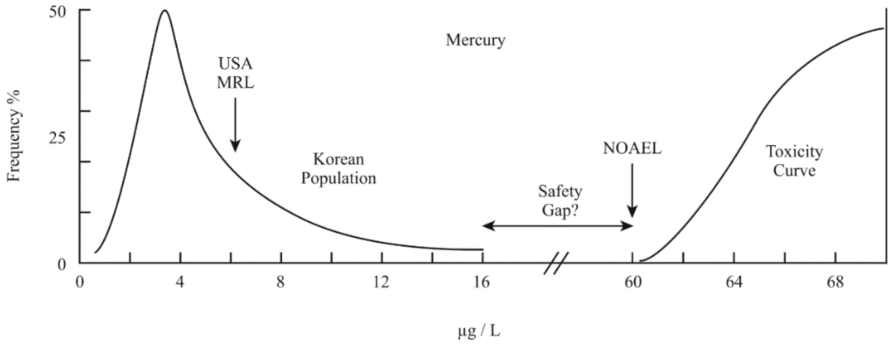
4. Distributions and Toxic Limits
5. Current Insightful Research
6. Recent Dietary and Medical Changes
7. The Current Role Now Played by Vaccines
8. Conclusions
Funding
Acknowledgments
Conflicts of Interest
References
- Jager, T. All individuals are not created equal; Accounting for inter-individual variation in fitting life-history responses to toxicants. Environ. Sci. Technol. 2013, 47, 1664–1669. [Google Scholar] [CrossRef] [PubMed]
- Everts, S. Brain barricade. Chem. Eng. News 2007, 85, 33–36. [Google Scholar] [CrossRef]
- Bauer, H.-C.; Krizbai, I.A.; Bauer, H.; Traweger, A. “You shall not pass”—Tight junctions of the blood brain barrier. Front. Neurosci. 2014, 8, 392. [Google Scholar] [CrossRef] [PubMed]
- Ostrea, E.M., Jr.; Mantaring, J.B., III; Silvestre, M.A. Drugs that affect the fetus and newborn infant via the placenta or breast milk. Pediatr. Clin. N. Am. 2004, 51, 539–579. [Google Scholar] [CrossRef] [PubMed]
- Mundy, W.R.; Padilla, S.; Breier, J.M.; Crofton, K.M.; Gilbert, M.E.; Herr, D.W.; Jensen, K.F.; Radio, N.M.; Raffaele, K.C.; Schumacher, K.; et al. Expanding the test set: Chemicals with potential to disrupt mammalian brain development. Neurotoxicol. Teratol. 2015, 52, 25–35. [Google Scholar] [CrossRef] [PubMed]
- Bondy, S.C. Metals and neuroinflammation. In Biometals in Neurodegenerative Diseases; White, A.R., Aschner, M., Costa, L.G., Bush, A.I., Eds.; Academic Press, Elsevier: London, UK, 2017; pp. 83–93. [Google Scholar]
- Kanninen, K.M.; White, A.R. Abnormal function of metalloproteins underlies most neurodegenerative diseases. In Biometals in Neurodegenerative Diseases; White, A.R., Aschner, M., Costa, L.G., Bush, A.I., Eds.; Academic Press, Elsevier: London, UK, 2017; pp. 415–438. [Google Scholar]
- Erikson, K.M.; Aschner, M. Manganese: Its role in disease and health. In Essential Metals in Medicine: Therapeutic Use and Toxicity of Metal Ions in the Clinic; Carver, P.L., Ed.; Walter de Gruyter GmbH: Berlin, Germany, 2019; Volume 19, pp. 253–266. [Google Scholar] [CrossRef]
- Ashley-Martin, J.; Dodds, L.; Arbuckle, T.E.; Ettinger, A.S.; Shapiro, G.D.; Fisher, M.; Monnier, P.; Morisset, A.-S.; Fraser, W.D.; Bouchard, M.F. Maternal and cord blood manganese (Mn) levels and birth weight: The MIREC birth cohort study. Int. J. Hyg. Environ. Health 2018, 221, 876–882. [Google Scholar] [CrossRef] [PubMed]
- Bouchard, M.F.; Sauve, S.; Barbeau, B.; Legrand, M.; Brodeur, M.-E.; Bouffard, T.; Limoges, E.; Bellinger, D.C.; Mergler, D. Intellectual impairment in school-age children exposed to manganese from drinking water. Environ. Health Perspect. 2011, 119, 138–143. [Google Scholar] [CrossRef]
- Yu, X.-D.; Zhang, J.; Yan, C.-H.; Shen, X.-M. Prenatal exposure to manganese at environment relevant level and neonatal neural behavioral development. Environ. Res. 2014, 133, 232–238. [Google Scholar] [CrossRef] [PubMed]
- Dos Santos, D.M.; Aschner, M.; dos Santos, A.P.M. Manganese and neurodegeneration. In Biometals in Neurodegenerative Diseases; White, A.R., Aschner, M., Costa, L.G., Bush, A.I., Eds.; Academic Press, Elsevier: London, UK, 2017; pp. 117–151. [Google Scholar]
- Chen, P.; Bornhorst, J.; Aschner, M. Manganese metabolism in humans. Front. Biosci. Landmark 2018, 23, 1655–1679. [Google Scholar] [CrossRef]
- Solovyev, N.D. Importance of selenium and selenoprotein for brain function: From antioxidant protection to neuronal signaling. J. Inorg. Biochem. 2015, 153, 1–12. [Google Scholar] [CrossRef]
- Kielczykowska, M.; Kocot, J.; Pazdzior, M.; Musik, I. Selenium—A fascinating antioxidant of protective properties. Adv. Clin. Exp. Med. 2018, 27, 245–255. [Google Scholar] [CrossRef] [PubMed]
- Sobolev, O.; Gutyj, B.; Petryshak, R.; Pivtorak, J.; Kovalskyi, Y.; Naumyuk, A.; Petryshak, O.; Semchuk, I.; Mateusz, V.; Shcherbatyy, A. Biological role of selenium in the organism of animals and humans. Ukrainian J. Ecol. 2018, 8, 654–665. [Google Scholar] [CrossRef]
- Karri, Z.; Schuhmacher, M.; Kumar, V. Heavy metals (Pb, Cd, As, and MeHg) as risk factors for cognitive dysfunction: A general review of metal mixture mechanism in brain. Environ. Toxic. Pharmacol. 2016, 48, 203–213. [Google Scholar] [CrossRef] [PubMed]
- Ollson, C.J.; Smith, E.; Herde, P.; Juhasz, A.L. Influence of co-contaminant exposure on the absorption of arsenic, cadmium and lead. Chemosphere 2017, 168, 658–666. [Google Scholar] [CrossRef] [PubMed]
- Martins, A.D., Jr.; Carneiro, M.F.; Grotto, D.; Adeyemi, J.A.; Barbosa, F., Jr. Arsenic, cadmium, and mercury-induced hypertension: Mechanisms and epidemiological findings. J. Tox. Environ. Health Part B Crit. Rev. 2018, 21, 61–82. [Google Scholar] [CrossRef] [PubMed]
- Pan, S.; Lin, L.; Zeng, F.; Zhang, J.; Dong, G.; Yang, B.; Jing, Y.; Chen, S.; Zhang, G.; Yu, Z.; et al. Effects of lead, cadmium, arsenic, and mercury co-exposure on children’s intelligence quotient in an industrialized area of southern China. Environ. Pollut. 2018, 235, 47–54. [Google Scholar] [CrossRef]
- Curren, M.S.; Liang, C.L.; Davis, K.; Kandola, K.; Brewster, J.; Potyrala, M.; Chan, H.M. Assessing determinants of maternal blood concentrations for persistent organic pollutants and metals in the Eastern and Western Canadian Arctic. Sci. Total Environ. 2015, 527–528, 150–158. [Google Scholar] [CrossRef]
- Long, M.; Knudsen, A.-K.S.; Pedersen, H.S.; Bonefeld-Jorgensen, E.C. Food intake and serum persistent organic pollutants in the Greenlandic pregnant women: The ACCEPT sub-study. Sci. Total Environ. 2015, 529, 198–212. [Google Scholar] [CrossRef]
- Miyashita, C.; Sasaki, S.; Saijo, Y.; Okada, E.; Kobayashi, S.; Baba, T.; Kajiwara, J.; Todaka, T.; Iwasaki, Y.; Nakazawa, H.; et al. Demographic, behavioral, dietary, and socioeconomic characteristics related to persistent organic pollutants and mercury levels in pregnant women in Japan. Chemosphere 2015, 133, 13–21. [Google Scholar] [CrossRef]
- Freeland-Graves, J.H.; Sanjeevi, N.; Lee, J.J. Global perspectives on trace element requirements. J. Trace Elem. Med. Biol. 2015, 31, 135–141. [Google Scholar] [CrossRef]
- Minoia, C.; Sabbioni, E.; Apostoli, P.; Pietra, R.; Gallorini, M.; Nicolaou, G.; Alessio, L.; Capodaglio, E. Trace element reference values in tissues from inhabitants of the European Community. I. A study of 46 elements in urine, blood and serum of Italian subjects. Sci. Total Environ. 1990, 95, 89–105. [Google Scholar] [CrossRef]
- Cesbron, A.; Saussereau, E.; Mahieu, L.; Couland, I.; Guerbet, M.; Goulle, J.-P. Metallic profile of whole blood and plasma in a series of 106 healthy volunteers. J. Anal. Toxicol. 2013, 37, 401–405. [Google Scholar] [CrossRef] [PubMed]
- Dlugaszek, M. Studies on relationships between essential and toxic elements in selected body fluids, cells and tissues. Chem. Biol. Interact. 2019, 297, 57–66. [Google Scholar] [CrossRef] [PubMed]
- Zeng, H.-L.; Li, H.-J.; Lu, J.; Guan, Q.; Cheng, L.-M. Assessment of 12 metals and metalloids in blood of general populations living in Wuhan of China by ICP-MS. Biolog. Trace Element Res. 2019, 189, 344–353. [Google Scholar] [CrossRef] [PubMed]
- Kalloo, G.; Wellenius, G.A.; McCandless, L.; Calafat, A.M.; Sjodin, A.; Karagas, M.; Chen, A.; Yolton, K.; Lanphear, B.P.; Braun, J.M. Profiles and predictors of environmental chemical mixture exposure among pregnant women: The health outcomes and measures of the environmental study. Environ. Sci. Technol. 2018, 52, 10104–10113. [Google Scholar] [CrossRef] [PubMed]
- Schofield, K. Autism, chemicals, probable cause and mitigation: A new examination. Autism Open Access 2016, 6, 3. [Google Scholar] [CrossRef]
- Kim, H.-J.; Lim, H.-S.; Lee, K.-R.; Choi, M.-H.; Kang, N.M.; Lee, C.H.; Oh, E.-J.; Park, H.-K. Determination of trace metal levels in the general population of Korea. Int. J. Environ. Res. Public Health 2017, 14, 702. [Google Scholar] [CrossRef] [PubMed]
- Eom, S.-Y.; Lee, Y.-S.; Lee, S.-G.; Seo, M.-N.; Choi, B.-S.; Kim, J.-D.; Lim, J.-A.; Hwang, M.-S.; Kwon, H.-J.; Kim, Y.-M.; et al. Lead, mercury and cadmium exposure in the Korean general population. J. Korean Med. Sci. 2018, 33, e9. [Google Scholar] [CrossRef]
- Schofield, K. The metal neurotoxins: An important role in current human neural epidemics? Int. J. Environ. Res. Public Health 2017, 14, 1511. [Google Scholar] [CrossRef]
- Sinha, D.; Prasad, P. Health effects inflicted by chronic low-level arsenic contamination in groundwater: A global public health challenge. J. Appl. Toxicol. 2019. [Google Scholar] [CrossRef]
- Signes-Pastor, A.J.; Vioque, J.; Navarrete-Munoz, E.M.; Carey, M.; Garcia-Villarino, M.; Fernandez-Somoano, A.; Tardon, A.; Santa-Marina, L.; Irizar, A.; Casas, M.; et al. Inorganic arsenic exposure and neuro-psychological development of children of 4 to 5 years of age living in Spain. Environ. Res. 2019, 174, 135–142. [Google Scholar] [CrossRef] [PubMed]
- Rahman, Z.; Singh, V.P. The relative impact of toxic heavy metals (arsenic, cadmium, chromium(VI), mercury and lead) on the total environment: An overview. Environ. Monit. Assess. 2019, 191, 419. [Google Scholar] [CrossRef] [PubMed]
- Vorvolakos, T.; Arseniou, S.; Samakouri, M. There is no safe threshold for lead exposure: A literature review. Psychiatriki 2016, 27, 204–214. [Google Scholar] [CrossRef] [PubMed]
- Shefa, S.T.; Heroux, P. Both physiology and epidemiology support zero tolerable blood lead levels. Toxicol. Lett. 2017, 280, 232–237. [Google Scholar] [CrossRef] [PubMed]
- Reuben, A.; Caspi, A.; Belsky, D.W.; Broadbent, J.; Harrington, H.; Sugden, K.; Houts, R.M.; Ramrakha, S.; Poulton, R.; Moffitt, T.E. Association of childhood blood levels with cognitive function and socioeconomic status at age 38 years and with IQ change and socioeconomic mobility between childhood and adulthood. J. Am. Med. Assoc. 2017, 317, 1244–1251. [Google Scholar] [CrossRef] [PubMed]
- Cardoso, B.R.; Roberts, B.R.; Bush, A.I.; Hare, D.J. Selenium, selenoproteins and neurodegenerative diseases. Metallomics 2015, 7, 1213–1228. [Google Scholar] [CrossRef] [PubMed]
- Cardoso, B.R.; Hare, D.J.; Bush, A.I. The role of selenium in neurodegenerative diseases. In Biometals in Neurodegenerative Diseases; White, A.R., Aschner, M., Costa, L.G., Bush, A.I., Eds.; Academic Press, Elsevier: London, UK, 2017; pp. 35–49. [Google Scholar]
- Dominiak, A.; Wilkaniec, A.; Wrocynski, P.; Adamczyk, A. Selenium in the therapy of neurological diseases. Where is it going? Curr. Neuropharmacol. 2016, 14, 282–299. [Google Scholar] [CrossRef]
- Pieczynska, J.; Grajeta, H. The role of selenium in human conception and pregnancy. J. Trace Elem. Med. Biol. 2015, 29, 31–38. [Google Scholar] [CrossRef]
- Varsi, K.; Bolann, B.; Torsvik, I.; Eik, T.C.R.; Hol, P.J.; Bjorke-Monsen, A.L. Impact of maternal selenium status on infant outcome during the first six months of life. Nutrients 2017, 9, 486. [Google Scholar] [CrossRef]
- Ambroziak, U.; Hybsier, S.; Shahnazaryan, U.; Krasnodebska-Kiljanska, M.; Rijntjes, E.; Bartoszewicz, Z.; Bednarczuk, T.; Schomburg, L. Severe selenium deficits in pregnant women irrespective of autoimmune thyroid disease in an area with marginal selenium intake. J. Trace Elem. Med. Biol. 2017, 44, 186–191. [Google Scholar] [CrossRef]
- Zachara, B.A. Selenium in complicated pregnancy: A review. In Advances in Clinical Chemistry; Makowski, G.S., Ed.; Academic Press, Elsevier: San Diego, CA, USA, 2018; Volume 86, pp. 157–178. [Google Scholar] [CrossRef]
- Krewski, D.; Yokel, R.A.; Nieboer, E.; Borchelt, D.; Cohen, J.; Harry, J.; Kacew, S.; Lindsay, J.; Mahfouz, A.M.; Rondeau, V. Human health risk assessment for aluminum, aluminum oxide and aluminum hydroxide. J. Toxicol. Environ. Health B 2007, 10, 1–269. [Google Scholar] [CrossRef] [PubMed]
- Mold, M.; Umar, D.; King, A.; Exley, C. Aluminum in brain tissue in autism. J. Trace Elem. Med. Biol. 2018, 46, 76–82. [Google Scholar] [CrossRef] [PubMed]
- Agency for Toxic Substances and Disease Registry. Toxicological Profile for Arsenic; US Department of Health and Human Services, Agency for Toxic Substances and Disease Registry: Atlanta, GA, USA, 2007; p. 559.
- Ishii, K.; Itoh, Y.; Iwasaki, N.; Shibata, Y.; Tamaoka, A. Detection of diphenylarsinic acid and its derivatives in human serum and cerebrospinal fluid. Clin. Chim. Acta 2014, 431, 227–231. [Google Scholar] [CrossRef] [PubMed]
- Ahmad, S.A.; Khan, M.H.; Haque, M. Arsenic contamination in groundwater in Bangladesh: Implications and challenges for healthcare policy. Risk Manag. Healthc. Policy 2018, 11, 251–261. [Google Scholar] [CrossRef] [PubMed]
- Du, X.-Y.; Tian, M.-P.; Wang, X.-X.; Zhang, J.; Huang, Q.-Y.; Liu, L.-P.; Shen, H.-Q. Cortex and hippocampus DNA epigenetic response to a long-term arsenic exposure via drinking water. Environ. Pollut. 2018, 234, 590–600. [Google Scholar] [CrossRef] [PubMed]
- World Health Organization, International Agency for Research on Cancer. Inorganic and Organic Lead Compounds; International Agency for Research on Cancer: Lyon, France, 2006; Volume 87, p. 16. [Google Scholar]
- Agency for Toxic Substances and Disease Registry. Toxicological Profile for Lead; US Department of Health and Human Services, Agency for Toxic Substances and Disease Registry: Atlanta, GA, USA, 2007; p. 582.
- United Nations. Final Review of Scientific Information on Lead; United Nations Environment Program Chemicals Branch; United Nations: Geneva, Switzerland, 2010; 332p. [Google Scholar]
- Rooney, J.P.K.; Woods, N.F.; Martin, M.D.; Woods, J.S. Genetic polymorphisms of GRIN2A and GRIN2B modify the neurobehavioral effects of low-level exposure in children. Environ. Res. 2018, 165, 1–10. [Google Scholar] [CrossRef] [PubMed]
- Fenga, C.; Liu, S.-S.; Zhou, F.-K.; Gao, Y.-Y.; Li, Y.-S.; Du, G.-H.; Chen, Y.; Jiao, H.; Feng, J.-G.; Zhang, Y.-Y.; et al. Oxidative stress in the neuro-degenerative brain following lifetime exposure to lead in rats: Changes in lifespan profiles. Toxicology 2019, 411, 101–109. [Google Scholar] [CrossRef] [PubMed]
- O’Connor, D.; Hou, D.-Y.; Ye, J.; Zhang, Y.-H.; Ok, Y.S.; Song, Y.-N.; Coulon, F.; Peng, T.-Y.; Tian, L. Lead-based paint remains a major public health concern: A critical review of global production, trade, use, exposure, health risk, and implications. Environ. Int. 2018, 121, 85–101. [Google Scholar] [CrossRef]
- Burbacher, T.M.; Shen, D.D.; Liberato, N.; Grant, K.S.; Cernichiari, E.; Clarkson, T. Comparison of blood and brain levels in infant monkeys exposed to methylmercury or vaccines containing Thimerosal. Environ. Health Perspect. 2005, 113, 1015–1021. [Google Scholar] [CrossRef]
- Rodrigues, J.L.; Serpeloni, J.M.; Batista, B.L.; Souza, S.S.; Barbosa, F., Jr. Identification and distribution of mercury species in rat tissues following administration of Thimerosal or methylmercury. Arch. Toxicol. 2010, 84, 891–896. [Google Scholar] [CrossRef]
- Dorea, J.G.; Farina, M.; Rocha, J.B.T. Toxicity of ethylmercury (and Thimerosal): A comparison with methylmercury. J. Appl. Toxicol. 2013, 33, 700–711. [Google Scholar] [CrossRef] [PubMed]
- Santana, L.N.D.; Bittencourt, L.O.; Nascimento, P.C.; Fernandes, R.M.; Teixeira, F.B.; Fernandes, L.M.P.; Silva, M.C.F.; Nogueira, L.S.; Amado, L.L.; Crespo-Lopez, M.E. Low doses of methylmercury exposure during adulthood in rats display oxidative stress, neurodegeneration in the motor cortex and lead to impairment of motor skills. J. Trace Elem. Med. Biol. 2019, 51, 19–27. [Google Scholar] [CrossRef] [PubMed]
- Agency for Toxic Substances and Disease Registry. Toxicological Profile for Manganese; US Department of Health and Human Services, Agency for Toxic Substances and Disease Registry: Atlanta, GA, USA, 2012; p. 556.
- Yokel, R.A. Brain uptake, retention, and efflux of aluminum and manganese. Environ. Health Perspect. 2002, 110 (Suppl. 5), 699–704. [Google Scholar] [CrossRef] [PubMed]
- Yokel, R.A. Manganese flux across the blood-brain barrier. Neuromol. Med. 2009, 11, 297–310. [Google Scholar] [CrossRef] [PubMed]
- Mahoney, J.P.; Small, W.J. Studies on manganese. III. The biological half-life of radio manganese in man and factors which affect this. J. Clin. Investig. 1968, 47, 643–653. [Google Scholar] [CrossRef]
- Davidsson, L.; Cederblad, A.; Lonnerdal, B.; Sandstrom, B. Manganese retention in man: A method for estimating manganese absorption in man. Am. J. Clin. Nutr. 1989, 49, 170–179. [Google Scholar] [CrossRef]
- O’Neal, S.L.; Hong, L.; Fu, S.; Jiang, W.; Jones, A.; Nie, L.H.; Zheng, W. Manganese accumulation in bone following chronic exposure in rats: Steady-state concentration and half-life in bone. Toxicol. Lett. 2014, 229, 93–100. [Google Scholar] [CrossRef]
- Grandjean, P.; Herz, K.T. Trace elements as paradigms of developmental neurotoxicants: Lead, methylmercury and arsenic. J. Trace Elem. Med. Biol. 2015, 31, 130–134. [Google Scholar] [CrossRef]
- Cobbina, S.J.; Chen, Y.; Zhou, Z.-X.; Wu, X.-S.; Feng, W.-W.; Wang, W.; Li, Q.; Zhao, T.; Mao, G.-H.; Wu, X.-Y. Interaction of four low-dose toxic metals with essential metals in the brain, liver and kidneys of mice on sub-chronic exposure. Environ. Toxicol. Pharmacol. 2015, 39, 280–291. [Google Scholar] [CrossRef]
- Cobbina, S.J.; Chen, Y.; Zhou, Z.-X.; Wu, X.; Feng, W.; Wang, W.; Mao, G.; Xu, H.; Zhang, Z.; Wu, X.; et al. Low concentration toxic metal mixture interactions: Effects on essential and non-essential metals in brain, liver, and kidneys of mice on sub-chronic exposure. Chemosphere 2015, 132, 79–86. [Google Scholar] [CrossRef]
- Bellinger, D.C. Applying methods of the global burden of diseases, injuries, and risk factors study to developmental neurotoxics: A commentary. Environ. Health 2018, 17, 53. [Google Scholar] [CrossRef] [PubMed]
- Valeri, L.; Mazumdar, M.M.; Bobb, J.F.; Hen, B.C.; Rodrigues, E.; Sharif, O.I.A.; Kile, M.L.; Quamruzzaman, Q.; Afroz, S.; Golam, M.; et al. The joint effect of pre-natal exposure to metal mixtures on neuro-developmental outcomes at 20–40 months of age: Evidence from rural Bangladesh. Environ. Health Perspect. 2017, 125, 067015. [Google Scholar] [CrossRef]
- Yu, H.-Y.; Zhang, K.-L. Links between environmental geochemistry and rate of birth defects: Shanxi Province, China. Sci. Total Environ. 2011, 409, 447–451. [Google Scholar] [CrossRef] [PubMed]
- Kim, I.; Kim, M.H.; Lim, S. Increased risk of spontaneous abortion and menstrual aberrations in female workers in semiconductor industry, South Korea. Occup. Environ. Med. 2014, 71 (Suppl. 1), A15. [Google Scholar] [CrossRef]
- Passini, R., Jr.; Cecatti, J.G.; Lajos, G.J.; Tedesco, R.P.; Nomura, M.L.; Dias, T.Z.; Haddad, S.M.; Rehder, P.M.; Pacagnella, R.C.; Costa, M.L.; et al. Brazilian multicenter study on preterm birth: Prevalence and factors associated with spontaneous preterm birth. PLoS ONE 2014, 9, e109069. [Google Scholar] [CrossRef] [PubMed]
- Ralston, N.V.C.; Raymond, L.J. Functional deletion of brain selenoenzymes by methylmercury. In Global Advance in Selenium Research from Theory to Application, Proceedings of the 4th International Conference on Selenium in the Environment and Human Health, Sao Paulo, Brazil, 8–21 October 2015; Banuelos, G.S., Lin, Z.-Q., Moraes, M.F., Guilherme, L.R.G., Reis, A.R., Eds.; CRC Press—Taylor and Francis Group: Leiden, The Netherlands, 2016; pp. 71–72. [Google Scholar]
- Amadi, C.N.; Igweze, Z.N.; Orisakwe, O.E. Heavy metals in miscarriages and stillbirths in developing nations. Middle East Fertil. Soc. J. 2017, 22, 91–100. [Google Scholar] [CrossRef]
- McClure, E.M.; Saleem, S.; Goudar, S.S.; Dhaded, S.; Guruprasad, G.; Kumar, Y.; Tikmani, S.S.; Kadir, M.; Raza, J.; Yasmin, H. The project to understand and research preterm pregnancy outcomes and stillbirths in South Asia: A protocol of a prospective, cohort study of causes of mortality among preterm births and stillbirths. Reprod. Health 2018, 15 (Suppl. 1), 89. [Google Scholar] [CrossRef] [PubMed]
- Spillar, H.A. Rethinking mercury: The role of selenium in the pathophysiology of mercury toxicity. Clin. Toxicol. 2018, 56, 313–326. [Google Scholar] [CrossRef] [PubMed]
- Demir, N.; Basaranoglu, M.; Huyut, Z.; Deger, I.; Karaman, K.; Sekeroglu, M.R.; Tuncer, O. The relationship between mother and infant plasma trace element and heavy metal levels and the risk of neural tube defect in infants. J. Matern. Fetal Neonatal Med. 2019, 32, 1433–1440. [Google Scholar] [CrossRef]
- Heimfarth, L.; Delgado, J.; Mingori, M.R.; Moresco, K.S.; Pureur, R.P.; Gelain, D.P.; Moreira, J.C.F. Delayed neurochemical effects of pre-natal exposure to MeHg in the cerebellum of developing rats. Toxicol. Lett. 2018, 284, 161–169. [Google Scholar] [CrossRef]
- Taylor, C.M.; Golding, J.; Emond, A.M. Lead, cadmium and mercury levels in pregnancy: The need for international consensus on levels of concern. J. Dev. Orig. Health Dis. 2014, 5, 16–30. [Google Scholar] [CrossRef]
- Brown, I.A.; Austin, D.W. Maternal transfer of mercury to the developing embryo/fetus: Is there a safe level? Toxicol. Environ. Chem. 2012, 94, 1610–1627. [Google Scholar] [CrossRef]
- Obi, E.; Okafor, C.; Igwebe, A.; Ebenebe, J.; Afonne, O.J.; Ifediata, F.; Orisakwe, O.E.; Nriagu, J.O.; Basu, N. Elevated prenatal methylmercury exposure in Nigeria: Evidence from maternal and cord blood. Chemosphere 2015, 119, 485–489. [Google Scholar] [CrossRef]
- Aylward, L.L.; Hays, S.M.; Kirman, C.R.; Marchitti, S.A.; Kenneke, J.F.; English, C.; Mattison, D.R.; Becker, R.A. Relationships of chemical concentrations in maternal and cord blood: A review of available data. J. Toxicol. Environ. Health B 2014, 17, 175–203. [Google Scholar] [CrossRef]
- Kosik-Bogacka, D.; Lanocha-Arendarczyk, N.; Kot, K.; Malinowski, W.; Szymanski, S.; Sipak-Szmigiel, O.; Pilarczyk, B.; Tomza-Marciniak, A.; Podlasinska, J.; Tomska, N.; et al. Concentrations of mercury and selenium in afterbirth and their relations with various factors. Environ. Geochem. Health 2018, 40, 1683–1695. [Google Scholar] [CrossRef]
- Sakamoto, M.; Chan, H.M.; Domingo, J.L.; Koriyama, C. Placental transfer and levels of mercury, selenium, vitamin E and docosahexaenoic acid in maternal and umbilical cord blood. Environ. Int. 2018, 111, 309–315. [Google Scholar] [CrossRef]
- Parajuli, R.P.; Fujiwara, T.; Umezaki, M.; Furusawa, H.; Ser, P.H.; Watanabe, C. Cord blood levels of toxic and essential trace elements and their determinants in the Terai region of Nepal: A birth cohort study. Biol. Trace Elem. Res. 2012, 147, 75–83. [Google Scholar] [CrossRef]
- Iwai-Shimada, M.; Kameo, S.; Nakai, K.; Yaginuma-Sakurai, K.; Tatsuta, N.; Kurokawa, N.; Nakayama, S.F.; Satoh, H. Exposure profile of mercury, lead, cadmium, arsenic, antimony, copper, selenium and zinc in maternal blood, cord blood and placenta: The Tohoku study of child development in Japan. Environ. Health Prev. Med. 2019, 24, 35. [Google Scholar] [CrossRef]
- Sakamoto, M.; Yasutake, A.; Kakita, A.; Ryufuku, M.; Chan, H.M.; Yamamoto, M.; Oumi, S.; Kobayashi, S.; Watanabe, C. Selenomethionine protects against neuronal degeneration by methylmercury in the developing rat cerebrum. Environ. Sci. Technol. 2013, 47, 2862–2868. [Google Scholar] [CrossRef]
- McKelvey, S.M.; Horgan, K.A.; Murphy, R.A. Chemical form of selenium differentially influences DNA repair pathways following exposure to lead nitrate. J. Trace Elem. Med. Biol. 2015, 29, 151–169. [Google Scholar] [CrossRef]
- Pacyna, J.M.; Sundseth, K.; Pacyna, E.G. Sources and fluxes of harmful metals. In Environmental Determinants of Human Health; Pacyna, J.M., Pacyna, E.G., Eds.; Part of the Molecular and Integrative Toxicology Series; Springer: Berlin, Germany, 2016; pp. 1–25. [Google Scholar] [CrossRef]
- Zhang, H.; Feng, X.-B.; Larssen, T.; Qiu, G.-L.; Vogt, R.D. In inland China, rice rather than fish is the major pathway for methylmercury exposure. Health Perspect. 2010, 118, 1183–1188. [Google Scholar] [CrossRef]
- Cui, W.-B.; Liu, G.-L.; Bezerra, M.; Lagos, D.A.; Li, Y.-B.; Cai, Y. Occurrence of methylmercury in rice based infant cereals and estimation of daily dietary intake of methylmercury for infants. J. Agric. Food Chem. 2017, 65, 9569–9578. [Google Scholar] [CrossRef]
- Al-Saleh, I.; Abduljabbar, M. Heavy metals (lead, cadmium, methyl mercury, arsenic) in commonly imported rice grains (Oryza sativa) sold in Saudi Arabia and their potential health risk. Int. J. Hyg. Environ. Health 2017, 220, 1168–1178. [Google Scholar] [CrossRef]
- Kwon, S.Y.; Selin, N.E.; Giang, A.; Karplus, V.J.; Zhang, D. Present and future mercury concentrations in Chinese rice: Insights from modeling. Glob. Biogeochem. Cycles 2018, 32, 437–462. [Google Scholar] [CrossRef]
- Xiao, G.-X.; Hu, Y.-L.; Li, N.; Yang, D.-J. Spatial autocorrelation analysis of monitoring data of heavy metals in rice in China. Food Control 2018, 89, 32–37. [Google Scholar] [CrossRef]
- Yokel, R.A.; Lasley, S.M.; Dorman, D.C. The speciation of metals in mammals influences their toxicokinetics and toxicodynamics and therefore human health risk assessment. Toxicol. Environ. Health B Crit. Rev. 2006, 9, 63–85. [Google Scholar] [CrossRef]
- Michalke, B.; Willkommen, D.; Drobyshev, E.; Solovyev, N. The importance of speciation analysis in neurodegeneration research. TrAC Trends Anal. Chem. 2018, 104, 160–170. [Google Scholar] [CrossRef]
- Witt, B.; Ebert, F.; Meyer, S.; Francesconi, K.A.; Schwerdtle, T. Assessing neuro-developmental effects of arsenolipids in pre-differentiated human neurons. Mol. Nutr. Food Res. 2017, 61, 1700199. [Google Scholar] [CrossRef]
- Stiboller, M.; Raber, G.; Lenters, V.; Gjengedal, E.L.F.; Eggesbo, M.; Francesconi, K.A. Arsenolipids detected in the milk of nursing mothers. Environ. Sci. Technol. Lett. 2017, 4, 273–279. [Google Scholar] [CrossRef]
- Reyes-Avila, A.D.; Laws, E.A.; Herrmann, A.D.; DeLaune, R.D.; Blanchard, T.P. Mercury and selenium levels, and Se:Hg molar ratios in freshwater fish from South Louisiana. J. Environ. Sci. Health. A Toxic Hazard. Sub. Environ. Eng. 2019, 54, 238–245. [Google Scholar] [CrossRef]
- Tindell, R.; Tipple, T. Selenium: Implications for outcomes in extremely preterm infants. J. Perinatol. 2018, 38, 197–202. [Google Scholar] [CrossRef]
- Lewandowska, M.; Sajdak, S.; Lubinski, J. Serum selenium level in early healthy pregnancy as a risk marker of pregnancy induced hypertension. Nutrients 2019, 11, 1028. [Google Scholar] [CrossRef]
- Phiri, F.P.; Ander, E.L.; Bailey, E.H.; Chilima, B.; Chilimba, A.D.C.; Gondwe, J.; Joy, E.J.M.; Kalimbira, A.A.; Kumssa, D.B.; Lark, R.M.; et al. The risk of selenium deficiency in Malawi is large and varies over multiple spatial scales. Sci. Rep. 2019, 9, 6566. [Google Scholar] [CrossRef]
- Stratton, K.R. Adverse Effects of Vaccines: Evidence and Causality; Institute of Medicine; The National Academies Press: Washington DC, USA, 2012; ISBN 978-0-309-21435-3. [Google Scholar]
- Goldman, G.S. Comparison of the vaccine adverse event reporting system (VAERS) fetal-loss reports during three consecutive influenza seasons: Was there a synergistic fetal toxicity associated with the two-vaccine 2009/2010 season? Human Exp. Toxicol. 2013, 32, 464–475. [Google Scholar] [CrossRef]
- Sanders, A.P.; Flood, K.; Chiang, S.; Herring, A.H.; Wolf, L.; Fry, R.C. Towards prenatal biomonitoring in North Carolina: Assessing arsenic, cadmium, mercury and lead levels in pregnant women. PLoS ONE 2012, 7, e31354. [Google Scholar] [CrossRef]
- Friedrich, M.J. High mercury levels found in women around the world. J. Am. Med. Assoc. 2017, 318, 1857. [Google Scholar] [CrossRef]
- Tratnik, J.S.; Falnoga, I.; Mazej, D.; Kocman, D.; Fajon, V.; Jadodic, M.; Stajnko, A.; Trdin, A.; Slejkovec, Z.; Jeran, Z.; et al. Results of the first national human biomonitoring in Slovenia: Trace elements in men and lactating women, predictors of exposure and reference values. Int. J. Hyg. Environ. Health 2019, 222, 563–582. [Google Scholar] [CrossRef]
- Wang, X.; Qi, L.; Peng, Y.; Xia, W.; Xu, S.-Q.; Li, Y.-Y.; Zhang, H.-L. Urinary concentrations of environmental metals and associating factors in pregnant women. Environ. Sci. Pollut. Res. 2019, 26, 13464–13475. [Google Scholar] [CrossRef]
- Shim, Y.K.; Lewin, M.D.; Ruiz, P.; Eichner, J.E.; Mumtaz, M.M. Prevalence and associated demographic characteristics of exposure to multiple metals and their species in human populations: The United States NHANES 2007–2012. J. Toxicol. Environ. Health A 2017, 80, 502–512. [Google Scholar] [CrossRef]
- Kim, Y.-M.; Chung, J.-Y.; An, H.S.; Park, S.Y.; Kim, B.-G.; Bae, J.W.; Han, M.; Cho, Y.J.; Hong, Y.-S. Biomonitoring of lead, cadmium, total mercury and methylmercury levels in maternal blood and in umbilical cord blood at birth in South Korea. Int. J. Environ. Res. Public Health 2015, 12, 13482–13493. [Google Scholar] [CrossRef]
- Arbuckle, T.E.; Liang, C.L.; Morisset, A.-S.; Fisher, M.; Weiler, H.; Cirtiu, C.M.; Legrand, M.; Davis, K.; Ettinger, A.S.; Fraser, W.D. Maternal and fetal exposure to cadmium, lead, manganese and mercury: The MIREC study. Chemosphere 2016, 163, 270–282. [Google Scholar] [CrossRef] [PubMed]
- Costa, L.G. Developmental exposure to metals and its contribution to age-related neurodegeneration. In Biometals in Neurodegenerative Diseases; White, A.R., Aschner, M., Costa, L.G., Bush, A.I., Eds.; Academic Press, Elsevier: London, UK, 2017; pp. 217–229. [Google Scholar]
- Schofield, K. Test Panel to Measure Blood Neurotoxin Levels in Pre-Maternal Women and for the General Public in Relation to Mental Disorders of the Aging. US Patent 10,049,767 B2, 14 August 2018. [Google Scholar]
- Gajek, R.; Barley, F.; She, J. Determination of essential and toxic metals in blood by ICP-MS calibration in synthetic matrix. Anal. Methods 2013, 5, 2193–2202. [Google Scholar] [CrossRef]
- Jones, D.R.; Jarrett, J.M.; Tevis, D.S.; Franklin, M.; Mullinix, N.J.; Wallon, K.L.; Quarles, C.D., Jr.; Caldwell, K.L.; Jones, R.L. Analysis of whole human blood for Pb, Cd, Hg, Se, and Mn by ICP-DRC-MS for biomonitoring and acute exposures. Talanta 2017, 162, 114–122. [Google Scholar] [CrossRef] [PubMed]
- Bello, G.A.; Arora, M.; Austin, C.; Horton, M.K.; Wright, R.O. Extending the Distributed Lag Model framework to handle chemical mixtures. Environ. Res. 2017, 156, 253–264. [Google Scholar] [CrossRef] [PubMed]
- Liu, S.H.; Bobb, J.F.; Henn, B.C.; Gennings, C.; Lourdes, S.; Tellez-Rojo, M.; Bellinger, D.; Arora, M.; Wright, R.O.; Coull, B.A. Bayesian varying coefficient kernel machine regression to assess neurodevelopmental trajectories associated with exposure to complex mixtures. Stat. Med. 2018, 37, 4680–4694. [Google Scholar] [CrossRef] [PubMed]

© 2019 by the author. Licensee MDPI, Basel, Switzerland. This article is an open access article distributed under the terms and conditions of the Creative Commons Attribution (CC BY) license (http://creativecommons.org/licenses/by/4.0/).
Share and Cite
Schofield, K. An Important Need to Monitor from an Early Age the Neurotoxins in the Blood or by an Equivalent Biomarker. Int. J. Environ. Res. Public Health 2019, 16, 3425. https://doi.org/10.3390/ijerph16183425
Schofield K. An Important Need to Monitor from an Early Age the Neurotoxins in the Blood or by an Equivalent Biomarker. International Journal of Environmental Research and Public Health. 2019; 16(18):3425. https://doi.org/10.3390/ijerph16183425
Chicago/Turabian StyleSchofield, Keith. 2019. "An Important Need to Monitor from an Early Age the Neurotoxins in the Blood or by an Equivalent Biomarker" International Journal of Environmental Research and Public Health 16, no. 18: 3425. https://doi.org/10.3390/ijerph16183425
APA StyleSchofield, K. (2019). An Important Need to Monitor from an Early Age the Neurotoxins in the Blood or by an Equivalent Biomarker. International Journal of Environmental Research and Public Health, 16(18), 3425. https://doi.org/10.3390/ijerph16183425