Article

Socio-Economic and Environmental Factors Related to Spatial Differences in Human Non-Tuberculous Mycobacterial Diseases in the Czech Republic

1
Faculty of Regional Development and International Studies, Mendel University in Brno, Tr. Generala Piky 7, 613 00 Brno, Czech Republic
2
Public Health Institute Ostrava, Partyzanske Nam. 7, 702 00 Ostrava, Czech Republic
3
IFCOR Klinicke Laboratore Ltd., Vinicni 235, 615 00 Brno, Czech Republic
4
Department of Biochemistry and Genetics, La Trobe Institute for Molecular Science, La Trobe University, Science Dr, Bundoora, Melbourne VIC 3086, Australia
*
Author to whom correspondence should be addressed.
Int. J. Environ. Res. Public Health 2019, 16(20), 3969; https://doi.org/10.3390/ijerph16203969
Submission received: 12 September 2019 / Accepted: 16 October 2019 / Published: 17 October 2019
(This article belongs to the Section Infectious Disease Epidemiology)

Abstract

:
Non-tuberculous mycobacteria (NTM) are ubiquitous environmental bacteria that can induce pulmonary and non-pulmonary diseases in susceptible persons. It is reported that the prevalence of NTM diseases is increasing in developed countries, but this differs by regions and countries. NTM species distribution and the rate of diseases caused by NTM vary widely in the historical territories of Moravia and Silesia (Czech Republic). This epidemiologic study of NTM diseases covers the period 2012–2018, reviews isolates obtained from patients with clinical disease and investigates correlations with related socio-economic and environmental factors. Individual NTM patients were included only once during the studied period and results were presented as incidence rate per year. The most frequently isolated NTM meeting the microbiological and clinical criteria in the study were the Mycobacterium avium-intracellulare complex, followed by Mycobacterium kansasii and Mycobacterium xenopi. A previously described endemic incidence of M. kansasii in the Karviná district and M. xenopi in the Ostrava district was also observed in this study. The incidence of NTM patients in the whole studied territory was 1.10/100,000 inhabitants (1.33/100,000 in men and 0.88/100,000 in women). The annual incidence of lymphadenitis in children (≤5 years of age) was 2.35/100,000 of the population of children during the 7 year period but increased in the year 2018 to 5.95/100,000. The rate of human tuberculosis in the studied area was 1.97/100,000 inhabitants. The incidence of NTM pulmonary diseases correlated with a lower socio-economic status (r = 0.63) and a higher concentration of benzo[a]pyrene pollution in the air (r = 0.64).

1. Introduction

Non-tuberculous mycobacteria (NTM) are ubiquitous environmental bacteria commonly found in water biofilms, soils, dust, and sediments [1,2,3,4]. Some NTM species are opportunistic pathogens that are known to cause lung diseases and non-pulmonary infections in susceptible individuals [5]. An increase in the prevalence of NTM pulmonary diseases has been recorded in developed countries [6,7,8,9]. This increase has caused a substantial financial burden due to patient treatment and hospitalization [10]. Probable explanations for the increase in the frequency of clinically relevant isolates of NTM are longer life expectancy and the improvement in mycobacterial DNA isolation techniques and other improvements in laboratory diagnostics [2]. Significant geographic differences in the distribution of different NTM species in human clinical isolates were recently described worldwide [11,12,13]. The factors associated with this phenomenon are climate differences, population density, host-related factors (genetic background and occupational exposure) and the presence of a specific environmental niches [11,14,15,16].
Important risk factors of NTM pulmonary diseases are immunodeficiency and various structural lung diseases, such as chronic obstructive pulmonary disease (COPD), bronchiectasis, and cystic fibrosis [16,17]. The development of COPD and/or similar diseases is manifested by diminishing bronchial epithelium integrity. It can be connected to smoking and airborne pollution comprising particulate matter (PM) [18,19]. PM of varying size, origin and chemistry can absorb harmful pollutants such as polycyclic aromatic hydrocarbons (PAHs) that induce the production of reactive oxygen species (ROS) and inflammatory responses through the interaction with the aryl hydrocarbon receptor and the subsequent activation of cytochrome P450. These mechanisms make airborne pollution a potential risk factor for pulmonary tissue impairment and consequently could increase the risk of NTM pulmonary diseases in polluted urban and industrial areas [20,21]. Exposure to aerosol contaminated by NTM is another possible factor, causing NTM lung diseases in miners and metalworkers who may already have impaired lung alveoli as a consequence of a polluted occupational environment [15,22,23].
The historical territory of Moravia and Silesia in the Eastern part of the Czech Republic includes the Ostrava and Karviná districts—the most polluted areas in Central Europe from airborne dust with high concentrations of dust particles (PM2.5 and PM10) and benzo[a]pyrene, which is one of the most harmful PAHs [24,25]. Historically, the environment of these districts has been polluted as a result of mining activities and heavy industry, and although presently, heavy industry and black coal mining are in decline, air pollution in this area still continues [26]. Currently, stationary combustion sources are the major emitters of pollutants with geographical and weather conditions also playing a significant role in maintaining the high level of air pollution in this area. A high endemic incidence of infections caused by Mycobacterium kansasii has been reported in the Karviná district since 1968. A correlation between a higher incidence of M. kansasii diseases and employment in the mining industry in this district was described during the period 1968–1995 [27]. During the last two decades, the mining industry has been in decline and, consequently, the number of people employed in the mining industry has significantly reduced from 32,000 in 1995 to 12,500 in 2015 [28]; however, there continues to be an enhanced NTM incidence in this area (unpublished data from the Public Health Institute Ostrava, Czech Republic, 2019).
We hypothesize that air pollution and the worsening socio-economic situation of the resident population are co-contributing factors, resulting in a higher NTM incidence in some parts of the Czech Republic. To verify this hypothesis, three research aims were assessed: (1) to find differences in the geographic distribution of the total number of NTM disease incidence and individual NTM species in the studied area; (2) to analyze the relationship of selected demographic and socio-economic factors to the incidence of NTM-infected patients; and (3) to investigate a connection between air pollution indicators and NTM lung disease incidence. We also compared the age and sex of patients with human tuberculosis caused by Mycobacterium tuberculosis (MTB) and NTM diseases in order to find differences in these demographic parameters. We included data from a 7 year period (2012–2018) in our study.

2. Materials and Methods

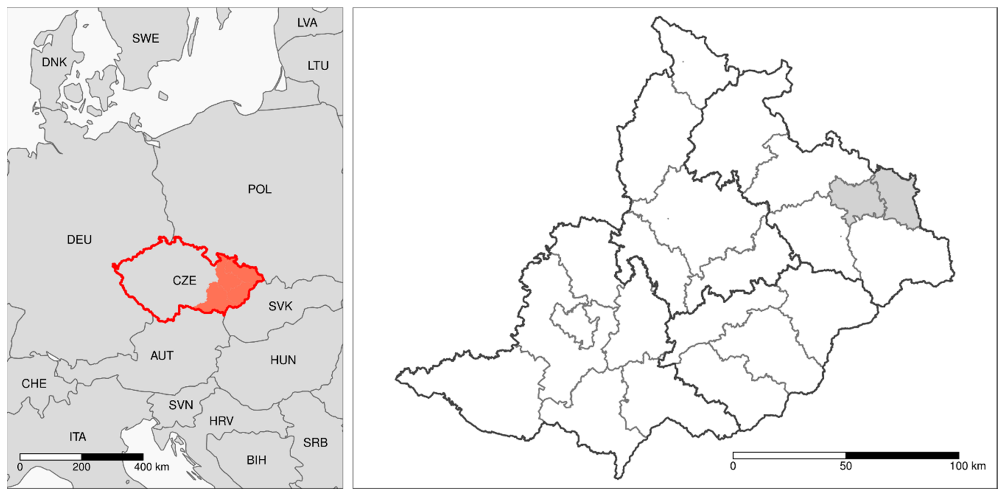
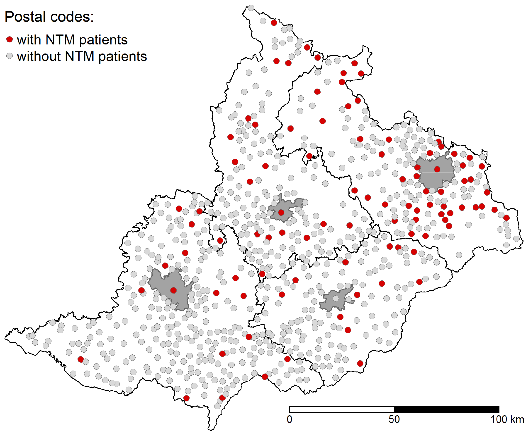
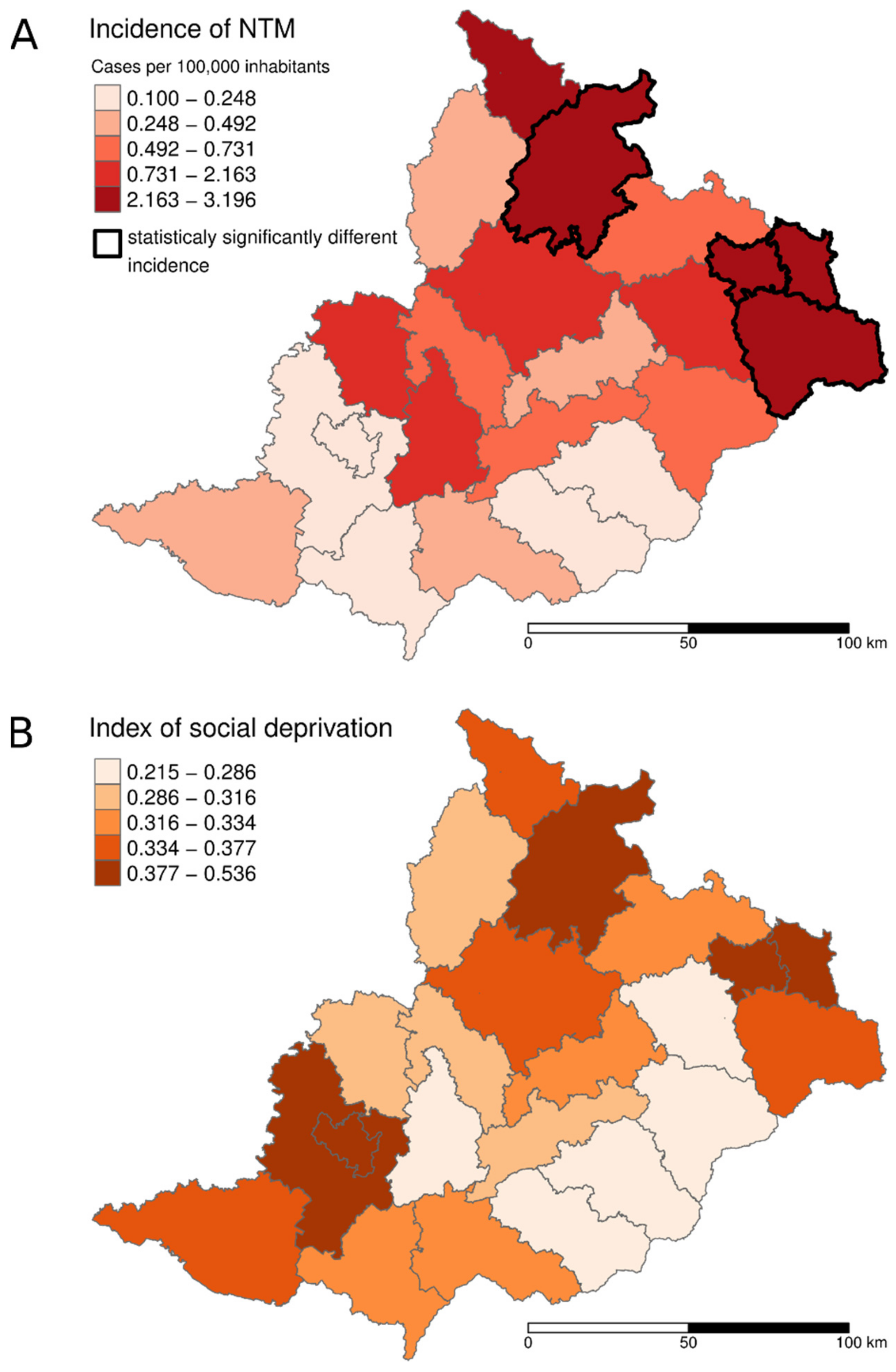
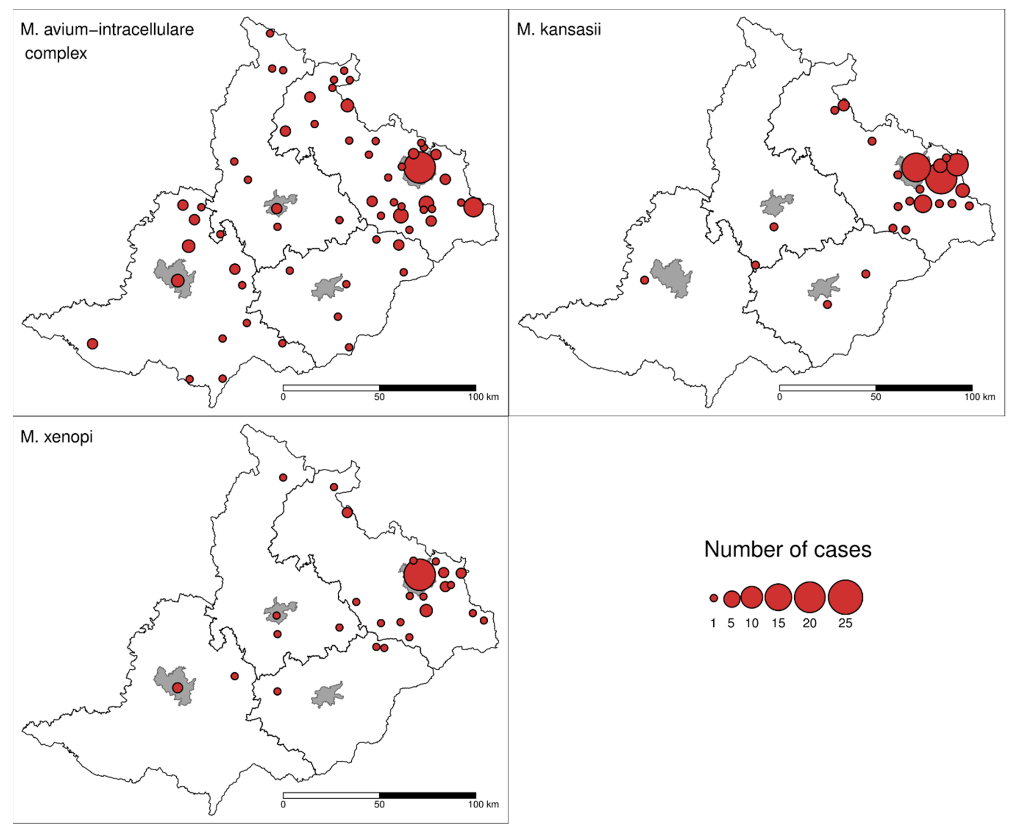
2.1. Area Description and Study Design

The Moravian and Silesian Regions (area 21,849 km2) of the Czech Republic (total area 78,867 km2) consist of 22 districts classified at the Local Administrative Unit (LAU) 1 level according to the European Union (Figure 1).
The total population in the studied area varied from 3.620 mil in 2012 to 3.604 mil in 2018. Within the studied area, territorial units (n = 710) were defined according to the postal codes of the home address of the patient from which the NTM was isolated and this was used to construct a study area map (Figure 2). The size of the population per territorial units ranged from 151 to 377,973 persons (median 1971) from 2012 to 2018.
To assess the relationship between NTM incidence and other factors (socio-economic indicators, benzo[a]pyrene, PM2.5 and PM10 air pollution), the NTM incidence data at the postal code level were integrated with data on other factors which were collected at the district level (LAU 1). The number of patients was adjusted (normalized) to the population of the given municipality using data provided by the Czech Statistical Office in the given year [29].

2.2. Data Source

Recorded identification of NTM human isolates was obtained from the Public Health Institute Ostrava (Czech Republic). Mycobacterial isolates were classified as the causative agent of disease according to the American Thoracic Society (ATS)/the Infectious Diseases Society of America (IDSA) microbiologic criteria as well as stricter microbiological criteria (multiple isolation of the same NTM species from respiratory samples, acid-fast stain-positive results, sole NTM isolation from bioptic tissues or sterile body fluids); and clinical criteria (significant progressive changes on a chest skiagram and/or HRCT; high-resolution computed tomography), nodular-bronchiectatic or cavernous lesions in association with a chronically worsened overall condition, and systemic or skin manifestation. The types of NTM diseases were divided into two groups: NTM pulmonary and NTM non-pulmonary diseases.
Individual NTM patients were included only once during the studied period and results were presented as an average incidence (new cases per population) per year.
The selected demographic indicators (sex and age) and socio-economic factors for every year (2012–2018) were obtained from the database of the Czech Statistical Office [29]. Four socio-economic indicators were selected: the crude divorce rate, the net migration rate (the difference between the number of people coming into an area and the number of people leaving an area throughout the year), the share of the paid housing allowance, and the percentage of unemployment. Based on these indicators an index of social deprivation was generated per region (LAU1).
The average concentrations of PM2.5, PM10 and benzo[a]pyrene in the air during the period 2012–2016 were obtained from the Czech Hydrometeorological Institute [30].

2.3. NTM Identification

All potential NTM isolates were first identified by macroscopic and Ziehl-Neelsen microscopic examinations. Mycobacterial isolate identification was carried out by AccuProbe Test (Hologic, San Diego, USA), by PCR with reverse hybridization on cellulose strips, GenoType Mycobacterium CM/AS assays (Hain Lifescience, Nehren, Germany) and/or by sequencing genes encoding the 16S ribosomal RNA subunit (Applied Biosystems Genetic Analyzer ABI3130 series, Thermo Fisher Scientific, Waltham, MA, USA).

2.4. Data Analysis and Statistical Methods

The incidence of each NTM species in patients every year was calculated and then presented as average annual incidence in the studied period 2012–2018. Demographic and socio-economic data for the year 2018 were not assessed due to the unavailability of this information on this date. Data on the four selected socio-economic indicators averaged over 6 years period (2012–2017) were converted to relative values, normalized with the Min–Max method and aggregated into an index of social deprivation [31].
The correlated data were assessed using the Pearson’s correlation coefficient. The comparisons of incidence distributions through the years were performed using the Kruskal–Wallis Rank Sum Test. All the data processing and statistical testing were performed in R [32], with the use of the packages dplyr [33] and ggplot2 [34] for data manipulation and visualization, and the packages sf [35] and tmap [36] for spatial data manipulation and visualization in the form of maps.

3. Results

In total, 2176 mycobacterial isolates belonging to 32 NTM species were recorded during the 7 year period. The identification of 66 (3.03%) clinical isolates was unsuccessful (Table 1). A total of 303 (13.92%) NTM isolates (one per patient) were identified as causative agents of disease according to ATS/IDSA microbiological and clinical criteria and consequently included in this study. None of the NTM or MTB patients who underwent human immunodeficiency virus (HIV) testing were positive.
M. avium-intracellulare complex (MAIC) members and ten NTM species meeting the microbiological and clinical criteria were encountered in this epidemiological study. Two NTM isolates from patients could not be identified. The most frequently isolated NTM were the MAIC members, followed by M. kansasii, and Mycobacterium xenopi. These NTM species accounted for almost 90% of all isolates from patients. Rapidly growing NTM (Mycobacterium chelonae, Mycobacterium fortuitum, Mycobacterium abscessus, and Mycobacterium mucogenicum) caused 6.6% of NTM diseases overall (Table 1).
The average incidence of NTM patients in the studied area was 1.10/100,000 inhabitants (1.33/100,000 in men and 0.88/100,000 in women) during the 7 year period. MAIC members’ pulmonary diseases were more frequent in women than men. However, M. kansasii and M. xenopi were significantly more often diagnosed in men (p < 0.05). Compared to the average, a statistically higher incidence of NTM in adult patients was found in four districts (p = 0.0012) located in the north part of the studied area: Bruntál, Jeseník, Karviná, and Ostrava districts (Figure 3A; Table A1). The correlation between NTM pulmonary disease incidence and the index of social deprivation was 0.63. A visualization of the level of social deprivation is shown in Figure 3B. NTM pulmonary disease incidence correlated with a higher than average benzo[a]pyrene concentration in the air (r = 0.64). The correlations between particle pollutions PM2.5 and PM10 were 0.41 and 0.42, respectively. The maps of benzo[a]pyrene, PM2.5 and PM10 concentrations in the air are available in Figure A1.
The occurrence of patients with MAIC members, M. kansasii and M. xenopi pulmonary diseases are shown in Figure 4. A significantly higher incidence of M. kansasii (p = 0.0006) was recorded in the Karviná district (1.90/100,000) compared to all other districts. A higher incidence was recorded in men (2.86/100,000) compared to women (0.99/100,000) in this district. A statistically higher incidence of disease caused by M. xenopi (p = 0.03) was detected in the Ostrava district (1.01/100,000; Table A1).
The distribution of the three most frequently recorded NTM causative agents (MAIC members, M. kansasii, and M. xenopi) in patients by age is shown in Figure 5. All 26 NTM patients under the age of 5 years had cervical lymphadenitis caused by MAIC members. In patients over 18 years of age, 98% of the cases of NTM disease were pulmonary infections. Non-pulmonary diseases were recorded in six adult patients: two cases were caused by M. chelonae and M. marinum and one case each by M. chimaera and Mycobacterium vulneris. M. chelonae was isolated from the eye of a patient who suffered from keratitis after surgery and the patient suffered from a periodontitis abscess. M. marinum and M. vulneris were isolated from cutaneous lesions of two patients, and M. chimaera was a causative agent of pleurisy after surgery in one patient.
The rate of lymphadenitis caused by MAIC members in children (<5 years) was 2.35/100,000 during the studied period; the rate was especially high in the final year of the study, at 5.92/100,000.
The median age of all NTM patients was 66.0 years. The median age of patients with MAIC members and M. xenopi infections was 68.0. M. kansasii infections were more frequently diagnosed in middle age patients (median age 56.0 years). The median age of children with lymphadenitis was 3.0 years. The number of patients, as well as their age and sex are shown in Figure 6. The overall incidence of MTB pulmonary diseases was 1.97/100,000. A higher number of MTB compared to NTM patients was found in adult men (age 15–64 years). Roughly, an equal number of MTB and NTM diseases were recorded in men and women older than 65 years (Figure 7).

4. Discussion

The reported incidence of clinically relevant NTM can differ depending on the methodology used to count NTM isolates, causing a discrepancy between isolation rate and actual disease diagnosis—specifically, whether the particular methods take into account multiple NTM isolates per patient, latent infections and transient presence of the NTM without clinical relevance [16]. In European countries, the prevalence of NTM pulmonary diseases meeting ATS/IDSA and stricter microbiological criteria make up only 5.5%–26.0% of the total number of NTM clinical isolates [12,37,38,39,40,41,42]. These findings are in accordance with the results of our study, where we confirmed that less than 14% of total NTM isolates were responsible for clinical disease (Table 1).
Environmental NTM is well adapted to outdoor survival in extreme conditions and their abundance in environment is influenced by environmental and geochemical characteristics [43,44,45,46]. NTMs are opportunistic pathogens with the potential to colonize tissues of immunocompromised or “susceptible” persons, especially from sources with higher numbers of infective units or sources with greater frequencies of human exposure [46,47,48]. Defensive mechanisms of healthy humans are capable of eliminating primary infection even in immunocompetent individuals in most cases. Natural self-cleaning/healing lung functions (debacilization), effective expectoration and ciliary transport may be the most effective elements of host defense against environmental NTM. The primary infectious dose of most NTM infections in immunocompetent and physiologically healthy individuals may even be eliminated by naive alveolar macrophages. Signaling cytokine pathways and the activation of macrophages are initiated within hours after NTM infection in comparison with the long delay seen in MTB infections [49].
The obligate pathogenic M. tuberculosis complex members and M. leprae have developed mechanisms (effective blockage of phagosome fusion with lysosomes by sulpholipide-1, deregulation of cytokine signaling pathways, and dormancy) to evade the host immune system, which allow them not only to survive phagocytosis but also to activate intracellular proliferation [50]. Compared to MTB, NTMs are equipped with much less effective virulence factors (cell wall structure, etc.) and their survival in the host organism depends on the degree of adaptability of that particular NTM species or even isolate. Factors for overcoming the interaction with the immune system vary significantly. The most basal mechanisms are wide range temperature tolerance and ability to produce compact cell formations: cording and clustering, as seen in M. kansasii and M. xenopi, or biofilm formation as seen in MAIC members [51]. Enzymatic and structural adaptations of the cell wall of NTM species associated with animal diseases (i.e., fish tank granuloma caused by M. marinum) can confer an advantage in the colonization of damaged human skin [52]. Pathological lesions in the pulmonary structure based on an underlying disease can create niches that allow further growth of NTM. Bronchiectasis, bronchial obstruction (asthma, COPD), fibrous tissue (silicosis, coniosis, other granulomatosis, and prior MTB) as well as cystic fibrosis can result in permanent deposition of air pollution, persistent mucus, and may also alter the effectiveness of neutrophils [53,54,55]. Some NTM can cause latent infections in lung tissue, and during the latency period, NTM can be isolated from expectorated sputum even when there are no apparent clinical symptoms. Long-term latency or inactivity of NTM and the graduating colonization of affected lung tissue is to be assumed. The patients in this period produce NTM cells in the expectorated sputum, but without significant apparent symptoms. Sufficient moisture and a microaerophilic environment in impaired areas of lungs can enable mycobacteria to multiply, leading to immune system hyperactivity and the progression of symptomatic disease [16].
The species distribution of NTM patient isolates in European countries was described recently [11]. However, this study did not include results from the Czech Republic. In this study, there was a significant difference between the geographical distribution of NTM isolation frequency and NTM diseases. If isolates of Mycobacterium gordonae and Mycobacterium fortuitum, which have a low clinical relevance anyway, are omitted, the MAIC members predominate in the North and West European regions [11,39,56]. This is different to what is seen in Eastern European countries, where M. kansasii is the predominant NTM in Poland and Slovakia and M. xenopi is the most isolated species in Hungary and Croatia [11,12,57]. These results correlate with our observations, where the north part of the studied area (near the border with Poland) has a higher prevalence of M. kansasii, and in the remainder of the studied area, MAIC is more prevalent (Figure 4). Rapidly growing NTM, common in the UK and Greece [11], formed less than 7% of the total NTM patients in our study (Table 1). Mycobacterium malmoense occurs mostly in Northern Europe [11,58]. In this study, it was recorded in six patients living in the north part of studied area (data not shown).
The annual incidence of children infected with MAIC members in our study (2.35/100,000 children’s population) is similar to estimates reported in other European countries such as the Netherlands [59] and Germany [60]. MAIC complex members have been known to be causative agents of lymphadenitis in children since the 1970s [61]. An increase in NTM disease incidence was documented in France after the discontinuation of mandatory Bacillus Calmette Guérin (BCG) immunization in 2007 [62]. We did not observe an increasing incidence of NTM disease during the period 2012–2017, although a sharp increase (5.92/100,000 population) was recorded during the year 2018.
The mean age of adult NTM patients in our study was similar to other European countries. The median age of NTM adult patients in Central Greece (2004–2006), Croatia (2006–2013), and Denmark (2015) was 66.1, 67.0, and 68.2 years, respectively [38,39,63]. The age of adult patients with M. kansasii pulmonary diseases was lower than the age of patients with M. xenopi and MAIC pulmonary diseases in other similar studies [63,64,65] which is in accord with the findings of this study (Figure 5). Generally, adult NTM patients are more often male [42,63,64,66] with the exception of MAIC pulmonary diseases, which are often diagnosed in older women [65,67].
It is thought that elderly patients are exposed to NTM in indoor environments through contaminated tap water. On the one hand, a link between potable water NTM isolates and human diseases using PCR methods has been recorded in MAIC members and M. chimaera infections. On the other hand, very few strains of M. kansasii isolates from tap water matched disease isolates [68,69]. Shower usage by elderly individuals is less frequent, which can lead to long term water stagnation and may allow NTM colonization of household plumbing. This fact in combination with other comorbidities may be one of the reasons why NTMs are more frequently isolated in elderly patients than the general population [70,71,72]. Higher NTM prevalence may also be linked to water supply practices, specifically chlorination of old municipal water supplies, which can concentrate the NTM in biofilms [73]. The importance of this factor is supported by the finding of a higher abundance of NTM in showerhead biofilms in USA domestic water systems compared to Europe, where a lower chlorine concentration is used during water treatment [74].
Pulmonary disease caused by M. kansasii has been endemic in the Karviná district since the 1970s [75]. During the period 1984–1989, the rate of NTM incidence in this district fluctuated between 13.5 and 17.6/100,000 inhabitants, which was much higher than the national average [75]. Subsequently, in the 1990s, there was a decrease in the prevalence of NTM patients being recorded; this coincided with and is probably related to mining restrictions that came into practice after the “velvet revolution” in 1989 [27]. The majority of cases were described in men (81%) during the period 1968–1995 and a link with the mining industry has been established in 56.4% of patients. Currently, there is still a statistically significantly higher incidence of M. kansasii pulmonary diseases in men from the Karviná district, at 2.86/100,000 of the population compared to the average of the entire studied area (0.44/100,000 population; Table A1). We also observed a higher incidence of M. kansasii pulmonary diseases from the Karviná district (0.99/100,000) in women compared to the overall incidence (0.16/100,000). This suggests that employment in the mining industry (which predominantly employs males) is not the only risk factor for M. kansasii infection as described in previous studies [27,76]. A relatively high level of M. kansasii detection in tap and surface water in the Karviná district could represent a possible risk factor [27,77]. However, a link between potable water isolates of M. kansasii and clinical isolates has not been confirmed up to now and point source contamination may occur from an alternative environmental source [69]. A higher incidence of M. kansasii infections has also been recorded in other regions with heavy industry and mining [76]. The higher mean age (56.0) of men with M. kansasii infection in the period 2012–2018 described in our study compared to the average age of 50.1 years during the period 1984–1989 [75] is probably the consequence of the aging of former miners. The mining industry in the Karviná district employs significantly less people then previously but former mineworkers are still at risk of occupational pneumoconiosis (unpublished data).
NTM can survive and persist in household and industrial plumbing systems due to the ability of the bacteria to grow at a low nutrient concentration, associate in biofilms, and chlorine resistance [3,78]. The high correlation of MAIC detected in household water with high MAIC disease incidence may be a reflection of the large number of water-based studies that have been completed [68]. A possible exposure route is aerosol inhalation during personal hygiene and/or aspiration of NTM contaminated water [6,79]. Our study could not confirm this because environmental isolates were not analyzed and compared to clinical isolates. The practice of screening households of patients with NTM diseases to confirm the presence of an NTM contamination is not yet conducted in the Czech Republic. Establishing this practice, especially in the case of pediatric patients or patients with a long history of NTM infection would be desirable. The availability and effectiveness of methods to accurately type isolate strains are limited presently. The most commonly used methods are Restriction Fragment Length Polymorphism (RFLP) with Pulsed-Field Gel electrophoresis (PFGE), Mycobacterial Interspersed Repetitive Units – Variable Number of Tandem Repeats (MIRU-VNTR) and Whole Genome Sequencing (WGS). Linking strains isolated from a patient’s clinical material with those isolated from environmental sources is difficult using these methods. The possibility of the restructuring of the genome of NTM species as a reaction to stress and adaptations associated with colonization of a new patient’s tissue environment makes this even more complicated [80,81,82,83]. A long period from infection and colonization to disease development may also affect differences in the genetic profile of the NTM isolates from patients compared to those from the environment [81]. Despite these difficulties, a study by Kaustova et al. (1993) showed a connection between M. xenopi human isolates and contaminated household water in the Ostrava district in a study covering the period 1990–1991 [84].
A lower income and socio-economic status are connected with higher morbidity, disability, and mortality [85,86]. In addition, some known risk factors for NTM pulmonary diseases (heavy smoking and alcohol misuse) are connected to worsened socio-economic status. Some of the northern parts of the studied area, which includes the Ostrava and Karviná districts, are classed as having lower socio-economic status [87]. However, data about smoking and alcoholism were only available at region level (NUTS 3, Nomenclature des Unites Territoriales Statistiques) and consequently a direct comparison of these factors with our results was not possible. For this reason, we used an indirect indicator (index of social deprivation) of socio-economic status. A deeper social deprivation in the north part of the studied area including the Ostrava and Karviná districts as well as a statistically higher incidence of NTM pulmonary diseases was evident in our study (Figure 3A,B). Our findings correspond to the results of other studies conducted in developed countries. A spatial epidemiological analysis of NTM infections in connection with socio-economic attributes was carried out by Chou, et al. [88] in Queensland, Australia. The risk of M. kansasii diseases was estimated to decrease with an increase in the earning level of a population. Similarly, a higher incidence of M. kansasii pulmonary diseases in patients with a lower median income was described in Northern California, USA [89]. It is essential in this context to state, that all types of factors—host susceptibility, environmental and socio-economic factors (median income level)—must be considered during the evaluation of NTM disease risk [44].
Although the socio-economic situation and environmental pollution levels are important predisposition factors of MTB prevalence [90,91,92], the influence of air pollution on NTM pulmonary infections has not been studied yet. Air pollution consists of various gases and particles. Air particles smaller than 10 µm in diameter deposited in the respiratory tract can cause serious adverse effects on health due to the potential of being biologically active [93]. The coarse fraction of air pollution (PM2.5–10) can penetrate into the upper airways, while the fine fraction (PM < 2.5) can be deposited in the lung alveoli—both of which lead to several pulmonary pathological processes and an increased risk of respiratory infections [20,94]. The level of adverse health effects of PM is also influenced by particle-associated contaminants, such as heavy metals, polychlorinated dibenzo-p-dioxins, and PAHs [95,96]. Most particulate phase PAHs are adsorbed onto PM2.5. Benzo[a]pyrene is a highly carcinogenic agent used worldwide as a criterion for air pollution measurement [97,98,99]. Both PM2.5 and benzo[a]pyrene induce markers of oxidative stress through the aryl hydrocarbon receptor (AhR) pathway and consequently increase the risk of COPD [18]. Thus, air pollution through pulmonary impairment and lung disease increases the risk of NTM pulmonary infection caused by M. kansasii, and this is evident in our study (Figure 4, Table A1).
The Czech Republic has a low MTB incidence (4.9/100,000 population) according to the ECDC European Centre for Disease Prevention and Control (ECDC)/WHO report from 2016 [100]. The higher rate of MTB documented in the whole territory of the Czech Republic compared to the studied area (1.97/100.000) was due to the high MTB rate in the population of Prague—the capital city of the Czech Republic [101]. The rate of MTB was higher than NTM in the age group 15–64 in both men and women, and a similar incidence of MTB and NTM was recorded in the age group >65 (Figure 7).
The interpretation of results in our study is limited by not knowing each patient´s predisposing conditions and the analysis of water samples from plumbing systems suspected of NTM contamination. Furthermore, some clinical isolates (estimated to be less than 5 per year) sampled in the studied area were examined in alternative laboratories and could not be included in our results. However, this is estimated to be less than 2% of the isolates per year.

5. Conclusions

Our study complements the results of other European studies researching the distribution of different NTM species in humans and the incidence of NTM diseases. Our results show the essential differences between NTM human isolates and NTM diseases in both, the total number of cases and the NTM species spectrum. The incidence of NTM pulmonary diseases caused by different NTM species significantly differed even in the small geographic units (districts) defined in our study. In terms of NTM ecology, it appears that M. xenopi and M. kansasii diseases are endemic in the northern parts of the studied area. Our study indicates that lung diseases connected to air pollution, occupational exposure and a worsened socio-economic situation could lead to an increase in the risk of NTM pulmonary diseases. Future research focused on the examination of NTM isolates from the environment in the studied area would need to match clinical and environmental isolates and determine more specific environmental sources of NTM.

Author Contributions

H.M. and V.U. were responsible for the overall conceptualization and analysis of the results. I.P. supervised the study and contributed to the finalization of the results. J.C. was responsible for statistical and spatial analysis. D.H. and O.K. performed the demographic analyses and edited the manuscript. J.S. and R.T.W. contributed to the acquisition of the data and editing the manuscript. H.M. wrote the paper.

Funding

This research was funded by the Czech Science Foundation (No. 16-13231S).

Acknowledgments

We thank the following persons working in the listed organizations in the Czech Republic: Leona Mejzlikova (Microbiology Clinic, St. Anne´s University Hospital, Brno); Natasa Bartonikova (Dept. of Microbiology, Hospital Zlin); Katerina Wezdenkova (Laboratory Mikrochem, Sumperk); Michaela Klementova (Dept. of Microbiology, Salesian Hospital in Opava); Yvona Loveckova (Microbiology Clinic, University Hospital in Olomouc); Ivan Tudik (TB Sanatorium, Jablunkov); Zbynek Vrba and Eva Poncova (Dept. of Pneumophtiseology, Hospital in Krnov); Gustav Ondrejka (Dept. of Pneumophtiseology, Hospital in Novy Jicin); Radovan Kozel (Dept. of Pneumophtiseology, Municipial Hospital in Ostrava); Tomas Skwarlo, Radomir Roubec, and Tomas Porzer (Pneumophtiseology Clinic, University Hospital in Ostrava). We also thank the laboratory staff (Anna Kracalikova, Eva Kalakayova, Anna Kubinova, Dagmar Sterbikova, and Lenka Krpcova from Public Health Institute Ostrava) for their excellent laboratory work.

Conflicts of Interest

The authors declare no conflict of interest.

Appendix A

Table A1. The incidence of NTM diseases per districts in the studied territory during the years 2012–2018.
DistrictNTMMAICM. kansasiiM. xenopi
TotalFemaleMaleTotalFemaleMaleTotalFemaleMaleTotalFemaleMale
Blansko1.0580.7831.3411.0580.7831.341000000
Brno-city0.2270.2190.2340.1130.0730.1570000.0750.1460
Brno0.1310.1300.1310000.06500.131000
Bruntál3.1963.0003.3991.8281.7991.8570.45500.9200.4590.2940.622
Břeclav0.2470.48600.2470.4860000000
Frýdek-Mistek2.4792.5072.4511.2721.8470.6800.6710.2641.0910.4020.2650.544
Hodonín0.3690.1830.5610.3690.1830.561000000
Jeseník2.1861.4452.9371.0911.4450.7330000.36700.739
Karviná2.7981.4274.2190.2760.2180.3371.9040.9872.8560.4500.1120.800
Kroměříž0.53901.1030.13400.2740000.13500.276
Nový Jičín2.0722.0432.1021.0361.1150.9560.2820.1860.3820.3760.3700.382
Olomouc0.7330.4751.0050.2440.3570.1250.06200.1260.12300.253
Opava0.7281.1110.3300.5670.7940.3300.0810.15800.0810.1580
Ostrava2.7701.8773.7191.0121.1940.8180.7040.3411.0881.0110.3391.722
Prostějov0.52601.0780.26300.5390.13200.270000
Přerov0.4340.2120.6640.11000.2230000.10900.222
Šumperk0.4700.2310.7180.1170.2310000000
Uh. Hradiště0.10000.2040.10000.204000000
Vsetín0.6950.9780.4030.3980.78300.09900.2020.1980.1940.201
Vyškov0.9451.2490.6330.4730.93600000.15600.316
Zlín0.2230.1460.3040.14900.3040.0750.1460000
Znojmo0.2520.49700.2520.4970000000
Studied area overall1.0960.8691.3330.4670.5350.3960.2850.1630.4360.2180.1520.331
NTM = non-tuberculous mycobacteria; MAIC = Mycobacterium avium-intracellulare complex.
Figure A1. Five-year (2012–2016) average concentrations of benzo[a]pyrene, PM10 and PM2.5 in the air in the studied regions based on the data from the Czech Hydrometeorological Institute [30].
Ijerph 16 03969 g0a1

References

  1. Falkinham, J.O., III. Surrounded by mycobacteria: Nontuberculous mycobacteria in the human environment. J. Appl. Microbiol. 2009, 107, 356–367. [Google Scholar] [CrossRef] [PubMed]
  2. Falkinham, J.O., III. Environmental sources of nontuberculous mycobacteria. Clin. Chest Med. 2015, 36, 35–41. [Google Scholar] [CrossRef] [PubMed]
  3. Revetta, R.P.; Gomez-Alvarez, V.; Gerke, T.L.; Santo Domingo, J.W.; Ashbolt, N.J. Changes in bacterial composition of biofilm in a metropolitan drinking water distribution system. J. Appl. Microbiol. 2016, 121, 294–305. [Google Scholar] [CrossRef] [PubMed] [Green Version]
  4. Roguet, A.; Therial, C.; Saad, M.; Boudahmane, L.; Moulin, L.; Lucas, F.S. High mycobacterial diversity in recreational lakes. Antonie Van Leeuwenhoek 2016, 109, 619–631. [Google Scholar] [CrossRef]
  5. Griffith, D.E.; Aksamit, T.; Brown-Elliott, B.A.; Catanzaro, A.; Daley, C.; Gordin, F.; Holland, S.M.; Horsburgh, R.; Huitt, G.; Iademarco, M.F.; et al. An official ATS/IDSA statement: Diagnosis, treatment, and prevention of nontuberculous mycobacterial diseases. Am. J. Respir. Crit. Care Med. 2007, 175, 367–416. [Google Scholar] [CrossRef] [PubMed]
  6. Donohue, M.J. Increasing nontuberculous mycobacteria reporting rates and species diversity identified in clinical laboratory reports. BMC Infect. Dis. 2018, 18, 163. [Google Scholar] [CrossRef] [PubMed]
  7. Henkle, E.; Hedberg, K.; Schafer, S.; Novosad, S.; Winthrop, L. Population-based incidence of pulmonary nontuberculous mycobacterial disease in Oregon 2007 to 2012. Ann. Am. Thorac. Soc. 2015, 12, 642–647. [Google Scholar] [CrossRef]
  8. Namkoong, H.; Kurashima, A.; Morimoto, K.; Hoshino, Y.; Hasegawa, N.; Ato, M.; Mitarai, S. Epidemiology of pulmonary nontuberculous mycobacterial disease in Japan. Emerg. Infect. Dis. 2016, 22, 1116–1117. [Google Scholar] [CrossRef]
  9. Shah, N.M.; Davidson, J.A.; Anderson, L.F.; Lator, M.K.; Kim, J.; Thomas, H.L.; Lipman, M.; Abubakar, I. Pulmonary Mycobacterium avium-intracellulare is the main driver of the rise in non-tuberculous mycobacteria incidence in England, Wales and Northern Ireland, 2007–2012. BMC Infect. Dis. 2016, 16, 195. [Google Scholar] [CrossRef]
  10. Diel, R.; Jacob, J.; Lampenius, N.; Loebinger, M.; Nienhaus, A.; Rabe, K.F.; Ringshausen, F.C. Burden of non-tuberculous mycobacterial pulmonary disease in Germany. Eur. Respir. J. 2017, 49, 1602109. [Google Scholar] [CrossRef] [Green Version]
  11. Hoefsloot, W.; van Ingen, J.; Andrejak, C.; Ängeby, K.; Bauriaud, R.; Bemer, P.; Beylis, N.; Boeree, M.J.; Cacho, J.; Chihota, V.; et al. The geographic diversity of nontuberculous mycobacteria isolated from pulmonary samples: An NTM-NET collaborative study. Eur. Respir. J. 2013, 42, 1604–1613. [Google Scholar] [CrossRef] [PubMed]
  12. Jankovic, M.; Samarzija, M.; Sabol, I.; Jakopovic, M.; Katalinic Jankovic, V.; Zmak, L.; Ticac, B.; Marusic, A.; Obrovac, M.; van Ingen, J. Geographical distribution and clinical relevance of non-tuberculous mycobacteria in Croatia. Int. J. Tuberc. Lung Dis. 2013, 17, 836–841. [Google Scholar] [CrossRef] [PubMed] [Green Version]
  13. Spaulding, A.B.; Lai, Y.L.; Zelazny, A.M.; Olivier, K.N.; Kadri, S.S.; Prevots, D.R.; Adjemian, J. Geographic distribution of nontuberculous mycobacterial species identified among clinical isolates in the United States, 2009–2013. Ann. Am. Thorac. Soc. 2017, 14, 1655–1661. [Google Scholar] [CrossRef] [PubMed]
  14. Adjemian, J.; Frankland, T.B.; Daida, Y.G.; Honda, J.R.; Olivier, K.N.; Zelazny, A.; Honda, S.; Prevots, D.R. Epidemiology of nontuberculous mycobacterial lung disease and tuberculosis, Hawaii, USA. Emerg. Infect. Dis. 2017, 23, 439–447. [Google Scholar] [CrossRef] [PubMed]
  15. Sonnenberg, P.; Murray, J.; Glynn, J.R.; Thomas, R.G.; Godfrey-Faussett, P.; Shearer, S. Risk factors for pulmonary disease due to culture-positive M. tuberculosis or nontuberculous mycobacteria in South African gold miners. Eur. Respir. J. 2000, 15, 291–296. [Google Scholar] [CrossRef] [PubMed]
  16. Stout, J.E.; Koh, W.-J.; Yew, W.W. Update on pulmonary disease due to non-tuberculous mycobacteria. Int. J. Infect. Dis. 2016, 45, 123–134. [Google Scholar] [CrossRef] [Green Version]
  17. Henkle, E.; Winthrop, K.L. Nontuberculous mycobacteria infections in immunosuppressed host. Clin. Chest Med. 2015, 36, 91–99. [Google Scholar] [CrossRef]
  18. Yang, L.; Wang, W.-C.; Lung, S.-C.C.; Sun, Z.; Chen, C.; Chen, J.-K.; Zou, Q.; Lin, Y.-H.; Lin, C.-H. Polycyclic aromatic hydrocarbons are associated with increased risk of chronic obstructive pulmonary disease during haze events in China. Sci. Total Environ. 2017, 574, 1649–1658. [Google Scholar] [CrossRef]
  19. Raudoniute, J.; Stasiulaitiene, I.; Kulvinskiene, I.; Bagdonas, E.; Garbaras, A.; Krugly, E.; Martuzevicius, D.; Bironaite, D.; Aldonyte, R. Pro-inflammatory effects of extracted urban fine particulate matter on human bronchial epithelial cells BEAS-2B. Environ. Sci. Pollut. Res. 2018, 25, 32277–32291. [Google Scholar] [CrossRef]
  20. Falcon-Rodriguez, C.I.; Osornio-Vargas, A.R.; Sada-Ovalle, I.; Segura-Medina, P. Aeroparticles, composition, and lung diseases. Front. Immunol. 2016, 7, 3. [Google Scholar] [CrossRef]
  21. Ranjit, S.; Midde, N.M.; Sinha, N.; Patters, B.J.; Rahman, M.A.; Cory, T.J.; Rao, P.S.S.; Kumar, S. Effect of polyaryl hydrocarbons on cytotoxicity in monocytic cells: Potential role of cytochromes P450 and oxidative stress pathways. PLoS ONE 2016, 11, e0163827. [Google Scholar] [CrossRef] [PubMed]
  22. Falkinham, J.O., III. Mycobacterial aerosols and respiratory disease. Emerg. Infect. Dis. 2003, 9, 763–767. [Google Scholar] [CrossRef] [PubMed]
  23. McGrath, E.E.; Bardsley, P. An association between Mycobacterium malmoense and coal workers´ pneumoconiosis. Lung 2009, 187, 51–54. [Google Scholar] [CrossRef]
  24. Jiřík, V.; Machaczka, O.; Miturová, H.; Tomášek, I.; Šlachtová, H.; Janoutová, J.; Velická, H.; Janout, V. Air pollution and potential health risk in Ostrava Region—A review. Cent. Eur. J. Public Health 2016, 24, S4–S17. [Google Scholar] [CrossRef] [PubMed]
  25. Pokorná, P.; Hovorka, J.; Klán, M.; Hopke, P.K. Source apportionment of size resolved particulate matter at a European air pollution hot spot. Sci. Total Environ. 2015, 502, 172–183. [Google Scholar] [CrossRef]
  26. OKD Ltd. Annual Reports 2005–2015. Available online: https://www.okd.cz/en/about-us/annual-reports (accessed on 10 September 2018).
  27. Chobot, S.; Mališ, J.; Šebáková, H.; Pelikán, M.; Zatloukal, O.; Palička, P.; Kocurová, D. Endemic incidence of infections caused by Mycobacterium kansasii in the Karviná District in 1968–1995. Cent. Eur. J. Public Health 1997, 5, 164–173. [Google Scholar]
  28. Sosnovcová, T. The Impacts of Coal Mining in the Ostrava Region. Bachelor’s Thesis, Faculty of Science, Masaryk University in Brno, Brno, Czech Republic, 2016. (In Czech). [Google Scholar]
  29. Czech Statistical Office. 2019. Available online: https://www.czso.cz/csu/czso/home (accessed on 12 May 2019).
  30. Czech Hydrometeorological Institute. 2019. Available online: http://portal.chmi.cz/?l=en (accessed on 18 May 2019).
  31. OECD. Handbook on Constructing Composite Indicators; Methodology and User Guide; OECD Publications: Paris, France, 2008. [Google Scholar]
  32. R Core Team. R: A Language and Environment for Statistical Computing; R Foundation for Statistical Computing: Vienna, Austria, 2018. Available online: https://www.R-project.org/ (accessed on 24 April 2019).
  33. Wickham, H.; François, R.; Henry, L.; Müller, K. Dplyr: A Grammar of Data Manipulation; R Package version 0.7.7. 2018. Available online: https://CRAN.R-project.org/package=dplyr (accessed on 24 April 2019).
  34. Wickham, H. Ggplot2: Elegant Graphics for Data Analysis; Springer: New York, NY, USA, 2016. [Google Scholar]
  35. Pebesma, E. Simple features for R: Standardized support for spatial vector data. R J. 2018, 10, 439–446. [Google Scholar] [CrossRef]
  36. Tennekes, M. Tmap: Thematic Maps in R. J. Stat. Softw. 2018, 84, 1–39. [Google Scholar] [CrossRef]
  37. Andréjak, C.; Thomsen, V.Ø.; Johansen, I.S.; Riis, A.; Benfield, T.L.; Duhaut, P.; Sørensen, H.T.; Lescure, F.-X.; Thomsen, R.W. Nontuberculous pulmonary mycobacteriosis in Denmark: Incidence and prognostic factor. Am. J. Respir. Crit. Care Med. 2010, 181, 514–521. [Google Scholar] [CrossRef]
  38. Gerogianni, I.; Papala, M.; Kostikas, K.; Petinaki, E.; Gourgoulianis, K.I. Epidemiology and clinical significance of mycobacterial respiratory infections in Central Greece. Int. J. Tuberc. Lung Dis. 2008, 12, 807–812. [Google Scholar]
  39. Jankovic, M.; Sabol, I.; Zmak, L.; Katalinic Jankovic, V.; Jakopovic, M.; Obrovac, M.; Ticac, B.; Kardum Bulat, L.; Grle, S.P.; Marekovic, I.; et al. Microbiological criteria in non-tuberculous mycobacteria pulmonary disease: A tool for diagnosis and epidemiology. Int. J. Tuberc. Lung Dis. 2016, 20, 934–940. [Google Scholar] [CrossRef] [PubMed]
  40. Kennedy, M.P.; O’Connor, T.M.; Ryan, C.; Sheehan, S.; Cryan, B.; Bredin, C. Nontuberculous mycobacteria: Incidence in Southwest Ireland from 1987 to 2000. Respir. Med. 2003, 97, 257–263. [Google Scholar] [CrossRef] [PubMed]
  41. Prevots, D.R.; Marras, T.K. Epidemiology of human pulmonary infection with non-tuberculous mycobacteria: A review. Clin. Chest Med. 2015, 36, 13–34. [Google Scholar] [CrossRef] [PubMed]
  42. Van Ingen, J.; Bendien, S.A.; de Lange, W.C.M.; Hoefsloot, W.; Dekhuijzen, P.N.R.; Boeree, M.J.; van Soolingen, D. Clinical relevance of non-tuberculous mycobacteria isolated in the Nijmegen-Arnhem region, The Netherlands. Thorax 2009, 64, 502–506. [Google Scholar] [CrossRef] [PubMed] [Green Version]
  43. Pavlik, I.; Gersl, M.; Bartos, M.; Ulmann, V.; Kaucka, P.; Caha, J.; Unc, A.; Hubelova, D.; Konecny, O.; Modra, H. Nontuberculous mycobacteria in the environment of Hranice Abyss, the world’s deepest flooded cave (Hranice karst, Czech Republic). Environ. Sci. Pollut. Res. 2018, 25, 23712–23724. [Google Scholar] [CrossRef]
  44. Adjemian, J.; Olivier, K.N.; Seitz, A.E.; Falkinham, J.O., III; Holland, S.M.; Prevots, D.R. Spatial clusters of nontuberculous mycobacterial lung disease in the United States. Am. J. Respir. Crit. Care Med. 2012, 186, 553–558. [Google Scholar] [CrossRef]
  45. Iivanainen, E.K.; Martikainen, P.J.; Räisänen, M.L.; Katila, M.-L. Mycobacteria in boreal coniferous forest soils. FEMS Microbiol. Ecol. 1997, 23, 325–332. [Google Scholar] [CrossRef]
  46. Kirschner, R.A.; Parker, B.C.; Falkinham, J.O., III. Epidemiology of infection by nontuberculous mycobacteria. Am. Rev. Respir. Dis. 1992, 145, 271–275. [Google Scholar] [CrossRef]
  47. Dirac, M.A.; Horan, K.L.; Doody, D.R.; Meschke, J.S.; Park, D.R.; Jackson, L.A.; Weiss, N.S.; Winthrop, K.L.; Cangelosi, G.A. Environment or host? A case-control study of risk factors for Mycobacterium avium complex lung disease. Am. J. Respir. Crit. Care Med. 2012, 186, 684–691. [Google Scholar] [CrossRef]
  48. Glassroth, J. Pulmonary disease due to nontuberculous mycobacteria. Chest 2008, 133, 243–251. [Google Scholar] [CrossRef]
  49. Lake, M.A.; Ambrose, L.R.; Lipman, M.C.I.; Lowe, D.M. “Why me, why now?” Using clinical immunology and epidemiology to explain who gets nontuberculous mycobacterial infection. BMC Med. 2016, 14, 54. [Google Scholar] [CrossRef] [PubMed]
  50. Gagneux, S. Ecology and evolution of Mycobacterium tuberculosis. Nat. Rev. Microbiol. 2018, 16, 202–213. [Google Scholar] [CrossRef] [PubMed]
  51. Claeys, T.A.; Robinson, R.T. The many lives of nontuberculous mycobacteria. J. Bacteriol. 2018, 200, e00739-e17. [Google Scholar] [CrossRef] [PubMed]
  52. Hashish, E.; Merwad, A.; Elgaml, S.; Amer, A.; Kamal, H.; Elsadek, A.; Marei, A.; Sitohy, M. Mycobacterium marinum infection in fish and man: Epidemiology, pathophysiology and management: A review. Vet. Q. 2018, 38, 35–46. [Google Scholar] [CrossRef] [PubMed]
  53. Adjemian, J.; Olivier, K.N.; Prevots, D.R. Epidemiology of pulmonary nontuberculous mycobacterial sputum positivity in patients with cystic fibrosis in the United States, 2010–2014. Ann. Am. Thorac. Soc. 2018, 15, 817–826. [Google Scholar] [CrossRef] [PubMed]
  54. Hodge, S.; Hodge, G.; Ahern, J.; Jersmann, H.; Holmes, M.; Reynolds, P.N. Smoking alters alveolar macrophage recognition and phagocytic ability: Implications in chronic obstructive pulmonary disease. Am. J. Respir. Cell. Mol. Biol. 2007, 37, 748–755. [Google Scholar] [CrossRef]
  55. Tan, Y.; Su, B.; Shu, W.; Cai, X.; Kuang, S.; Kuang, H.; Liu, J.; Pang, Y. Epidemiology of pulmonary disease due to nontuberculous mycobacteria in Southern China, 2013–2016. BMC Pulm. Med. 2018, 18, 168. [Google Scholar] [CrossRef]
  56. Kaustová, J.; Málková, V. Results of ten-year follow-up of patients with repeated isolation of M. gordonae in the Ostrava District. Stud. Pneumol. Phtiseol. Cech. 1982, 42, 443–447. [Google Scholar]
  57. Bakuła, Z.; Kościuch, J.; Safianowska, A.; Proboszcz, M.; Bielecki, J.; van Ingen, J.; Krenke, R.; Jagielski, T. Clinical, radiological and molecular features of Mycobacterium kansasii pulmonary disease. Respir. Med. 2018, 139, 91–100. [Google Scholar] [CrossRef]
  58. Hoefsloot, W.; Boeree, M.J.; van Ingen, J.; Bendien, S.; Magis, C.; de Lange, W.; Dekhuijzen, P.N.R.; van Soolingen, D. The rising incidence and clinical relevance of Mycobacterium malmoense: A review of the literature. Int. J. Tuberc. Lung Dis. 2008, 12, 987–993. [Google Scholar]
  59. Haverkamp, M.H.; Arend, S.M.; Lindeboom, J.A.; Hartwig, N.G.; van Dissel, J.T. Nontuberculous mycobacterial infection in children: A 2-year prospective surveillance study in the Netherlands. Clin. Infect. Dis. 2004, 39, 450–456. [Google Scholar] [CrossRef] [PubMed]
  60. Reuss, M.A.; Wiese-Posselt, M.; Weißmann, N.; Siedler, A.; Zuschneid, I.; An der Heiden, M.; Claus, H.; von Kries, R.; Haas, W.H. Incidence rate of nontuberculous mycobacterial disease in immunocompetent children: A prospective nationwide surveillance study in Germany. Pediatr. Infect. Dis. J. 2009, 28, 642–644. [Google Scholar] [CrossRef] [PubMed]
  61. Wolinsky, E. Mycobacterial lymphadenitis in children: A prospective study of 105 nontuberculous cases with long-term follow-up. Clin. Infect. Dis. 1995, 20, 954–963. [Google Scholar] [CrossRef] [PubMed]
  62. Lacroix, A.; Piau, C.; Lanotte, P.; Carricajo, A.; Guillouzouic, A.; Peuchant, O.; Cady, A.; Dupin, C.; Fangous, M.-S.; Martin, C.; et al. Emergence of nontuberculous mycobacterial lymphadenitis in children after the discontinuation of mandatory Bacillus Calmette and Guérin immunization in France. Pediatr. Infect. Dis. J. 2018, 37, e257–e260. [Google Scholar] [CrossRef] [PubMed]
  63. Hermansen, T.S.; Ravn, P.; Svensson, E.; Lillebaek, T. Nontuberculous mycobacteria in Denmark, incidence and clinical importance during the last quarter-century. Sci. Rep. 2017, 7, 6696. [Google Scholar] [CrossRef] [PubMed]
  64. Moore, J.E.; Kruijshaar, M.E.; Ormerod, L.P.; Drobniewski, F.; Abubakar, I. Increasing reports of non-tuberculous mycobacteria in England, Wales and Northern Ireland, 1995–2006. BMC Public Health 2010, 10, 612. [Google Scholar] [CrossRef]
  65. Dailloux, M.; Abalain, M.L.; Laurain, C.; Lebrun, L.; Loos-Ayav, C.; Lozniewski, A.; Maugein, J. Respiratory infections associated with nontuberculous mycobacteria in non-HIV patients. Eur. Resp. J. 2006, 28, 1211–1215. [Google Scholar] [CrossRef]
  66. Marras, T.K.; Daley, C.L. Epidemiology of human pulmonary infections with nontuberculous mycobacteria. Clin. Chest Med. 2002, 23, 553–568. [Google Scholar] [CrossRef]
  67. Chan, E.D.; Iseman, M.D. Slender, older women appear to be more susceptible to nontuberculous mycobacterial lung disease. Gend. Med. 2010, 7, 5–18. [Google Scholar] [CrossRef]
  68. Halstrom, S.; Price, P.; Thomson, R. Review: Environmental mycobacteria as a cause of human infection. Int. J. Mycobacteriol. 2015, 4, 81–91. [Google Scholar] [CrossRef]
  69. Thomson, R.; Tolson, C.; Huygens, F.; Hargreaves, M. Strain variation amongst clinical and potable water isolates of M. kansasii using automated repetitive unit PCR. Int. J. Med. Microbiol. 2014, 304, 484–489. [Google Scholar] [CrossRef] [PubMed]
  70. Ling, F.; Whitaker, R.; Lechevallier, M.W.; Wen-Tso Liu, W.-L. Drinking water microbiome assembly induced by water stagnation. ISME J. 2018, 12, 1520–1531. [Google Scholar] [CrossRef] [PubMed] [Green Version]
  71. Bédard, E.; Laferrière, C.; Déziel, E.; Prévost, M.; Li, M. Impact of stagnation and sampling volume on water microbial quality monitoring in large buildings. PLoS ONE 2018, 13, e0199429. [Google Scholar] [CrossRef] [PubMed]
  72. Mirsaedi, M.; Farshidpour, M.; Ebrahimi, G.; Aliberti, S.; Falkinham, J.O., III. Management of nontuberculous mycobacterial infection in the elderly. Eur. J. Intern. Med. 2014, 25, 356–363. [Google Scholar] [CrossRef] [PubMed] [Green Version]
  73. Loret, J.F.; Dumoutier, N. Non-tuberculous mycobacteria in drinking water systems: A review of prevalence data and control means. Int. J. Hyg. Environ. Health 2019, 222, 628–634. [Google Scholar] [CrossRef]
  74. Gebert, M.J.; Delgado-Baquerizo, M.; Oliverio, A.M.; Webster, T.M.; Nichols, L.M.; Honda, J.R.; Chan, E.D.; Adjemian, J.; Dunn, R.R.; Fierer, N. Ecological analyses of mycobacteria in showerhead biofilms and their relevance to human health. MBio 2018, 9, e01614–e01618. [Google Scholar] [CrossRef]
  75. Kaustová, J.; Chmelík, M.; Ettlová, D.; Hudec, V.; Lazarevá, H.; Richtrová, S. Disease due to Mycobacterium kansasii in the Czech Republic: 1984–89. Tuber. Lung Dis. 1995, 76, 205–209. [Google Scholar] [CrossRef]
  76. Corbett, E.L.; Hay, M.; Churchyard, G.J.; Herselman, P.; Clayton, T.; Williams, B.G.; Hayes, R.; Mulder, D.; De Cock, K.M. Mycobacterium kansasii and M. scrofulaceum isolates from HIV-negative South African gold miners: Incidence, clinical significance and radiology. Int. J. Tuberc. Lung Dis. 1999, 3, 501–507. [Google Scholar]
  77. Ulmann, V.; Kracalikova, A.; Dziedzinska, R. Mycobacteria in water used for personal hygiene in heavy industry and collieries: A potential risk for employees. Int. J. Environ. Public Health 2015, 12, 2870–2877. [Google Scholar] [CrossRef]
  78. Vaerewijck, M.J.M.; Huys, G.; Palomino, J.C.; Swings, J.; Portaels, F. Mycobacteria in drinking water distribution systems: Ecology and significance for human health. FEMS Microbiol. Rev. 2005, 29, 911–934. [Google Scholar] [CrossRef]
  79. Bentham, R.; Whiley, H. Quantitative microbial risk assessment and opportunist waterborne infections—Are there too many gaps to fill? Int. J. Environ. Public Health 2018, 15, 1150. [Google Scholar] [CrossRef] [PubMed]
  80. Danelishvili, L.; Stang, B.; Bermudez, L.E. Identification of Mycobacterium avium genes expressed during in vivo infection and the role of the Oligopeptide transporter OppA in virulence. Microb. Pathog. 2014, 76, 67–76. [Google Scholar] [CrossRef] [PubMed]
  81. Dvorska, L.; Bartos, M.; Ostadal, O.; Kaustova, J.; Matlova, J.; Pavlik, I. IS1311 and IS1245 restriction fragment length polymorphism analyses, serotypes, and drug susceptibilities of Mycobacterium avium complex isolates obtained from a human immunodeficiency virus-negative patient. J. Clin. Microbiol. 2002, 40, 3712–3719. [Google Scholar] [CrossRef] [PubMed]
  82. Gupta, S.; Chatterji, D. Stress responses in mycobacteria. Life 2005, 57, 149–159. [Google Scholar] [CrossRef]
  83. Yano, H.; Iwamoto, T.; Nishiuchi, Y.; Nakajima, C.; Starkova, D.A.; Mokrousov, I.; Narvskaya, O.; Yoshida, S.; Arikawa, K.; Nakanishi, N.; et al. Population structure and local adaptation of MAC lung disease agent Mycobacterium avium subsp. hominissuis. Genome Biol. Evol. 2017, 9, 2403–2417. [Google Scholar] [CrossRef]
  84. Kaustová, J.; Charvát, B.; Mudra, R.; Holendová, E. Ostrava—A new endemic focus of Mycobacteria xenopi in the Czech Republic. Cent. Eur. J. Public Health 1993, 1, 35–37. [Google Scholar]
  85. Marmot, M. Social justice, epidemiology and health inequalities. Eur. J. Epidemiol. 2017, 32, 537–546. [Google Scholar] [CrossRef]
  86. Marmot, M.; Bell, R. Fair society, healthy lives. Public Health 2012, 126, S4–S10. [Google Scholar] [CrossRef]
  87. Ouředníček, M.; Temelová, J.; Pospíšilová, L. (Eds.) Atlas of Socio-Spatial Differentiation of the Czech Republic, 1st ed.; Carolinum: Prague, Czech Republic, 2011; p. 137, (In Czech). ISBN 978-8-02-461889-0. [Google Scholar]
  88. Chou, M.P.; Clements, A.C.A.; Thomson, R. A spatial epidemiological analysis of nontuberculous mycobacterial infections in Queensland, Australia. BMC Infect. Dis. 2014, 14, 279. [Google Scholar] [CrossRef]
  89. Bloch, K.C.; Zwerling, L.; Pletcher, M.J.; Hahn, J.A.; Gerberding, J.L.; Osroff, S.M.; Vugia, D.J.; Reingold, A.L. Incidence and clinical implication of isolation of Mycobacterium kansasii: Results of a 5-year, population-based study. Ann. Intern. Med. 1998, 129, 698–704. [Google Scholar] [CrossRef]
  90. Alene, K.A.; Viney, K.; McBryde, E.S.; Clements, A.C.A. Spatial patterns of multidrug resistant tuberculosis and relationships to socioeconomic, demographic and household factors in northwest Ethiopia. PLoS ONE 2017, 12, e0171800. [Google Scholar] [CrossRef] [PubMed]
  91. Munch, Z.; Van Lill, S.; Booysen, C.; Zietsman, H.; Enarson, D.; Beyers, N. Tuberculosis transmission patterns in a high- incidence area: A spatial analysis. Int. J. Tuberc. Lung Dis. 2003, 7, 271–277. [Google Scholar] [PubMed]
  92. Wei, W.; Yuan-Yuan, J.; Ci, Y.; Ahan, A.; Ming-Qin, C. Local spatial variations analysis of smear-positive tuberculosis in Xinjiang using Geographically Weighted Regression model. BMC Public Health 2016, 16, 1–9. [Google Scholar] [CrossRef] [PubMed]
  93. Heyder, J. Deposition of inhaled particles in the human respiratory tract and consequences for regional targeting in respiratory drug delivery. Proc. Am. Thorac. Soc. USA 2004, 1, 315–320. [Google Scholar] [CrossRef] [PubMed]
  94. Cohen, A.; Brauer, M.; Burnett, R.; Anderson, H.R.; Frostad, J.; Estep, K.; Balakrishnan, K.; Brunekreef, B.; Dandona, L.; Dandona, R.; et al. Estimates and 25-year trends of the global burden of disease attributable to ambient air pollution: An analysis of data from the Global Burden of Diseases Study 2015. Lancet 2017, 389, 1907–1918. [Google Scholar] [CrossRef]
  95. Srogi, K. Monitoring of environmental exposure of polycyclic aromatic hydrocarbons: A review. Environ. Chem. Lett. 2007, 5, 169–195. [Google Scholar]
  96. Li, Y.; Juhasz, A.L.; Ma, L.Q.; Cui, X. Inhalation bioaccessibility of PAHs in PM2.5: Implications for risk assessment and toxicity prediction. Sci. Total Environ. 2019, 650, 56–64. [Google Scholar] [CrossRef]
  97. Boström, C.-E.; Gerde, P.; Hanberg, A.; Jernström, B.; Johansson, C.; Kyrklund, T.; Rannug, A.; Törnquist, M.; Victorin, K.; Westerholm, R. Cancer risk assessment, indicators, and guidelines for polycyclic aromatic hydrocarbons in the ambient air. Environ Health Perspect. 2002, 110, 451–488. [Google Scholar]
  98. Delgado-Saborit, J.M.; Stark, C.; Harrison, R.M. Carcinogenic potential, levels and sources of polycyclic aromatic hydrocarbon mixtures in indoor and outdoor environments and their implications for air quality standards. Environ. Int. 2011, 37, 383–392. [Google Scholar] [CrossRef]
  99. Shrivastava, M.; Lou, S.; Zelenyuk, A.; Easter, R.C.; Corley, R.A.; Thrall, B.D.; Rasch, P.J.; Fast, J.D.; Simonich, S.L.M.; Shen, H.; et al. Global long-range transport and lung cancer risk from polycyclic aromatic hydrocarbons shielded by coating of organic aerosol. Proc. Natl. Acad. Sci. USA 2017, 114, 1246–1251. [Google Scholar] [CrossRef]
  100. ECDC/WHO European Centre for Disease Prevention and Control; Regional Office for Europe. Tuberculosis Surveillance and Monitoring in Europe 2018. 2016 Data; ECDC: Stockholm, Sweden; WHO: Geneva, Switzerland, 2018; p. 195. [Google Scholar]
  101. ÚZIS CR (Institute of Health Information and Statistics of the Czech Republic). Basic Overview of Tuberculosis Epidemiology in the Czech Republic in 2016; ÚZIS CR: Prague, Czech Republic, 2017; p. 13. [Google Scholar]
Figure 1. Left: location of the Czech Republic (CZE) in Central Europe and the studied area is colored in red. Right: individual districts (n = 22) within the studied area. The Karviná and Ostrava districts are shaded grey.
Ijerph 16 03969 g001
Figure 2. Postal codes within the studied area. Postal codes, from where non-tuberculous mycobacteria (NTM) patients originated are colored red.
Ijerph 16 03969 g002
Figure 3. (A) Average incidence of NTM pulmonary diseases; (B) Index of social deprivation in the districts of the studied area during the period 2012–2018.
Ijerph 16 03969 g003
Figure 4. Location and number of patients with the three most frequent NTM species in the studied area during the period 2012–2018.
Ijerph 16 03969 g004
Figure 5. Type of the diseases, number of patients with the most frequent NTM complex and species, and the number of cases by age. * 100% of patients under 5 years of age suffered from cervical lymphadenitis; ** 98.02% of patients older than 18 years suffered from NTM lung diseases in the period 2012–2018. MAIC: M. avium intracellulare complex.
Ijerph 16 03969 g005
Figure 6. Histogram of NTM diseases by age and sex in the period 2012–2018.
Ijerph 16 03969 g006
Figure 7. Sankey diagram: Comparison of the number of human tuberculosis (MTB, red color) and non-tuberculous mycobacteria (NTM, green color) patients with respect to gender (left) and age (right) in Moravian and Silesian territories of the Czech Republic (2012–2018). Males in the category 15–64 years suffered from MTB more often than NTM, but males 65 years and over had equal incidence of MTB and NTM. Total number of both MTB and NTM patients was lower in females in comparison to males.
Ijerph 16 03969 g007
Table 1. Species distribution in human isolates of non-tuberculous mycobacteria (NTM) in Moravian and Silesian territories (Czech Republic) during the years 2012–2018. NTM species in the column are ordered by the highest number of causative agents of the diseases. (Numbered NTM species were clinically important.)
No.NTM SpeciesTotal Individual Patient Isolates n (%)Number of Isolates from Patients that Met ATS Criteria n (%)
1.MAIC *386 (17.74)143 (47.19)
2.Mycobacterium kansasii134 (6.16)72 (23.76)
3.M. xenopi792 (36.40)55 (18.15)
4.M. chelonae **41 (1.88)7 (2.31)
5.M. malmoense10 (0.46)6 (1.98)
6.M. fortuitum **210 (9.65)6 (1.98)
7.M. abscessus **12 (0.55)4 (1.32)
8.M. mucogenicum **45 (2.07)3 (0.99)
9.M. marinum2 (0.09)2 (0.66)
10.M. chimaera3 (0.14)2 (0.66)
11.M. vulneris1 (0.05)1 (0.33)
M. sp.66 (3.03)2 (0.66)
Subtotal1702 (78.22)303 (100)
M. gordonae341 (15.67)0
M. smegmatis **55 (2.53)0
M. celatum22 (1.01)0
M. goodie **12 (0.55)0
M. lentiflavum10 (0.46)0
M. peregrinum **8 (0.37)0
M. neoaurum **5 (0.23)0
M. arupense3 (0.14)0
M. scrofulaceum3 (0.14)0
Others #15 (0.69)0
Subtotal474 (21.78)0 (0)
Total number2176 (100)303 (100)
* Mycobacterium avium-intracellulare complex, ** Rapidly growing NTM, and # NTM species in number ≤2. each patient was included only once during the studied period.

