Article

Klebsiella and Enterobacter Isolated from Mangrove Wetland Soils in Thailand and Their Application in Biological Decolorization of Textile Reactive Dyes

by
Aiya Chantarasiri
Faculty of Science, Energy and Environment, King Mongkut’s University of Technology North Bangkok, Rayong Campus, Rayong 21120, Thailand
Int. J. Environ. Res. Public Health 2020, 17(20), 7531; https://doi.org/10.3390/ijerph17207531
Submission received: 24 September 2020 / Revised: 10 October 2020 / Accepted: 14 October 2020 / Published: 16 October 2020
(This article belongs to the Section Environmental Microbiology)

Abstract

:
Wastewater released from textile and dye-based industries is one of the major concerns for human and aquatic beings. Biological decolorization using ligninolytic bacteria has been considered as an effective and alternative approach for the treatment of dyeing wastewater. This study aimed to assess the isolation, characterization and application of soil bacteria isolated from mangrove wetlands in Thailand. Four active bacteria were genetically identified and designated as Klebsiella pneumoniae strain RY10302, Enterobacter sp. strain RY10402, Enterobacter sp. strain RY11902 and Enterobacter sp. strain RY11903. They were observed for ligninolytic activity and decolorization of nine reactive dyes under experimental conditions. All bacteria exhibited strong decolorization efficiency within 72 h of incubation at 0.01% (w/v) of reactive dyes. The decolorization percentage varied from 20% (C.I. Reactive Red 195 decolorized by K. pneumoniae strain RY10302) to 92% (C.I. Reactive Blue 194 decolorized by Enterobacter sp. strain RY11902) in the case of bacterial monoculture, whereas the decolorization percentage for a mixed culture of four bacteria varied from 58% (C.I. Reactive Blue 19) to 94% (C.I. Reactive Black 1). These findings confer the possibility of using these bacteria for the biological decolorization of dyeing wastewater.

1. Introduction

Reactive dyes are soluble chromophores comprising chemical reactive groups that chemically form covalent bonds with the hydroxyl groups of the cotton fiber under alkaline pH conditions [1,2]. Azo, anthraquinone and phthalocyanine dyes are the most widely used classes of reactive dyes in the color-based industries [3]. The dyes remain in the fiber after washing off, being considered dye-fixed on the fiber, and have fixation efficiencies in the range of 50–80% [2]. There are 20–50% of dye contents lost in the effluent depending on the class of the dye and the discharge to aquatic environments [2,4]. The wastewater released from fiber and textile manufacturing is incredibly complex in terms of volume and pollutant composition [5,6]. The industrialization of the dye-utilizing industries worldwide has been accompanied by a rise in wastewater pollution by dyestuffs, which have a significant effect on the surface water quality [7] and contaminate the surrounding environment as a consequence, such as ground water and soil. About 17–20% of freshwater pollution is caused by textile effluents that contain a large variety of dyes [8]. Azo dyes are problematic for biodegradability because of the electron-withdrawing groups in their structures [9,10]. Several studies have reported that azo dyes and their metabolites are toxic, carcinogenic and mutagenic [4,11]. The anthraquinone dyes are weakly toxic to aquatic organisms. However, they are highly resistant to biological degradation due to their fused aromatic structures [12]. Phthalocyanine dyes are metallic complexes and persist in biological degradation. The metal copper in their structure is potentially mutagenic and toxic [13]. The presence of these textile reactive dyes in aquatic ecosystems causes serious environmental problems and human health concerns.
The existing physicochemical and photochemical approaches for decolorizing dye effluents have the disadvantages of being expensive, being associated with operational difficulties and producing large amounts of polluting sludge [14,15]. Therefore, there is an urgent need for cost-effective, non-toxic and eco-friendly decolorizing methods. Biological decolorization by various microorganisms is considered as a potentially alternative approach [15,16]. Bacteria isolated from different sources can process the decolorization and complete mineralization of dyes under optimal conditions [16]. Several previous studies on the decolorization of textile reactive dyes have indicated the involvement of bacterial ligninolytic enzymes with oxidative activity, such as lignin peroxidase (LiP, EC 1.11.1.14), laccase (Lac, EC 1.10.3.2) and manganese peroxidase (MnP, EC 1.11.1.13) [14,17,18,19]. For this reason, the isolation of novel and effective decolorizing bacteria from various environments is an important challenge.
Mangrove ecosystems offer a promising habitat for the isolation of ligninolytic bacteria which are able to decolorize textile reactive dyes. Mangrove is a unique coastal wetland ecosystem that can be found along tropical and subtropical coastlines dominated by many halophilic plants [20,21]. They provide many ecological services such as the degradation of contaminants, nutrient cycling, nurseries for myriad aquatic fauna, pollution trapping, reduction of coastal erosion and the storage of surface runoff [21,22,23]. The mangrove soils are lignin-rich resources. Lignin is a complex heterogeneous biopolymer formed by phenylpropanoid units derived from three monolignols [24]. The source of lignin can be of plant origin that represents a significant part of plant litters input into the soil [25]. A previous report showed that there were four mangrove plants that had total lignin content more than that of the normal range of hardwood [26]. Moreover, mangrove soil is considered as a natural pollution trap for dyestuffs and related chemicals released from local communities, community enterprises and dye-utilizing industries. Previous studies found that several ligninolytic bacteria isolated from mangroves and aquatic ecosystems were indicated as dye-decoloring bacteria, such as Lysinibacillus sp. [14], Paenibacillus sp. [27] and Roseobacter sp. [28]. Most mangrove bacteria are naturally tolerant of saline conditions that are profitable for the treatment of dyeing wastewater. In dyeing processes, salt molecules help in the migration of dye molecules from the dye bath to the cotton fiber. They are usually released in the effluent and are harmful to freshwater ecosystems. However, little is known about the decolorization potential of mangrove bacteria.
This study aimed to isolate and characterize mangrove wetland bacteria from Rayong Province, Thailand, for the decolorization of textile reactive dyes by their ligninolytic enzymes. The selected bacteria were genetically identified using 16S rRNA nucleotide sequencing analysis. The active bacteria were observed for biological decolorization of the textile reactive dyes under experimental conditions. The knowledge gained from this study could possibly be used for wastewater treatment in dye-based industries.

2. Materials and Methods

2.1. Study Area

The study area was the “Phra Chedi Klang Nam” mangrove wetland in Rayong River Estuary, Rayong Province, Thailand (12°39′ N, 101°14′ E), which covers an area of 75,400 m2 that is dominantly grown over by mangroves Avicennia sp. and Rhizophora sp. [21,29]. Rayong Province is an important ecological area covered with mangrove wetlands and other aquatic ecosystems situated in the eastern region of the country. Several regions of this province are important agriculture, domestic and petrochemical industry zones for Thailand, which are situated near the riverine. Therefore, this estuarine has become chronically contaminated by many pollutants and dyestuffs, which have been trapped by the mangrove wetlands. The location of the study mangrove wetland is shown in Figure 1.

2.2. Sampling of Mangrove Wetland Soils

Soil samples from the mangrove wetland were collected during the late winter season in October 2016. Forty samples were taken randomly at a depth of 0–5 cm to obtain aerobic bacteria. Soil temperatures were measured at the site by a needle probe thermometer (Extech 39240, Extech Instruments, Waltham, MA, USA). The samples were kept at 4 °C and taken for isolation within 24 h of sampling.

2.3. Enrichment and Isolation of Bacteria from Mangrove Wetland Soils

The enrichment procedure for wetland bacteria and BSGYP culture medium for bacterial enrichment were previously described by Chantarasiri and Boontanom [14] and Chang et al. [30], respectively. The BSGYP medium contained 0.2 g of CaCl2·2H2O, 0.169 mg of CoCl2·6H2O, 0.085 mg of CuCl2·2H2O, 0.6 mg of H3BO3, 0.05 g of K2HPO4, 0.05 g of KH2PO4, 0.3 g of MgSO4·7H2O, 0.099 mg of MnCl2·4H2O, 0.1 g of NaCl, 0.22 mg of ZnCl2, 10.0 g of glucose, 0.18 g of yeast extract and 5.0 g of peptone in 1000 mL of distilled water. One gram of each collected mangrove soil sample was inoculated in 10 mL of BSGYP medium, pH 7.0. All cultures were incubated at 28 °C (the average temperature of sampling sites) for 48 h in an orbital shaking incubator at 120 rpm (LSI-3016R, Daihan Labtech, Gagok-ri, Korea). Subsequently, the culture media were serially diluted with sterilized 0.85% (w/v) NaCl solution supplemented with 0.1% (w/v) buffered peptone (Merck, Ponda, India) to obtain 1:10,000 dilutions. One hundred microliters of each diluted sample was spread plated on BSGYP medium agar and incubated at 28 °C for 24 h. The isolated bacteria were selected based on their morphological dissimilarities and named for the isolation numbers.

2.4. Screening of Dye-Decolorizing Bacteria by Rapid RBBR Method

The rapid screening method for the detection of ligninolytic bacteria was applied to screened dye-decolorizing bacteria. The rapid RBBR method described previously by Chang et al. [30] was used with slight modifications. Each isolated bacterium was inoculated and cultured in 5 mL of BSGYP medium supplemented with 0.01% (w/v) Remazol Brilliant Blue R reactive dye (RBBR, C.I. Reactive Blue 19, Sigma-Aldrich, Steinheim, Germany) at 28 °C for 48 h in the orbital shaking incubator at 120 rpm. All cultures were incubated in dark to avoid photo-decolorization of the dye. Bacterial cells were discarded from the culture medium by centrifugation at 10,000× g for 10 min (Digicen 21 R, Ortoalresa, Spain). The concentration of RBBR was determined in the cell-free supernatant using a spectrophotometer at 592 nm, which is the maximum wavelength (λmax) of RBBR. The uninoculated BSGYP medium supplemented with the respective dye was considered as a reference.
The decolorization percentage was calculated using the following formula: decolorization percentage (%) = ((Absi − Absf)/Absi) × 100. Absi is the initial absorbance of cell-free supernatant, while Absf is the final absorbance of cell-free supernatant after 48 h of incubation. All experiments were assayed in triplicate.

2.5. Growth Characterization of Dye-Decolorizing Bacteria

Dye-decolorizing bacteria were grown and characterized by culture in tryptone soya broth (HiMedia, Mumbai, India) under different pH, temperature and salinity conditions. The parameter values were: pH of 2.0–10.0, temperature of 25–50 °C and NaCl concentration of 0.05–5.00% (w/v). All cultures were incubated for 24 h in an orbital shaking incubator at 120 rpm. Bacterial growth was determined as optical density (OD) using a GENESYS 10S Vis spectrophotometer (Thermo Scientific, Waltham, MA, USA) at 600 nm after incubation. All experiments were assayed in triplicate.

2.6. Identification of Dye-Decolorizing Bacteria

Genetic identification of dye-decolorizing bacteria was performed by 16S rRNA gene sequence analysis. The genomic DNA of bacteria was extracted by the Genomic DNA Isolation Kit (Bio-Helix, Keelung City, Taiwan) according to the protocol described by Bio-Helix. Polymerase chain reaction (PCR) of 16S rRNA gene was carried out using the OnePCR reaction mixture (Bio-Helix, Keelung City, Taiwan) in the Mastercycler Nexus (Eppendorf, Hamburg, Germany). The universal primers used in PCR amplification were 27F (5′-AGAGTTTGATCMTGGCTCAG-3′) and 1492R (5′-TACGGYTACCTTGTTACGACTT-3′) (Sigma-Aldrich, Biopolis-Nucleos, Singapore) [31]. The PCR reaction was conducted according to Boontanom and Chantarasiri [32] and Chantarasiri [33]. PCR was performed for 35 amplification cycles, which involved a preheating step at 94 °C for 4 min, a denaturation step at 94 °C for 40 s, an annealing step at 55 °C for 60 s, an extension step at 72 °C for 1 min 10 s and a final extension step at 72 °C for 10 min. Approximately 1500-bp PCR products were analyzed using a 1.5% (w/v) OmniPur agarose gel (Calbiochem, Darmstadt, Germany) and visualized by Novel Juice (Bio-Helix, Keelung City, Taiwan). The PCR products were subsequently purified using the PCR Clean-Up Kit (Bio-Helix, Keelung City, Taiwan) and sequenced by the nucleotide sequencing service of Macrogen Inc. (Seoul, Korea). Sequence similarity analysis of the amplified 16S rRNA genes was aligned through the BLASTn program of the National Center for Biotechnology Information (NCBI) database [34]. The phylogenetic tree was inferred using the SeaView program version 5.0.2 [35] and visualized by the FigTree program version 1.4.4 (Institute of Evolutionary Biology, University of Edinburgh, Edinburgh, UK). The phylogenetic tree in terms of cladogram was generated by the BIONJ algorithm [36] with 100,000 bootstrap replications. All nucleotide sequences of 16S rRNA gene from this study were deposited in the GenBank database of NCBI under the accession numbers MT355793, MT355794, MT355797 and MT355798.

2.7. Preparation of Crude Peroxidases and Laccase

The crude ligninolytic enzymes with oxidative activity including peroxidases and laccase were prepared by culture of the effective bacterial isolates in BSGYP medium supplemented with 0.01% (w/v) Kraft lignin (Sigma-Aldrich, St. Louis, MO, USA). All dye-decolorizing bacteria were shake-flask cultured under the optimum growth conditions of each bacterium for 48 h in the orbital shaking incubator at 120 rpm. The bacterial cultures were centrifuged at 10,000× g at 4 °C for 10 min to obtain cell-free supernatants. The supernatants were concentrated by 10 kDa Amicon ultra centrifugal filter units (Millipore, Cork, Ireland) and the concentrated supernatants finally served as the crude enzyme solution. All crude enzymes were kept at 4 °C.

2.8. Determination of Bacterial Peroxidase and Laccase Activities

The activity of crude peroxidases and laccase was determined following the methods of Chang et al. [30] and Chantarasiri et al. [19]. Lignin peroxidase was measured by incubating 1.5 mL of crude enzyme solution with 0.5 mL of reaction mixture containing 2.5 mM 3,4-dimethoxybenzyl alcohol (Sigma-Aldrich, St. Louis, MO, USA) and 0.5 mM H2O2 (QRëC, New Zealand) in an assay buffer. The lignin peroxidase reaction was spectrophotometrically measured at 310 nm for 5 min. Manganese peroxidase activity was measured by incubating 1.5 mL of crude enzyme solution with 0.5 mL of reaction mixture containing 0.1 mM MnSO4, 0.1 mM H2O2 and 0.25 mM phenol red (LabChem, Zelienople, PA, USA) in an assay buffer. Manganese peroxidase reaction was spectrophotometrically measured at 610 nm for 5 min. Laccase activity was measured in 1 mL of reaction mixture by incubating 0.2 mL of crude enzyme solution with 75 mM 1,2-dihydroxybenzene (Sigma-Aldrich, St. Louis, MO, USA) in an assay buffer. Laccase reaction was spectrophotometrically measured at 440 nm for 10 min. The assay buffer used in this study was 50 mM sodium phosphate buffer at pH 7.0. One unit (U) of enzyme activity was defined as the amount of enzyme catalyzing in 1 µmol of substrates oxidized per minute of reaction. All experiments were assayed in triplicate.

2.9. Dye Decolorization Efficiency of Bacterial Monoculture and Mixed Culture

Dye decolorization efficiency was analyzed with nine textile reactive dyes as follows: C.I. Reactive Black 1, C.I. Reactive Brown 1, C.I. Reactive Blue 19, C.I. Reactive Blue 194, C.I. Reactive Blue 21, C.I. Reactive Green 19, C.I. Reactive Orange 122, C.I. Reactive Red 195 and C.I. Reactive Yellow 167 (INDAFIX, Bangkok, Thailand; Sigma-Aldrich, Steinheim, Germany). The maximum wavelength (λmax) of each reactive dye in this study was obtained by spectrophotometric analysis of a GENESYS 10S Vis spectrophotometer (Thermo Scientific, Waltham, MA, USA) in the wavelength scanning mode. The properties of the textile reactive dyes used in this study are shown in Table 1.
Each bacterial isolate was pre-cultured in tryptone soya broth under the optimum growth conditions for 24 h in the orbital shaking incubator at 120 rpm. Bacterial cells were harvested by centrifugation at 10,000× g at 4 °C for 10 min and then resuspended in sterilized 0.85% (w/v) NaCl to get the concentration of 0.5 McFarland standard (1.5 × 10⁸ cells/mL). These were used as the inoculums for bacterial monoculture. The inoculum of bacterial mixed culture was prepared by mixing all monoculture inoculum in a 1:1 ratio and resuspended to get the concentration of 0.5 McFarland standard.
A baffled flask containing 100 mL of BSGYP medium supplemented with 0.01% (w/v) textile reactive dye was seeded with 1% (v/v) of each bacterial inoculum. All monocultures were incubated under their optimum growth conditions for 72 h in the orbital shaking incubator at 120 rpm. The mixed culture was seeded and incubated under sharing growth conditions involving pH 8.0, 30 °C and 0.5% NaCl (w/v) for the aforementioned incubation time and agitation rate.
Five hundred microliters of culture suspension was aseptically collected to measure dye decolorization efficiency every 12 h. Decolorization of textile reactive dyes was observed in the cell-free supernatant using a spectrophotometer at λmax of each dye. The decolorization percentage was calculated using the formula mentioned earlier. All experiments were performed in triplicate. The control was the BSGYP medium supplemented with 0.01% (w/v) textile reactive dye inoculated with Escherichia coli strain TISTR 073. This bacterium is a well-known enteric bacterium and is a member of the family Enterobacteriaceae in the class Gammaproteobacteria. It was employed in the decolorization of several commercial anthraquinone and azo dyes [37].

2.10. Data Analysis

The statistical analysis in this study was performed by one-way ANOVA followed by Tukey’s test with a 95% confidence interval (p < 0.05) using R software version 4.0.0 (R Foundation for Statistical Computing, Vienna, Austria).

3. Results and Discussion

3.1. Sampling of Mangrove Soils

Forty mangrove soil samples were randomly collected from the Phra Chedi Klang Nam mangrove wetland in Rayong Province, Thailand. The soil samples were likely rich in mangrove lignin due to the decayed mangrove plant litters. A previous report showed that several mangrove plants had higher total lignin content than that of other plants [26]. Moreover, the mangrove wetland soils have been impacted by chronic chemical and dyestuff contaminations from anthropogenic activities, as mentioned above in the “Introduction” section. The texture of all soil samples was muddy clay with a brownish-black color. In Thailand, most mangrove wetland soil is composed of fine clay particles and the color is grey-dark reddish brown [38]. The average temperature measured at the site using a needle probe thermometer was 28.10 ± 0.20 °C.

3.2. Enrichment and Isolation of Bacteria from Mangrove Soil

Soil bacteria were enriched, isolated and purified by the BSGYP medium. There were 190 dissimilar bacterial colonies isolated from the mangrove soil. Most isolated bacteria had white pigmentation, circular shape, entire margin and raised elevation. The morphology of isolated bacteria in terms of percentage is shown in Table 2.

3.3. Screening of Dye-Decolorizing Bacteria by Rapid RBBR Method

The bacterial isolates (n = 190) were screened for dye decolorization by the modified rapid RBBR method. They were cultured in the BSGYP medium amended with 0.01% (w/v) RBBR reactive dye (C.I. Reactive Blue 19). Almost all isolated bacteria showed vigorous growth in this BSGYP medium. However, they could not decolorize RBBR in the medium. The result revealed that only four bacterial isolates were able to decolorize RBBR with more than 20% of decolorization percentage. They were assigned as bacterium strains RY10302, RY10402, RY11902 and RY11903. The bacterial strain RY11903 exhibited maximum RBBR decolorization of 59.14 ± 0.87% within 48 h of incubation. All dye-decolorizing bacteria retained the original colony color, opaque white, after incubation in the BSGYP medium supplemented with RBBR. It was indicated that dye decolorization resulted from the enzymatic processes rather than cell adsorption of dye molecules [15].

3.4. Growth Characterization of Dye-Decolorizing Bacteria

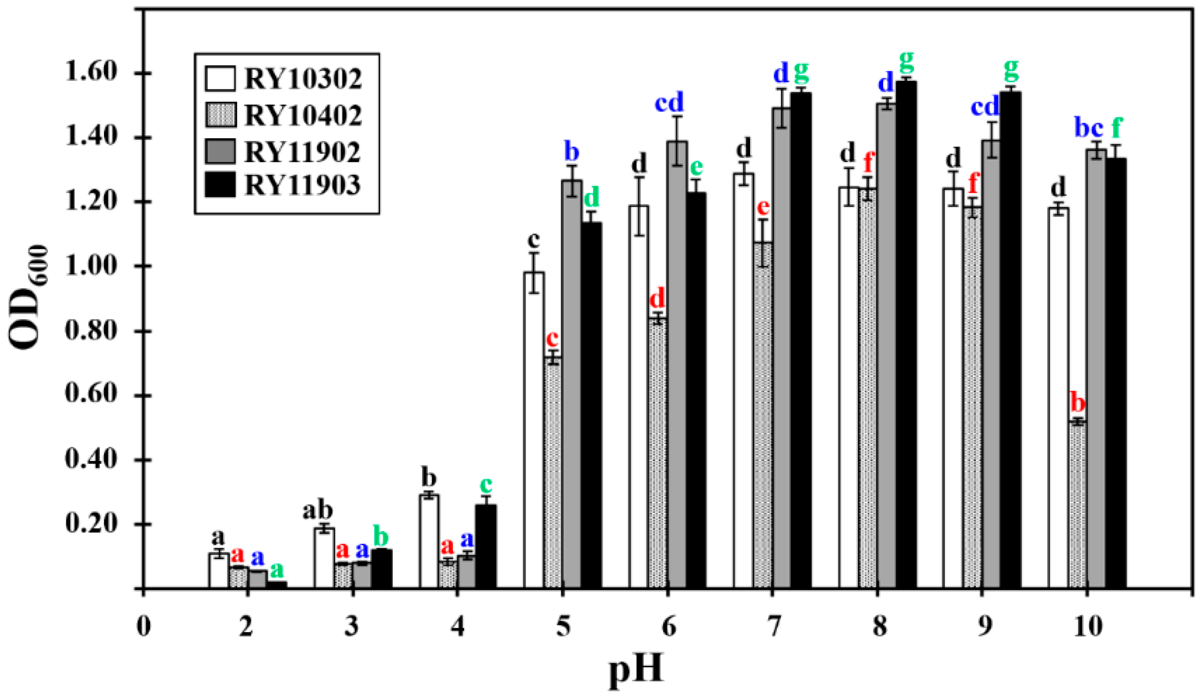
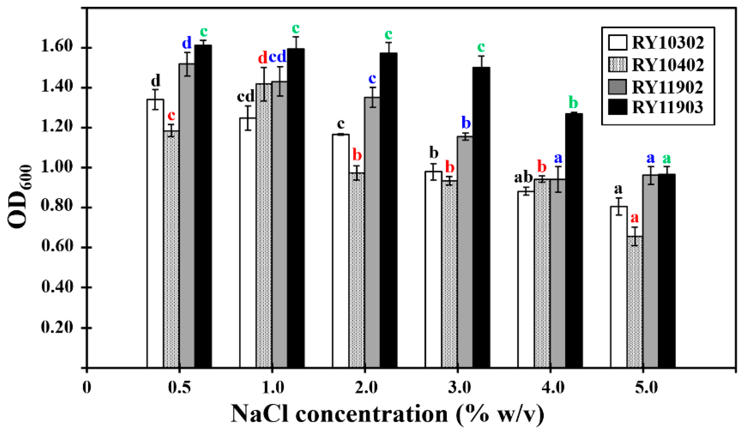
The growth characteristics of four bacterial candidates were determined by their optical density values at 600 nm (OD600) after 24 h of incubation in tryptone soya broth under various conditions. The results showed that they could grow at pH values from 5.0 to 10.0, temperatures between 25 and 40 °C and NaCl concentrations of 0.5–5.0% (w/v). The optimum growth conditions of dye-decolorizing bacteria were different, as shown in Figure 2, Figure 3 and Figure 4. The optimal pH for bacterial growth ranged between weakly acidic and weakly alkaline. Therefore, the dye-decolorizing bacteria were considered to be neutrophilic bacteria. The optimal temperature for bacterial growth was 30 °C, except for the bacterial strain RY10302, which was 35 °C. All strains in this study were designated as mesophilic bacteria. The optimal pH and temperature for bacterial growth are reasoned by the climate conditions of mangrove wetlands. The pH of mangrove soil in Thailand is neutral or weakly acidic and the temperature ranges from 25.5 to 29.9 °C [38,39]. The optimal salinity for bacterial growth was 0.5–1.0% NaCl based on bacterial strains. The bacterial strain RY11903 optimally grew in 3.0% NaCl and could be considered as a slight halophilic bacterium.
The characteristics of textile industry wastewater vary according to the different processes, mills and countries. Yaseen and Scholz [40] reviewed the typical characteristics of real textile wastewater reported from many studies [41,42,43]. The dye effluents released from textile industries are mainly between a weakly acidic pH of 6.0 and an alkaline pH of 10.0. The temperature ranges from 35 to 40 °C. The salinity of textile wastewater after fiber scouring and rinsing processes is 2000 mg/L (0.2% w/v). Finally, the salinity increases after the soaping process to 5000 mg/L (0.5% w/v).
All growth characteristics of the four bacterial isolates were favorable in reported textile wastewater parameters. Therefore, these isolated dye-decolorizing bacteria were believed to be useful in the biological decolorization of real textile wastewater.

3.5. Genotypic Identification of Dye-Decolorizing Bacteria

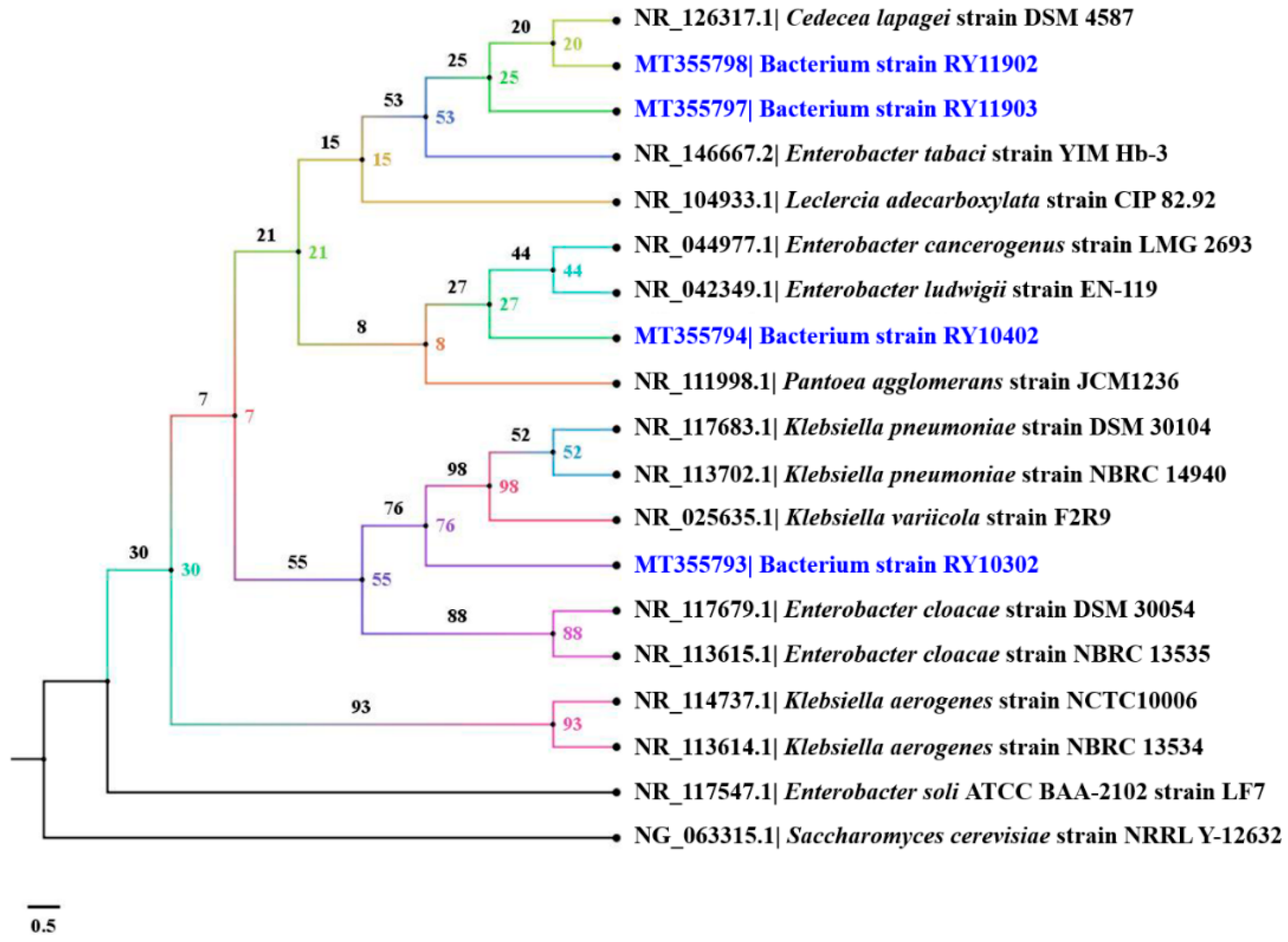
Dye-decolorizing bacteria were genetically identified by PCR amplification and nucleotide analysis of 16S rRNA gene. Alignment using the BLASTn program revealed that the bacterial strain RY10302 was closely similar to Klebsiella pneumoniae strain DSM 30104 with 97% query coverage and 99.02% identity. Three strains of dye-decolorizing bacteria, including strains RY10402, RY11902 and RY11903, were closely similar to Enterobacter tabaci strain YIM Hb-3 with 98–100% query coverage and 97.38–98.36% identity. The genotypic identification of dye-decolorizing bacteria is shown in Table 3. The E values of all BLASTn alignment results were zero. A phylogenetic tree in terms of cladogram using the BIONJ algorithm with 100,000 bootstrap replications is shown in Figure 5. All nucleotide sequences of 16S rRNA genes from this study were deposited in the GenBank database of NCBI under the accession numbers MT355793, MT355794, MT355797 and MT355798, as previously mentioned in the “Materials and Methods” section. Indeed, the bacterial strain RY10302 was confidently designated as K. pneumoniae strain RY10302. The other three strains were designated in the genus level as Enterobacter sp. due to their percentages of identity obtained from BLASTn program being lower than 99%. These dye-decolorizing bacteria were categorized in class Gammaproteobacteria of phylum Proteobacteria. All bacteria were stored as frozen stocks in 15% (v/v) glycerol and kept at the Faculty of Science, Energy and Environment, King Mongkut’s University of Technology North Bangkok, Thailand.
Klebsiella and Enterobacter are considered as members of total coliforms belonging to the family Enterobacteriaceae in the class Gammaproteobacteria. K. pneumoniae is an opportunistic pathogen causing nosocomial infections. It is widespread in various environments such as drinking water, industrial effluent, plants, sewage, soil and surface water [44]. It could be isolated from water and sediment samples from tropical estuaries and mangrove wetland ecosystems [44,45]. It complies with the habitat of K. pneumoniae strain RY10302 isolated from mangrove wetland soil in this study. Enterobacter species are common nosocomial pathogenic bacteria [46]. They are distributed in a diverse range of environments including soil, vegetation, water and human feces [47]. The most closely related Enterobacter of the three isolated bacteria in this study was E. tabaci strain YIM Hb-3. It was first isolated from the stems of cultivated tobacco (Nicotiana tabacum L.) collected from China in 2015 [48]. The 16S rRNA gene sequence of E. tabaci strain YIM Hb-3 was closely similar to six species of Enterobacter including Enterobacter mori, Enterobacter asburiae, Enterobacter hormaechei, Enterobacter ludwigii, Enterobacter cloacae and Enterobacter cancerogenus. Several Enterobacter species could be isolated from the rhizosphere of black mangroves (Avicennia germinans L.) [49] and the branches of red mangroves (Rhizophora mangle) [50]. They were considered as phosphate-solubilizing bacteria and mangrove-associated endophytic bacteria. It likely conforms to the nature and habitat of isolated Enterobacter sp. strains RY10402, RY11902 and RY11903.

3.6. Determination of Bacterial Peroxidase and Laccase Activities

Ligninolytic enzymes are extracellular oxidative enzymes commonly produced from white-rot fungi and primarily include lignin peroxidases, manganese peroxidases, versatile peroxidases (VP, EC 1.11.1.16) and laccases. Only very recently, dye-decolorizing peroxidases (DyPs, EC 1.11.1.19) have been identified in some ligninolytic fungi and bacteria [25]. However, knowledge about this group of peroxidases is very limited [51]. These ligninolytic enzymes synergistically and efficiently degrade non-phenolic and phenolic subunits of lignin polymer structure [52,53]. The ligninolytic enzymes can oxidize various pollutants such as aromatic dyes, chlorophenols, polycyclic aromatic hydrocarbons (PAHs), synthetic dyes and related model compounds [54,55,56]. Ligninolytic bacteria and their enzymes have been employed as a potentially alternative approach and cost-effective method for the bioremediation of textile dyes. Unfortunately, bacterial ligninolytic enzymes have not been studied extensively and the ligninolytic species are limited. They have only been found among some proteobacteria and actinomycetes [18].
In this study, the four dye-decolorizing bacteria of Gammaproteobacteria were examined for peroxidase and laccase activities. The results showed that K. pneumoniae strain RY10302 was not a peroxidase-producing bacterium. It could only produce laccase with enzymatic activity of 0.45 U/mL. The enzymatic assays exhibited that the three strains of Enterobacter sp. could yield 0.13–0.86 U/mL of lignin peroxidase activity and 0.66–1.47 U/mL of laccase activity. Enterobacter sp. strain RY11903 was considered as the most effective bacterium with significant lignin peroxidase and laccase activities among the four dye-decolorizing bacteria. All dye-decolorizing bacteria isolated from the mangrove wetland ecosystem were designated as laccase-producing bacteria. Interestingly, they could not produce manganese peroxidase. Their peroxidase and laccase activities are shown in Table 4.
Immobilized ligninolytic enzymes offer greater decolorization of dye-based pollutants in textile effluents and are considered as an environmental responsive technology [57]. This innovation requires purified ligninolytic enzymes. Therefore, the purification and polishing of ligninolytic enzymes are suggested for further study.
Various mangrove-isolated bacteria are able to produce ligninolytic enzymes because they are adapted to the lignin-rich soil found in mangroves and related aquatic ecosystems. An actinomycete bacteria, Streptomyces psammoticus strain NJP 49, was isolated from mangrove sediments in the west coast of India. It was capable of producing the three major ligninolytic enzymes, including approximately 2.00 U/mL of lignin peroxidase, 0.25 U/mL of manganese peroxidase and 1.50 U/mL of laccase [55]. A recent study found that Lysinibacillus sphaericus strain JD1103, isolated from a soil sample in the Rayong River Estuary in Thailand, near the sampling area in this study, had ligninolytic activity of crude lignin peroxidase by 0.90 U/mL and crude laccase by 1.13 U/mL [14].
Knowledge of K. pneumoniae and Enterobacter sp. isolated from mangrove soil with their ligninolytic activity remains limited. Besides the mangrove environment, there are several studies reported for ligninolytic K. pneumoniae. K. pneumoniae strain IITRC13 was isolated from the pulp industry in India and found to be associated with lignin degradation by its lignin peroxidase, manganese peroxidase and laccase [58]. K. pneumoniae strain 601 isolated from soil in China showed laccase activity with both thermal and broad pH stability [59]. K. pneumoniae strain NX-1 isolated from leaf mold samples in China could efficiently produce lignin peroxidase and laccase [60]. Four strains of K. pneumoniae isolated from the pulp and paper industry in India were designated as manganese peroxidase and laccase producing bacterium [61]. It is evidenced that Klebsiella species represent a promising source of ligninolytic enzymes. However, lignin and manganese peroxidase activities were not detected in K. pneumoniae strain RY10302, as shown in Table 4.
Ligninolytic Enterobacter species were isolated from various soil samples. Enterobacter lignolyticus strain SCF1 isolated from tropical forest soil in the USA was determined to be a ligninolytic bacterium. Its genome revealed two putative laccases and a putative peroxidase [62]. A laccase-producing bacterium was isolated from soil and decaying wood samples in Nigeria and molecularly identified as E. ludwigii [63]. E. cloaceae strain IITRCS11 was isolated from a dumping site in India. The function of ligninolytic enzymes was observed in decolorization and showed a high content of manganese peroxidase and laccase [64]. E. cancerogenus and E. ludwigii were isolated from petroleum-contaminated soil and animal manure samples in Turkey. They showed ligninolytic activities by degradation of Kraft lignin and RBBR decolorization [65]. Several Enterobacter species were employed in ligninolytic applications and showed successful performance. In this study, three isolated Enterobacter strains did not produce manganese peroxidase, as shown in Table 4.
The lack of peroxidases in the environmental strains from the studied mangrove wetlands is not well-understood. It might be explained by the inappropriate conditions for peroxidase preparations of Klebsiella and Enterobacter strains in this study. According to a recent previous study, it was reported that ligninolytic Streptomyces sp. strain S6 produced the highest enzyme activity for lignin peroxidase, slight laccase activity and no manganese peroxidase activity when grown on W minimal medium supplemented with Kraft lignin [66]. Likewise, several strong lignin degraders only produced manganese peroxidase and laccase, and no lignin peroxidase activity was detected when bacteria such as Pandoraea strain sp. B-6, Comamonas sp. strain B-9 and Novosphingobium sp. strain B-7 were grown on culture supplemented with Kraft lignin [67,68,69].

3.7. Dye Decolorization Efficiency of Bacterial Monoculture and Mixed Culture

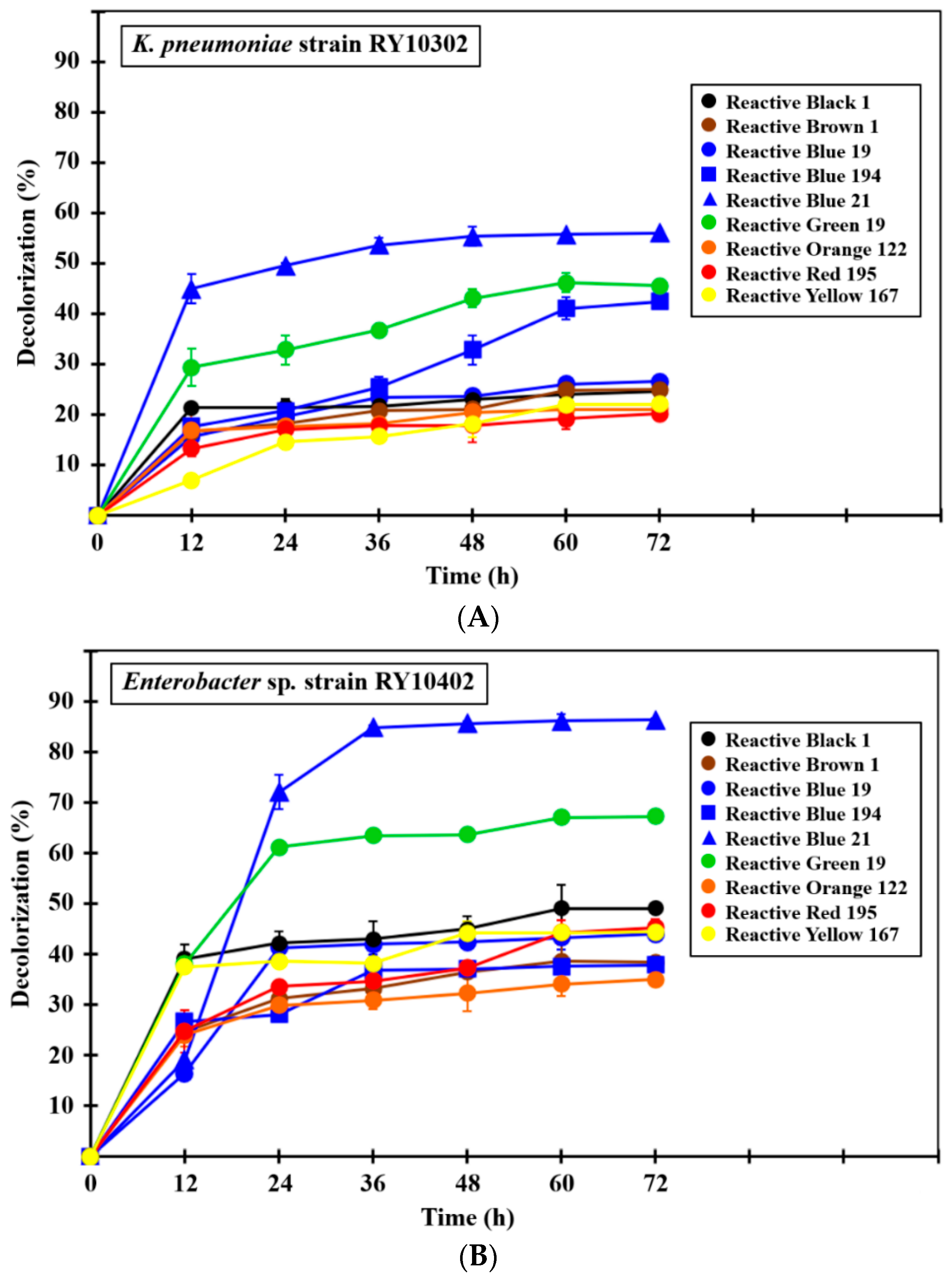
The dye decolorization efficiency of the four dye-decolorizing bacteria were studied in monoculture and mixed-culture systems under experimental conditions. The final reactive dye concentration used in this study was 0.01% (w/v) amended in BSGYP medium. It was caused by a dye mass concentration suspended in the textile effluent that reported up to 50 mg/L (0.005%, w/v) [15]. Satisfactory results of dye decolorization by monoculture and mixed-culture bacteria were obtained after 36 h of incubation (Figure 6A–E).
In the monoculture experiment, all dye-decolorizing bacteria were able to decolorize nine reactive dyes by more than 20% within 72 h (Figure 6A–D), while the control bacterium, E. coli strain TISTR 073, showed a slight level of decolorization (Figure 6F). Three strains of Enterobacter evidentially had better dye decolorization efficiency than K. pneumoniae strain RY10302 at the same incubation time. Enterobacter sp. strain RY11902 was considered as the most effective decolorizing bacterium in this study because it productively decolorized four reactive dyes including C.I. Reactive Black 1 (azo), C.I. Reactive Blue 194 (azo), C.I. Reactive Blue 21 (phthalocyanine) and C.I. Reactive Green 19 (azo) by 88–92% within 72 h (Figure 6C and Table 5). Enterobacter sp. strain RY11903 was the second most effective decolorizing bacterium. It could decolorize C.I. Reactive Black 1, C.I. Reactive Blue 194, C.I. Reactive Blue 21 and C.I. Reactive Green 19 by 70–81% within 72 h (Figure 6D and Table 5). The decolorization efficiency of Enterobacter strains 11902 and 11903 conformed to their lignin peroxidase and laccase activities, as mentioned above in Table 4.
Interestingly, the mixed culture of dye-decolorizing bacteria was found to decolorize more efficiently than monoculture. It could decolorize six reactive dyes at 83–94% within 72 h, including C.I. Reactive Black 1, C.I. Reactive Brown 1 (azo), C.I. Reactive Blue 194, C.I. Reactive Blue 21, C.I. Reactive Green 19 and C.I. Reactive Red 195 (azo) (Figure 6E, Table 5). According to Karim et al. [15], the decolorization percentage of textile reactive dyes by bacterial monoculture and mixed culture was reported. The results showed 0–90% of dye decolorization in the case of bacterial monoculture and 65–90% of dye decolorization in the case of mixed culture. It was believed that bacterial species in the mixed culture synergistically interacted and co-metabolized to decolorize the reactive dye [70,71]. The different bacterial strains have different ability to break down the dye structure. Alternatively, they may use some harmful intermediates produced by co-existing strains for further degradation [72,73]. Therefore, the mixed culture of dye-decolorizing bacteria should be employed in the biological decolorization of various reactive dyes in textile wastewater. However, the compatibility of bacterial strains in the mixed culture should be of concern and antagonistic bacteria need to be avoided.
C.I. Reactive Black 1 was the most decolorized dye. It was decolorized by the mixed culture of dye-decolorizing bacteria by 94.20 ± 0.83% within 72 h (p < 0.0001) (Table 5). The second and third most decolorized dyes were C.I. Reactive Blue 194 and C.I. Reactive Green 19, respectively. They were decolorized by Enterobacter sp. strain RY11902 by 92.45 ± 0.40% (p < 0.001) and the mixed bacterial culture by 92.05 ± 1.78% (p < 0.0001) within 72 h (Table 5). Finally, the study found that C.I. Reactive Blue 19 (anthraquinone), C.I. Reactive Orange 122 (azo) and C.I. Reactive Yellow 167 (azo) were persistent in the decolorization of both bacterial monoculture and mixed culture. The differences in the decolorization pattern for each reactive dye is due to the dissimilarity in specificities, structure and complexity of dyes [15].
Textile reactive dyes in this study were believed to be decolorized by bacterial lignin peroxidase and laccase. Lignin peroxidase is an important enzyme for the treatment of colored industrial effluents and other xenobiotics [74]. It has been used to oxidize a variety of recalcitrant aromatic compounds including dyes in the class of azo [75]. Lignin peroxidase absolutely needs H2O2 as a terminal electron acceptor in decolorizing reactions [7]. Laccase could oxidize lignin, aromatic amines, polyphenols and related aromatic compounds. It has broad substrate specificity and is capable of decolorizing wastewater containing structurally different azo dyes [76]. Laccase requires oxygen and redox mediators during enzyme mechanisms [7]. It was demonstrated by relatively complete decolorization of many azo-reactive dyes in this study, such as C.I. Reactive Black 1, C.I. Reactive Blue 21 and C.I. Reactive Green 19. The strength of these enzymes is in the non-generation of hazardous intermediates and toxic byproducts after decolorizing, such as toxic aromatic amines [7]. Therefore, the byproducts and metabolites formed as a result of dye decolorization by bacteria were found to be less toxic compared with untreated wastewater.
Many reactive dyes were studied for biological decolorization by various effective dye-decolorizing bacteria, as shown in Table 6. The mixed cultures of four bacterial strains in this study are promising candidates for the decolorization of C.I. Reactive Black 1 and C.I. Reactive Green 19 when compared to previous studies. They have also been used successfully for the decolorization of C.I. Reactive Red 195 in weakly alkaline wastewater supplemented with a high concentration of the reactive dye. Enterobacter sp. strain RY11903 is suggested for the decolorization of C.I. Reactive Blue 19 in saline-alkaline wastewater since other bacteria in several studies favored decolorizing under weakly acidic wastewater without salt supplementation.
Finally, K. pneumoniae strain RY10302, Enterobacter sp. strain RY10402, Enterobacter sp. strain RY11902 and Enterobacter sp. strain RY11903 should be employed for wastewater treatment in dye-based industries. The growth characteristics of these bacteria are suitable for real textile wastewater. A proposed wastewater-treatment process uses an aerobic basin and related aerobic methods due to the limitation of their oxidative ligninolytic enzymes. Carbon and nitrogen sources are indicated as the obligate requirement for bacterial growth and the induction of dye decolorization [15]. A previous study reported that only a few bacteria are capable of utilizing dyes as a sole carbon source [82]. Nutrient feeding units are recommended for the proposed wastewater-treatment process. A major concern for bacterial application is clinical awareness because K. pneumoniae and Enterobacter sp. are considered as pathogens capable of causing nosocomial infections. Therefore, the effluent should be disinfected using chlorine or ultraviolet light after decolorization.

4. Conclusions

A variety of reactive dyes released by the textile and dye-based industries are of significant concern for human beings and living organisms in aquatic environments. The mangrove wetland is a unique ecosystem rich in mangrove lignin. It is recorded as a potential source for the isolation of ligninolytic bacteria. Ligninolytic bacteria are able to decolorize various textile reactive dyes by the oxidative reactions of peroxidases and laccase. Therefore, ligninolytic bacteria are promising candidates for the biological decolorization of reactive dyes. In this study, there were four strains of effective ligninolytic bacteria isolated from the Phra Chedi Klang Nam mangrove wetland in Thailand. Their growth characteristics possibly thrived under textile wastewater parameters. They were genetically identified by 16S rRNA nucleotide sequencing and assigned as K. pneumoniae strain RY10302, Enterobacter sp. strain RY10402, Enterobacter sp. strain RY11902 and Enterobacter sp. strain RY11903. Interestingly, they could only produce lignin peroxidase and laccase. These ligninolytic enzymes were believed to be responsible for the decolorization of reactive dyes. All bacterial strains exhibited decolorization of nine reactive dyes within 72 h, both in the case of bacterial monoculture and mixed culture experiments. Finally, the four dye-decolorizing bacteria obtained in this study could possibly be used in the biological decolorization of textile reactive dyes. Further studies are suggested concerning the purification of ligninolytic enzymes, enzyme immobilization for wastewater treatment applications, identification and toxicity of metabolizes after decolorization reactions and decolorization experiments under a natural environment.

Funding

This research was funded by King Mongkut’s University of Technology North Bangkok (contract no. KMUTNB-62-KNOW-05).

Acknowledgments

I am grateful to Ms. Parima Boontanom (ORCID 0000-0002-0278-9688) for kindly editing this manuscript.

Conflicts of Interest

The authors declare no conflict of interest.

References

  1. Chakraborty, J.N. Fundamentals and Practices in Colouration of Textiles; Woodhead Publishing: New Delhi, India, 2010; pp. 57–75. [Google Scholar]
  2. Khatri, A.; Peerzada, M.H.; Mohsin, M.; White, M. A review on developments in dyeing cotton fabrics with reactive dyes for reducing effluent pollution. J. Clean. Prod. 2015, 87, 50–57. [Google Scholar] [CrossRef]
  3. Ekanayake, E.M.M.S.; Manage, P.M. Decolorization of CI Direct Blue 201 textile dye by native bacteria. Int. J. Multidiscip. Stud. 2017, 4, 48–57. [Google Scholar] [CrossRef]
  4. Saratale, R.G.; Saratale, G.D.; Chang, J.S.; Govindwar, S.P. Decolorization and biodegradation of reactive dyes and dye wastewater by a developed bacterial consortium. Biodegradation 2010, 21, 999–1015. [Google Scholar] [CrossRef] [PubMed]
  5. Bayar, S.; Erdogan, M. Removal of COD and color from Reactive Red 45 azo dye wastewater using Fenton and Fenton-like oxidation processes: Kinetic studies. Appl. Ecol. Environ. Res. 2019, 17, 1517–1529. [Google Scholar] [CrossRef]
  6. Jegan, J.; Praveen, S.; Bhagavathi Pushpa, T.; Gokulan, R. Sorption kinetics and isotherm studies of cationic dyes using groundnut (Arachis hypogaea) shell derived biochar a low-cost adsorbent. Appl. Ecol. Environ. Res. 2020, 18, 1925–1939. [Google Scholar] [CrossRef]
  7. Imran, M.; Crowley, D.E.; Khalid, A.; Hussain, S.; Mumtaz, M.W.; Arshad, M. Microbial biotechnology for decolorization of textile wastewaters. Rev. Environ. Sci. Biotechnol. 2015, 14, 73–92. [Google Scholar] [CrossRef]
  8. Gupta, V.K.; Khamparia, S.; Tyagi, I.; Jaspal, D.; Malviya, A. Decolorization of mixture of dyes: A critical review. Glob. J. Environ. Sci. Manag. 2015, 1, 71–94. [Google Scholar]
  9. Singh, R.P.; Singh, P.K.; Singh, R.L. Bacterial decolorization of textile azo dye Acid Orange by Staphylococcus hominis RMLRT03. Toxicol. Int. 2014, 21, 160–166. [Google Scholar] [CrossRef] [Green Version]
  10. Luo, X.; Liang, C.; Hu, Y. Comparison of different enhanced coagulation methods for Azo Dye removal from wastewater. Sustainability 2019, 11, 4760. [Google Scholar] [CrossRef] [Green Version]
  11. Saranraj, P.; Manigandan, M. Enzymes involved in bacterial decolourization and degradation of textile azo dyes. Indo-Asian J. Multidiscip. Res. 2018, 4, 1369–1376. [Google Scholar]
  12. Leme, D.M.; de Rodrigues Oliveira, G.A.; Meireles, G.; Brito, L.B.; Rodrigues, L.B.; de Palma Oliveira, D. Eco-and genotoxicological assessments of two reactive textile dyes. J. Toxicol. Environ. Health Part A 2015, 78, 287–300. [Google Scholar] [CrossRef] [PubMed]
  13. Silva, M.C.; Corrêa, A.D.; Amorim, M.T.S.P.; Parpot, P.; Torres, J.A.; Chagas, P.M.B. Decolorization of the phthalocyanine dye reactive blue 21 by turnip peroxidase and assessment of its oxidation products. J. Mol. Catal. B Enzym. 2012, 77, 9–14. [Google Scholar] [CrossRef] [Green Version]
  14. Chantarasiri, A.; Boontanom, P. Decolorization of synthetic dyes by ligninolytic Lysinibacillus sphaericus JD1103 isolated from Thai wetland ecosystems. AACL Bioflux 2017, 10, 814–819. [Google Scholar]
  15. Karim, M.E.; Dhar, K.; Hossain, M.T. Decolorization of textile reactive dyes by bacterial monoculture and consortium screened from textile dyeing effluent. J. Genet. Eng. Biotechnol. 2018, 16, 375–380. [Google Scholar] [CrossRef] [PubMed]
  16. Roy, D.C.; Biswas, S.K.; Saha, A.K.; Sikdar, B.; Rahman, M.; Roy, A.K.; Prodhan, Z.H.; Tang, S.S. Biodegradation of Crystal Violet dye by bacteria isolated from textile industry effluents. Peer J. 2018, 6, e5015. [Google Scholar] [CrossRef]
  17. Ghodake, G.S.; Kalme, S.D.; Jadhav, J.P.; Govindwar, S.P. Purification and partial characterization of lignin peroxidase from Acinetobacter calcoaceticus NCIM 2890 and its application in decolorization of textile dyes. Appl. Biochem. Biotechnol. 2009, 152, 6–14. [Google Scholar] [CrossRef]
  18. Bandounas, L.; Wierckx, N.J.P.; de Winde, J.H.; Ruijssenaars, H.J. Isolation and characterization of novel bacterial strains exhibiting ligninolytic potential. BMC Biotechnol. 2011, 11, 94. [Google Scholar] [CrossRef] [Green Version]
  19. Chantarasiri, A.; Boontanom, P.; Nuiplot, N. Isolation and characterization of Lysinibacillus sphaericus BR2308 from coastal wetland in Thailand for the biodegradation of lignin. AACL Bioflux 2017, 10, 200–209. [Google Scholar]
  20. Mitsch, W.J.; Gosselink, J.G. Wetlands, 5th ed.; Wiley: New Jersey, NJ, USA, 2015; pp. 27–44. [Google Scholar]
  21. Chantarasiri, A. Aquatic Bacillus cereus JD0404 isolated from the muddy sediments of mangrove swamps in Thailand and characterization of its cellulolytic activity. Egypt J. Aquat. Res. 2015, 41, 257–264. [Google Scholar] [CrossRef] [Green Version]
  22. Sandilyan, S.; Kathiresan, K. Decline of mangroves-a threat of heavy metal poisoning in Asia. Ocean. Coast. Manag. 2014, 102, 161–168. [Google Scholar] [CrossRef]
  23. Kaewtubtim, P.; Meeinkuirt, W.; Seepom, S.; Pichtel, J. Heavy metal phytoremediation potential of plant species in a mangrove ecosystem in Pattani Bay, Thailand. Appl. Ecol. Environ. Res. 2016, 14, 367–382. [Google Scholar] [CrossRef]
  24. Abejón, R.; Pérez-Acebo, H.; Clavijo, L. Alternatives for chemical and biochemical lignin valorization: Hot topics from a bibliometric analysis of the research published during the 2000–2016 period. Processes 2018, 6, 98. [Google Scholar] [CrossRef] [Green Version]
  25. Datta, R.; Kelkar, A.; Baraniya, D.; Molaei, A.; Moulick, A.; Meena, R.S.; Formanek, P. Enzymatic degradation of lignin in soil: A review. Sustainability 2017, 9, 1163. [Google Scholar] [CrossRef] [Green Version]
  26. Mun, S.P.; Jahan, M.S.; Al-Maruf, A.; Chowdhury, D.A.N. Chemical characterization of six mangrove species in Bangladesh. Wood Sci. Technol. 2011, 45, 281–288. [Google Scholar] [CrossRef]
  27. Srinivasan, G.P.; Sikkanthar, A.; Elamaran, A.; Delma, C.R.; Subramaniyan, K.; Somasundaram, S.T. Biodegradation of carcinogenic textile azo dyes using bacterial isolates of mangrove sediment. J. Coast. Life Med. 2014, 2, 154–162. [Google Scholar]
  28. Fang, Z.; Li, T.; Wang, Q.; Zhang, X.; Peng, H.; Fang, W.; Hong, Y.; Ge, H.; Xiao, Y. A bacterial laccase from marine microbial metagenome exhibiting chloride tolerance and dye decolorization ability. Appl. Microbiol. Biotechnol. 2011, 89, 1103–1110. [Google Scholar] [CrossRef]
  29. Chantarasiri, A. Enrichment and identification of phenanthrene-degrading bacteria isolated from the oil-stained engine sediment in the mangrove swamps of Thailand. Appl. Sci. Eng. Prog 2020, in press. [Google Scholar] [CrossRef]
  30. Chang, Y.C.; Choi, D.; Takamizawa, K.; Kikuchi, S. Isolation of Bacillus sp. strains capable of decomposing alkali lignin and their application in combination with lactic acid bacteria for enhancing cellulase performance. Bioresour. Technol. 2014, 152, 429–436. [Google Scholar] [CrossRef] [Green Version]
  31. Ferbiyanto, A.; Rusmana, I.; Raffiudin, R. Characterization and identification of cellulolytic bacteria from gut of worker Macrotermes gilvus. Hayati 2015, 22, 197–200. [Google Scholar] [CrossRef] [Green Version]
  32. Boontanom, P.; Chantarasiri, A. Short communication: Diversity of culturable epiphytic bacteria isolated from seagrass (Halodule uninervis) in Thailand and their preliminary antibacterial activity. Biodiversitas 2020, 21, 2907–2913. [Google Scholar] [CrossRef]
  33. Chantarasiri, A. Diversity of cellulolytic bacteria isolated from a freshwater wetland reserve in Thailand and their cellulolytic activity. Appl. Ecol. Environ. Res. 2020, 18, 5965–5983. [Google Scholar]
  34. Camacho, C.; Coulouris, G.; Avagyan, V.; Ma, N.; Papadopoulos, J.; Bealer, K.; Madden, T.L. BLAST+: Architecture and applications. BMC Bioinform. 2009, 10, 421. [Google Scholar] [CrossRef] [PubMed] [Green Version]
  35. Gouy, M.; Guindon, S.; Gascuel, O. SeaView version 4: A multiplatform graphical user interface for sequence alignment and phylogenetic tree building. Mol. Biol. Evol. 2010, 27, 221–224. [Google Scholar] [CrossRef] [PubMed] [Green Version]
  36. Gascuel, O. BIONJ: An improved version of the NJ algorithm based on a simple model of sequence data. Mol. Biol. Evol. 1997, 14, 685–695. [Google Scholar] [CrossRef] [PubMed] [Green Version]
  37. Cerboneschi, M.; Corsi, M.; Bianchini, R.; Bonanni, M.; Tegli, S. Decolorization of acid and basic dyes: Understanding the metabolic degradation and cell-induced adsorption/precipitation by Escherichia coli. Appl. Microbiol. Biotechnol. 2015, 99, 8235–8245. [Google Scholar] [CrossRef] [Green Version]
  38. Pumijumnong, N. Mangrove forests in Thailand. In Mangrove Ecosystems of Asia; Faridah-Hanum, I., Ed.; Springer Science + Business Media: New York, NY, USA, 2014; pp. 61–79. [Google Scholar]
  39. Srisunont, C.; Jaiyen, T.; Tenrung, M.; Likitchaikul, M. Nutrient accumulation by litterfall in mangrove forest at Klong Khone, Thailand. Thammasat Int. J. Sci. Tech. 2017, 22, 9–18. [Google Scholar]
  40. Yaseen, D.A.; Scholz, M. Textile dye wastewater characteristics and constituents of synthetic effluents: A critical review. Int. J. Environ. Sci. Technol. 2019, 16, 1193–1226. [Google Scholar] [CrossRef] [Green Version]
  41. Upadhye, V.B.; Joshi, S.S. Advances in wastewater treatment-a review. Int. J. Chem. Sci. Appl. 2012, 3, 264–268. [Google Scholar]
  42. Zhang, W.; Liu, W.; Zhang, J.; Zhao, H.; Zhang, Y.; Quan, X.; Jin, Y. Characterisation of acute toxicity, genotoxicity and oxidative stress posed by textile effluent on zebrafish. J. Environ. Sci. 2012, 24, 2019–2027. [Google Scholar] [CrossRef]
  43. Ghaly, A.; Ananthashankar, R.; Alhattab, M.; Ramakrishnan, V. Production, characterization and treatment of textile effluents: A critical review. J. Chem. Eng. Proc. Technol. 2014, 5, 1–18. [Google Scholar]
  44. Barati, A.; Ghaderpour, A.; Chew, L.L.; Bong, C.W.; Thong, K.L.; Chong, V.C.; Chai, L.C. Isolation and characterization of aquatic-borne Klebsiella pneumoniae from tropical estuaries in Malaysia. Int. J. Environ. Res. Public Health 2016, 13, 426. [Google Scholar] [CrossRef] [PubMed] [Green Version]
  45. Zhou, S.; Li, L.; Perseke, M.; Huang, Y.; Wei, J.; Qin, Q. Isolation and characterization of a Klebsiella pneumoniae strain from mangrove sediment for efficient biosynthesis of 1,3-propanediol. Sci. Bull. 2015, 60, 511–521. [Google Scholar] [CrossRef] [Green Version]
  46. Cosgrove, S.E.; Kaye, K.S.; Eliopoulous, G.M.; Carmeli, Y. Health and economic outcomes of the emergence of third-generation cephalosporin resistance in Enterobacter species. Arch. Intern. Med. 2002, 162, 185–190. [Google Scholar] [CrossRef] [PubMed]
  47. Cooney, S.; O’ Brien, S.; Iversen, C.; Fanning, S. Bacteria: Other pathogenic Enterobacteriaceae-Enterobacter and other genera. In Encyclopedia of Food Safety; Motarjemi, Y., Ed.; Academic Press: Cambridge, MA, USA, 2014; Volume 1, pp. 433–441. [Google Scholar]
  48. Duan, Y.Q.; Zhou, X.K.; Di-Yan, L.; Li, Q.Q.; Dang, L.Z.; Zhang, Y.G.; Qiu, L.H.; Nimaichand, S.; Li, W.J. Enterobacter tabaci sp. nov., a novel member of the genus Enterobacter isolated from a tobacco stem. Antonie Van Leeuwenhoek 2015, 108, 1161–1169. [Google Scholar] [CrossRef]
  49. Vazquez, P.; Holguin, G.; Puente, M.E.; Lopez-Cortes, A.; Bashan, Y. Phosphate-solubilizing microorganisms associated with the rhizosphere of mangroves in a semiarid coastal lagoon. Biol. Fertil. Soils. 2000, 30, 460–468. [Google Scholar] [CrossRef]
  50. Castro, R.A.; Quecine, M.C.; Lacava, P.T.; Batista, B.D.; Luvizotto, D.M.; Marcon, J.; Ferreira, A.; Melo, I.S.; Azevedo, J.L. Isolation and enzyme bioprospection of endophytic bacteria associated with plants of Brazilian mangrove ecosystem. Springer Plus 2014, 3, 382. [Google Scholar] [CrossRef] [Green Version]
  51. Chen, C.; Li, T. Bacterial dye-decolorizing peroxidases: Biochemical properties and biotechnological opportunities. Phys. Sci. Rev. 2016, 1, 20160051. [Google Scholar] [CrossRef] [Green Version]
  52. Wong, D.W.S. Structure and action mechanism of ligninolytic enzymes. Appl. Biochem. Biotechnol. 2009, 157, 174–209. [Google Scholar] [CrossRef]
  53. Lambertz, C.; Ece, S.; Fischer, R.; Commandeur, U. Progress and obstacles in the production and application of recombinant lignin-degrading peroxidases. Bioengineered 2016, 7, 145–154. [Google Scholar] [CrossRef] [Green Version]
  54. Mester, T.; Tien, M. Oxidation mechanism of ligninolytic enzymes involved in the degradation of environmental pollutants. Int. Biodeterior. Biodegrad. 2000, 46, 51–59. [Google Scholar] [CrossRef]
  55. Niladevi, K.N.; Prema, P. Mangrove actinomyces as the source of ligninolytic enzymes. Actinomycetologica 2005, 19, 40–47. [Google Scholar] [CrossRef] [Green Version]
  56. De Gonzalo, G.; Colpa, D.I.; Habib, M.H.M.; Fraaije, M.W. Bacterial enzymes involved in lignin degradation. J. Biotechnol. 2016, 236, 110–119. [Google Scholar] [CrossRef] [PubMed] [Green Version]
  57. Bilal, M.; Asgher, M.; Parra-Saldivar, R.; Hu, H.; Wang, W.; Zhang, X.; Iqbal, H.M.N. Immobilized ligninolytic enzymes: An innovative and environmental responsive technology to tackle dye-based industrial pollutants-a review. Sci. Total Environ. 2017, 576, 646–659. [Google Scholar] [CrossRef] [PubMed]
  58. Yadav, S.; Chandra, R. Syntrophic co-culture of Bacillus subtilis and Klebsiella pneumonia for degradation of kraft lignin discharged from rayon grade pulp industry. J. Environ. Sci. 2015, 33, 229–238. [Google Scholar] [CrossRef]
  59. Liu, Y.; Huang, L.; Guo, W.; Jia, L.; Fu, Y.; Gui, S.; Lu, F. Cloning, expression, and characterization of a thermostable and pH-stable laccase from Klebsiella pneumoniae and its application to dye decolorization. Process. Biochem. 2017, 53, 125–134. [Google Scholar] [CrossRef]
  60. Xu, Z.; Qin, L.; Cai, M.; Hua, W.; Jin, M. Biodegradation of kraft lignin by newly isolated Klebsiella pneumoniae, Pseudomonas putida, and Ochrobactrum tritici strains. Environ. Sci. Pollut. Res. 2018, 25, 14171–14181. [Google Scholar] [CrossRef]
  61. Gaur, N.; Narasimhulu, K.; Setty, Y.P. Extraction of ligninolytic enzymes from novel Klebsiella pneumoniae strains and its application in wastewater treatment. Appl. Water Sci. 2018, 8, 111. [Google Scholar] [CrossRef] [Green Version]
  62. DeAngelis, K.M.; D’Haeseleer, P.; Chivian, D.; Fortney, J.L.; Khudyakov, J.; Simmons, B.; Woo, H.; Arkin, A.P.; Davenport, K.W.; Goodwin, L.; et al. Complete genome sequence of “Enterobacter lignolyticus” SCF1. Stand. Genom. Sci. 2011, 5, 69–85. [Google Scholar] [CrossRef] [Green Version]
  63. Odeniyi, O.A.; Unuofin, J.O.; Adebayo-Tayo, B.C.; Wakil, S.M.; Onilude, A.A. Production characteristics, activity patterns and biodecolourisation applications of thermostable laccases from Corynebacterium efficiens and Enterobacter ludwigii. J. Sci. Ind. Res. 2017, 76, 562–569. [Google Scholar]
  64. Kumar, V.; Chandra, R. Characterisation of manganese peroxidase and laccase producing bacteria capable for degradation of sucrose glutamic acid-Maillard reaction products at different nutritional and environmental conditions. World J. Microbiol. Biotechnol. 2018, 34, 32. [Google Scholar] [CrossRef] [Green Version]
  65. Ozer, A.; Rakici, E.; Bektas, K.I.; Canakci, S.; Belduz, A.O. Isolation of lignin-degrading bacteria from different sources and testing of their ligninolytic activities. J. Apitherapy Nat. 2019, 2, 30–45. [Google Scholar]
  66. Riyadi, F.A.; Tahir, A.A.; Yusof, N.; Sabri, N.S.A.; Noor, M.J.M.M.; Akhir, F.N.M.D.; Othman, N.; Zakaria, Z.; Hara, H. Enzymatic and genetic characterization of lignin depolymerization by Streptomyces sp. S6 isolated from a tropical environment. Sci. Rep. 2020, 10, 7813. [Google Scholar] [CrossRef] [PubMed]
  67. Shi, Y.; Chai, L.; Tang, C.; Yang, Z.; Zheng, Y.; Chen, Y.; Jing, Q. Biochemical investigation of kraft lignin degradation by Pandoraea sp. B-6 isolated from bamboo slips. Bioprocess. Biosyst. Eng. 2013, 36, 1957–1965. [Google Scholar] [CrossRef] [PubMed] [Green Version]
  68. Chen, Y.H.; Chai, L.Y.; Zhu, Y.H.; Yang, Z.H.; Zheng, Y.; Zhang, H. Biodegradation of kraft lignin by a bacterial strain Comamonas sp. B-9 isolated from eroded bamboo slips. J. Appl. Microbiol. 2012, 112, 900–906. [Google Scholar] [CrossRef] [PubMed]
  69. Chai, L.Y.; Chen, Y.H.; Tang, C.J.; Yang, Z.H.; Zheng, Y.; Shi, Y. Depolymerization and decolorization of kraft lignin by bacterium Comamonas sp. B-9. Appl. Microbiol. Biotechnol. 2014, 98, 1907–1912. [Google Scholar] [CrossRef] [PubMed]
  70. Asgher, M.; Bhatti, H.N.; Shah, S.A.H.; Asad, M.J.; Legge, R.L. Decolorization potential of mixed microbial consortia for reactive and disperse textile dyestuffs. Biodegradation 2007, 18, 311–316. [Google Scholar] [CrossRef]
  71. Sghaier, I.; Ouertani, R.; Mahjoubi, M.; El-Hidri, D.; Hassen, W.; Chamkhi, A.; Chouchane, H.; Jaouani, A.; Cherif, A.; Neifar, M. Application of a mixture design to optimize textile azo-dye decolorization using a bacterial consortium. Biom Biostat. Int. J. 2019, 8, 58–63. [Google Scholar] [CrossRef]
  72. Chattaraj, S.; Johnson, J.; Madamwar, D. Biotransformation of mixture of dyes by enriched bacterial consortium ASD. Desalin. Water Treat. 2016, 57, 21585–21597. [Google Scholar] [CrossRef]
  73. Balapure, K.; Aghera, P.; Bhatt, N.; Madamwar, D. Community synergism: Degradation of triazine dye Reactive Black 1 by mixed bacterial cultures KND_PR under microaerophilic and aerobic conditions. Environ. Process. 2019, 6, 713–739. [Google Scholar] [CrossRef]
  74. Jadhav, P.U.; Bholay, A.D.; Shindikar, M. Bacterial lignin peroxidase in biobleaching of lignin-mimicking indicator dyes. Int. J. Pure Appl. Biosci. 2016, 4, 84–92. [Google Scholar] [CrossRef]
  75. Gottlieb, A.; Shaw, C.; Smith, A.; Wheatley, A.; Forsythe, S. The toxicity of textile reactive dyes after hydrolysis and decolourisation. J. Biotechnol. 2003, 101, 49–56. [Google Scholar] [CrossRef]
  76. Saparrat, N.; Carlos, M.; Hammer, E. Decolorization of synthetic dyes by the deuteromycete Pestalotiopsis guepinii CLPS no. 786 strain. J. Basic Microbiol. 2006, 46, 28–33. [Google Scholar] [CrossRef] [PubMed]
  77. Falade, A.; Mabinya, L.; Okoh, A.; Nwodo, U. Peroxidases produced by new ligninolytic Bacillus strains isolated from marsh and grassland decolourized anthraquinone and azo dyes. Pol. J. Environ. Stud. 2019, 28, 3163–3172. [Google Scholar] [CrossRef]
  78. Saratale, R.G.; Saratale, G.D.; Chang, J.S.; Govindwar, S.P. Ecofriendly degradation of sulfonated diazo dye C.I. Reactive Green 19A using Micrococcus glutamicus NCIM-2168. Bioresour. Technol. 2009, 100, 3897–3905. [Google Scholar] [CrossRef] [PubMed]
  79. Meerbergen, K.; Willems, K.A.; Dewil, R.; Impe, J.V.; Appels, L.; Lievens, B. Isolation and screening of bacterial isolates from wastewater treatment plants to decolorize azo dyes. J. Biosci. Bioeng. 2018, 125, 448–456. [Google Scholar] [CrossRef] [PubMed]
  80. Sahasrabudhe, M.; Pathade, G. Decolourization and degradation of C.I. Reactive Red 195 by Georgenia sp. CC-NMPT-T3. Indian J. Exp. Biol. 2012, 50, 290–299. [Google Scholar]
  81. Mate, M.S.; Pathade, G. Biodegradation of C.I. Reactive Red 195 by Enterococcus faecalis strain YZ66. World J. Microbiol. Biotechnol. 2012, 28, 815–826. [Google Scholar] [CrossRef]
  82. Sarnaik, S.; Kanekar, P. Biodegradation of methyl violet by Pseudomonas mendocina MCM B-402. Appl. Microbiol. Biotechnol. 1999, 52, 1–4. [Google Scholar] [CrossRef]
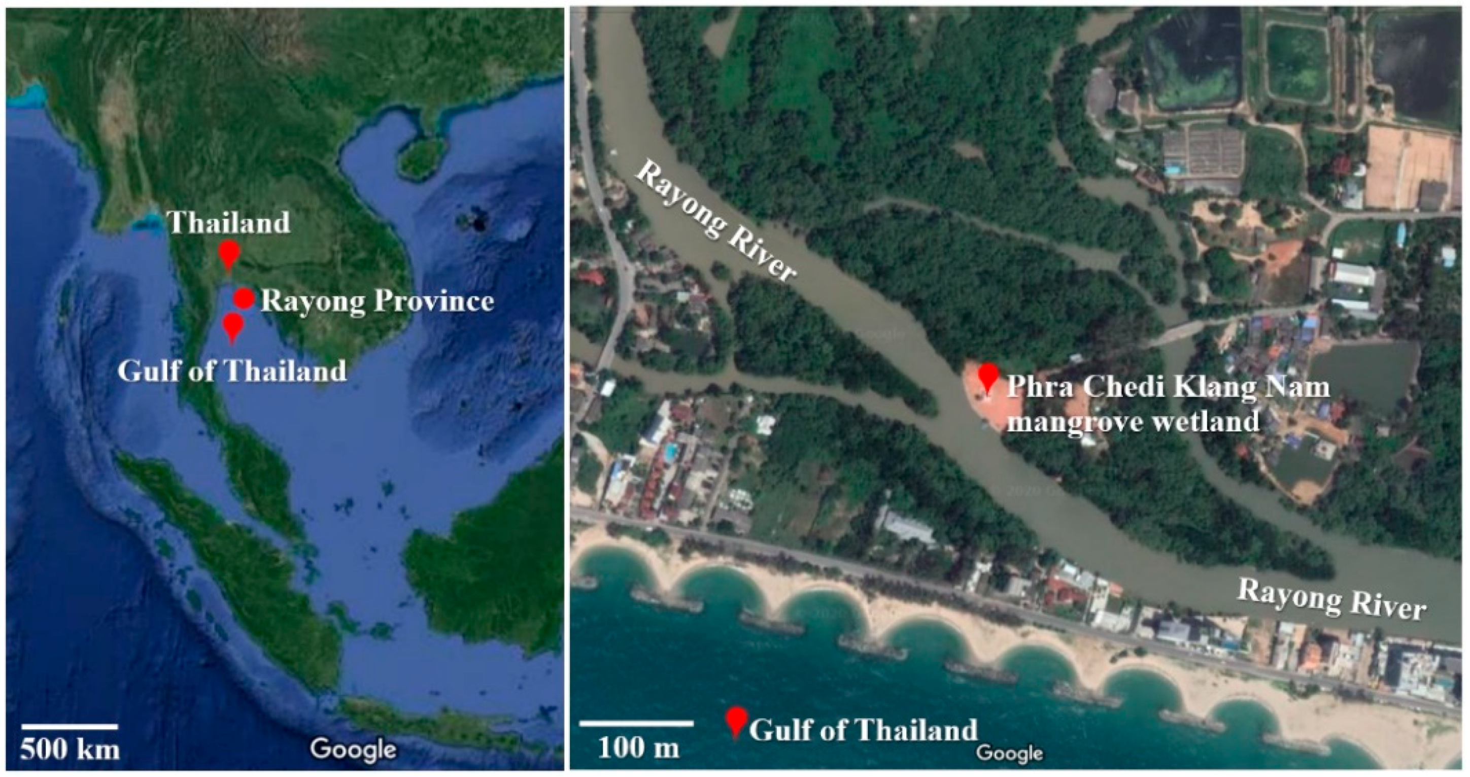
Figure 1. Location of the Phra Chedi Klang Nam mangrove wetland situated in Rayong Province, Thailand. (12°39′ N, 101°14′ E) (Source: Google Maps).
Ijerph 17 07531 g001
Figure 2. Effect of pH on the growth of four dye-decolorizing bacteria expressed as optical density measured at 600 nm. All experiments were performed in triplicate. Different letters with the same color on the top of SD bars indicate significant differences (p < 0.05).
Ijerph 17 07531 g002
Figure 3. Effect of temperature on the growth of four dye-decolorizing bacteria expressed as optical density measured at 600 nm. All experiments were performed in triplicate. Different letters with the same color on the top of SD bars indicate significant differences (p < 0.05).
Ijerph 17 07531 g003
Figure 4. Effect of salinity on the growth of four dye-decolorizing bacteria expressed as optical density measure at 600 nm. All experiments were performed in triplicate. Different letters with the same color on the top of SD bars indicate significant differences (p < 0.05).
Ijerph 17 07531 g004
Figure 5. Phylogenetic tree is shown in terms of cladogram of four dye-decolorizing bacteria using the BIONJ algorithm with 100,000 bootstrap replications. The phylogenetic tree was generated by the SeaView program version 5.0.2 and visualized by the FigTree program version 1.4.4.
Ijerph 17 07531 g005
Figure 6. Decolorization of nine textile reactive dyes by various dye-decolorizing bacteria under experimental conditions. (A) Dye decolorization by Klebsiella pneumoniae strain RY10302. (B) Dye decolorization by Enterobacter sp. strain RY10402. (C) Dye decolorization by Enterobacter sp. strain RY11902. (D) Dye decolorization by Enterobacter sp. strain RY11903. (E) Dye decolorization by mixed bacterial culture of four isolated strains. (F) Dye decolorization by Escherichia coli strain TISTR 073 (a control dye-decolorizing bacterium). All experiments were performed in triplicate.
Ijerph 17 07531 g006aIjerph 17 07531 g006bIjerph 17 07531 g006c
Table 1. Properties of the nine reactive dyes used in this study.
Textile Reactive DyeClass of Textile Reactive Dyeλmax (nm)
C.I. Reactive Black 1Azo606
C.I. Reactive Brown 1Azo489
C.I. Reactive Blue 19Anthraquinone592
C.I. Reactive Blue 194Azo594
C.I. Reactive Blue 21Phthalocyanine670
C.I. Reactive Green 19Azo636
C.I. Reactive Orange 122Azo489
C.I. Reactive Red 195Azo544
C.I. Reactive Yellow 167Azo408
Table 2. Percentage of the morphology of the isolated bacteria from the mangrove wetland soil.
Pigmentation
(Percentage)
Shape
(Percentage)
Margin
(Percentage)
Elevation
(Percentage)
White49.47Circular94.22Entire71.57Raised42.63
Yellow23.16Filamentous3.68Erose12.11Convex42.11
Red10.00Irregular1.58Undulate11.58Umbonate10.52
Colorless17.37Punctiform0.52Filamentous4.74Flat4.74
Total100.00Total100.00Total100.00Total100.00
Table 3. Identity percentage of 16S rRNA gene sequences for the four dye-decolorizing bacteria.
Bacterial IsolateClose BacteriaGenBank Accession Number
(References)
Query Cover (%)Identity
(%) *
GenBank Accession Number (Deposited)
RY10302Klebsiella pneumoniaeNR_117683.19799.02MT355793
strain DSM 30104
RY10402Enterobacter tabaciNR_146667.29897.38MT355794
strain YIM Hb-3
RY11902Enterobacter tabaciNR_146667.29997.91MT355798
strain YIM Hb-3
RY11903Enterobacter tabaciNR_146667.210098.36MT355797
strain YIM Hb-3
Note: * Identity results were analyzed on 19 April 2020.
Table 4. Peroxidase and laccase activities of the four dye-decolorizing bacteria.
Bacterial StrainEnzymatic Activities (U/mL)
Lignin PeroxidaseManganese PeroxidaseLaccase
K. pneumoniaeNo activityNo activity0.45 ± 0.03 a
(p < 0.001)
strain RY10302
Enterobacter sp.0.13 ± 0.02 a
(p < 0.001)
No activity0.81 ± 0.02 c
(p < 0.001)
strain RY10402
Enterobacter sp.0.62 ± 0.03 b
(p < 0.001)
No activity0.66 ± 0.02 b
(p < 0.001)
strain RY11902
Enterobacter sp.0.86 ± 0.06 c
(p < 0.001)
No activity1.47 ± 0.03 d
(p < 0.001)
strain RY11903
Note: The mean values in the same enzymatic activity followed by the same letter were not significantly different according to Tukey’s test (p < 0.05) among the bacteria. All experiments were assayed in triplicate.
Table 5. Decolorization of textile reactive dyes by various dye-decolorizing bacteria under experimental conditions at 72 h of incubation.
Bacterial StrainDecolorization of Textile Reactive Dye (%) at 72 h of Incubation
Black 1Brown 1Blue 19Blue 194Blue 21Green 19Orange 122Red 195Yellow 167
K. pneumoniae24.65 ± 0.74 b24.96 ± 0.26 b26.51 ± 0.44 b42.43 ± 0.35 b55.97 ± 0.05 b45.49 ± 0.60 b21.00 ± 0.01 b20.10 ± 0.00 b22.00 ± 0.01 b
strain
RY10302
Enterobacter sp.49.00 ± 0.00 c38.30 ± 0.26 c43.94 ± 0.07 d37.83 ± 0.29 b86.23 ± 0.32 d67.23 ± 0.40 c34.96 ± 1.12 c45.23 ± 1.71 c44.29 ± 0.66 c
strain
RY10402
Enterobacter sp.89.52 ± 1.41 e66.27 ± 4.70 d37.27 ± 3.96 c92.45 ± 0.40 d88.67 ± 0.42 e91.09 ± 0.68 e54.50 ± 2.17 e60.27 ± 2.18d 40.86 ± 3.71 c
strain
RY11902
Enterobacter sp. strain73.85 ± 3.18 d72.32 ± 1.71 d60.34 ± 0.94 e69.91 ± 5.24 c82.70 ± 0.76 c81.35 ± 0.84 d40.38 ± 2.02 d64.22 ± 2.48 d50.17 ± 2.12 d
RY11903
Mixed94.20 ± 0.83 f83.03 ± 2.36 e58.25 ± 1.86 e86.81 ± 1.19 d88.32 ± 0.67 de92.05 ± 1.78 e63.01 ± 1.10 f84.32 ± 1.03 e68.26 ± 1.37 e
culture
E. coli4.67 ± 0.22 a12.38 ± 0.44a 16.56 ± 2.91 a5.92 ± 0.81 a13.37 ± 1.70 a14.53 ± 0.36 a15.10 ± 0.94 a11.07 ± 0.81 a14.36 ± 1.07 a
strain
TISTR 073
Note: The mean values in the same column (decolorization efficiency of reactive dye) followed by the same letter were not significantly different according to Tukey’s test (p < 0.05) among the bacteria. All experiments were assayed in triplicate.
Table 6. Decolorization of some reactive dyes by isolated decolorizing-bacteria and other bacteria isolated from various environments.
Reactive DyeDecolorizing BacteriaSource of IsolationDye Concentration /Decolorization PercentageConditions for DecolorizationReferences
C.I. Reactive Black 1Mixed bacterial cultureDye contaminated soils, India100 mg/L,37 °C, 12 h[73]
(Lysinibacillus sp. strains BAB-4931 and BAB-4935, Raoultella sp. strain BAB-4932, Enterococcus sp. strain BAB-4933 and Citrobacter sp. strain BAB-4934)65.0%
Mixed bacterial cultureMangrove wetland soils, Thailand100 mg/L,pH 8.0, 30 °C, 0.5% (w/v) NaCl, 72 hThis study
94.2%
C.I. Reactive Blue 19 (RBBR)Bacillus sp.Mountain soils, Japan100 mg/L,pH 6.0, 30 °C,[30]
strain CS-1100.0%72 h
Bacillus sp.Mountain soils, Japan100 mg/L,pH 6.0, 30 °C,[30]
strain CS-295.0%72 h
Lysinibacillus sphaericus strain BR2308Coastal wetland soils, Thailand100 mg/L,pH 6.0, 30 °C,[19]
58.1%72 h
L. sphaericusWetland and estuary soils, Thailand50 mg/L,pH 6.0, 28 °C,[14]
strain JD110350.0%72 h
Bacillus sp.Marsh and grassland samples, South Africa100 mg/L,pH 6.0, 25 °C,[77]
strain NWODO-372.1%30 min
Enterobacter sp.Mangrove wetland soils, Thailand100 mg/L, pH 9.0, 30 °C, 3% (w/v) NaCl, 72 hThis study
strain RY1190360.3%
C.I. Reactive Green 19Micrococcus glutamicus strain NCIM-2168National Chemical Laboratory, India50 mg/L,pH 8.0, 37 °C,[78]
100.0%42 h
Acinetobacter sp.Activated sludges, Belgium100 mg/L,30 °C, 72 h[79]
strain ST16.16/16493.4%
Mixed bacterial cultureMangrove wetland soils, Thailand100 mg/L,pH 8.0, 30 °C, 0.5% (w/v) NaCl, 72 hThis study
92.1%
C.I. Reactive Red 195Georgenia sp.India50 mg/L,pH 7.0, 40 °C, 5 h[80]
strain CC-NMPT-T395.9%
Enterococcus faecalisDye contaminated soils, India50 mg/L,pH 5.0, 40 °C,[81]
strain YZ6699.5%1.5 h
Mixed bacterial cultureMangrove wetland soils, Thailand100 mg/L,pH 8.0, 30 °C, 0.5% (w/v) NaCl, 72 hThis study
84.3%
Publisher’s Note: MDPI stays neutral with regard to jurisdictional claims in published maps and institutional affiliations.