Article

Occurrence of Extended Spectrum Beta Lactamase (ESBL) Producers, Quinolone and Carbapenem Resistant Enterobacteriaceae Isolated from Environmental Samples along Msimbazi River Basin Ecosystem in Tanzania

by
Zuhura I. Kimera
1,2,*,
Fauster X. Mgaya
1,
Stephen E. Mshana
3,4,
Esron D. Karimuribo
4,5 and
Mecky I. N. Matee
1,4
1
Department of Microbiology and Immunology, Muhimbili University of Health and Allied Sciences, P.O. Box 65001, Dar es Salaam 11103, Tanzania
2
Ministry of Livestock and Fisheries, Mtumba Area, P.O. Box 2182, Dodoma 40487, Tanzania
3
Department of Microbiology and Immunology, Catholic University of Health and Allied Sciences, P.O. Box 1464, Mwanza 33109, Tanzania
4
SACIDS Africa Centre of Excellence for Infectious Diseases, Sokoine University of Agriculture, P.O. Box 3297, Morogoro 67125, Tanzania
5
Department of Veterinary Medicine and Public Health, Sokoine University of Agriculture, P.O. Box 3021, Morogoro 67125, Tanzania
*
Author to whom correspondence should be addressed.
Int. J. Environ. Res. Public Health 2021, 18(16), 8264; https://doi.org/10.3390/ijerph18168264
Submission received: 18 June 2021 / Revised: 24 July 2021 / Accepted: 28 July 2021 / Published: 4 August 2021

Abstract

:
We conducted environmental surveillance of antimicrobial resistance (AMR) bacteria in the Msimbazi river basin in Tanzania to determine the occurrence of extended-spectrum β-lactamase (ESBL)-producing, carbapenem resistant Enterobacteriaceae (CRE) and quinolone resistant Escherichia coli and Klebsiella spp. A total of 213 Enterobacteriaceae isolates were recovered from 219 samples. Out of the recovered isolates, 45.5% (n = 97) were Klebsiella pneumoniae and 29.6% (n = 63) were Escherichia coli. K. pneumoniae isolates were more resistant in effluent (27.9%) compared to the E. coli (26.6%). The E. coli had a higher resistance in river water, sediment and crop soil than the K. pneumoniae (35 versus 25%), respectively. Higher resistance in K. pneumoniae was found in nalidixic acid (54.6%) and ciprofloxacin (33.3%) while the E. coli isolates were highly resistant to ciprofloxacin (39.7%) and trimethoprim/sulfamethoxazole (38%). Resistance increased from 28.3% in Kisarawe, where the river originates, to 59.9% in Jangwani (the middle section) and 66.7% in Upanga West, where the river enters the Indian Ocean. Out of 160 E. coli and K. pneumoniae isolates, 53.2% (n = 85) were resistant to more than three classes of the antibiotic tested, occurrence being higher among ESBL producers, quinolone resistant and carbapenem resistant strains. There is an urgent need to curb environmental contamination with antimicrobial agents in the Msimbazi Basin.

1. Introduction

Antimicrobial resistance (AMR) presents a major threat to public and animal health, the global economy and security [1]. The World Health Organisation considers AMR to be one of the top ten threats to global health [2]. Without interventions, by 2050 at least 50 million people will die annually [3]. Due to the nature and complexity of the drivers of AMR, efforts to control AMR must utilise a One Health based approach [1,4,5]. Although tackling AMR requires surveillance of human–animal environment compartments, the latter is relatively under-investigated, especially in sub-Saharan African countries [6]. This is largely due to a lack of proper understanding of the role of the environment as a conduit and reservoir of AMR pathogens and genes, with potential spillover to animals and humans [7]. Our previous study indicated spillage of resistant environmental isolates into Lake Victoria through the sewage system in the city of Mwanza, involving clonal spread of resistant strains as well as dissemination by commonly occurring IncY plasmids [8].
Although the presence of AMR micro-organisms in the environment is of global concern, the situation in Africa is more critical due to weak regulation in the use of antimicrobials, weak surveillance systems for antimicrobial use (AMU) and AMR, unregulated disposal of waste and poor sanitation infrastructure [9,10,11,12]. This is coupled with a lack of basic knowledge of the concept of antimicrobial resistance [13,14] and a lack of continuing medical/veterinary education for prescribers [15]. In addition, the use of antimicrobials as pesticides in crops is very common [16].
Yet in our review of AMR studies conducted in Africa between 2005 and 2018, we found only ten (5.68%) out of 176 published investigations on AMR involved the environment [6]. Four of these studies were conducted in South Africa, two in Algeria and one in each of the following countries: Ethiopia, Egypt, Tunisia and Tanzania. These studies, which sampled domestic and biomedical waste, wastewater, river sediments, surface and drinking water, treated wastewater, river water and vegetables, found the prevalence of MDR E. coli ranging from 33.3 to 100%, with resistance to 16 different antimicrobial agents. The variations in the findings could be attributed, at least in part, to methodological differences [6].
Recently, a framework for environmental surveillance of antibiotics and antibiotic resistance has been developed, with a choice of markers and sampling sites for harmonizing surveillance systems under different scenarios [7]. For each scenario, the framework provides suggestions on different phenotypic and genotypic microbial surveillance markers, as well as antibiotic residues and sites where monitoring would be particularly informative. Such an approach ensures good quality data that can be shared between countries and contribute toward the protection of human, animal and ecosystem health [17].
Using this framework, we conducted environmental surveillance of AMR bacteria in the Msimbazi river basin in Dar es Salaam, Tanzania’s largest city and one of Africa’s fastest growing metropolitan areas [18]. The city’s growth is largely centred along the Msimbazi Basin, with 27% of its population living in the basin and along its tributaries, which flow through the heart of Dar es Salaam [19]. The basin, which is important to the city’s development, environment and economy, has many human activities of significance with respect to the emergence and spread of AMR in the environment. Unfortunately, service provision in the basin has not kept up with the rate of urbanisation, resulting in unplanned settlements, sanitation challenges, pollution, inadequate infrastructure and erosion. Our focus was on the occurrence and distribution of extended-spectrum β-lactamase (ESBL)-producing and carbapenem resistant Enterobacteriaceae (CRE) and quinolone resistant Escherichia coli and Klebsiella spp. in environmental samples. Many enterobacteria that have been isolated from environments, such as E. coli and Klebsiella spp., are often associated with ESBL production and carbapenem and quinolone resistance, often occurring together in most multidrug resistant phenotypes [20,21,22]. Resistance to these classes of antibiotics is very significant, since it renders most of the drugs used for human and veterinary medicine ineffective [6,14].

2. Materials and Methods
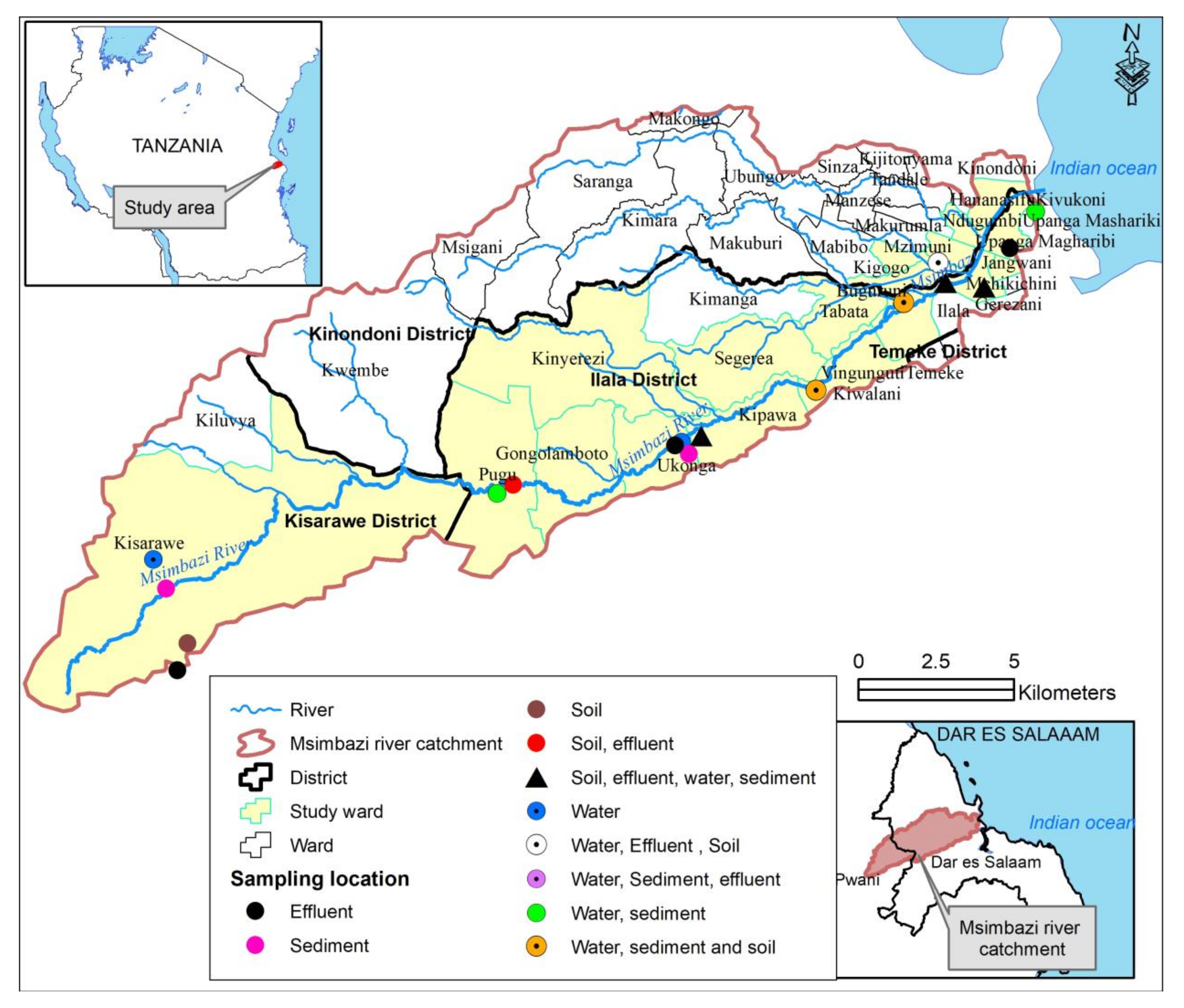
2.1. Study Area

This study was conducted between February and March 2021 in the Msimbazi River basin in Tanzania. The river originates from the Kisarawe highlands in the Pwani Region and discharges its water into the Indian Ocean as previously described [23]. The Msimbazi River serves as an important drainage system for rainwater, runoff water and wastewater from community, medical, industrial and animal farming and agricultural activities along the basin.

2.2. Sampling Frame

Using integrated surveillance for the collection of AMR data in the environment as described in [7], a sampling frame from the study area was generated. The specified sampling frame for the study included poultry and domestic pig farms, crops and vegetable farms that use manure from the domesticated animals, abattoirs, commercial factories, river water, community settings that directly discharge effluents to the river and river sediments.

2.3. Sampling Strategy and Sample Sites

Based on the characteristics of the study area, the Msimbazi River basin was categorized into three segments. The upper part where the river originates is slightly populated, less prone to flooding with agriculture, irrigation and drinking points for livestock activities. The middle part with moderate to dense population, multiple activities such as agriculture and irrigation, animal farming, drinking points for livestock, abattoirs and commercial factories is prone to frequent flooding. The lower part is characterised by prolonged flooding, a high-density population, agricultural activities, fishing and discharge from community, industrial and the national hospital effluents, and it is the discharge point of the river. From the three categories, ten sampling sites were selected: two from the upper part, six from the middle and two from the lower part of the basin (Figure 1). Samples of river water, river sediments, crop soil and effluent from the community, factory, abattoir, hospital and veterinary settings that are directly discharged onto the river were collected. During sampling, information about the localities where samples were collected (sampling sites), dates of sampling, type of sample, sample origin, weight of the sample, transportation conditions, test parameters to be conducted, organism of interest, time of sample collection and analysis was recorded in a special form.

2.4. Sample Collection and Processing

For each site, between three and seven samples were collected from different positions, making a total of 219 liquid and solid samples. The liquid samples were collected in a sterile 50 mL falcon tube (BD, Nairobi, Kenya). For river water, samples were collected from a distance of at least 0.5 m from the shore and at a depth of 20 and 50 cm. The community, industrial and hospital effluents were collected from the point where the final effluents entered into the river. Soil samples were collected at up to five different points per site using a sterile trowel and placed in a 50 mL sterile falcon tube (BD, Nairobi, Kenya). The tube was then placed in a zip-top bag. Liquid and solid samples were labelled according to the site of collection, placed in a cooler box containing ice packs and transferred to the laboratory for analysis within 3 h of collection. In each site the frequency of sampling was conducted once. The collection and processing of solid samples was conducted following the procedure described by [24], with some modifications. The liquid samples were processed as per [8], in which each individual sample was mixed with sterile 0.9% saline at a ratio of 1:1 and mixed to produce a homogenous solution. Approximately 1 g of the soil sample was made into suspension by adding 4 mL of normal saline and mixed thoroughly by vortexing.

2.5. Isolation and Identification of Bacterial Strains

A loopful of sample suspension was inoculated onto MacConkey agar (Oxoid, Basingstoke, UK) without antibiotics and incubated at 37 °C aerobically for 24 h. A single colony from predominant morphologically similar colonies was picked from each plain MacConkey agar plate and subcultured in a Nutrient agar (Hi media, Mumbai, India). Colonies on the Nutrient agar were identified by colonial morphology, Gram stain, catalase and oxidase production [25] and various biochemical tests (Indole, Methyl red, Voges Proskauer, and Citrate utilisation tests) and were later confirmed by API 20E following the manufacturer’s recommendations (BioMérieux, Marcyl’Etoile, France) [26]. Briefly, a single colony was emulsified into sterile saline and filled in the compartments, then incubated at 37 °C for 18 to 24 h aerobically in a wet chamber of API 20 E strips. Escherichia coli and Klebsiella pneumoniae were identified to the species level.

2.6. Antimicrobial Susceptibility Testing

The antimicrobial susceptibility testing was done using the Kirby–Bauer disc diffusion method on Mueller Hinton agar (Becton, Dickinson and Company, New Jersey, USA) based on the Clinical Laboratory Standard Institute (CLSI) standards [27]. The antibiotics tested were ampicillin (10 µg), cefotaxime (30 µg), ceftriaxone (30 µg), nalidixic acid (30 µg), ciprofloxacin (5 µg), tetracycline (30 µg), chloramphenicol (30 µg), meropenem (10 µg) and imipenem (10 µg). One to two colonies from the pure culture of the identified lactose fermenters were emulsified into 5 mL of sterile saline. The suspension was adjusted to achieve turbidity equivalent to 0.5 McFarland standard solutions, emulsified using sterile cotton swabs onto Mueller Hinton agar plate and incubated at 37 °C for 16 to 18 h. The inhibition zone of each antimicrobial agent was measured using a ruler and the results were interpreted according to the CLSI standards 2019 [27]. E. coli strain ATCC 29522 and K. pneumoniae strain ATCC 700603 were used as controls.

2.7. Screening and Confirmation of ESBL Production

Confirmed E. coli isolates were inoculated onto MacConkey agar containing 2 µg/mL cefotaxime for the preliminary screening of extended spectrum beta lactamase (ESBL) production [8]. Confirmation of ESBL production was performed using a combination disk diffusion method of cefotaxime (30 µg) alone and in combination with cefotaxime-clavulanic acid (10 µg) and ceftazidime (30 µg) alone and combination with ceftazidime-clavulanic acid (10 µg). Klebsiella pneumoniae (ATCC 700603) was used as a positive control (ESBL positive strain) and E. coli (ATCC 25922) was used as an ESBL negative strain; results were interpreted as per CLSI standards 2019 [27].

3. Results

3.1. Detection of Enterobacteriaceae Isolates and the Prevalence of Resistance from Effluent, River Water, River Sediment and Crop Soil

A total of 213 Enterobacteriaceae isolates were recovered from 219 samples (171, 27, 12 and 9 of effluents, river water, river sediments and crop soil, respectively). Approximately 60 samples yielded more than one isolate. Out of the recovered isolates, 45.5% (n = 97) were Klebsiella pneumoniae and 29.6% (n = 63) were Escherichia coli. Other Enterobacteriaceae (24.8%, n = 53) detected were Serratia odorifera, Enterobacter aerogenes, Enterobacter cloacae, Klebsiella oxytoca, Pantoea spp., Citrobacter spp., Aeromonas hydrophila and Kluyvera spp. (Table 1).
As shown in Figure 2, K. pneumoniae isolates were more resistant in effluent (27.9%) compared to the E. coli (26.6%) while the E. coli had a higher resistance in river water, sediment and crop soil than the K. pneumoniae (35 versus 25%), respectively. The K. pneumoniae isolates had higher resistance against nalidixic acid (54.6%) and ciprofloxacin (33.3%). The E. coli isolates were more resistant to ciprofloxacin (39.7%), ampicillin and trimethoprim/sulfamethoxazole (38%), respectively.

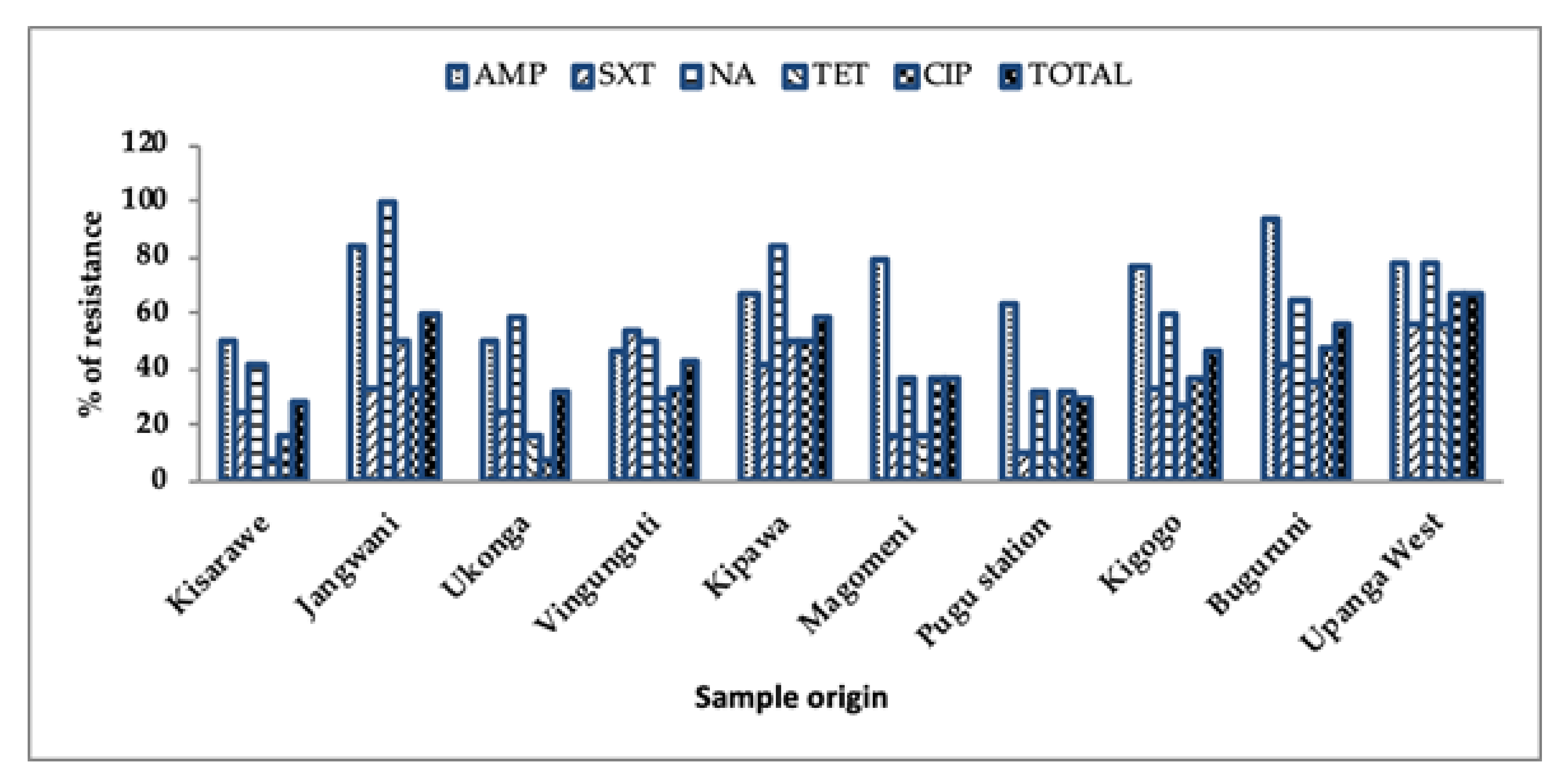
3.2. Prevalence of Antibiotic Resistance from Different Sample Locations

Overall, the level of resistance from the K. pneumoniae and E. coli isolates were higher in ampicillin (68.9%), nalidixic acid (60.4%), ciprofloxacin (36%) and trimethoprim/sulfamethoxazole (33.6%) throughout the sample locations. As shown in Figure 3, the level of antibiotic resistance increased from 28.3% in Kisarawe, where the river originates, to (59.9%) in Jangwani (the middle section) to 66.7% in Upanga West, where the river enters the Indian Ocean.

3.3. Multidrug Resistant E. coli and K. pneumoniae Isolates in Effluent, River Water, River Sediment and Crop Soil

Table 2 shows that out of 160 E. coli and K. pneumoniae isolates, 53.2% (n = 85) were multidrug resistant [28]. The most common resistance pattern determined was QNL/PEN/SUL (15 isolates), followed by QNL/PEN/TET (11 isolates) and CEP/QNL/PEN (nine isolates). Some of the E. coli and K. pneumoniae isolates were resistant to more than five classes of the antibiotic tested.

3.4. Prevalence of Quinolone Resistance, ESBL Producers and Carbapenem Resistant E. coli and K. pneumoniae from the Effluent, River Water, Sediment and Crop Soil

Out of 160 E. coli and K. pneumoniae isolates, 48.6% (n = 57) were found to be quinolone resistant [29], 14.4% (n = 23) were confirmed to be ESBL producers and 8.8% (n = 14) were carbapenem resistant. The ESBL producers were significantly resistant (p < 0.05) against trimethoprim/sulfamethoxazole and tetracycline and those that were quinolone resistant were significantly resistant to trimethoprim/sulfamethoxazole and tetracycline. The carbapenem resistant isolates had no significant resistance to the tested drugs except for tetracycline. The ESBL producers, quinolone resistant and carbapenem resistant isolates were more resistant to the tested antibiotics compared to the non-ESBL producers, non-quinolone resistant and non-carbapenem resistant isolates (Table 3). As shown in Table 4, both ESBL producers, quinolone resistant and carbapenem resistant isolates depicted various levels of resistance to the tested antibiotics and were significantly resistant compared to the isolates that were sensitive.

4. Discussion

This study was conducted in an ecosystem that is characterised by a high population, intensive agricultural and farming practices involving the use of manures, pesticides and antimicrobial agents, and is polluted with effluents and wastes from the largest pharmaceutical and commercial industries in the country [18,30,31]. The basin is located in Dar es Salaam, the largest city in Tanzania, with the highest population density of humans (3,133 humans/square kilometre) [32] and livestock in the country [33], and is the largest destination of livestock and livestock products from almost all areas of the country. In Africa, studies dealing with surveillance of the environment for AMR are very few [6,34] and to the best of our knowledge this is the first such study in Tanzania. In this study we used the recently described integrated systematic sampling approach [7] that ensures good quality data that can be shared and compared with data reported in other countries and contribute to the global picture.
Overall, K. pneumoniae isolates were more resistant in effluent compared to the E. coli isolates which had higher resistance in river water, river sediment and crop soil. Both the K. pneumoniae and E. coli organisms are widely distributed in the community and wastewater settings and can easily acquire multiple resistance mechanisms as previously reported [26,34,35,36,37,38,39]. The levels of resistance to the tested antibiotics were higher in nalidixic acid (54.6%) and ciprofloxacin (39.7%) for K. pneumoniae and trimethoprim/sulfamethoxazole (38%) for E. coli, probably due to extensive use of these drugs in both animals and humans, uncontrolled disposal of drug leftovers and discharge of effluent into the environment and water bodies [23,40,41]. The levels of nalidixic acid, ciprofloxacin and trimethoprim/sulfamethoxazole found in this study are comparable with the one in Ethiopia and Tanzania, which reported that waste effluent discharged to the environment contains isolates of E. coli and K. pneumoniae that are resistant to multiple antibiotics [35,42].
In this study, more than half (53.2%) of the E. coli and K. pneumonia isolates from the effluent, river water, river sediment and crop soil exhibited multidrug resistance against three to seven classes of the antibiotic tested. The most frequent combination was observed in QNL/PEN/SUL, QNL/PEN/TET and CEP/QNL/PEN and might be attributed to extensive use in animals and humans and their release to the environment [41,43,44]. A previous study from Tanzania, Mozambique and Zambia reported quinolone, tetracycline, penicillin and sulfamethoxazole antibiotics were among the commonly used antimicrobial in animals and humans [14,45,46,47,48] with the consequence of increasing MDR organisms across the human, animal and environmental compartments.
We found significantly lower levels of antibiotic resistance in Kisarawe and Pugu station, where the river originates, compared to the lower part of the basin (Jangwani and Upanga West areas), which discharges into the Indian Ocean. A possible explanation of these findings is that the basin is less contaminated at the upper part compared to the middle and lower basin due to anthropogenic activities and human settlement [31,49].
The overall prevalence of ESBL producers, quinolone and carbapenem resistance were 14.4, 48.6 and 8.8%, respectively, suggesting widespread use and release of these antimicrobial drugs and their active metabolites to the environment [50,51]. The acquisition of the gene encoding for resistance is through plasmids, transposons and integrons, which can spread very rapidly, thus posing a problem of treatment failure [34,50]. Our previous findings from the study area that compared genotypic and phenotypic results of MDR E. coli isolates, ESBL producers and quinolone resistance found that 80% of the isolates harboured blaCTX-M, 15% aac(6)-lb-cr, 10% qnrB and 5% qepA. None harboured TEM, SHV, qnrA, qnrS, qnrC, or qnrD [46,52]. The ESBL level found in this study was higher than that reported in soil and water samples in Tunisia and DRC Congo [36,41] and lower than previous findings from Tanzania and in Angola [8,53]. The level of quinolone in this study was lower (85%) than that reported from similar studies in Algeria and Togo [34,44]. These variations may be due to differences in relative use of the antibiotics between countries.
Our results, which show 27.9 and 35% of resistant K.pneumoniae and E. coli isolates, respectively, compare with other environmental surveillance studies that showed AMR levels between 33 and 100% in samples collected from rivers and streams, effluent and solid wastes [6]. Collectively, these results show a high level of environmental contamination with AMR and MDR bacteria in Africa, due to a high intensity of AMU in animals and humans, agriculture activities and weakness in regulation and disposal of antimicrobials in the environment [54].
We hypothesize that the sources of AMR bacteria in the basin are mainly the uncontrolled use of a wide range of antibiotics in humans and animals, use of manure and pesticides in agriculture, the release of effluents from different setups ranging from community, hospital and veterinary healthcare and commercial industries.
Therefore, we do recommend the following measures be instituted to address pollution of the environment with AMR bacteria in the Msimbazi basin: strengthen implementation of the available legislation, such as the national Environmental Management Act 2004 [55] to control for environmental contamination, the Animal Welfare Act 2008 [56] and the Veterinary Act 2003 [57] on proper animal husbandry and the Tanzania Food and Drugs Authority (TFDA) Act 2003 [58] on the proper handling and sale of both human and veterinary drugs; preventing the use of contaminated water for crop irrigation and establishment of multisectoral intervention and focusing on training and creating awareness of the magnitude and consequences of the AMR problem. We are also advocating for continuous monitoring of these measures to evaluate their impact on curbing environmental contamination with AMR organisms and antimicrobial agents.

5. Conclusions

The level of AMR and MDR bacteria, including ESBL producers and quinolone resistant strains, seen in the Msimbazi basin is very high, which poses a risk to both human and animal health. Curbing AMR in the basin will require comprehensive and well-coordinated approaches that include continuous surveillance and stewardship on AMU and AMR in animals and humans; revising the approach on the implementation of regulatory bodies governing handling, distribution and sale of human and veterinary drugs; proper disposal of waste and effluents and improved agricultural practices along the river basin. Modelling will be required to assess the most effective set of approaches to embark on.

Author Contributions

Study design, sample collection, laboratory work and analysis, original draft preparation, Z.I.K.; organization and supervision of laboratory work, F.X.M.; design of the outline, review of the manuscript and supervision of the manuscript writing, M.I.N.M.; coordination of data analysis and interpretation and manuscript editing, E.D.K.; manuscript review and editing, S.E.M. All authors have read and agreed to the published version of the manuscript.

Funding

This study was funded by the Government of the United Republic of Tanzania through the World Bank [WB-ACE II Grant PAD1436, IDA [Credit 5799-TZ] to the SACIDS Africa Centre of Excellence for Infectious Diseases at Sokoine University of Agriculture.

Institutional Review Board Statement

The ethical clearance was provided by the Medical Research Coordinating Committee of the National Institute for Medical Research (NIMR) of Tanzania (Reference No. NIMR/HQ/R.8a/Vol. IX/3133) and Muhimbili University of Health and Allied Sciences (Permit No. DA.282/298/01.C). The permission was sought from the relevant authorities that are the municipal directors at the three districts.

Informed Consent Statement

Not applicable since there were no human subjects involved in this study.

Data Availability Statement

The data generated are contained within the article.

Acknowledgments

The authors acknowledge the support provided by the SACIDS foundation for One Health for supporting the study through provision of grants. We appreciate the technical assistance provided by the local government authorities and the SACIDS staff during data collection and molecular analysis.

Conflicts of Interest

The authors declare no conflict of interest.

References

  1. O’Neill, J. Tackling Drug-Resistant Infections Globally: Final Report and Recommendations; AMR Review: London, UK, 2016; p. 84. Available online: https://amr-review.org (accessed on 9 January 2018).
  2. World Health Organization. Global Action Plan on Antimicrobial Resistance. 2017. Available online: http://www.who.int/drugresistance/global_action_plan/en/ (accessed on 12 May 2019).
  3. World Health Organization. Report on Surveillance of Antibiotic Consumption. 2018. Available online: http://apps.who.int/iris%0Ahttps://apps.who.int/iris/bitstream/handle/10665/277359/9789241514880-eng.pdf (accessed on 3 April 2021).
  4. Robinson, T.P.; Bu, D.P.; Carrique-Mas, J.; Fèvre, E.M.; Gilbert, M.; Grace, D.; Hay, S.I.; Jiwakanon, J.; Kakkar, M.; Kariuki, S.; et al. Antibiotic resistance is the quintessential One Health issue. Trans. R. Soc. Trop. Med. Hyg. 2016, 110, 377–380. [Google Scholar] [CrossRef]
  5. Tamhankar, A.J.; Lundborg, C.S. Antimicrobials and antimicrobial resistance in the environment and its remediation: A global one health perspective. Int. J. Environ. Res. Public Health 2019, 16, 4614. [Google Scholar] [CrossRef] [Green Version]
  6. Kimera, Z.I.; Mshana, S.E.; Rweyemamu, M.M.; Mboera, L.E.G.; Matee, M.I.N. Antimicrobial use and resistance in food-producing animals and the environment: An African perspective. Antimicrob. Resist. Infect. Control 2020, 9, 37. [Google Scholar] [CrossRef] [Green Version]
  7. Huijbers, P.M.; Flach, C.F.; Larsson, D.J. A conceptual framework for the environmental surveillance of antibiotics and antibiotic resistance. Environ. Int. 2019, 130, 104880. [Google Scholar] [CrossRef]
  8. Moremi, N.; Manda, E.V.; Falgenhauer, L.; Ghosh, H.; Imirzalioglu, C.; Matee, M.; Chakraborty, T.; Mshana, S.E. Predominance of CTX-M-15 among ESBL producers from environment and fish gut from the shores of Lake Victoria in Mwanza, Tanzania. Front. Microbiol. 2016, 7, 1862. [Google Scholar] [CrossRef] [Green Version]
  9. Nonga, H.E.; Simon, C.; Karimuribo, E.D.; Mdegela, R.H. Assessment of antimicrobial usage and residues in commercial chicken eggs from smallholder poultry keepers in Morogoro municipality, Tanzania. Zoonoses Public Health 2010, 57, 339–344. [Google Scholar] [CrossRef] [PubMed]
  10. Caudell, M.A.; Quinlan, M.B.; Subbiah, M.; Call, D.R.; Roulette, C.J.; Roulette, J.W.; Roth, A.; Matthews, L.; Quinlan, R.J. Antimicrobial use and veterinary care among agro-pastoralists in Northern Tanzania. PLoS ONE 2017, 12, e0170328. [Google Scholar] [CrossRef] [PubMed] [Green Version]
  11. Afema, J.A.; Byarugaba, D.K.; Shah, D.H.; Atukwase, E.; Nambi, M.; Sischo, W.M. Potential sources and transmission of Salmonella and antimicrobial resistance in Kampala, Uganda. PLoS ONE 2016, 11, e0152130. [Google Scholar] [CrossRef] [PubMed] [Green Version]
  12. Mainda, G.; Bessell, P.B.; Muma, J.B.; McAteer, S.P.; Chase-Topping, M.E.; Gibbons, J.; Stevens, M.P.; Gally, D.L.; Bronsvoort, B.M. Prevalence and patterns of antimicrobial resistance among Escherichia coli isolated from Zambian dairy cattle across different production systems. Sci. Rep. 2015, 5, 12439. [Google Scholar] [CrossRef] [Green Version]
  13. Katakweba, A.A.S.; Mtambo, M.M.A.; Olsen, J.E.; Muhairwe, A.P. Awareness of human health risks associated with the use of antimicrobials among livestock keepers and factors that contribute to selection of antibiotic resistance bacteria within livestock in Tanzania. Livestock Res. Rural Dev. 2012, 24, 170. [Google Scholar]
  14. Sindato, C.; Mboera, L.E.; Katale, B.Z.; Frumence, G.; Kimera, S.; Clark, T.G.; Legido-Quigley, H.; Mshana, S.E.; Rweyemamu, M.M.; Matee, M. Knowledge, attitudes and practices regarding antimicrobial use and resistance among communities of Ilala, Kilosa and Kibaha districts of Tanzania. Antimicrob. Resist. Infect. Control 2020, 9, 194. [Google Scholar] [CrossRef]
  15. Tacconelli, E.; Magrini, N.; World Health Organisation. Global Priority List of Antibiotic-Resistant Bacteria to Guide Research, Discovery, and Development of New Antibiotics. 2017. Available online: https://www.aidsdatahub.org/sites/default/files/resource/who-global-priority-list-antibiotic-resistant-bacteria.pdf (accessed on 13 May 2021).
  16. Taylor, P.; Reeder, R. Antibiotic use on crops in low and middle-income countries based on recommendations made by agricultural advisors. CABI Agric. Biosci. 2020, 1, 1. [Google Scholar] [CrossRef]
  17. Wuijts, S.; van den Berg, H.H.; Miller, J.; Abebe, L.; Sobsey, M.; Andremont, A.; Medlicott, K.O.; van Passel, M.W.; de Roda Husman, A.M. Towards a research agenda for water, sanitation and antimicrobial resistance. J. Water Health 2017, 15, 175–184. [Google Scholar] [CrossRef] [PubMed] [Green Version]
  18. Mrutu, A.; Nkotagu, H.; Luilo, G. Spatial distribution of heavy metals in Msimbazi River mangrove sediments in Dar es Salaam coastal zone, Tanzania. Int. J. Environ. Sci. 2013, 3, 1641–1655. [Google Scholar]
  19. World Bank. The Msimbazi Opportunity: Transforming the Msimbazi Basin into a Beacon of Urban Resilience, Strategy and Management Framework. 2019. Available online: https://documents.worldbank.org/en/publication/documents-reports/documentdetail/694491555396781552/volume-a-strategy-and-management-framework (accessed on 30 March 2020).
  20. Taggar, G.; Rheman, M.A.; Boerlin, P.; Diarra, M.S. Molecular epidemiology of carbapenemases in enterobacteriales from humans, animals, food and the environment. Antibiotics 2020, 9, 693. [Google Scholar] [CrossRef] [PubMed]
  21. Hijazi, S.M.; Fawzi, M.A.; Ali, F.M.; Abd, E.L.; Galil, K.H. Prevalence and characterization of extended-spectrum beta-lactamases producing Enterobacteriaceae in healthy children and associated risk factors. Ann. Clin. Microbiol. Antimicrob. 2016, 15, 3. [Google Scholar] [CrossRef] [PubMed] [Green Version]
  22. Manaia, C.M. Assessing the Risk of Antibiotic Resistance Transmission from the Environment to Humans: Non-Direct Proportionality between Abundance and Risk. Trends Microbiol. 2017, 25, 173–181. [Google Scholar] [CrossRef] [Green Version]
  23. Kimera, Z.I.; Frumence, G.; Mboera, L.E.G.; Rweyemamu, M.M.; Mshana, S.E.; Matee, M.I.N. Assessment of drivers of antimicrobial use and resistance in pig and poultry farming in the Msimbazi River Basin in Tanzania. Antibiotics 2020, 9, 838. [Google Scholar] [CrossRef]
  24. Lupindu, A.M.; Dalsgaard, A.; Msoffe, P.L.M.; Ngowi, H.A.; Mtambo, M.M.; Olsen, J.E. Transmission of antibiotic-resistant Escherichia coli between cattle, humans and the environment in peri-urban livestock keeping communities in Morogoro, Tanzania. Prev. Vet. Med. 2015, 118, 477–482. [Google Scholar] [CrossRef]
  25. Moffat, J.; Chalmers, G.; Reid-Smith, R.; Mulvey, M.R.; Agunos, A.; Calvert, J.; Cormier, A.; Ricker, N.; Scott-Weese, J.; Boerlin, P. Resistance to extended-spectrum cephalosporins in Escherichia coli and other Enterobacterales from Canadian turkeys. PLoS ONE 2020, 15, e0236442. [Google Scholar] [CrossRef]
  26. Hamza, E.; Dorgham, S.M.; Hamza, D.A. Carbapenemase-producing Klebsiella pneumoniae in broiler poultry farming in Egypt. Integr. Med. Res. 2016, 7, 8–10. [Google Scholar] [CrossRef]
  27. Clinical and Laboratory Standards Institute—CLSI. Performance Standards for Antimicrobial Susceptibility Testing. 2019. Available online: http://www.emeraldinsight.com/doi/10.1108/08876049410065598 (accessed on 18 May 2020).
  28. Magiorakos, A.; Srinivasan, A.; Carey, R.B.; Carmeli, Y.; Falagas, M.E.; Giske, C.G.; Harbath, S.; Hindler, J.F.; Kahlmeter, G.; Olsson-Liljequist, B.; et al. Multidrug-resistant, extensively drug-resistant and pandrug-resistant bacteria: An internatiojnal expert proposal for interim standard definitions for acquired resistance. Clin. Microbiol. Infect. 2012, 18, 268–281. [Google Scholar] [CrossRef] [Green Version]
  29. Dellgren, L.; Claesson, C.; Högdahl, M.; Forsberg, J.; Hanberger, H.; Nilsson, L.E.; Hällgren, A. Phenotypic screening for quinolone resistance in Escherichia coli. Eur. J. Clin. Microbiol. Infect. Dis. 2019, 38, 1765–1771. [Google Scholar] [CrossRef] [PubMed] [Green Version]
  30. Mwegoha, W.J.S.; Leonard, L.S.; Kihampa, C. Heavy metal pollutions and urban agriculture in Msimbazi River valley: Health risk and public awareness. Int. J. Plants Anim. Environ. Stud. 2012, 2, 107–118. [Google Scholar]
  31. Kayombo, M.C.; Mayo, A.W. Assessment of Microbial Quality of Vegetables Irrigated with Polluted Waters in Dar es Salaam City, Tanzania. Environ. Ecol. Res. 2018, 6, 229–239. [Google Scholar] [CrossRef]
  32. Tanzania URT of the 2012 Population and Housing Census. Population Distribution by Administrative Areas. 2013. Available online: http://www.tzdpg.or.tz/fileadmin/documents/dpg_internal/dpg_working_groups_clusters/cluster_2/water/WSDP/Background_information/2012_Census_General_Report.pdf (accessed on 18 April 2021).
  33. Tanzania URT of the National Sample Census of Agriculture: Small holder Agriculture. Ministry of Livestock and Fisheries Development. 2012. Available online: https://www.instepp.umn.edu/sites/instepp.umn.edu/files/product/downloadable/Tanzania_2007-8_Vol_3.pdf (accessed on 12 June 2018).
  34. Alouache, S.; Estepa, V.; Messai, Y.; Ruiz, E.; Torres, C.; Bakour, R. Characterization of ESBLs and Associated Quinolone Resistance in Escherichia coli and Klebsiella pneumoniae Isolates from an Urban Wastewater Treatment Plant in Algeria. Microb. Drug Resist. 2013, 20, 30–38. [Google Scholar] [CrossRef]
  35. Moges, F.; Endris, M.; Belyhun, Y.; Worku, W. Isolation and characterization of multiple drug resistance bacterial pathogens from waste water in hospital and non-hospital environments, Northwest Ethiopia. BMC Res. Notes 2014, 7, 2–7. [Google Scholar] [CrossRef]
  36. De Boeck, H.; Lunguya, O.; Muyembe, J.J.; Glupczynski, Y.; Jacobs, J. Presence of extended-spectrum beta-lactamase-producing Enterobacteriaceae in waste waters, Kinshasa, the Democratic Republic of the Congo. Eur. J. Clin. Microbiol. Infect. Dis. 2012, 31, 3085–3088. [Google Scholar] [CrossRef]
  37. Chah, K.F.; Ugwu, I.C.; Okpala, A.; Adamu, K.Y.; Alonso, C.A.; Ceballos, S.; Nwanta, J.N.; Torres, C. Detection and molecular characterisation of extended-spectrum Beta—lactamase-producing enteric bacteria from pigs and chickens in Nsukka, Nigeria. Glob. Antimicrob. Res. 2018, 15, 36–40. [Google Scholar] [CrossRef] [PubMed]
  38. Skandalis, N.; Maeusli, M.; Papafotis, D.; Miller, S.; Lee, B.; Theologidis, I.; Luna, B. Environmental Spread of Antibiotic Resistance. Antibiotics 2021, 10, 640. [Google Scholar] [CrossRef]
  39. Cheng, J.; Tang, X.; Liu, C. Occurrence and distribution of antibiotic resistance genes in various rural environmental media. Environ. Sci. Pollut. Res. 2020, 27, 29191–29203. [Google Scholar] [CrossRef]
  40. Mervat, A.A.; Hesham, M.M.; Safaa, M.E.; Essam, H.A.; Mostafa, A.E. Antimicrobial Resistance Profiles of Enterobacteriaceae Isolated from Rosetta Branch of River Nile, Egypt. World Appl. Sci. J. 2012, 19, 1234–1243. [Google Scholar]
  41. Said, L.B.; Jouini, A.; Klibi, N.; Dziri, R.; Alonso, C.A.; Boudabous, A.; Slama, K.B.; Torres, C. Detection of extended-spectrum beta-lactamase (ESBL)-producing Enterobacteriaceae in vegetables, soil and water of the farm environment in Tunisia. Int. J. Food Microbiol. 2015, 203, 86–92. [Google Scholar] [CrossRef]
  42. Mwaikono, K.S.; Maina, S.; Gwakisa, P. Prevalence and Antimicrobial Resistance Phenotype of Enteric Bacteria from a Municipal Dumpsite. J. Appl. Environ. Microbiol. 2015, 3, 82–94. [Google Scholar]
  43. Ma, Y.; Chen, J.; Fong, K.; Nadya, S.; Allen, K.; Laing, C.; Ziebell, K.; Topp, E.; Carroll, L.M.; Wiedmann, M.; et al. Antibiotic resistance in shiga toxigenic Escherichia coli isolates from surface waters and sediments in a mixed use urban agricultural landscape. Antibiotics 2021, 10, 237. [Google Scholar] [CrossRef] [PubMed]
  44. Salah, F.D.; Soubeiga, S.T.; Ouattara, A.K.; Sadji, A.Y.; Metuor-Dabire, A.; Obiri-Yeboah, D.; Banla-Kere, A.; Karou, S.; Simpore, J. Distribution of quinolone resistance gene (qnr) in ESBL-producing Escherichia coli and Klebsiella spp. in Lomé, Togo. Antimicrob. Resist. Infect. Control 2019, 8, 104. [Google Scholar] [CrossRef] [PubMed]
  45. Zurfluh, K.; Abgottspon, H.; Haċhler, H.; Nuësch-Inderbinen, M.; Stephan, R. Quinolone resistance mechanisms among Extended- Spectrum Beta-Lactamase (ESBL) producing Escherichia coli isolated from rivers and lakes in Switzerland. PLoS ONE 2014, 9, e95864. [Google Scholar] [CrossRef] [PubMed] [Green Version]
  46. Kimera, Z.I.; Mgaya, F.X.; Misinzo, G.; Mshana, S.E.; Moremi, N.; Matee, M.I.N. Multidrug-resistant, including extended-spectrum beta lactamase-producing and quinolone-resistant, Escherichia coli isolated from poultry and domestic pigs in dar es salaam, Tanzania. Antibiotics 2021, 10, 406. [Google Scholar] [CrossRef]
  47. Hounmanou, Y.M.G.; Mdegela, R.H. Current situation for antimicrobial use, antimicrobial resistance and antimicrobial residues in the food and agriculture sectors in Tanzania: A review. Tanzania Vet. J. 2017, 35, 58–62. [Google Scholar]
  48. Mshana, S.E.; Matee, M.; Rweyemamu, M. Antimicrobial resistance in human and animal pathogens in Zambia, Democratic Republic of Congo, Mozambique and Tanzania: An urgent need of a sustainable surveillance system. Ann. Clin. Microbiol. Antimicrob. 2013, 12, 28. [Google Scholar] [CrossRef] [PubMed] [Green Version]
  49. Ebomah, K.E.; Adefisoye, M.A.; Okoh, A.I. Pathogenic Escherichia coli strains recovered from selected aquatic resources in the eastern cape, South Africa, and its significance to public health. Int. J. Environ. Res. Public Health 2018, 15, 1506. [Google Scholar] [CrossRef] [PubMed] [Green Version]
  50. Athanasakopoulou, Z.; Reinicke, M.; Diezel, C.; Sofia, M.; Chatzopoulos, D.C.; Braun, S.D.; Reissig, A.; Spyrou, V.; Monecke, S.; Ehricht, R.; et al. Antimicrobial resistance genes in ESBL-producing Escherichia coli isolates from animals in Greece. Antibiotics 2021, 10, 389. [Google Scholar] [CrossRef]
  51. Bonardi, S.; Pitino, R. Carbapenemase-producing bacteria in food-producing animals, wildlife and environment: A challenge for human health. Ital. J. Food Saf. 2019, 8, 2. [Google Scholar] [CrossRef] [PubMed]
  52. Mgaya, F.X.; Matee, M.I.; Muhairwa, A.P.; Hoza, A.S. Occurrence of multidrug resistant Escherichia coli in raw meat and cloaca swabs in poultry processed in slaughter slabs in Dar es Salaam, Tanzania. Antibiotics 2021, 10, 343. [Google Scholar] [CrossRef] [PubMed]
  53. Ribeiro, T.G.; Novais, A.; Peixe, L.; Machado, E. Atypical epidemiology of CTX-M-15 among Enterobacteriaceae from a high diversity of non-clinical niches in Angola. J. Antimicrob. Chemother. 2016, 71, 1169–1173. [Google Scholar] [CrossRef] [PubMed] [Green Version]
  54. Pormohammad, A.; Nasiri, M.J.; Azimi, T. Prevalence of antibiotic resistance in Escherichia coli strains simultaneously isolated from humans, animals, food, and the environment: A systematic review and meta-analysis. Infect. Drug Resist. 2019, 12, 1181–1197. [Google Scholar] [CrossRef] [Green Version]
  55. Tanzania UR of the Environmental Management Act No 20. 2004. Available online: https://www.vpo.go.tz/publications/legislations (accessed on 14 April 2020).
  56. Tanzania UR of the Animal Welfare Act No. 19. 2008. Available online: https://www.mifugouvuvi.go.tz/publications/45 (accessed on 1 April 2020).
  57. Tanzania UR of the Veterinary Act No.16. Tanzania. 2003. Available online: https://www.mifugouvuvi.go.tz/publications/45 (accessed on 1 April 2020).
  58. Tanzania UR of the Tanzania Food, Drugs and Cosmetics Act No. 1 of 2003. 2003. Available online: https://www.tanzania.go.tz/egov_uploads/documents/The_Tanzania_Food,_Drugs_and_Cosmetics_Act,_1-2003_en.pdf (accessed on 1 April 2020).
Figure 1. Map of Msimbazi river basin showing sample collection points and the sample type.
Ijerph 18 08264 g001
Figure 2. Percentage of antibiotic resistance from the effluent, water, sediments and soil isolates. Key: CRO, Ceftriaxone; AMP, ampicillin; GEN, gentamycin; SXT, trimethoprim/sulfamethoxazole; CHL, chloramphenicol; NAL, nalidixic acid; TET, tetracycline; IMP, imipenem; CIP, ciprofloxacin; CTX, cefotaxime; WSS, river water, sediment and crop soil.
Ijerph 18 08264 g002
Figure 3. Resistance pattern of the antibiotics by samples’ geographical locations. Key: AMP, ampicillin; SXT, trimethoprim/sulfamethoxazole; NAL, nalidixic acid; TET, tetracycline; CIP, ciprofloxacin.
Ijerph 18 08264 g003
Table 1. Distribution of bacterial species isolated from 219 effluent, water, sediment and soil samples.
Organism Effluent (n = 176)Water (n = 22)Sediment (n = 10)Soil (n = 5)Total
No (%) No (%)No (%)No (%)No (%)
Klebsiella pneumoniae83 (85.6)8 (8.3)3 (3.1)3 (3.1)97 (100.0)
Escherichia coli53 (84.1)8 (12.7)1 (1.6)1 (1.6)63 (100.0)
Other organisms40 (75.5)6 (11.3)6 (11.3)1 (1.9)53 (100.0)
Table 2. Overall resistance pattern of MDR isolates from community effluent, river water, river sediments and soil.
No of Antibiotics ClassesResistant PatternNo. of IsolatesPrevalence (%)
3 CEP/QNL/PEN95.63
SUL/PHE/PEN31.88
CEP/QNL/CAR21.25
PHE/TET/PEN31.88
QNL/PEN/SUL159.34
TET/CAR/QNL31.88
QNL/PEN/TET116.88
PEN/AMN/SUL21.25
CAR/CEP/PHE31.88
SUL/PHE/AMN10.63
CEP/QNL/AMN21.25
4 PHE/TET/CAR/PEN21.25
PHE/TET/CAR/SUL42.5
CEP/QNL/PEN/TET21.25
CEP/QNL/PEN/CAR31.88
TET/CAR/CEP/PEN10.65
SUL/PHE/TET/QNL31.88
PEN/AMN/SUL/TET31.88
5 SUL/PHE/TET/CAR/CEP21.25
TET/CAR/CEP/QNL/SUL31.88
CAR/CEP/QNL/PEN/SUL10.65
6 CEP/QNL/PEN/AMN/SUL/TET21.25
SUL/PHE/TET/CAR/CEP/QNL10.63
TET/CAR/CEP/QNL/PEN/SUL31.88
7 SUL/PHE/TET/CAR/CEP/QNL/PEN10.63
Total 8553.2
Key: QNL, quinolones; PHE, phenicols; AMN, aminoglycosides; PEN, penicillins; TET, tetracyclines; SUL, sulfonamides; CEP, cephalosporins; CAR, carbapenems.
Table 3. Comparative distribution of quinolone resistant E. coli and K.pneumoniae isolated from different sample sources.
Antibiotic% of Resistance E. coli and K. pneumoniae Isolatesp-Value % of Resistance E. coli and K. pneumoniae Isolatesp-Value% of Resistance E. coli and K. pneumoniae Isolatesp-Value
ESBL Producers (n = 23)Non-ESBL Producers (n = 137)Quinolone Resistant (n = 57)Non-Quinolone Resistant (n = 103)Carbapenem Resistant (n = 14)Non-Carbapenem Resistant (n = 146)
AMPNANANA41 (71.9)71 (68.9)0.692NANANA
GEN14 (60.9)12 (8.7)0.99214 (24.6)9 (8.7)0.99413 (92.9)13 (8.9)0.824
SXT17 (73.9)41 (29.7)0.03631 (54.4)22 (21.4)0.0008 (57.1)47 (32.2)0.419
CHL12 (52.2)11 (8.0)0.43213 (22.8)8 (7.8)0.55512 (85.7)12 (8.2)0.444
TET13 (56.5)33 (24.1)0.05331 (54.4)12 (11.7)0.0009 (64.3)38 (26)0.436
Key: AMP, ampicillin; GEN, gentamycin; SXT, trimethoprim/sulfamethoxazole; CHL, chloramphenicol; TET, tetracycline.
Table 4. Comparison resistance levels between ESBL versus non-ESBL producers, quinolone resistant versus quinolone sensitive and carbapenem resistant versus carbapenem E. coli and K.pneumoniae.
Antibiotic ESBL Producers (n = 23)Quinolone Resistant (n = 57)Carbapenem Resistant (n = 14)
RSRSRS
AMPNANA41 (71.9)16 (28.3)14 (100)0 (0.0)
GEN14 (60.9)9 (39.1)14 (24.6)43 (75.4)13 (92.9)1 (7.1)
SXT17 (73.9)6 (26.1)31 (54.4)26 (45.6)8 (57.1)6 (42.9)
CHL12 (52.2)11 (47.8)13 (22.8)44 (77.2)12 (85.7)2 (14.3)
TET13 (56.5)10 (43.5)31 (54.4)26 (45.6)9 (64.3)5 (35.7)
Key: AMP, ampicillin; GEN, gentamycin; SXT, trimethoprim/sulfamethoxazole; CHL, chloramphenicol; TET, tetracycline.
Publisher’s Note: MDPI stays neutral with regard to jurisdictional claims in published maps and institutional affiliations.

