Article

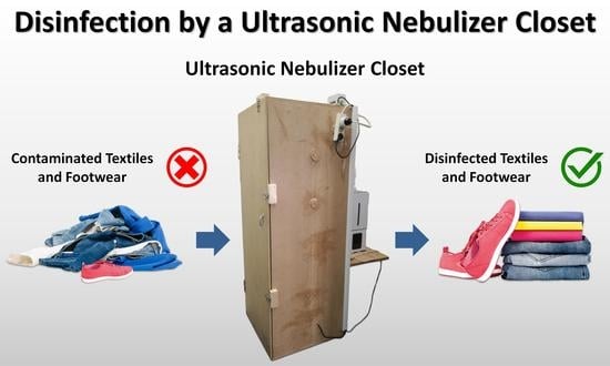
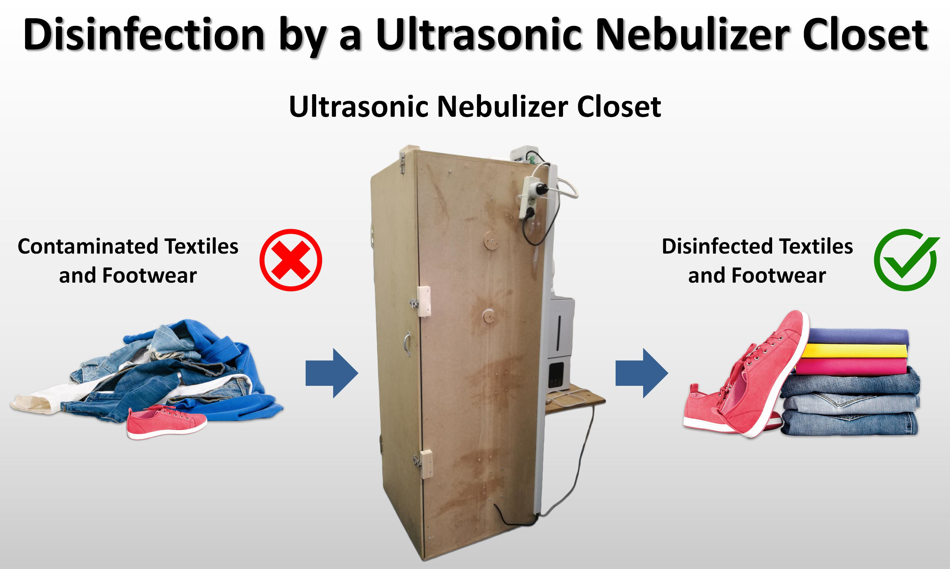
Application of an Ultrasonic Nebulizer Closet in the Disinfection of Textiles and Footwear

1
UCCCB—University of Coimbra Bacteria Culture Collection, Department of Life Science, University of Coimbra, 3000-456 Coimbra, Portugal
2
IATV—Instituto do Ambiente Tecnologia e Vida, 3030-790 Coimbra, Portugal
3
University of Coimbra, Centre for Mechanical Engineering, Materials and Processes, Department of Life Sciences, 3000-456 Coimbra, Portugal
*
Author to whom correspondence should be addressed.
Int. J. Environ. Res. Public Health 2022, 19(17), 10472; https://doi.org/10.3390/ijerph191710472
Submission received: 5 July 2022 / Revised: 17 August 2022 / Accepted: 18 August 2022 / Published: 23 August 2022
(This article belongs to the Section Environmental Health)

Abstract

:
The emergence of the coronavirus disease 2019 (COVID-19) pandemic highlighted the importance of disinfection processes in health safety. Textiles and footwear have been identified as vectors for spreading infections. Therefore, their disinfection can be crucial to controlling pathogens’ dissemination. The present work aimed to evaluate the effectiveness of a commercial disinfectant aerosolized by an ultrasonic nebulizer closet as an effective method for disinfecting textiles and footwear. The disinfection was evaluated in three steps: suspension tests; nebulization in a 0.08 m3 closet; nebulization in the upscaled 0.58 m3 closet. The disinfection process of textiles and footwear was followed by the use of bacteriophages, bacterial spores, and bacterial cells. The disinfection in the 0.58 m3 closet was efficient for textiles (4 log reduction) when bacteriophage Lambda, Pseudomonas aeruginosa, and Bacillus subtilis were used. The footwear disinfection was achieved (4 log reduction) in the 0.08 m3 closet for Escherichia coli and Staphylococcus aureus. Disinfection in an ultrasonic nebulization closet has advantages such as being quick, not wetting, being efficient on porous surfaces, and is performed at room temperature. Ultrasonic nebulization disinfection in a closet proves to be useful in clothing and footwear stores to prevent pathogen transmission by the items’ widespread handling.

Graphical Abstract

1. Introduction

The emergence of the coronavirus disease 2019 (COVID-19) pandemic caused by the severe acute respiratory syndrome coronavirus 2 (SARS-CoV-2) highlighted the importance of disinfection processes in health safety. Moreover, the risk of the emergence of new variants of SARS-CoV-2 and new pandemics [1], along with the growing problem of multidrug-resistant microorganisms [2], makes the development of new and better disinfection methods necessary.
Manually executed disinfection processes are operator-dependent and prone to failure [3]. Furthermore, many of the contaminated areas are not accessible by conventional manual disinfection methods. On the other hand, automatic disinfection using ultraviolet radiation is not efficient on porous surfaces [4]. Thus, ultrasonic disinfectant nebulization is an automated, easy-to-use alternative that, owing to the small size of the aerosol particles generated, has a high penetration into contaminated areas with difficult access [5]. Moreover, ultrasonic nebulization has advantages over conventional mechanical nebulization methods, such as pressure- or gas-assisted systems. Firstly, the disinfectant aerosol droplets generated by the ultrasonic system are smaller in size. Second, as the ultrasonic system operates at ambient pressure and does not need high speed to produce the nebulization, the ultrasonic generation of the aerosol does not require as much direct mechanical energy as conventional nebulization systems [6].
Textiles, such as clothes or household linens, and footwear, such as shoes or slippers, have been identified by several studies as vectors for spreading infections [7,8,9,10,11]. Indeed, many microorganisms are transferred to textiles and footwear through contact with skin and body excretions. On the other hand, microorganisms can also be transferred from the environment through textiles and footwear contact with, for example, dust, soil, furniture, or food [7,9,10]. Most microorganisms present in textiles and footwear do not pose a health risk since many are part of skin microbiota [7]. However, the contact of textiles and footwear with an infected person or a contaminated environment causes these materials to carry pathogens, thus making them act as fomites and pose a risk to public health [7,8,9,10,11]. Effectively, in healthcare facilities such as hospitals, pathogens are found on hospital linens and on the clothing of both patients and healthcare professionals [7,8]. In the context of the COVID-19 pandemic, Aumeran et al. [12] detected the presence of SARS-CoV-2 on the gowns of healthcare workers who treated COVID-19 patients, and Redmond et al. [13] detected the presence of SARS-CoV-2 on personnel shoes of a COVID-19 ward. Furthermore, pathogens can remain viable in textiles and footwear for long periods of time [14,15,16,17,18,19,20,21,22]. At room temperature, Owen et al. [15] showed that HCoV-OC43 remains infectious on polyester, cotton, and polycotton for at least 72 h, 24 h, and 6 h, respectively, and HCoV-229E remains infectious on polyester, cotton, and polycotton for at least 24 h, 2 h, and 2 h, respectively. For their part, Shivkumar et al. [16] detected HCoV-OC43 for 6 h, 24 h, and 48 h on patent leather, finished leather, and calf leather, respectively. In another study, Harbourt et al. [18] investigated the stability of SARS-CoV-2 in clothing at different temperatures and 40–50% relative humidity. Despite not detecting viable viruses at 37 °C after initial exposure, the authors found that at 4 °C the virus remained viable for at least 96 h and that at 22 °C the virus remained viable for at least 4 h. In turn, Chin et al. [19] investigated the stability of SARS-CoV-2 on cloth and surgical masks at room temperature with a relative humidity of around 65%. These authors found infectious viruses being detectable on the cloth after 1 day, on the inner mask layer after 4 days, and on the outer mask layer after 7 days [19]. For their part, Fijan et al. [20] found that Enterococcus faecium, Staphylococcus aureus, and Pseudomonas aeruginosa survive several days on cotton textile swatches at 25 °C. Riley et al. [21] also found that Escherichia coli and S. aureus survive several days in cotton and polyester textile swatches at 23 °C and 47% relative humidity. In another study, Hanczvikkel and Tóth [22] found several species of multidrug-resistant bacteria that survive several days on a cotton towel at 25 °C and 52% relative humidity. Therefore, laundering is usually employed in textiles and footwear, not only to clean but also to disinfect [7,8,9,10,11,23,24,25]. Nevertheless, the washing machine itself can be a source of contamination for textiles and footwear [7,10,26]. In this case, contamination can occur due to biofilm formation inside the washing machine, which can work as a reservoir of pathogens, or cross-contamination between materials, which can occur when a contaminated item is put in the washing machine along with uncontaminated laundry items [7]. In addition to possibly contaminating clothes and footwear, laundering is also a time-consuming process that requires drying. Therefore, laundering is not a practical process for disinfecting clothes and footwear in contexts where rapid disinfection without wetting is required. One such context is the disinfection of clothing and footwear in stores after customer handling.
Some studies have investigated the disinfection of textiles and footwear through coarse spray disinfectant application [27,28,29,30,31,32,33,34,35,36]. However, few studies have explored the disinfection of textiles and footwear through fine aerosol disinfectant application inside a disinfection chamber. Recently, as part of fighting the spread of COVID-19, Abu-Zidan et al. [37] proposed a prefabricated portable chamber that sprays individuals with fine mist sanitizing fluid to disinfect the clothing and exposed surfaces of people. For their part, Khan et al. [38] studied a solar-powered disinfection tunnel mist spray to disinfect individuals’ clothing in large gatherings. Previously, in order to disinfect various surfaces, including synthetic-fiber carpets and cotton fabric, Callahan et al. [39] developed a chamber for the application of a nebulized disinfectant. In addition, some patents also describe apparatus for ultrasonic nebulization of disinfectants [40,41,42,43,44,45]. Nevertheless, to the best of our knowledge, the use of an ultrasonic nebulizer closet for the disinfection of textiles and footwear has never been studied. Therefore, the present work aimed to evaluate the effectiveness of a commercial disinfectant aerosolized by an ultrasonic nebulizer closet as an effective method to quickly disinfect textiles and footwear after customers handle it in stores. The disinfection process was followed by the use of Bacillus atrophaeus DSM 2277 spores as an indicator of chemical sterilization efficiency, bacteriophage Escherichia virus Lambda DSM 4499 as an indicator of virucidal activity, and vegetative cells of E. coli DSM 30083, P. aeruginosa DSM 1117, S. aureus DSM 20231, and Bacillus subtilis DSM 10 as indicators of bactericidal activity.

2. Materials and Methods

2.1. Microbiological Indicators Production and Preservation

B. atrophaeus DSM 2277, bacteriophage Lambda DSM 4499, E. coli DSM 4230 (bacteriophage Lambda host), E. coli DSM 30083, P. aeruginosa DSM 1117, S. aureus DSM 20231, and B. subtilis DSM 10 were obtained from the Leibniz Institute DSMZ-German Collection of Microorganisms and Cell Cultures GmbH, Braunschweig, Germany. Bacteria were grown on agar medium plates according to the incubation conditions shown in Table 1 and preserved at −80 °C in nutrient broth (NB) medium supplemented with 15% (v/v) glycerol.
For bacteriophage Lambda DSM 4499 multiplication, the host bacterium E. coli DSM 4230 was plated on Luria–Bertani (LB) agar medium through the top agar layer method. For that, 100 μL of host bacterium E. coli DSM 4230 culture in 0.85% (w/v) NaCl suspension with 0.3 optical density (OD) at 600 nm was mixed with 5 mL of LB soft agar medium (0.75% (w/v) agar) at 50 °C and plated over LB agar medium. A filter paper containing the dried bacteriophage suspension was placed in the center of the host plate, 100 μL of LB broth medium was added to the surface, and the plate was incubated overnight under the conditions of the host bacterium (37 °C). After incubation, a halo was visible around the filter paper, which resulted from the lysis of the host cells. To prepare a bacteriophage stock suspension, 5 mL of LB broth medium was added to the plate and then placed on a slowly rotating shaker (GFL® 3005, GFL Gesellschaft für Labortechnik mbH, Burgwedel, Germany) at room temperature for at least 4 h. After this period, the bacteriophage suspension was centrifuged at 5000× g for 20 min at 4 °C (VWR® MicroStar 17R, VWR International BV, Leuven, Belgium). The resulting supernatant was filtrated with a sterile cellulose acetate syringe filter of 0.45 μm porous size (VWR International LLC., Radnor, PA, USA) to remove the remaining bacteria, and the filtrate (bacteriophage stock suspension) was stored at 4 °C.
To obtain spores of B. atrophaeus DSM 2277, cells were inoculated in a 300 mL Erlenmeyer flask containing 120 mL of NB medium and incubated at 130 rpm for 3 days at 30 °C in an orbital shaker (S200D, Comecta S.A., Barcelona, Spain). After growth, cells were harvested by centrifugation at 8801× g for 30 min at 4 °C and resuspended in 15 mL. The cell suspension was pasteurized at 85 °C for 15 min and placed on ice for more than 15 min. The suspension was passed through a high-pressure homogenizer (EmulsiFlex® C3, Avestin Inc., Ottawa, ON, Canada) (15,000–20,000 psi) for 1 min. The lysate was concentrated using Vivaspin® 6 centrifugal concentrator with a molecular weight cut-off of 100 kDa (Sartorius Stedim Biotech GmbH, Göttingen, Germany) at 3150 g for 30 min at 4 °C (Centrifuge 5810R, Eppendorf AG, Hamburg, Germany). This stock spore suspension was stored at 4 °C.
Inocula of B. atrophaeus DSM 2277 spores and bacteriophage Lambda DSM 4499 were obtained from the respective stock suspensions. Inocula of E. coli DSM 30083, P. aeruginosa DSM 1117, S. aureus DSM 20231, and B. subtilis DSM 10 were obtained from vegetative cell suspensions prepared in 5 mL sterile 0.85% (w/v) NaCl. The inoculum turbidity of the microbiological indicator suspensions was adjusted to five on the McFarland scale.

2.2. Disinfectant and Ultrasonic Nebulizer Closets

VIRCOV BAC 360 (Inokem S.A., Forte da Casa, Portugal) is a disinfectant formed by three biocidal active substances: benzalkonium chloride (BAC), which is a quaternary ammonium compound, glycolic acid, and ethanol. All these three substances are included in Article 95 List of European Chemicals Agency (ECHA) for industrial disinfection [46]. Additionally, VIRCOV BAC 360 also contains tetrasodium glutamate diacetate, which acts as a chelating and wetting agent.
Nebulization disinfection tests were carried out in a 0.08 m3 closet of 605 mm (height) × 420 mm (width) × 325 mm (length) (Supplementary Materials Figure S1) and in a 0.58 m3 closet of 1600 mm (height) × 600 mm (width) × 600 mm (length) (Supplementary Materials Figure S2). The 0.58 m3 closet was equipped with a dehumidifier, a heater, and an exhaust fan with activated carbon filters. During the nebulization disinfection tests, the dehumidifier was off, the temperature was between 35 °C and 40 °C, and the exhaust fan was only turned on in the final stage of disinfection tests to remove the aerosol inside the closet through the activated carbon filters. This 0.58 m3 closet consisted of a closet scale up and was a prototype of NovirBox from Dynasys—Engenharia e Telecomunicações, S.A., Setúbal, Portugal.
In both closets, nebulization of the disinfectant solution was carried out by a piezoelectric ultrasonic nebulizer. Briefly, the nebulizer had three piezoelectric transducers at the bottom of the disinfectant solution container that converted electrical energy into high-frequency mechanical vibrations. These vibrations caused the liquid above the piezoelectric transducers to be rarefied and compressed at a high-frequency cycle, which led to the formation of a micron-size aerosol from the disinfectant solution [6]. The aerosol formed was conducted into the disinfection closet by a forced draft fan installed at the top of the disinfectant solution container. All nebulization disinfection tests were performed at a nebulization flow rate of 10.4 ± 1.4 mL·min−1.

2.3. Experimental Design

The disinfection was evaluated in three steps: (1) suspension tests; (2) nebulization tests in a 0.08 m3 closet; (3) nebulization tests in the upscaled 0.58 m3 closet.
Evaluation of VIRCOV BAC 360 sporicidal and virucidal activity was performed by suspension test, in a 48-well microplate, at 100% concentration and serial 2-fold dilutions in sterile deionized water up to 1/16 (v/v) dilution. The control was performed with sterile deionized water without disinfectant. B. atrophaeus DSM 2277 spores and bacteriophage Lambda DSM 4499 were used as indicators with a contact time of 2 min. The suspension tests were carried out in 100 μL of the respective disinfectant dilution inoculated with 5 μL of the indicator suspension.
To evaluate nebulization disinfection effectiveness in both closets, fabric swatches (ca. 6 cm2) composed of 88% polyester and 12% elastane (Supplementary Materials Figure S3) were inoculated with 30 μL of each indicator suspension.
In the 0.08 m3 closet, the fabric swatches were placed at the bottom of the chamber and subjected to 2 min disinfectant nebulization. Afterward, the fabric swatches were removed 5 min after stopping the nebulization and analyzed. The disinfectant dilutions of 1/2 (v/v) and 1/3 (v/v) were used to evaluate the elimination of B. atrophaeus DSM 2277 spores and bacteriophage Lambda DSM 4499. In the 0.08 m3 closet, the disinfection of a folded cotton towel of 460 × 720 mm and of a footwear item was also tested. For that, fabric swatches (ca. 6 cm2) inoculated with 30 μL of each indicator suspension were placed inside the folded towel (Supplementary Materials Figure S4) and glass slides (24 × 50 mm) were placed inside the footwear item and then also inoculated with 30 μL of each indicator suspension. In these experiments, the disinfectant dilution of 1/3 (v/v) was used to evaluate the elimination of B. atrophaeus DSM 2277 spores, bacteriophage Lambda DSM 4499, E. coli DSM 30083, and S. aureus DSM 20231. The folded towel and the footwear item were also subjected to 2 min disinfectant nebulization. Five minutes after stopping the nebulization, the fabric swatches and the glass slides were taken and utilized to evaluate the presence of the indicators.
To evaluate the nebulization disinfection effectiveness in the upscaled 0.58 m3 closet, the disinfectant was used at 1/3 (v/v) dilution and the indicators were B. atrophaeus DSM 2277 spores, bacteriophage Lambda DSM 4499, E. coli DSM 30083, P. aeruginosa DSM 1117, S. aureus DSM 20231, and B. subtilis DSM 10. The fabric swatches were placed in the middle of the chamber over a wire mesh. In this closet, a short disinfection cycle and a long disinfection cycle were tested. The short disinfection cycle consisted of 2 min of nebulization, 2 min of rest without nebulization, and 2 min of aerosol extraction from the closet. The long disinfection cycle consisted of 4 min of nebulization, 4 min of rest without nebulization, and 2 min of aerosol extraction from the closet.

2.4. Determination of Cell Survival

In suspension disinfection experiments, after the 2 min contact time, serial 10-fold dilutions in sterile 0.85% (w/v) NaCl were performed and 100 μL were spread on agar plates for colony-forming unit (CFU) enumeration, or in the case of bacteriophages, plaque-forming units (PFU). The plates were incubated according to the conditions shown in Table 1.
In nebulization disinfection experiments, the materials were placed inside the closet, the chamber was closed, and the disinfection cycle proceeded. Immediately after the disinfection cycle was completed, the fabric swatches or the glass slides were placed into 50 mL tubes with 5 mL of sterile 0.85% (w/v) NaCl, shaken manually, and allowed to incubate for 1 h at room temperature. Serial 10-fold dilutions were made and 100 μL plated for CFU or PFU quantification. The plates were incubated according to the conditions shown in Table 1.
PFU quantification was performed using the 100 μL of the 10-fold serial dilutions mixed with 100 μL of host suspension (0.3 OD600nm in 0.85% (w/v) NaCl) E. coli DSM 4230. After blending the 10-fold serial dilution with the host bacteria, the 200 μL was plated through the top agar layer method, i.e., the 200 μL blend was mixed with 5 mL of LB soft agar medium (0.75% (w/v) agar) at 50 °C and immediately poured over an LB agar medium plate. Plates were incubated at 37 °C for 24 h. Bacteriophage plaques were counted and calculated to PFU·mL−1.
In the nebulization experiments, the control tests were performed by transferring the 30 μL of indicator suspension directly into the 50 mL tubes with 5 mL of sterile 0.85% (w/v) NaCl.

2.5. Data Analysis

In all experiments, the tests with disinfectant were compared to the control tests. The results of the comparison were expressed as log10 reduction and elimination rate (%). The log10 reduction was calculated through Equation (1):
log10 reduction = log10(N0) − log10(N1)
where N0 is the mean of the CFU·mL−1 or PFU·mL−1 replicate values of the control test in a given experiment, and N1 is the CFU·mL−1 or PFU·mL−1 value of one of the replicates of the disinfection test of a given experiment. For its part, the elimination rate was calculated through Equation (2):
Elimination   rate   ( % ) = N 0   N 1 N 0   ×   100
Results were presented as mean ± standard deviation and the number of replicates performed in each test is indicated in the respective figure caption.
Statistical differences between groups were evaluated by the employment of one-way or two-way analysis of variance (ANOVA) where appropriate. After ANOVAs, post hoc comparisons were made by applying the Tukey test. Differences were considered statistically significant when the associated p-values were lower than 0.05. The caption of each figure indicates which statistical test was used. Statistical analysis was performed by use of GraphPad Prism 9 for Windows 64-bit, version 9.3.1 (GraphPad Software Inc., San Diego, CA, USA).

2.6. Criteria for Disinfection Acceptance

The present study follows the disinfection acceptance criteria presented by ECHA in Guidance on the Biocidal Products Regulation: Volume II Efficacy—Assessment and Evaluation (Parts B + C) [47]. This guide Appendix 4 of [47] presents the available standards for testing the efficiency of biocides when applied in the disinfection of textiles.These standards include suspension tests and carrier tests Appendix 4 of [47]. In suspension tests, the standards EN 13727 (medical applications) [48] and EN 1276 (non-medical applications) [49] are identified for bacterial disinfection, and the standard EN 14476 (medical applications) [50] is identified for virus disinfection. Both EN 13727 and EN 1276 require 5 log10 reduction of vegetative bacteria as a pass criterion [47]. For its part, EN 14476 only requires 4 log10 reduction of viruses as a pass criterion [47]. Likewise, the guideline of the German Association for the Control of Virus Diseases (DVV) and the Robert Koch Institute (RKI) also requires 4 log10 reduction of viruses as a pass criterion for suspension tests [51]. In carrier tests, Appendix 4 of the ECHA’s guidance [47] identifies the standards EN 16616 [52], ASTM E2406 [53], and ASTM E2274 [54] for both bacterial and virus disinfection. For vegetative bacteria, the standards EN 16616, ASTM E2406, and ASTM E2274 require, respectively, 7, 4, and 4 log10 reduction as pass criteria [47]. Nevertheless, for viruses, these three standards do not define any pass criterion [47]. Furthermore, none of the standards for textile disinfection mentioned in Appendix 4 of the ECHA’s guidance provide criteria for bacterial spore disinfection [47]. Therefore, in the criteria for disinfection acceptance of the present study, the EN 13704 standard [55] was also considered because this standard provides a quantitative suspension test for the evaluation of the bacterial sporicidal activity of chemical disinfectants used in food, industrial, domestic, and institutional sectors [56]. As a pass criterion, EN 13704 requires a 3 log10 reduction of bacterial spores [56].
In the present study, in addition to the criteria mentioned above, the disinfection acceptance criteria presented in the U.S. Pharmacopeia [57] were also considered. According to the U.S. Pharmacopeia, a coupon surface disinfection process (no materials specified) is considered effective if it allows a log10 reduction of at least 2 for bacterial spores and 3 for vegetative bacterial cells [57]. However, for viruses, the U.S. Pharmacopeia does not clarify the log10 reduction value required for the disinfectant to be considered effective [57].

3. Results

3.1. Disinfection by Suspension Test

Disinfection of B. atrophaeus DSM 2277 spores with VIRCOV BAC 360 by suspension test is shown in Figure 1. With disinfectant at both 100% concentration and 1/2 (v/v) dilution, the B. atrophaeus DSM 2277 spores log10 reduction and elimination rate were, respectively, 9.90 ± 0 and 100% (Figure 1). Further disinfectant dilutions (1/4 (v/v), 1/8 (v/v), and 1/16 (v/v) dilution) significantly reduced the number of viable spores (p-value < 0.0001) (Supplementary Materials Figure S5), reaching a log10 reduction of 2.27 ± 0.01, 2.00 ± 0.05, and 0.91 ± 0 for 1/4 (v/v), 1/8 (v/v), and 1/16 (v/v) dilutions, respectively (Figure 1). These log10 reductions corresponded to elimination rates of, respectively, 99.5%, 99.0%, and 87.6%. Minimum bactericidal concentration (MBC) is defined as the minimum concentration of an antimicrobial agent required to eradicate 99.9% of the microorganism isolates under testing by culturing in an antimicrobial agent-free medium [58,59]. Therefore, in accordance with the elimination rates obtained for the various dilutions, the MBC of VIRCOV BAC 360 for B. atrophaeus DSM 2277 spores with a contact time of 2 min is 50% of its concentration. For bacteriophage Lambda DSM 4499, the elimination rates for the various dilutions of VIRCOV BAC 360 were not possible to obtain because the host E. coli DSM 4230 was sensitive to the residual disinfectant carried in the bacteriophage suspension.

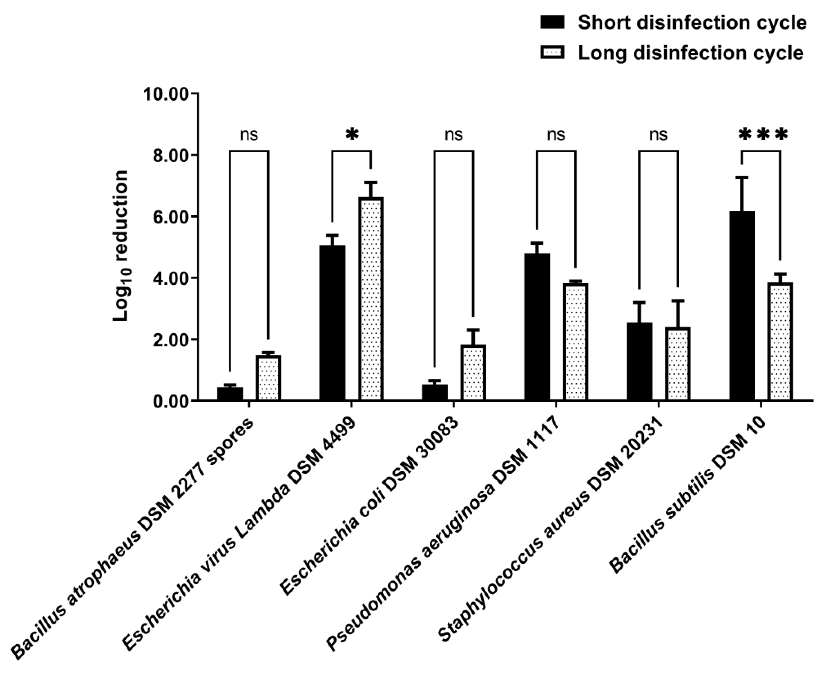
3.2. Disinfection by Nebulization in a 0.08 m3 Closet

Disinfection by nebulization of fabric swatches, a folded cotton towel, and a footwear item was tested in a 0.08 m3 closet.
Fabric swatch disinfection was tested with nebulization of VIRCOV BAC 360 at 1/2 (v/v) dilution, which corresponds to the MBC found by the suspension test, and 1/3 (v/v) dilution, which, according to the disinfectant manufacturer, is a dilution that should continue to be effective. Both disinfectant dilutions allowed a significant reduction in the number of viable spores and bacteriophages (p-value < 0.0001) (Supplementary Materials Figure S6). Fabric swatches nebulization with the 1/2 (v/v) disinfectant dilution resulted in a log10 reduction of 3.75 ± 0.16 (99.98% elimination rate) and 3.94 ± 0.01 (99.989% elimination rate) for B. atrophaeus DSM 2277 spores and bacteriophage Lambda DSM 4499, respectively (Figure 2). For its part, nebulization with the 1/3 (v/v) disinfectant dilution resulted in a log10 reduction of 2.20 ± 0.09 (99.4% elimination rate) and 3.28 ± 0.01 (99.95% elimination rate) for B. atrophaeus DSM 2277 spores and bacteriophage Lambda DSM 4499, respectively (Figure 2). The 1/3 (v/v) disinfectant dilution yielded a significantly lower log10 reduction than the 1/2 (v/v) disinfectant dilution for both spores (p-value < 0.0001) and bacteriophages (p-value < 0.001) (Figure 2). In the spore tests, the log10 reduction with the 1/3 (v/v) disinfectant dilution was 1.55 log10 lower than that with the 1/2 (v/v) disinfectant dilution, and, in the bacteriophage tests, the log10 reduction with the 1/3 (v/v) disinfectant dilution was 0.66 log10 lower than that with the 1/2 (v/v) disinfectant dilution.
For the folded cotton towel disinfection, inoculated fabric swatches were placed inside the folded towel (Supplementary Materials Figure S4), and then the towel was disinfected by nebulization of disinfectant at 1/3 (v/v) dilution. The disinfection by nebulization of the folded cotton towel significantly reduced the number of viable Lambda DSM 4499 bacteriophages (p-value < 0.0001) and S. aureus DSM 20231 cells (p-value < 0.0001) (Supplementary Materials Figure S7). The log10 reduction of bacteriophage Lambda DSM 4499 and S. aureus DSM 20231 was 5.38 ± 1.31 (99.998% elimination rate) and 6.30 ± 0.001 (100% elimination rate), respectively (Figure 3). However, for B. atrophaeus DSM 2277 spores and E. coli DSM 30083, disinfection by nebulization did not significantly reduce the number of viable spores (p-value > 0.05) and cells (p-value > 0.05) (Supplementary Materials Figure S7).
The disinfection by nebulization of the footwear item significantly reduced the number of viable E. coli DSM 30083 (p-value < 0.0001) and S. aureus DSM 20231 cells (p-value < 0.0001) (Supplementary Materials Figure S8). The log10 reductions of E. coli DSM 30083 and S. aureus DSM 20231 were 5.91 ± 2.55 (99.96% elimination rate) and 6.30 ± 0.001 (100% elimination rate), respectively (Figure 4). However, for B. atrophaeus DSM 2277 spores and bacteriophage Lambda DSM 4499, disinfection by nebulization in the 0.08 m3 closet did not significantly reduce their numbers (p-value > 0.05 for both) (Supplementary Materials Figure S8).

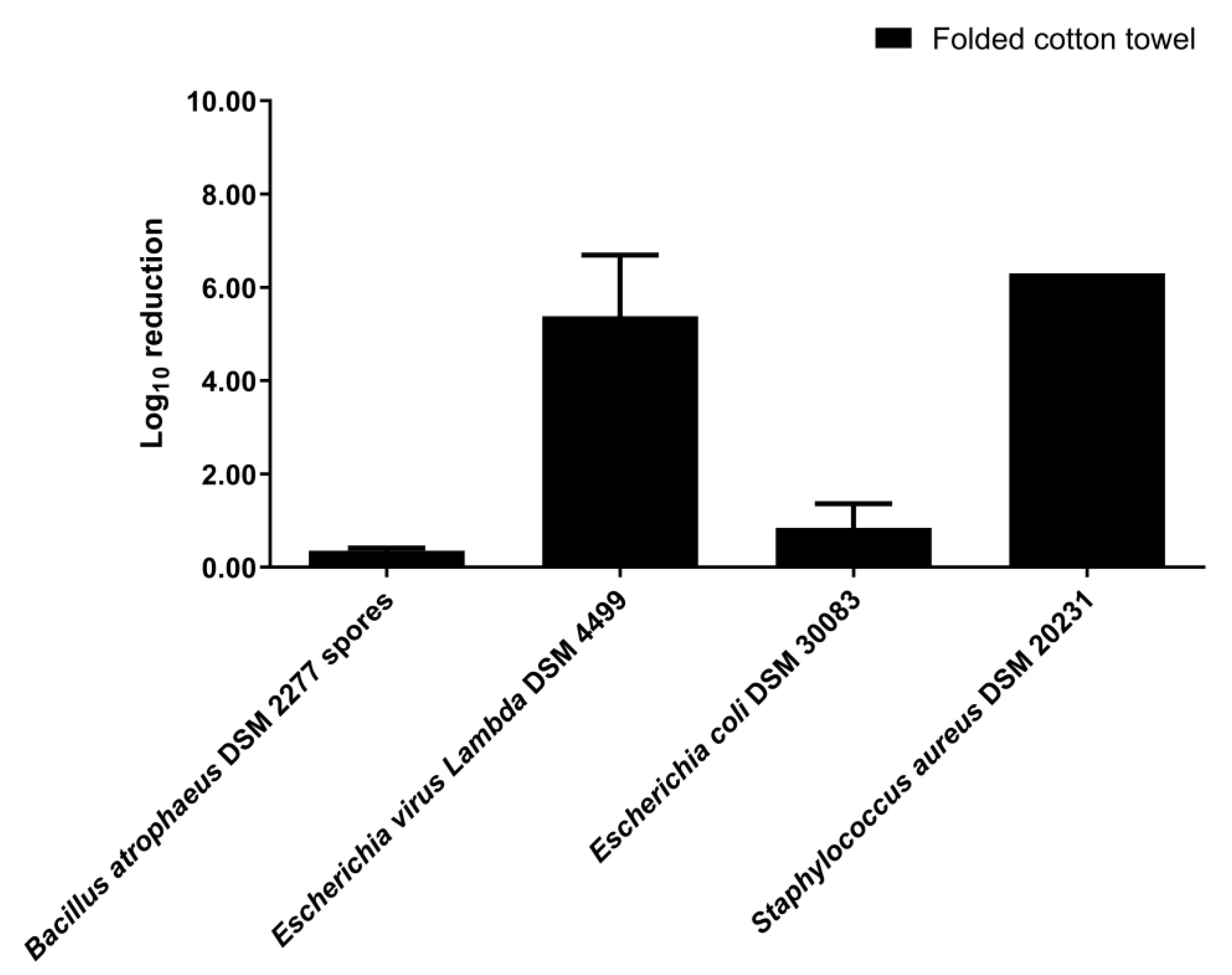
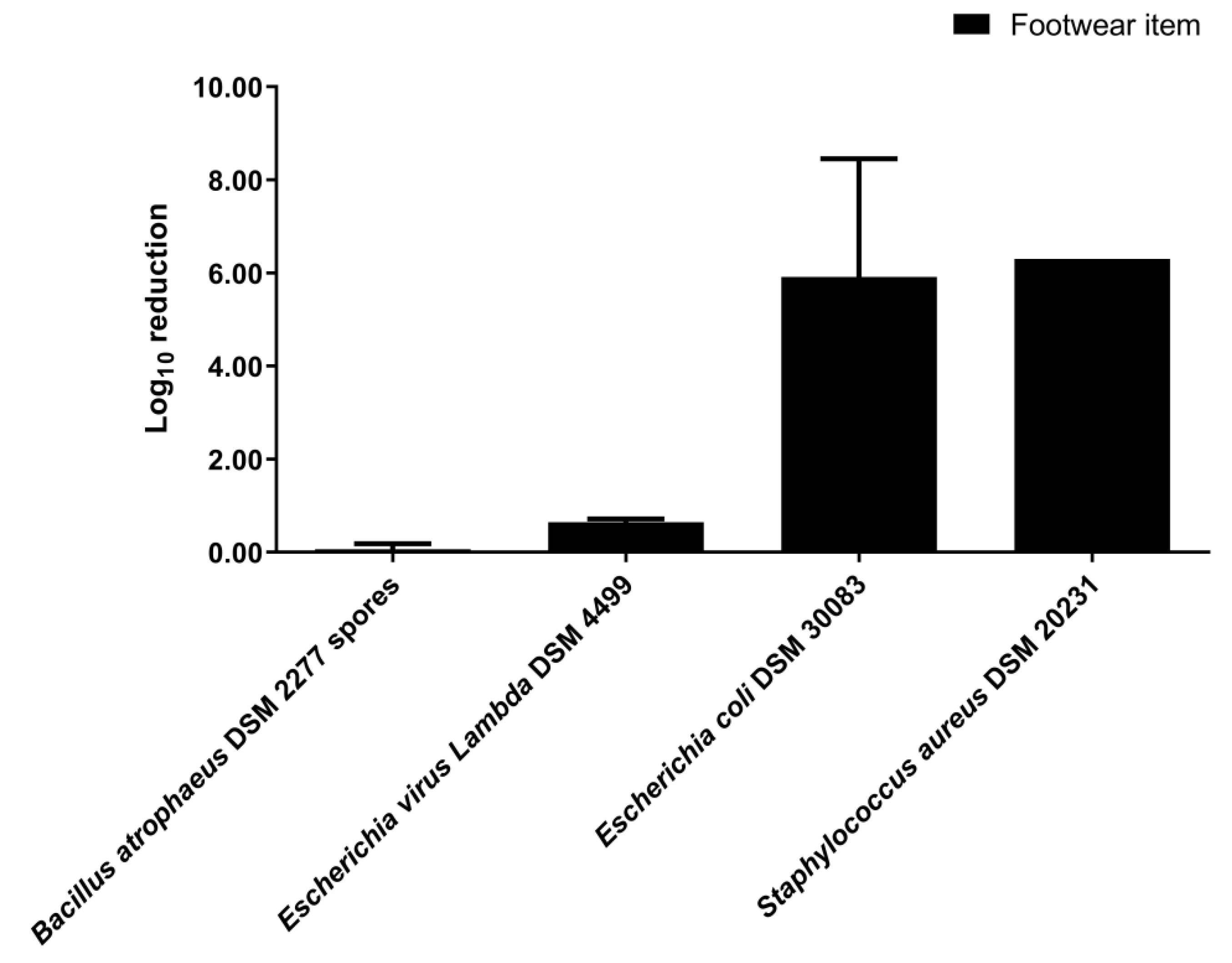
3.3. Disinfection by Nebulization in a 0.58 m3 Closet

Disinfection by nebulization of fabric swatches was also tested in the upscaled 0.58 m3 closet with a short disinfection cycle and a long disinfection cycle. The use of a short disinfection cycle significantly reduced the number of viable Lambda DSM 4499 bacteriophages (p-value < 0.0001) and the number of viable P. aeruginosa DSM 1117, S. aureus DSM 20231, and B. subtilis DSM 10 cells (p-value < 0.0001) inoculated in the fabric swatches (Supplementary Materials Figure S9). However, the short disinfection cycle did not significantly reduce the number of viable B. atrophaeus DSM 2277 spores (p-value > 0.05) and E. coli DSM 30083 cells (p-value > 0.05) (Supplementary Materials Figure S9). In the short disinfection cycle experiments, the log10 reductions of bacteriophage Lambda DSM 4499, P. aeruginosa DSM 1117, S. aureus DSM 20231, and B. subtilis DSM 10 were 5.07 ± 0.31 (99.999% elimination rate), 4.80 ± 0.33 (99.999% elimination rate), 2.54 ± 0.66 (99.4% elimination rate), and 6.17 ± 1.09 (99.9997% elimination rate), respectively (Figure 5). For its part, the use of a long disinfection cycle significantly reduced the number of viable microorganisms inoculated in the fabric swatches for all indicators tested (p-value < 0.01) (Supplementary Materials Figure S10). In the long disinfection cycle experiments, the log10 reductions of B. atrophaeus DSM 2277 spores, bacteriophage Lambda DSM 4499, E. coli DSM 30083, P. aeruginosa DSM 1117, S. aureus DSM 20231, and B. subtilis DSM 10 were 1.48 ± 0.09 (96.7% elimination rate), 6.63 ± 0.48 (99.99997% elimination rate), 1.83 ± 0.47 (98.2% elimination rate), 3.83 ± 0.07 (99.99% elimination rate), 2.40 ± 0.86 (99.4% elimination rate), and 3.85 ± 0.28 (99.98% elimination rate), respectively (Figure 5). For the indicators B. atrophaeus DSM 2277 spores, E. coli DSM 30083, P. aeruginosa DSM 1117, and S. aureus DSM 20231, the log10 reductions of the experiments with a short disinfection cycle was not significantly different from the log10 reduction of the experiments with a long disinfection cycle (p-value > 0.05) (Figure 5). However, for bacteriophage Lambda DSM 4499, the log10 reduction of the long disinfection cycle was significantly greater than the log10 reduction of the short disinfection cycle (p-value < 0.05), with the log10 reduction of the long disinfection cycle being 1.56 log10 greater than the log10 reduction of the short disinfection cycle (Figure 5). On the other hand, for B. subtilis DSM 10, the result was the opposite (p-value < 0.001), with the log10 reduction of the long disinfection cycle being 2.32 log10 lower than the log10 reduction of the short disinfection cycle (Figure 5).

4. Discussion

Pathogens can lodge on clothing and footwear, and lead to the spread of infections, putting public health at risk [7,8,9,10,11]. Clothing and footwear are commonly disinfected by laundering [7,8,9,10,11,23,24,25]. However, laundering is not practicable in stores where materials are shared between customers. Therefore, the availability of an automatic, fast, and effective disinfection system is relevant.
In the present study, the disinfection of textiles and footwear in an ultrasonic nebulizer closet was evaluated by the use of several indicators. B. atrophaeus spores are commonly used as indicators of sterilization and biocidal activity of chemical agents [60], and here they were used as an indicator of chemical sterilization efficiency. In the context of the COVID-19 pandemic, the virucidal capacity of disinfection systems has a further interest. SARS-CoV-2 is reported in the literature as being sensitive to the biocidal active substances present in VIRCOV BAC 360 [61,62,63,64,65,66,67,68,69]. Since the bacteriophage Lambda DSM 4499, used in the present study as a virucidal indicator, is non-enveloped [70], it is considered less susceptible to disinfection with this disinfectant than enveloped SARS-CoV-2 [71,72,73]. Therefore, the bacteriophage elimination suggests a possible action of VIRCOV BAC 360 on SARS-CoV-2. However, as pointed out by Nims and Zhou [74], caution should be taken when viral inactivation susceptibilities are extrapolated from one virus to another. In the present study, several species of bacteria were also used as indicators of bactericidal activity. Thus, vegetative cells of E. coli DSM 30083 and P. aeruginosa DSM 1117 were used as indicators of Gram-negative bactericidal activity, and vegetative cells of S. aureus DSM 20231 and B. subtilis DSM 10 were used as indicators of Gram-positive bactericidal activity.
In the present study, the disinfectant VIRCOV BAC 360 in the suspension tests with B. atrophaeus DSM 2277 spores exceeded the 5 log10 reduction required by EN 13727 and EN 1276 standards [47] when applied at 100% concentration and at 1/2 dilution, but not to other dilutions. Nonetheless, it should be noted that these standards refer to the elimination of vegetative bacteria [47], not bacterial spores, as was the case of the suspension tests of the present study. Bacterial spores are substantially more resistant to disinfection than vegetative bacterial cells [73,75]. Thus, here, the EN 13704 standard [55], which provides a quantitative suspension test for the evaluation of the bacterial sporicidal activity of chemical disinfectants used in food, industrial, domestic, and institutional sectors [56], was also considered. Following this standard, the disinfectant VIRCOV BAC 360 in the suspension test with B. atrophaeus DSM 2277 spores also reached the pass criterion (3 log10 reduction) when it was applied at 100% concentration and at 1/2 dilution.
The nebulization disinfection experiments of the present study did not reach the 7 log10 reduction of bacteria indicators required by the EN 16616 standard as a pass criterion [47]. However, according to ASTM E2406 and ASTM E2274 standards (4 log10 reduction as a pass criterion [47]), the disinfection process was validated for the following conditions and bacteria indicators: (1) folded cotton towel disinfection in the 0.08 m3 closet (S. aureus DSM 20231); (2) footwear item disinfection in the 0.08 m3 closet (E. coli DSM 30083 and S. aureus DSM 20231); (3) fabric disinfection with a short disinfection cycle in the 0.58 m3 closet (P. aeruginosa DSM 1117 and B. subtilis DSM 10). These three standards do not define pass criteria for viruses [47]. Therefore, considering the same criteria as the ones of the ASTM E2406 and ASTM E2274 standards adopted above for bacteria indicators [47], the disinfection process was effective for the bacteriophage in all nebulization experiments, except for the fabric and the footwear item experiments in the 0.08 m3 closet. The EN 16616 [52], ASTM E2406 [53], and ASTM E2274 [54] standards are directed to processes conducted in washing machines [7]. Hence, the fact of the nebulization experiments of the present study have reached the pass criteria of ASTM E2406 and ASTM E2274 standards [47] is remarkable and shows that disinfection of clothing and footwear in an ultrasonic nebulization closet can be as effective as in a washing machine.
In view of the U.S. Pharmacopeia pass criteria (2 log10 reduction for bacterial spores and 3 log10 reduction for vegetative bacteria) [57], the disinfection process was effective for the following conditions and indicators: (1) fabric disinfection with the 0.08 m3 closet using both 1/2 (v/v) and 1/3 (v/v) disinfectant dilution (B. atrophaeus DSM 2277 spores); (2) folded cotton towel disinfection with the 0.08 m3 closet (S. aureus DSM 20231); (3) footwear item disinfection with the 0.08 m3 closet (E. coli DSM 30083 and S. aureus DSM 20231); (4) fabric disinfection with both short and long disinfection cycle in the 0.58 m3 closet (P. aeruginosa DSM 1117 and B. subtilis DSM 10). Therefore, according to the U.S. Pharmacopeia [57], the disinfection of clothing and footwear in an ultrasonic nebulization closet is attained.
As noted by Zonta et al. [76] and Callahan et al. [39], the comparison of disinfection results between different studies is difficult because of differences between disinfectants, carrier materials, application methods and conditions, and indicator strains. Nevertheless, as in the present study, Callahan et al. [39] also developed a disinfection chamber for the application of a nebulized disinfectant. The log10 reductions obtained in the 0.58 m3 closet for the disinfection of S. aureus DSM 20231 on fabric are in line with the log10 reductions obtained by Callahan et al. [39] for methicillin-resistant S. aureus (MRSA) on both carpet and fabric. Furthermore, the log10 reductions obtained in the 0.58 m3 closet for the disinfection of B. subtilis DSM 10 were higher than those obtained by these authors for the Gram-positive indicators (MRSA and vancomycin-resistant Enterococci) used on both carpet and fabric. Additionally, in the 0.58 m3 closet, the 6 min disinfection time of the short disinfection cycle experiment was considerably shorter than the 1 h disinfection time of the system used by Callahan et al. [39]. Therefore, the present system can be applied for the quick disinfection of clothing and footwear in stores after customer handling.
Clothing and footwear disinfection in an ultrasonic nebulization closet such as the 0.58 m3 closet presents advantages, such as quick disinfection without wetting the materials; application of disinfectant in the aerosol form, which allows the disinfectant to penetrate porous surfaces, such as clothing; reaching all parts of an irregular surface, such as shoes; disinfection of clothes and footwear that cannot withstand high temperatures or cannot be washed, and it can be performed at room temperature. Moreover, during operation, the 0.58 m3 closet can be operated in the presence of people since it is completely sealed, and, at the end of the disinfection cycle, the aerosol present inside the closet is extracted through activated carbon filters.
The disinfectant used in the present study also has advantages, namely, it does not stain or damage clothes or footwear and the quaternary ammonium compound included in its formulation interacts with the surface of negatively charged textiles [7].
Some limitations to the disinfection assessment can be pointed out, such as the non-use of human viruses, the non-use of unclean materials, and the non-use of a neutralizer to inactivate the residual disinfectant. Nevertheless, in the nebulization experiments, the effect of the residual disinfectant was not expected because the indicators were removed from the closet after disinfection, being no longer in contact with the aerosol. Additionally, in the nebulization experiments, the indicators were placed immediately into sterile 0.85% (w/v) NaCl after disinfection, which diluted the residual disinfectant.

5. Conclusions

Finding an efficient method for the quick disinfection of textiles and footwear can be decisive for controlling the spread of infections. The disinfection of textiles and footwear by disinfectant aerosolization in an ultrasonic nebulizer closet is an automated easy-to-use alternative. The results of the present study show that, in a 0.08 m3 closet, the ultrasonic nebulization of the disinfectant used in this work at a 1/3 dilution allowed efficient disinfection of fabric inoculated with B. atrophaeus DSM 2277 spores in 7 min. In the same 0.08 m3 closet and under the same conditions, the ultrasonic nebulization disinfection of a folded cotton towel was achieved for bacteriophage Lambda DSM 4499 and S. aureus DSM 20231. Similarly, the disinfection of a footwear item was also achieved for E. coli DSM 30083 and S. aureus DSM 20231 in the 0.08 m3 closet under the same conditions. In the upscaled 0.58 m3 closet, the ultrasonic nebulization of the disinfectant at 1/3 dilution allowed efficient disinfection of fabric inoculated with bacteriophage Lambda DSM 4499, P. aeruginosa DSM 1117, and B. subtilis DSM 10 in 6 min. Disinfection by ultrasonic nebulization in a closet was shown to be as effective as laundering methods, with the advantage of not wetting the materials and being much faster. Furthermore, the ultrasonic nebulization provides a very fine aerosol that penetrates the pores of the fabric and reaches all parts of footwear. In addition, a sealed closet equipped with aerosol exhaust with activated carbon filters, such as the 0.58 m3 closet used in the present study, can be operated in the presence of people. Thus, ultrasonic nebulization disinfection in a closet system proves to be useful in clothing and footwear stores to prevent pathogen transmission by the items’ widespread handling.

Supplementary Materials

The following are available online at https://www.mdpi.com/article/10.3390/ijerph191710472/s1, Figure S1: Closet of 0.08 m3 with the following dimensions: 605 mm (height) × 420 mm (width) × 325 mm (length); Figure S2: Closet of 0.58 m3 with the following dimensions: 1600 mm (height) × 600 mm (width) × 600 mm (length); Figure S3: Example of fabric swatches of approximately 6 cm2 composed of 88% polyester and 12% elastane used in nebulization disinfection experiments; Figure S4: Example of the preparation of the folded cotton towel of 460 × 720 mm for nebulization disinfection experiments in the 0.08 m3 closet; Figure S5: Log10(CFU·mL−1) obtained in disinfection of Bacillus atrophaeus DSM 2277 spores by suspension test with different disinfectant concentrations; Figure S6: Log10(CFU or PFU·mL−1) obtained in control and disinfection tests of disinfection of fabric by nebulization in a 0.08 m3 closet; Figure S7: Log10(CFU or PFU·mL−1) obtained in control and disinfection tests of disinfection of a folded cotton towel of 460 × 720 mm by nebulization in a 0.08 m3 closet; Figure S8: Log10(CFU or PFU·mL−1) obtained in control and disinfection tests of disinfection of a footwear item by nebulization in a 0.08 m3 closet; Figure S9: Log10(CFU or PFU·mL−1) obtained in control and disinfection tests of disinfection of fabric by nebulization in the upscaled 0.58 m3 closet by employing a short disinfection cycle; Figure S10: Log10(CFU or PFU·mL−1) obtained in control and disinfection tests of disinfection of fabric by nebulization in the upscaled 0.58 m3 closet by employing a long disinfection cycle.

Author Contributions

T.M.H.: Executed benchwork, performed data curation, analyzed all data using bioinformatic and statistical analyses, and wrote the original draft. B.R.: Performed benchwork and data analysis. D.N.P.: Supervised benchwork and contributed to the methodology. P.V.M.: Conceptualized the whole study, secured funding, supervised the laboratory, co-wrote the original draft, and revised the manuscript. All authors have read and agreed to the published version of the manuscript.

Funding

This research was supported by the projects Biorecover under grant agreement no. 821096, funded by the European Union Horizon 2020, REVIVING—Revisiting mine tailings to innovate metals biorecovery—ERAMIN_2_67 and MicroMiner—Microbiological technologies in mining and recycling of high-tech critical metals—PTDC/CTA-AMB/31820/2017. This research is sponsored by FEDER funds through the program COMPETE and by national funds through FCT, under the project UID/EMS/00285/2020.

Institutional Review Board Statement

Not applicable.

Informed Consent Statement

Not applicable.

Data Availability Statement

The data reported in the present study are available within this article and in the Supplementary Materials.

Acknowledgments

The authors would like to acknowledge Dynasys—Engenharia e Telecomunicações, S.A., Portugal. Dynasys (www.dynasys.pt), who have developed germicidal equipment based on ultrasonic nebulizers and quaternary ammonium compounds (for porous surfaces), funded by the Portuguese State (Lisb@2020 and Portugal 2020) and the EU (European Regional Development Fund). Prototypes for the present study were supplied by Dynasys and disinfectants supplied by Inokem (www.inokem.pt). The action of the disinfection process on the fabrics was analyzed by Citeve—Textile Technology (www.citeve.pt). The authors also acknowledge A.P. Simões for decontaminating, washing, and arranging the material, and M.C. Ferreira for cleaning the laboratory.

Conflicts of Interest

The authors declare no conflict of interest.

References

  1. Høiby, N. Pandemics: Past, present, future: That is like choosing between cholera and plague. APMIS 2021, 129, 352–371. [Google Scholar] [CrossRef] [PubMed]
  2. Grossi, P.A.; Tebini, A.; Dalla Gasperina, D. Novel multidrug resistant microorganisms in critically ill: A potential threat. Minerva Anestesiol. 2015, 81, 52–64. [Google Scholar] [PubMed]
  3. Boyce, J.M. Modern technologies for improving cleaning and disinfection of environmental surfaces in hospitals. Antimicrob. Resist. Infect. Control 2016, 5, 10. [Google Scholar] [CrossRef]
  4. Wolfgruber, S.; Loibner, M.; Puff, M.; Melischnig, A.; Zatloukal, K. SARS-CoV2 neutralizing activity of ozone on porous and non-porous materials. New Biotechnol. 2022, 66, 36–45. [Google Scholar] [CrossRef] [PubMed]
  5. Unal, N.; Yanik, K.; Karadag, A.; Odabasi, H.; Esen, S.; Günaydin, M. Evaluation of the efficacy of akacid plus® fogging in eradicating causative microorganism in nosocomial infections. Int. J. Clin. Exp. Med. 2014, 7, 5867–5871. [Google Scholar]
  6. Murugan, S.S.; Vijayakumar, P. Identification of ultrasonic frequency for water mist generation using piezoelectric transducer. Arch. Mater. Sci. Eng. 2017, 83, 74–78. [Google Scholar] [CrossRef]
  7. Bockmühl, D.P.; Schages, J.; Rehberg, L. Laundry and textile hygiene in healthcare and beyond. Microb. Cell 2019, 6, 299–306. [Google Scholar] [CrossRef]
  8. Fijan, S.; Turk, S.S. Hospital textiles, are they a possible vehicle for healthcare-associated infections? Int. J. Environ. Res. Public Health 2012, 9, 3330–3343. [Google Scholar] [CrossRef]
  9. Bloomfield, S.F.; Exner, M.; Signorelli, C.; Nath, K.J.; Scott, E.A. The Infection Risks Associated with Clothing and Household Linens in Home and Everyday Life Settings, and the Role of Laundry; International Scientific Forum on Home Hygiene (IFH): Somerset, UK, 2011; pp. 1–43. [Google Scholar]
  10. Bockmühl, D.P. Laundry hygiene—how to get more than clean. J. Appl. Microbiol. 2017, 122, 1124–1133. [Google Scholar] [CrossRef]
  11. Gupta, A.K.; Versteeg, S.G. The role of shoe and sock sanitization in the management of superficial fungal infections of the feet. J. Am. Podiatr. Med. Assoc. 2019, 109, 141–149. [Google Scholar] [CrossRef]
  12. Aumeran, C.; Henquell, C.; Brebion, A.; Noureddine, J.; Traore, O.; Lesens, O. Isolation gown contamination during healthcare of confirmed SARS-CoV-2-infected patients. J. Hosp. Infect. 2021, 107, 111–113. [Google Scholar] [CrossRef] [PubMed]
  13. Redmond, S.N.; Dousa, K.M.; Jones, L.D.; Li, D.F.; Cadnum, J.L.; Navas, M.E.; Kachaluba, N.M.; Silva, S.Y.; Zabarsky, T.F.; Eckstein, E.C.; et al. Severe acute respiratory syndrome coronavirus 2 (SARS-CoV-2) nucleic acid contamination of surfaces on a coronavirus disease 2019 (COVID-19) ward and intensive care unit. Infect. Control Hosp. Epidemiol. 2021, 42, 215–217. [Google Scholar] [CrossRef] [PubMed]
  14. Owen, L.; Laird, K. The role of textiles as fomites in the healthcare environment: A review of the infection control risk. PeerJ 2020, 8, e9790. [Google Scholar] [CrossRef]
  15. Owen, L.; Shivkumar, M.; Laird, K. The stability of model human coronaviruses on textiles in the environment and during health care laundering. mSphere 2021, 6, e00316-21. [Google Scholar] [CrossRef] [PubMed]
  16. Shivkumar, M.; Adkin, P.; Owen, L.; Laird, K. Investigation of the stability and risks of fomite transmission of human coronavirus OC43 on leather. FEMS Microbiol. Lett. 2021, 368, fnab112. [Google Scholar] [CrossRef]
  17. Katoh, T. Dermatomycosis and environment. Jpn. J. Med. Mycol. 2006, 47, 63–67. [Google Scholar] [CrossRef]
  18. Harbourt, D.E.; Haddow, A.D.; Piper, A.E.; Bloomfield, H.; Kearney, B.J.; Fetterer, D.; Gibson, K.; Minogue, T. Modeling the stability of severe acute respiratory syndrome coronavirus 2 (SARS-CoV-2) on skin, currency, and clothing. PLoS Negl. Trop. Dis. 2020, 14, e0008831. [Google Scholar] [CrossRef]
  19. Chin, A.W.H.; Chu, J.T.S.; Perera, M.R.A.; Hui, K.P.Y.; Yen, H.-L.; Chan, M.C.W.; Peiris, M.; Poon, L.L.M. Stability of SARS-CoV-2 in different environmental conditions. Lancet Microbe 2020, 1, e10. [Google Scholar] [CrossRef]
  20. Fijan, S.; Pahor, D.; Šostar Turk, S. Survival of Enterococcus faecium, Staphylococcus aureus and Pseudomonas aeruginosa on cotton. Text. Res. J. 2017, 87, 1711–1721. [Google Scholar] [CrossRef]
  21. Riley, K.; Williams, J.; Owen, L.; Shen, J.; Davies, A.; Laird, K. The effect of low-temperature laundering and detergents on the survival of Escherichia coli and Staphylococcus aureus on textiles used in healthcare uniforms. J. Appl. Microbiol. 2017, 123, 280–286. [Google Scholar] [CrossRef]
  22. Hanczvikkel, A.; Tóth, Á. Quantitative study about the role of environmental conditions in the survival capability of multidrug-resistant bacteria. J. Infect. Public Health 2018, 11, 801–806. [Google Scholar] [CrossRef]
  23. Agarwal, M.; Hamilton-Stewart, P.; Dixon, R.A. Contaminated operating room boots: The potential for infection. Am. J. Infect. Control 2002, 30, 179–183. [Google Scholar] [CrossRef]
  24. Tanaka, K.; Katoh, T.; Irimajiri, J.; Taniguchi, H.; Yokozeki, H. Preventive effects of various types of footwear and cleaning methods on dermatophyte adhesion. J. Dermatol. 2006, 33, 528–536. [Google Scholar] [CrossRef]
  25. Aljohani, Y.; Almutadares, M.; Alfaifi, K.; El Madhoun, M.; Albahiti, M.H.; Al-Hazmi, N. Uniform-related infection control practices of dental students. Infect. Drug Resist. 2017, 10, 135–142. [Google Scholar] [CrossRef]
  26. Lucassen, R.; Blümke, H.; Born, L.; Fritz, A.; Geurtz, P.; Hofmann, N.; Hoffmann, L.; Steiner, R.; Merettig, N.; Bockmühl, D. The washing machine as a source of microbial contamination of domestic laundry—A case study. Househ. Pers. Care. Today 2014, 9, 54–57. [Google Scholar]
  27. Koganti, S.; Alhmidi, H.; Tomas, M.E.; Cadnum, J.L.; Sass, C.; Jencson, A.L.; Donskey, C.J. Evaluation of an ethanol-based spray disinfectant for decontamination of cover gowns prior to removal. Infect. Control Hosp. Epidemiol. 2017, 38, 364–366. [Google Scholar] [CrossRef]
  28. Robinson, G.L.; Hitchcock, S.; Kpadeh-Rogers, Z.; Karikari, N.; Johnson, J.K.; Blanco, N.; Morgan, D.J.; Harris, A.D.; Leekha, S. Preventing viral contamination: Effects of wipe and spray-based decontamination of gloves and gowns. Clin. Infect. Dis. 2019, 69, S228–S230. [Google Scholar] [CrossRef]
  29. Neely, A.N.; Maley, M.P. 3% hydrogen peroxide for the Gram-positive disinfection of fabrics. J. Burn Care Rehabil. 1999, 20, 471–477. [Google Scholar] [CrossRef]
  30. DeQueiroz, G.A.; Day, D.F. Disinfection of Bacillus subtilis spore-contaminated surface materials with a sodium hypochlorite and a hydrogen peroxide-based sanitizer. Lett. Appl. Microbiol. 2008, 46, 176–180. [Google Scholar] [CrossRef]
  31. String, G.M.; Gutiérrez, E.V.; Lantagne, D.S. Laboratory efficacy of surface disinfection using chlorine against Vibrio cholerae. J. Water Health 2020, 18, 1009–1019. [Google Scholar] [CrossRef]
  32. Chabrelie, A.; Mitchell, J.; Rose, J.; Charbonneau, D.; Ishida, Y. Evaluation of the influenza risk reduction from antimicrobial spray application on porous surfaces. Risk Anal. 2018, 38, 1502–1517. [Google Scholar] [CrossRef]
  33. Moriello, K.A.; Kunder, D.; Hondzo, H. Efficacy of eight commercial disinfectants against Microsporum canis and Trichophyton spp. infective spores on an experimentally contaminated textile surface. Vet. Dermatol. 2013, 24, 621-e152. [Google Scholar] [CrossRef]
  34. Burnett, S.L.; Egland, S.J.; McKelvey, P.J.; Cook, F.K. Chemical decontamination of footwear soles to limit microbial transfer in a dry environment. Food Prot. Trends 2013, 33, 74–81. [Google Scholar]
  35. Feuilhade De Chauvin, M. A study on the decontamination of insoles colonized by Trichophyton rubrum: Effect of terbinafine spray powder 1% and terbinafine spray solution 1%. J. Eur. Acad. Dermatol. Venereol. 2012, 26, 875–878. [Google Scholar] [CrossRef]
  36. Alhmidi, H.; Koganti, S.; Cadnum, J.L.; Rai, H.; Jencson, A.L.; Donskey, C.J. Evaluation of a novel alcohol-based surface disinfectant for disinfection of hard and soft surfaces in healthcare facilities. Open Forum Infect. Dis. 2017, 4, ofx054. [Google Scholar] [CrossRef]
  37. Abu-Zidan, Y.; Nguyen, K.; Mendis, P.; Setunge, S.; Adeli, H. Design of a smart prefabricated sanitising chamber for COVID-19 using computational fluid dynamics. J. Civ. Eng. Manag. 2021, 27, 139–148. [Google Scholar] [CrossRef]
  38. Khan, O.; Khan, M.Z.; Khan, M.E.; Goyal, A.; Bhatt, B.K.; Khan, A.; Parvez, M. Experimental analysis of solar powered disinfection tunnel mist spray system for coronavirus prevention in public and remote places. Mater. Today Proc. 2020, 46, 6852–6858. [Google Scholar] [CrossRef]
  39. Callahan, K.L.; Beck, N.K.; Duffield, E.A.; Shin, G.; Meschke, J.S. Inactivation of methicillin-resistant Staphylococcus aureus (MRSA) and vancomycin-resistant Enterococcus faecium (VRE) on various environmental surfaces by mist application of a stabilized chlorine dioxide and quaternary ammonium compound-based disinfectant. J. Occup. Environ. Hyg. 2010, 7, 529–534. [Google Scholar] [CrossRef]
  40. Berentsveig, V.; Weinberger, R.; Potas, M. Sub-cycle based aerosol disinfection system. US 9358315 B2, 7 June 2016. [Google Scholar]
  41. Berentsveig, V.; Erickson, G.; Weinberger, R. Aerosol. US 9241491 B2, 26 January 2016. [Google Scholar]
  42. Kritzler, S.; Sava, A. Improved Disinfection. AU 2010200764 B2, 1 March 2011. [Google Scholar]
  43. Ricciardi, J.J.; Ricciardi, C.L.; Swidler, H.J. Methods and Apparatuses for Applying Agent to Objects. US 8062590 B1, 22 November 2011. [Google Scholar]
  44. Klaassen, F.O.; Alma, S.A.; Musters, C.B.H.; Plantinga, P.; Timmerman, C.P. Method and Device for Disinfecting a Space. US 9616148 B2, 11 April 2017. [Google Scholar]
  45. Pendred, C.J. Method of Generating a Dry Fog. GB 2483552 A, 14 March 2012. [Google Scholar]
  46. Wypych, A.; Wypych, G. Databook of Biocides: Biocides Included in Article 95 List; Elsevier Inc.: Amsterdam, The Netherlands, 2015; pp. 1–464. [Google Scholar]
  47. ECHA. Guidance on the BPR: Volume II Efficacy—Assessment and Evaluation (Parts B+C)—Version 4.1; European Chemicals Agency: Helsinki, Finland, 2022. [Google Scholar] [CrossRef]
  48. EN 13727:2012+A2:2015; Chemical Disinfectants and Antiseptics—Quantitative Suspension Test for the Evaluation of Bactericidal Activity in the Medical Area—Test Method and Requirements (Phase 2, Step 1). European Committee for Standardization: Brussels, Belgium, 2015.
  49. EN 1276:2019; Chemical Disinfectants and Antiseptics—Quantitative Suspension Test for the Evaluation of Bactericidal Activity of Chemical Disinfectants and Antiseptics Used in Food, Industrial, Domestic and Institutional Areas—Test Method and Requirements (Phase 2, Step 1). European Committee for Standardization: Brussels, Belgium, 2019.
  50. EN 14476:2013+A2:2019; Chemical Disinfectants and Antiseptics—Quantitative Suspension Test for the Evaluation of Virucidal Activity in the Medical Area—Test method and Requirements (Phase 2/Step 1). European Committee for Standardization: Brussels, Belgium, 2019.
  51. Rabenau, H.F.; Schwebke, I.; Blümel, J.; Eggers, M.; Glebe, D.; Rapp, I.; Sauerbrei, A.; Steinmann, E.; Steinmann, J.; Willkommen, H.; et al. Guideline for testing chemical disinfectants regarding their virucidal activity within the field of human medicine: As of December 1st, 2014 Prepared by the German Association for the Control of Virus Diseases (DVV) and the Robert Koch Institute (RKI). Bundesgesundheitsblatt Gesundh. Gesundh. 2020, 63, 645–655. [Google Scholar] [CrossRef]
  52. EN 16616:2015; Chemical Disinfectants and Antiseptics—Chemical-Thermal Textile Disinfection—Test Method and Requirements (Phase 2, Step 2). European Committee for Standardization: Brussels, Belgium, 2015.
  53. ASTM E2406-16; Standard Test Method for Evaluation of Laundry Sanitizers and Disinfectants for Use in High Efficiency Washing Operations. ASTM International: West Conshohocken, PA, USA, 2016.
  54. ASTM E2274-16; Standard Test Method for Evaluation of Laundry Sanitizers and Disinfectants. ASTM International: West Conshohocken, PA, USA, 2016.
  55. EN 13704:2018; Chemical Disinfectants—Quantitative Suspension Test for the Evaluation of Sporicidal Activity of Chemical Disinfectants Used in Food, Industrial, Domestic and Institutional Areas—Test Method and Requirements (Phase 2, Step 1). European Committee for Standardization: Brussels, Belgium, 2018.
  56. Humphreys, P.N. Testing standards for sporicides. J. Hosp. Infect. 2011, 77, 193–198. [Google Scholar] [CrossRef]
  57. General Chapter <1072> Disinfectants and Antiseptics. In U.S. Pharmacopeia 42—NF 37; The United States Pharmacopeial Convention: North Bethesda, MD, USA, 2019.
  58. Hoseinzadeh, E.; Makhdoumi, P.; Taha, P.; Hossini, H.; Pirsaheb, M.; Omid Rastegar, S.; Stelling, J. A review of available techniques for determination of nano-antimicrobials activity. Toxin Rev. 2017, 36, 18–32. [Google Scholar] [CrossRef]
  59. Dafale, N.A.; Semwal, U.P.; Rajput, R.K.; Singh, G.N. Selection of appropriate analytical tools to determine the potency and bioactivity of antibiotics and antibiotic resistance. J. Pharmaceut. Analy. 2016, 6, 207–213. [Google Scholar] [CrossRef]
  60. Sella, S.R.B.R.; Vandenberghe, L.P.S.; Soccol, C.R. Bacillus atrophaeus: Main characteristics and biotechnological applications—A review. Crit. Rev. Biotechnol. 2015, 35, 533–545. [Google Scholar] [CrossRef] [PubMed]
  61. Nomura, T.; Nazmul, T.; Yoshimoto, R.; Higashiura, A.; Oda, K.; Sakaguchi, T. Ethanol susceptibility of SARS-CoV-2 and other enveloped viruses. Biocontrol Sci. 2021, 26, 177–180. [Google Scholar] [CrossRef] [PubMed]
  62. Takeda, Y.; Jamsransuren, D.; Makita, Y.; Kaneko, A.; Matsuda, S.; Ogawa, H.; Oh, H. Inactivation activities of ozonated water, slightly acidic electrolyzed water and ethanol against SARS-CoV-2. Molecules 2021, 26, 5465. [Google Scholar] [CrossRef]
  63. Huang, Y.; Xiao, S.; Song, D.; Yuan, Z. Evaluating the virucidal activity of four disinfectants against SARS-CoV-2. Am. J. Infect. Control 2022, 50, 319–324. [Google Scholar] [CrossRef]
  64. Xiling, G.; Yin, C.; Ling, W.; Xiaosong, W.; Jingjing, F.; Fang, L.; Xiaoyan, Z.; Yiyue, G.; Ying, C.; Lunbiao, C.; et al. In vitro inactivation of SARS-CoV-2 by commonly used disinfection products and methods. Sci. Rep. 2021, 11, 2418. [Google Scholar] [CrossRef]
  65. Hirose, R.; Bandou, R.; Ikegaya, H.; Watanabe, N.; Yoshida, T.; Daidoji, T.; Naito, Y.; Itoh, Y.; Nakaya, T. Disinfectant effectiveness against SARS-CoV-2 and influenza viruses present on human skin: Model-based evaluation. Clin. Microbiol. Infect. 2021, 27, 1042.e1041–1042.e1044. [Google Scholar] [CrossRef]
  66. Herdt, B.L.; Black, E.P.; Zhou, S.S.; Wilde, C.J. Inactivation of SARS-CoV-2 by 2 commercially available Benzalkonium chloride-based hand sanitizers in comparison with an 80% ethanol-based hand sanitizer. Infect. Preven. Prac. 2021, 3, 100191. [Google Scholar] [CrossRef]
  67. Ijaz, M.K.; Nims, R.W.; Zhou, S.S.; Whitehead, K.; Srinivasan, V.; Kapes, T.; Fanuel, S.; Epstein, J.H.; Daszak, P.; Rubino, J.R.; et al. Microbicidal actives with virucidal efficacy against SARS-CoV-2 and other beta- and alpha-coronaviruses and implications for future emerging coronaviruses and other enveloped viruses. Sci. Rep. 2021, 11, 5626. [Google Scholar] [CrossRef]
  68. Ogilvie, B.H.; Solis-Leal, A.; Lopez, J.B.; Poole, B.D.; Robison, R.A.; Berges, B.K. Alcohol-free hand sanitizer and other quaternary ammonium disinfectants quickly and effectively inactivate SARS-CoV-2. J. Hosp. Infect. 2021, 108, 142–145. [Google Scholar] [CrossRef] [PubMed]
  69. Welch, J.L.; Xiang, J.; MacKin, S.R.; Perlman, S.; Thorne, P.; O’Shaughnessy, P.; Strzelecki, B.; Aubin, P.; Ortiz-Hernandez, M.; Stapleton, J.T. Inactivation of severe acute respiratory coronavirus virus 2 (SARS-CoV-2) and diverse RNA and DNA viruses on three-dimensionally printed surgical mask materials. Infect. Control Hosp. Epidemiol. 2021, 42, 253–260. [Google Scholar] [CrossRef] [PubMed]
  70. Catalano, C.E. Bacteriophage lambda: The path from biology to theranostic agent. Wiley Interdiscip. Rev. Nanomed. Nanobiotechnol. 2018, 10, e1517. [Google Scholar] [CrossRef] [PubMed]
  71. Wang, M.Y.; Zhao, R.; Gao, L.J.; Gao, X.F.; Wang, D.P.; Cao, J.M. SARS-CoV-2: Structure, biology, and structure-based therapeutics development. Front. Cell. Infect. Microbiol. 2020, 10, 587269. [Google Scholar] [CrossRef] [PubMed]
  72. Bayarri, B.; Cruz-Alcalde, A.; López-Vinent, N.; Micó, M.M.; Sans, C. Can ozone inactivate SARS-CoV-2? A review of mechanisms and performance on viruses. J. Hazard. Mater. 2021, 415, 125658. [Google Scholar] [CrossRef]
  73. Filipe, H.A.L.; Fiuza, S.M.; Henriques, C.A.; Antunes, F.E. Antiviral and antibacterial activity of hand sanitizer and surface disinfectant formulations. Int. J. Pharm. 2021, 609, 121139. [Google Scholar] [CrossRef]
  74. Nims, R.W.; Zhou, S.S. Intra-family differences in efficacy of inactivation of small, non-enveloped viruses. Biologicals 2016, 44, 456–462. [Google Scholar] [CrossRef]
  75. Leggett, M.J.; McDonnell, G.; Denyer, S.P.; Setlow, P.; Maillard, J.Y. Bacterial spore structures and their protective role in biocide resistance. J. Appl. Microbiol. 2012, 113, 485–498. [Google Scholar] [CrossRef]
  76. Zonta, W.; Mauroy, A.; Farnir, F.; Thiry, E. Comparative virucidal efficacy of seven disinfectants against murine norovirus and feline calicivirus, surrogates of human norovirus. Food Environ. Virol. 2016, 8, 1–12. [Google Scholar] [CrossRef]
Figure 1. Log10 reduction achieved in disinfection of Bacillus atrophaeus DSM 2277 spores by suspension test with different disinfectant concentrations. VIRCOV BAC 360 disinfectant was tested at the following dilutions: 100% disinfectant; 1/2 (v/v) dilution; 1/4 (v/v) dilution; 1/8 (v/v) dilution; 1/16 (v/v) dilution. The contact time was 2 min. The error bars in the graph represent the standard deviation of the mean of three replicates (n = 3). Statistical difference between groups was evaluated by one-way analysis of variance (ANOVA) with post hoc comparisons made by the Tukey test. ns: not significant with p-value > 0.05; ****: significant with p-value < 0.0001.
Ijerph 19 10472 g001
Figure 2. Log10 reduction achieved in disinfection of fabric by nebulization in a 0.08 m3 closet. VIRCOV BAC 360 disinfectant was tested at 1/2 (v/v) and 1/3 (v/v) dilutions. The indicators tested were B. atrophaeus DSM 2277 spores and bacteriophage Escherichia virus Lambda DSM 4499. The disinfection time consisted of 2 min of disinfectant nebulization plus 5 min of rest without nebulization. The error bars in the graph represent the standard deviation of the mean of two replicates (n = 2) for B. atrophaeus DSM 2277 spores tests and three replicates (n = 3) for bacteriophage Lambda DSM 4499 tests. Statistical difference between groups was evaluated by two-way ANOVA with post hoc comparisons made by the Tukey test. ***: significant with p-value < 0.001; ****: significant with p-value < 0.0001.
Ijerph 19 10472 g002
Figure 3. Log10 reduction achieved in disinfection of a folded cotton towel of 460 × 720 mm by nebulization in a 0.08 m3 closet. VIRCOV BAC 360 disinfectant was used at 1/3 (v/v) dilution. The indicators tested were B. atrophaeus DSM 2277 spores, bacteriophage Lambda DSM 4499, Escherichia coli DSM 30083, and Staphylococcus aureus DSM 20231. The disinfection time consisted of 2 min of disinfectant nebulization plus 5 min of rest without nebulization. The error bars in the graph represent the standard deviation of the mean of three replicates (n = 3).
Ijerph 19 10472 g003
Figure 4. Log10 reduction achieved in disinfection of a footwear item by nebulization in a 0.08 m3 closet. VIRCOV BAC 360 disinfectant was used at 1/3 (v/v) dilution. The indicators tested were B. atrophaeus DSM 2277 spores, bacteriophage Lambda DSM 4499, E. coli DSM 30083, and S. aureus DSM 20231. The disinfection time consisted of 2 min of disinfectant nebulization plus 5 min of rest without nebulization. The error bars in the graph represent the standard deviation of the mean of three replicates (n = 3).
Ijerph 19 10472 g004
Figure 5. Log10 reduction achieved in disinfection of fabric by nebulization in the upscaled 0.58 m3 closet. VIRCOV BAC 360 disinfectant was used at a 1/3 (v/v) dilution. The indicators tested were B. atrophaeus DSM 2277 spores, bacteriophage Lambda DSM 4499, E. coli DSM 30083, Pseudomonas aeruginosa DSM 1117, S. aureus DSM 20231, and Bacillus subtilis DSM 10. For the disinfection time, a short disinfection cycle and a long disinfection cycle were tested. The short disinfection cycle consisted of 2 min of nebulization, 2 min of rest without nebulization, and 2 min of aerosol extraction from the closet interior. For its part, the long disinfection cycle consisted of 4 min of nebulization, 4 min of rest without nebulization, and 2 min of aerosol extraction from the closet interior. The error bars in the graph represent the standard deviation of the mean of three replicates (n = 3). Statistical difference between groups was evaluated by two-way ANOVA with post hoc comparisons made by the Tukey test. ns: not significant with p-value > 0.05; *: significant with p-value < 0.05; ***: significant with p-value < 0.001.
Ijerph 19 10472 g005
Table 1. Incubation conditions for bacteria cultivation in agar medium plates.
BacteriumAgar Growing
Medium
Incubation
Temperature (°C)
Incubation Time (h)
Bacillus atrophaeus DSM 2277NA 13048
Escherichia coli DSM 4230LB agar 23724
Escherichia coli DSM 30083NA 13724
Pseudomonas aeruginosa DSM 1117NA 13724
Staphylococcus aureus DSM 20231NA 13748
Bacillus subtilis DSM 10NA 13024
1 NA: nutrient agar medium. 2 LB agar: Luria–Bertani agar medium.
Publisher’s Note: MDPI stays neutral with regard to jurisdictional claims in published maps and institutional affiliations.

