Article

Insights into Asparaginase from Endophytic Fungus Lasiodiplodia theobromae: Purification, Characterization and Antileukemic Activity

by
Hani A. Moubasher
1,
Bassem A. Balbool
2,*,
Yosra A. Helmy
3,
Amnah Mohammed Alsuhaibani
4,
Ahmed A. Atta
5,
Donia H. Sheir
6 and
Ahmed M. Abdel-Azeem
7,*
1
Botany and Microbiology Department, Faculty of Science, Cairo University, Giza 12613, Egypt
2
Biotechnology Department, Faculty of Biotechnology, October University for Modern Sciences and Arts, 6th October 12451, Egypt
3
Department of Animal Hygiene, Zoonoses and Animal Ethology, Faculty of Veterinary Medicine, Suez Canal University, Ismailia 41522, Egypt
4
Department of Physical Sport Science, College of Education, Princess Nourah bint Abdulrahman University, P.O. Box 84428, Riyadh 11671, Saudi Arabia
5
Department of Physics, College of Science, Taif University, P.O. Box 11099, Taif 21944, Saudi Arabia
6
Chemistry of Natural and Microbial Products Department, National Research Center, Giza 12622, Egypt
7
Botany and Microbiology Department, Faculty of Science, Suez Canal University, Ismailia 41522, Egypt
*
Authors to whom correspondence should be addressed.
Int. J. Environ. Res. Public Health 2022, 19(2), 680; https://doi.org/10.3390/ijerph19020680
Submission received: 10 November 2021 / Revised: 19 December 2021 / Accepted: 24 December 2021 / Published: 7 January 2022
(This article belongs to the Special Issue New Advances in Environmental Microbiology)

Abstract

:
Endobiotic fungi are considered as a reservoir of numerous active metabolites. Asparaginase is used as an antileukemic drug specially to treat acute lymphoblastic leukaemia. The presented study aims to optimize the media conditions, purify, characterize, and test the antileukemic activity of the asparaginase induced from Lasiodiplodia theobromae. The culture medium was optimized using an experiment designed by The Taguchi model with an activity ranging from 10 to 175 IU/mL. Asparaginase was induced with an activity of 315 IU/mL. Asparaginase was purified with a specific activity of 468.03 U/mg and total activity of 84.4 IU/mL. The purified asparaginase showed an approximate size of 70 kDa. The purified asparaginase showed an optimum temperature of 37 °C and an optimum pH of 6. SDS reduced the activity of asparaginase to 0.65 U/mL while the used ionic surfactants enhanced the enzyme activity up to 151.92 IU/mL. The purified asparaginase showed a Km of 9.37 µM and Vmax of 127.00 µM/mL/min. The purified asparaginase showed an IC50 of 35.2 ± 0.7 IU/mL with leukemic M-NFS-60 cell lines and CC50 of 79.4 ± 1.9 IU/mL with the normal WI-38 cell line. The presented study suggests the use of endophytic fungi as a sustainable source for metabolites such as asparaginase, provides an opportunity to develop a facile, eco-friendly, cost-effective, and rapid synthesis of antileukemic drugs, which have the potential to be used as alternative and reliable sources for potent anticancer agents.

1. Introduction

Endobiotic (endophytic) fungi are microfungi that live inside the host plant tissue intercellularly and/or intracellularly without any apparent pathological symptoms [1]. To be able to sustain steady symbiosis, endophytes secrete chemical substances to help the plants to acclimatize better to the harsh environment [2]. As a treasure mine of bioactive metabolites, endophytic fungi are a sustainable source of various natural products viz: quinones, saponins, alkaloids, steroids, phenolic acids, terpenoids, and tannins that exhibit antimicrobial and anticancer properties [3].
Asparaginase (EC 3.5.1.1) is classified among amidohydrolases, it hydrolyzes the asparagine into aspartic acid (aspartate) and ammonium (NH4+) [4]. Clementi [5] reported asparaginase for the first time in the serum of guinea pig. Furthermore, Mashburn [6] extracted asparaginase from E.coli and reported its antileukemic properties for the first time.
Clinical data published over the last two decades suggests that asparaginase is a vital element in the treatment of acute lymphoblastic leukaemia (ALL) [6,7]. Both normal and leukemic cells require the amino acid asparagine for the proliferation process. Normal cells can synthesize asparagine for their growth with the help of asparagine synthetase (AS, EC 6.3.5.4). While, neoplastic cells lack asparagine synthetase [8], thus they are dependent on the serum asparagine to grow and proliferate. The administration of asparaginase to ALL patients depletes the serum asparagine due to its hydrolytic activity, which will lead to inhibition of neoplastic cells proliferation process and in return it will die off and arrest in G-1 phase [9].
The extracellular production of asparaginases is mainly dependent on the optimization of cultural conditions, to produce it in a bulk amount with a cheap method. The production is mainly based on optimizing the carbon and nitrogen sources, other parameters as; pH, temperature and inoculum size [10].
Different methods were reported to produce asparaginase from microorganisms including solid state fermentation and submerged fermentation—induction and purification methods vary between microorganisms [7,8,9,10]. Several trials were designed by scientists to purify asparaginase with a high purity and yield in order to use asparaginase as a drug [11,12,13,14]. The trials included the precipitation of total protein using ammonium sulphate followed by ion exchange column chromatography [15].Other reports used ammonium sulphate followed by size exclusion gel filtration chromatography [16,17]. Other researchers used the affinity columns for direct purification of asparaginase [18].
Purified asparaginase from E. coli and Erwinia chrysanthemi was used clinically to treat the acute lymphoblastic leukemia, but it showed allergic side effects which led to death [19,20,21]. The harsh side effects associated with the bacterial asparaginase was related to the prokaryotic origin of the purified enzyme [19]. Hence, researchers started to target fungi as a safe alternative eukaryotic source to produce asparaginase to decrease the associated allergic reactions [22]. Asparaginases were recorded from several fungal species such as Mucor hiemalis, Aspergillus niger, A. flavus, A. nidulans, A. terreus [23,24].
Due to the ability of endophytes to produce several enzymes and other natural products with high activity, researchers targeted endophytic fungi as a source of asparaginase. Balbool et al. screened the ability of 25 endophytic fungal isolates to produce asparaginase, 7 isolates showed positive asparaginase results. Lasiodiplodia theobromae recorded the highest asparaginase activity out of the tested isolates [24].
The present study is focusing on the production of asparaginase with maximum activity by optimizing the medium condition and induction of asparaginase from Lasidiplodia theobromae, followed by the purification of asparaginase and the characterization of the purified asparaginase. Furthermore, the antileukemic activity of the purified asparaginase was tested against the mouse myelogenous leukemia carcinoma cells (M-NFS-60).

2. Materials and Methods

2.1. Microorganism

Lasiodiplodia theobromae was obtained from the Suez Canal University Fungarium, Ismailia, Egypt (SCUF-TP2016). It was selected based on a continuing work from previous studies carried out by Balbool [24].

2.2. Optimization of Media Conditions and Asparaginase Induction

To optimize the culture conditions to produce asparaginase with maximum activity, a Taguchi statistical model was used [25]. The experimental design created several conditions as orthogonal arrays to ensure an efficient and reproducible experiment with minimal errors. During the presented experimental design three variables were studied under temperature of 28 °C; asparagine (nitrogen source), glucose (carbon source), and pH. Each of the three variables is represented by three variation levels. The experiment size was nine trials (layout L9) with a degree of freedom 8 [26], (for more details please check supplementary materials).
To produce asparaginase with maximum activity; 5 mm disks of L. theobromae were inoculated in 10 flasks containing 100 mL Malt Extract Agar media (MEA) and incubated for 7 days at 28 °C. One liter of 1% asparagine (substrate) was prepared in bi distilled water and distributed into 10 flasks (100 mL), and then autoclaved at 121 °C for 15 min. Grown fungal mats (average 0.2 g for each flask) were transferred into each flask and incubated at 28 °C for 48 h.
Enzyme activity was estimated with directed Nesslerization according to Wriston [27] using asparagine as substrate. The enzyme activity was expressed as international unit (IU); IU is defined as the amount of enzyme needed to liberate one micromole of ammonium per minute at pH = 7.00 and 37 °C. The activity was estimated and compared to the results out of Taguchi design.

2.3. Purification of Asparaginase

2.3.1. Total Protein Precipitation

The extracellular protein precipitation was carried out by cold acetone precipitation method [28]. Acetone was kept at −80 °C prior to precipitation, 2× cold acetone was added to the culture filtrate and incubated at −20 °C for 2 h. The samples were centrifuged at 13,000 rpm for 15 min at 4 °C. The supernatant was discarded, and the pellet obtained were dissolved in a suitable volume of 50 mM Tris buffer (pH 7) and stored at −80 °C till further use.

2.3.2. Ion Exchange Chromatography

The collected precipitate of asparaginase was further fractionated by prepacked ion exchange column (Hi-trap-Q-FF, 5 mL, GE). The column was equilibrated by 5× of column volume of Tris buffer (till reaching UV baseline stability) (50 mM, pH 7). The enzyme was eluted by gradient concentrations of NaCl (0–1 M) with a rate of 1.5 mL/min. The activity was estimated. The fractions that showed high enzymatic activity was collected together, concentrated using chilled acetone and the enzyme activity and total protein were estimated. The collected fractions were further purified using gel filtration chromatography using Sephadex G-100.

2.3.3. Size Exclusion Gel-Filtration Chromatography

The collected precipitate of asparaginase from the ion exchange column was further fractionated by gel- filtration Sephadex G-100 column. After 2 days swelling of Sephadex G100 particles, the resin was poured to the column (2 × 40 cm), and the column was equilibrated by Tris buffer (50 mM, pH 7), with a flow rate of 12 mL/h. The enzyme was loaded to the top of the column, eluted by the same buffer, and the activity, protein contents, and molecular homogeneity of the eluted fractions were assessed. The most active fractions collected together to be checked on SDS–PAGE.

2.4. Estimation of Total Protein

Total protein concentration was estimated by measuring the absorbance at 280 nm. First, the standard curve of Bovine serum albumin (BSA) was created. Different dilutions of BSA ranging from 50 to 450 µg/mL were prepared by adding the adequate volume of BSA stock solution (1 mg/mL) and double distilled water in test tubes. The absorbance versus concentration graph was used to estimate the protein content of the unknown protein sample (Supplementary Materials).

Estimation of Enzyme Molecular Weight Using SDS PAGE

The homogeneity of the bands and the molecular weight of the purified asparaginase were estimated using SDS-PAGE according to the instructions of Laemmli [29], using 12% separating gel (6.8) and 5% stacking gel (pH 8.8). The molecular weight of the appeared protein bands was calculated from the inference of authentic protein marker (BLUEstain™ 2 Protein ladder, 5-245 kDa). The marker consists of approximately 0.1–0.4 mg/mL of each protein in the buffer (20 mM Tris- phosphate, pH 7.5 at 25 °C), 2% SDS, 0.2 mM Dithiothreitol, 3.6 M Urea, and 15% (v/v) Glycerol). The protein bands were stained with Coomassie brilliant blue R-250.

2.5. Enzyme Characterization

2.5.1. Determination of the Optimum Reaction Temperature and Stability

To study the effect of temperature on the purified asparaginase, 0.25 mL of the purified enzyme was pre-incubated with 0.75 mL of 50 mM Tris buffer (pH 7) at temperature range from 20 °C to 70 °C. Followed with assaying the asparaginase activity under the standard reaction conditions [30,31]. The temperature stability was studied by mixing 1.5 mL of purified enzyme and 4.5 mL buffer (pH 7) together and incubated at 37 °C. Removed each time at different time intervals (0 min, 30 min, 1 h, 2 h, 4 h, 5 h and 24 h) was 1 mL of the mixture, and the enzyme activity was estimated according to Mashburn and Wriston [6].

2.5.2. Effect of pH on Asparaginase Activity

The effect of pH on asparaginase activity was studied over a pH range of 3 to 10 using different buffers; acetate buffer (pH 3, 4 and 5), Phosphate buffer (pH 6–6.5), and Tris base buffer (pH 7–10) [11]. The assay was performed by pre-incubating 0.25 mL of purified enzyme with 0.75 mL of each of the buffers at 37 °C for 30 min. The enzyme activity was estimated according to Mashburn and Wriston [6].

2.5.3. Effect of Inhibitors, Activators on Asparaginase Activity

The influence of inhibitors and activators (2 mM) was analyzed using ethylenediaminetetraacetate (EDTA), N-ethylemaleimide (NEM), phenylmethylsulphonylfluoride (PMSF), Triton-X 100, Tween-80, sodium dodecyl sulphate (SDS), and β-mercaptoethanol (β-ME) [32]. A pre-incubation was conducted for 30 min after mixing 0.25 mL of the enzyme with 0.75 mL of the concerned inhibitor/activator. The activity was determined as previously mentioned.

2.5.4. Enzyme Kinetics

The kinetic parameters (Km and Vmax) of the purified asparaginase were determined by linear regression from Lineweaver-Burk and Michaelis-Menten plots with different concentrations of asparagine (substrate) (0 mM–20 mM) [11].

2.6. Testing the Antileukemic Activity

Cytotoxicity Evaluation Using the Viability Assay

Cell line propagation
WI-38 cells (human lung fibroblast normal cells) were propagated in Dulbecco’s modified Eagle’s medium (DMEM) supplemented with 10% heat-inactivated fetal bovine serum, 50 µg/mL gentamycin, 1% L-glutamine and 4-(2-hydroxyethyl)-1-piperazinethanesulfonic acid (HEPES) buffer. All cells were maintained at 37 °C in humidified atmosphere with 5% CO2 and were sub-cultured two times a week [33].
M-NFS-60 (mouse myelogenous leukemia carcinoma cells) were propagated in RPMI-1640, supplemented with 10% heat-inactivated fetal bovine serum, 1% L-glutamine, HEPES buffer, and 50 µg/mL gentamycin. All cells were maintained at 37 °C in a humidified atmosphere with 5% CO2 and were sub-cultured two times a week [34].
Cytotoxicity evaluation
Cytotoxicity of the purified asparaginase was tested on mouse leukemia cell line: M-NFS-60 (mouse myelogenous leukemia carcinoma cells) to determine the Median Minimal Inhibitory Concentration (MIC50) and the mammalian cell line: WI-38 cells (human lung fibroblast normal cells) to verify the Median Minimal Cytotoxic Concentration (MCC50), according to methods described by [35]. Cells were seeded in 96-well plate at a cell concentration of 1 × 10⁴ cells per well in 100 µL of growth medium. Fresh medium containing serial two-fold dilutions of the purified asparaginase were prepared. After 24 h of seeding, the media containing serial asparaginase were added to confluent monolayer cells dispended into 96-well, flat bottomed micro-titer plates (Falcon, NJ, USA) using multichannel pipette. Three wells were used for each concentration of the test sample (experiments were carried in triplicates), control cells were incubated without test sample. The micro-titer plates were incubated at 37 °C in humidified incubator with 5% CO2 for a period of 24 h. After the end of incubation period, media were aspirated, and 1% crystal violet solution was added to each well for 30 min. The stain was removed, and the plates were rinsed using tap water until all excess stain is removed. Glacial acetic acid (30%) was then added to all wells and mixed thoroughly, and then the absorbance of the plates were measured after gently shaken using microplate reader (Tecan Trading AG, Switzerland.) at a wavelength of 490 nm. All the results were corrected for background absorbance detected in wells without adding stain. Treated samples were compared with the cell control in the absence of the purified asparaginase.
Cell cytotoxicity effect of the purified asparaginase was calculated to determine the number of viable cells and the percentage of viability was calculated as.
Viability   percentage = ODt ODc × 100
where ODt is the mean optical density of the three wells treated with each concentration of asparaginase, ODc is the mean optical density of untreated cells.
The relation between the surviving cells and the asparaginase used concentrations was plotted to get the survival curve of each tumor cell line after treatment with the purified asparaginase. The cytotoxic concentration (MCC50), the concentration required to cause toxic effect in 50% of intact cells, was estimated from graphic plots of the dose response curve for each concentration, using prism software (San Diego, CA, USA).

2.7. Statistics

The data were presented in tables using the Microsoft excel 365, figures were created using the Prism ver. 8. All experiments were carried out in triplicates, all data were reported with mean ± SEM.

3. Results

3.1. Optimization of Media Conditions

Among the nine different experiments that were performed according to Taguchi model, two of them; experiment 9 and 8 showed a high asparaginase activity with activities of 175 IU/mL, 135 IU/mL respectively, other experiments showed activities ranging from 75–10 IU/mL (Figure 1). However, on inducing asparaginase the enzyme activity was estimated to be 315 IU/mL.

3.2. Purification of Asparaginase

The purification of asparaginase from L. theobromae was carried out with a combination of some steps, starting with total protein precipitation using chilled acetone, ion-exchange chromatography on Q-FF column, and gel filtration Sephadex G-100. After precipitation of the induced enzyme with chilled acetone, asparaginase showed a specific activity of 36.71 IU/mg with a total activity of 250 IU/mL in a 6.81 mg/mL total protein (Table 1). Concentrated enzyme was loaded into pre-equilibrated Q-FF ion exchange column and eluted using gradient concentrations of NaCl (0–1 M). All fractions eluted only with 0.3 M NaCl showed asparaginase activity (Figure 2). All fractions showed high asparaginase activity were collected together and concentrated, the asparaginase activity was estimated to be 138.91 IU/mL and the specific activity was 105.88 U/mg. For further purification, concentrated fractions were loaded into a pre-equilibrated Sephadex G-100 column. All positive fractions were collected together and concentrated. Collected fraction showed a total activity of 84.4 IU/mL, a specific activity of 468.03 U/mg compared to the induced enzyme specific activity (Table 1) (for more details please check supplementary materials).

Estimation of Enzyme Molecular Weight

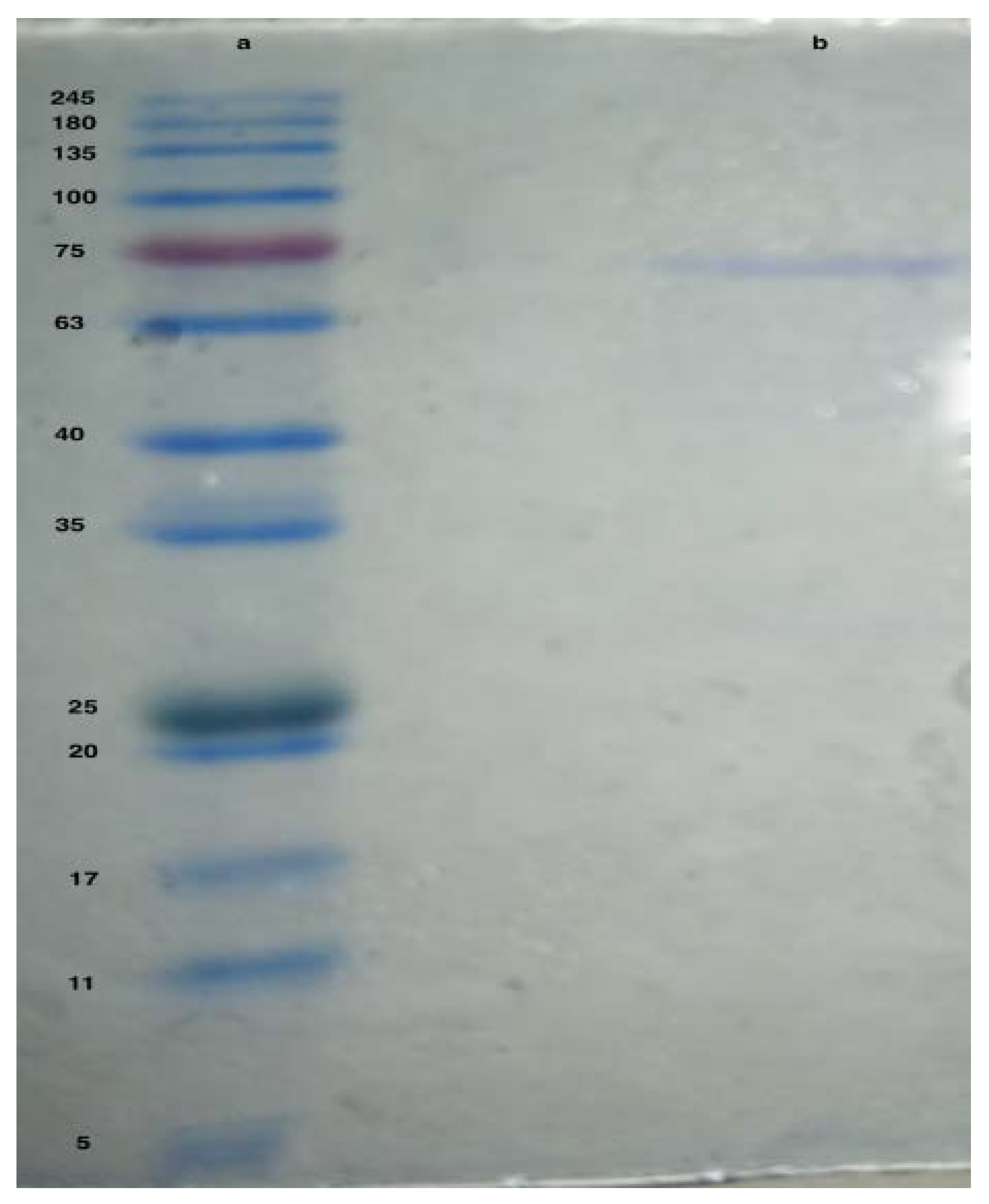
The purified asparaginase showed a band around 70 kDa compared to the reference protein ladder (Figure 2).

3.3. Enzyme Characterization

3.3.1. Effect of pH on Asparaginase Activity

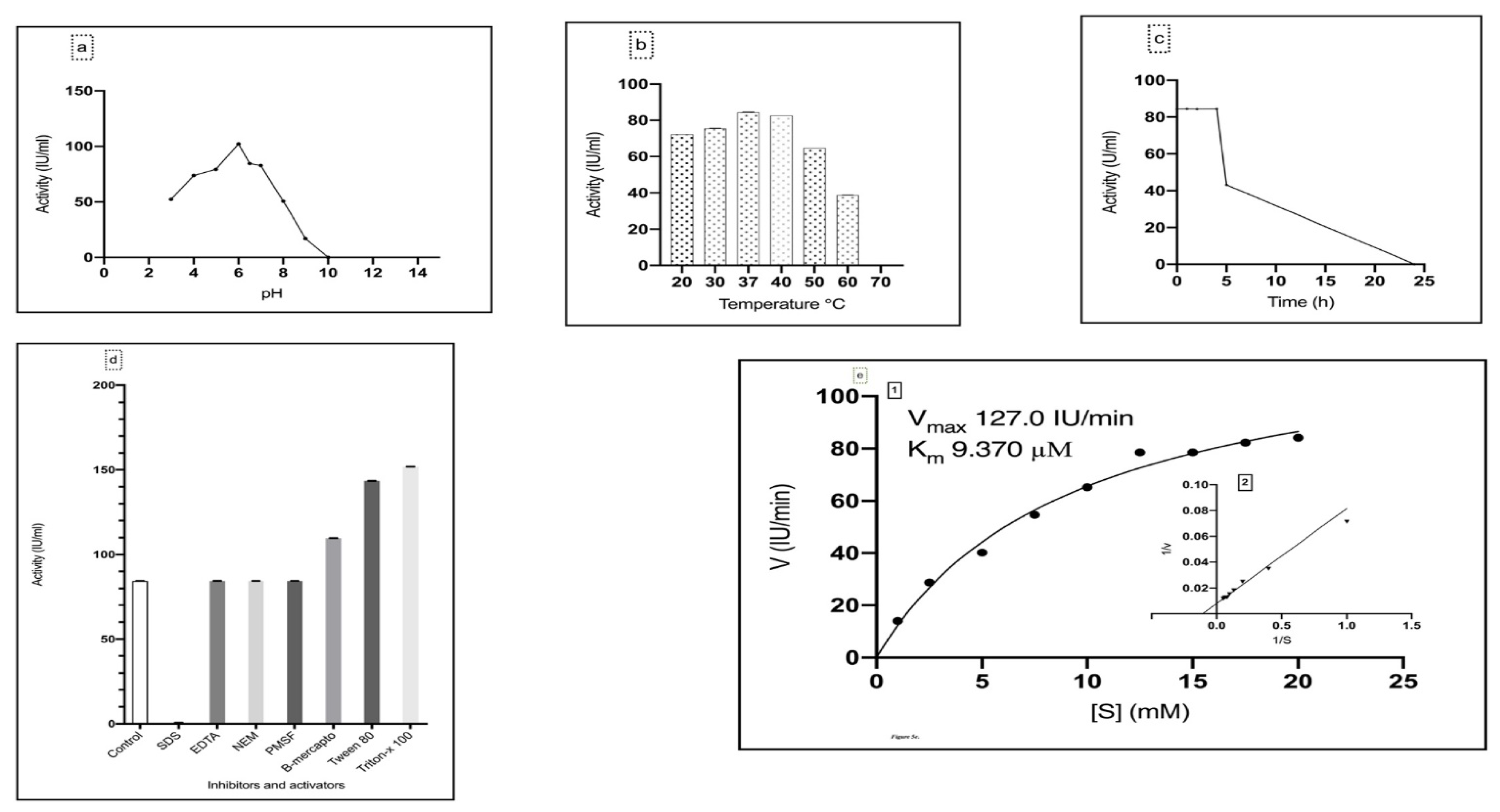
According to the results presented in Figure 3a, the optimum pH for the purified asparaginase was 6, while at pH 10 the enzyme lost its activity, starting from pH 3 the enzyme retained 51.22% of its activity, while at pH 4, 5 retained 72.22%, 77.43% respectively of its activity, the enzyme activity started to decrease simultaneously with increasing the pH value.

3.3.2. Effect of Temperature on Asparaginase Activity

The purified asparaginase showed an activity over a wide range of temperatures (20–60 °C). The optimum temperature was 37 °C with an activity of 84.41 IU/mL, followed by 40 °C with 82.52 IU/mL (Figure 3b).
The purified asparaginase showed complete stability at 37 °C for 1, 2 and 4 h, while the activity decreased to 43.2 IU/mL after 5 h. The enzyme lost its activity totally after 24 h (Figure 3c).

3.3.3. Effect of Inhibitors and Activators on Asparaginase Activity

The effect of different inhibitors and activators was studied on the purified asparaginase, where SDS showed a reduction in the enzyme activity to 0.65 IU/mL (10.35% relative activity), while EDTA, NEM, and PMSF showed no effect on asparaginase activity, in contrast β-mercaptoethanol, Tween 80, and Triton-x 100 enhanced the enzyme specific activity to 130%, 143.48% and 151.92% respectively regarding the control (Figure 3d).

3.3.4. Determination of Enzyme Kinetics

According to results in Figure 3e 1, 2, Michaelis constant Km and maximum velocity Vmax of the purified asparaginase from L. theobromae were 9.370 µM and 127.00 IU min−1 respectively.

3.4. Estimation of Asparaginase Antileukemic Activity

3.4.1. Evaluation of Cytotoxicity against M-NFS-60 Cell Line

Different concentrations of the purified asparaginase starting with 84.00 IU/mL to 2.62 IU/mL were tested against mouse myelogenous leukemia carcinoma cells to estimate the MIC50 value, results revealed that the MIC50 was 35.2 ± 0.7 IU/mL (Figure 4).

3.4.2. Evaluation of Cytotoxicity against WI-38 Cell Line

Different concentrations of the purified asparaginase starting with 84.00 IU/mL to 2.62 IU/mL were tested against WI-38 cells (human lung fibroblast normal cells) to estimate MCC50 value, results revealed that the MCC50 was 79.4 ± 1.9 IU/mL (Figure 5).

4. Discussion

Zhao et al. [36] and Jia et al. [37] reported that the activity of the bioactive metabolites produced by endophytes is considered similar or higher than those produced by host plants. Schulz et al. [38] reported that plant defense mechanisms and fungal virulence factors develop a balance mechanism by secreting different novel bioactive metabolites.
Lasiodiplodia theobromae was reported to produce several bioactive compounds used as anticancer, antifungal, antioxidant, anti-inflammatory such as: Lasiodiplodin [39], amylase [40], Paclitaxel [41], Felix [42] reported L. theobromae strain with the ability to produce: (3S,4S)-4-acetyl-3-methyl-2-dihydrofuranone, (2R/2S,3S,4S)-3-epibotryodiplodin, and jasmonic acid.
Our results showed that L. theobromae produces asparaginase extracellularly with a high enzymatic activity, while, Narayana et al. [43] reported that most of the asparaginase with microbial sources is intracellular. Extracellular asparaginase interacts minimally with the cellular constituents and is easier to extract; hence, the induction of extracellular asparaginases with high activity would be more advantageous.
To produce asparaginase with maximum activity, nine experiments were designed using Taguchi model. Taguchi model results showed a variation of asparaginase activity from 175 to 10 IU/mL, the variation of the results obtained were compatible with Ali et al. [34] who designed a model of 19 experiments for the optimization of asparaginase from A. sydowii and F. oxysporum with asparaginase activity ranging from 37-146 IU/mL and 33 to 143 IU/mL respectively. Ashok et al. [41] used Taguchi design to simulate nine experiments with asparaginase activities ranging from 3.94 to 20.57 IU/mL from Trichosporon asahii.
Another experiment designed for the induction of asparaginase with a maximum activity by incubating a five-days-old culture mat of L. theobromae with asparagine for two days and the asparaginase activity was estimated as 315 IU/mL. The incubation of the grown L. theobromae mat with asparagine solely as a substrate for asparaginase enhanced the selective release of asparaginase with a maximum activity with other extracellular enzymes, Nossal et al. [44] designed a similar experiment for the selective release of some degrading enzymes viz: alkaline phosphatase, cyclic phosphodiesterase, 5’-nucleotidase, acid phosphatase from E.coli by osmotic shock using different salts concentrations.
Asparaginase from L. theobromae was purified with a total activity of 84.40 IU/mL, specific activity of 468.03 IU/mg, and 13.68 purification folds. The estimated activity is considered low compared to Thakur et al. [11] who reported an activity of 222.18 IU/mL and a specific activity of 69.43 IU/mg, Mohamed et al. [45] reported an activity of 127 IU/mL and a specific activity of 846.IU/mg, and it was higher than the activity reported by Abbas [46] who reported an activity of 13.1 IU/mL and 0.4 IU/mg specific activity. The variation of activities would be related to the growth condition of each organism, the variation of extraction protocols.
To estimate the molecular weight of the purified asparaginase using SDS-PAGE, it was compared against known protein marker. SDS-PAGE showed a band of asparaginase by around 70 kDa. The presented result is similar to Huang [47] who reported a weight of 72 kDa and Hong [48] who reported an asparaginase of 71 kDa. Asparaginases showed a variation on the molecular weights where, Cedar and Schwartz [49] and Shifrin et al. [50] reported asparaginase from E. coli with 141 kDa, Scheetz et al. [51] reported asparaginase from Fusarium tricinctum with 161–170 kDa, Manna et al. [52] reported asparaginase from Pseudomonas stutzeri with 34 kDa and El-Bessouny [13] who recorded asparaginase from Pseudomonas aeruginosa at 160 kDa.
The purified asparaginase was characterized, where it showed a working pH range of 3–9, with an optimum pH of 6; Abbas [46] reported a working pH of 3–12 and an optimum pH of 10, on the other hand, Thakur [11] reported a working pH range of 3–10 and optimum pH of 7.
On testing the working temperature range, purified asparaginase showed activity over a temperature range of 20–60 °C with an optimum temperature of 37 °C which is in agreement with [11,45,53]. On the other hand, Gaffar and Shethna [54] reported an asparaginase isolated from Azotobacter vinelandii with optimum temperature of 48 °C and Mesas [55] reported an optimum temperature of 40 °C for asparaginase isolated from Corynebacterium glutamicum.
During the presented study some inhibitors and activators were studied against the purified asparaginase. SDS was the only reagent that showed reduction in the enzyme activity and retained 0.65% of the relative activity which was reported earlier by Thakur [11]. Which would be related to the SDS mode of action. Where it disturbs the structure of the protein which brings the folded protein structure into a linear model and coats the protein with uniform negative charges; leading to masking of the protein charges.
EDTA showed no effect on the purified enzyme which elucidated the fact that asparaginase is not an metalloenzyme, which is similar to data on Candida utilits by Sakamoto [56]. Regardless the fact of the non-metalloenzyme nature of the purified asparaginase some reports suggested that asparaginase is a metal activated enzyme where, El-Naggar et al. [8] reported that asparaginase from Sterptomyces brollosae showed an increase on activity in the presence of Mg2+, Mn2+ and Co2+ ions. Sonaimuthu [57] showed a metal activating characters with Mn2+ with asparaginase purified from Fusarium culmorum.
Tween 80 and Triton X-100 enhanced the activity with a relative activity of 170% and 180%, β-mercaptoethanol enhanced the enzyme activity with a relative activity of 130% our result was in agreement with Raha et al. [58]. The native protein structure mainly stabilized by the formation of disulfide bond [59], β-mercaptoethanol is a strong reducing agent that inhibit the formation of disulfide bonds in cysteine residues [60]. L-asparaginase was activated by the addition of β-mercaptoethanol. Hamza and Engel [61] reported the presence of free sulphydryl groups in the enzymes, such as cysteine residues will make it unstable and will aggregate the enzymes and the addition of β-mercaptoethanol and other ionic surfactants viz; Tween 80 and Triton X-100 will enhance the enzymatic activity.
NEM and PMSF showed an insignificant effect on the enzyme activity; which may be due to the absence of cysteine or serine on the active site of the purified asparaginase, our results are compatible with Thakur [11].
The activity of the purified asparaginase was assayed against different concentrations of asparagine (0–20 mM) to determine the kinetic properties, the purified enzyme showed a very low Km of 9.37 µM and Vmax of 127.00 U min−1. The recorded Km value considered lower than the recorded Km of Mucor hiemalis; 4 mM [11], Penicillium; 4 mM [62], such low Km value reflects the high affinity of the purified asparaginase towards asparagine as substrate.
The anti-leukemic effect of the purified asparaginase was studied against mouse myelogenous leukemia carcinoma cells to estimate the median minimal inhibitory concentration (MIC50). Results revealed that the MIC50 was 35.2 ± 0.7 U/mL which is lower than the MIC50 of A. sydowii (50 U/mL) and F. oxysporum (62.5 U/mL) [31]. It is also considered higher than the MIC50 of Phaseolus vulgaris (0.75 U/mg) [45]. The lower the MIC value, the lower the doses needed and the better the efficacy of the anticancer drug [63].
Tanfous et al. [64] related the antileukemic activity of asparaginase to its capability to hydrolyze asparagine in the serum, thus depleting serum asparagine; which is essential for the proliferation of leukemic cells. Several trials were carried out by scientists to discover asparaginase and test its role in the inhibition of leukemic cells [5,65,66,67]. Asparagine is produced by asparagine synthetase (ASNS), which is encoded by ASNS gene [68]. Gervasini and Vgace [69] hypothesized that ASNS genes in the malignant lymphoblasts cells have low expression or cannot regulate the expression of ASNS when exposed to asparaginase. Thus, we can hypothesize that the leukemic cells are more dependent on the extracellular sources of asparagine, thus the administration of asparaginase will selectively depletes serum asparagine leading to suppression of the growth of leukemic cells.
The presented results showed that the MIC50 (35.2 ± 0.7 IU/mL) of the purified asparaginase was around double the MCC50 (79.4 ± 1.9 IU/mL) where MCC50 value represents half the cytotoxic concentration of the tested asparaginase on normal cells. Such ratio indicates that the tested asparaginase is considered safe where it requires double of the asparaginase concentration to reach the MCC50. With treating the leukemic cell line with the purified asparaginase, our results revealed that the carcinoma cell line reached cell death up to 83.05% with the highest concentration of the purified enzyme (84 IU/mL) which is equal to about double the MIC50; respectively, leukemic cells require a high concentration of asparagine to keep viable [64].

5. Conclusions

In conclusion, the presented results proved that endophytic mycobiota especially isolated from extreme environment is considered as a source of safe novel bioactive compounds. This study sheds the light on a new application of endophytic native taxa and it recommends the production of the extracellular induced endophytic asparaginase on a high scale to be used as a safe antileukemic drug and in food industries. Future studies must focus on the screening for bioactive compounds secreted by Egyptian endophytic mycobiota to find a safe alternative for the traditional pharmaceutical compounds or discovering a novel compound to be used in industrial, agricultural, and medical applications.

Supplementary Materials

The following are available online at https://www.mdpi.com/article/10.3390/ijerph19020680/s1, Figure S1. Standard curve of different concentrations of BSA, Figure S2. Elution profile of asparaginase from L. theobromae from the Q-FF ion exchange column. The column was pre-equilibred with 50 mM Tris buffer (pH 7) and fractions were eluted with a gradient solution of NaCl (0-1M) prepared in same buffer, and Figure S3. Elution profile of asparaginase from L. theobromae from the gel-filtration column (Sephadex G-100). Fractions were eluted with 50 mM Tris buffer (pH 7). Table S1. The nine models generated by Taguchi design.

Author Contributions

Conceptualization, H.A.M., B.A.B. and A.M.A.-A.; methodology, H.A.M., D.H.S. and B.A.B.; software, B.A.B., Y.A.H., A.M.A., D.H.S. and A.A.A.; validation, H.A.M., B.A.B., Y.A.H., A.M.A., A.A.A., D.H.S. and A.M.A.-A.; formal analysis, H.A.M., B.A.B., Y.A.H., A.M.A., D.H.S. and A.M.A.-A.; investigation, H.A.M., B.A.B., D.H.S. and A.M.A.-A.; resources, A.A.A.; writing—original draft preparation, H.A.M., B.A.B. and A.M.A.-A.; writing—review and editing, H.A.M., B.A.B., Y.A.H., A.M.A., D.H.S., A.A.A. and A.M.A.-A.; funding acquisition, A.A.A. All authors have read and agreed to the published version of the manuscript.

Funding

This research received no external funding.

Institutional Review Board Statement

Not Applicable.

Informed Consent Statement

Not Applicable.

Data Availability Statement

Not Applicable

Acknowledgments

We appreciate and thank Taif University for the financial support for Taif University Re-searchers Supporting Project (TURSP-2020/22), Taif University, Taif, Saudi Arabia. We would like to present their deep appreciation to Abdel Ghaffar Abu El Saoud, Botany and Microbiology Department, Faculty of Science, Suez Canal University for his support and help during the data analysis of the results. Also, to the members of Saint Katherine Protectorate for their support during the samples collection in the early stages of the research work progress.

Conflicts of Interest

The authors have no conflicts of interest to declare. All co-authors have seen and agreed with the contents of the manuscript.

References

  1. Rodriguez, R.J.; White, J.F., Jr.; Arnold, A.E.; Redman, R.S. Fungal endophytes: Diversity and functional roles. New Phytol. 2009, 182, 314–330. [Google Scholar] [CrossRef] [PubMed]
  2. Gouda, S.; Kerry, R.G.; Das, G.; Paramithiotis, S.; Shin, H.-S.; Patra, J.K. Revitalization of plant growth promoting rhizobacteria for sustainable development in agriculture. Microbiol. Res. 2018, 206, 131–140. [Google Scholar] [CrossRef]
  3. Abdel-Azeem, A.M.; Zaki, S.M.; Khalil, W.F.; Makhlouf, N.A.; Farghaly, L.M. Anti-Rheumatoid Activity of Secondary Metabolites Produced by Endophytic Chaetomium globosum. Front. Microbiol. 2016, 7, 1477. [Google Scholar] [CrossRef] [PubMed] [Green Version]
  4. Kumar, D.; Sobha, K. L-Asparaginase from Microbes: A Comprehensive Review. Adv. Biores. 2012, 3, 137–157. [Google Scholar]
  5. Clementi, A. La Désamidation Enzymatique De L’asparagine Chez Les Différentes Espéces Animales Et La Signification Physio Logique De Sa Presence Dans L’organisme. Arch. Int. Physiol. 1922, 19, 369–398. [Google Scholar] [CrossRef]
  6. Mashburn, L.T.; Wriston, J.C. Tumor inhibitory effect of l-asparaginase from Escherichia coli. Arch. Biochem. Biophys. 1964, 105, 450–453. [Google Scholar] [CrossRef]
  7. Pieters, R.; Hunger, S.P.; Boos, J.; Rizzari, C.; Silverman, L.; Baruchel, A.; Goekbuget, N.; Schrappe, M.; Pui, C.-H. L-asparaginase treatment in acute lymphoblastic leukemia: A Focus on Erwinia Asparaginase. Cancer 2011, 117, 238–249. [Google Scholar] [CrossRef] [PubMed] [Green Version]
  8. El-Naggar, N.E.-A.; El-Ewasy, S.M.; El-Shweihy, N.M. Microbial L-Asparaginase as a Potential Therapeutic Agent for the Treatment of Acute Lymphoblastic Leukemia: The Pros and Cons. Int. J. Pharmacol. 2014, 10, 182–199. [Google Scholar] [CrossRef]
  9. Zhang, J.; Fan, J.; Venneti, S.; Cross, J.R.; Takagi, T.; Bhinder, B.; Djaballah, H.; Kanai, M.; Cheng, E.H.; Judkins, A.R.; et al. Asparagine Plays a Critical Role in Regulating Cellular Adaptation to Glutamine Depletion. Mol. Cell 2014, 56, 205–218. [Google Scholar] [CrossRef] [Green Version]
  10. Fung, M.K.L.; Chan, G.C.-F. Drug-induced amino acid deprivation as strategy for cancer therapy. J. Hematol. Oncol. 2017, 10, 1–18. [Google Scholar] [CrossRef]
  11. More, S.S.; Thakur, L.L.M. Isolation, Purification and Characterization of Fungal Extracellular L-Asparaginase from Mucor hiemalis. J. Biocatal. Biotransform. 2013, 2. [Google Scholar] [CrossRef]
  12. Verma, N.; Kumar, K.; Kaur, G.; Anand, S. L-Asparaginase: A Promising Chemotherapeutic Agent. Crit. Rev. Biotechnol. 2007, 27, 45–62. [Google Scholar] [CrossRef]
  13. El-Bessoumy, A.A.; Sarhan, M.; Mansour, J. Production, isolation, and purification of L-asparaginase from Pseudomonas aeruginosa 50071 using solid-state fermentation. J. Biochem. Mol. Biol. 2004, 37, 387–393. [Google Scholar] [CrossRef] [Green Version]
  14. Kafkewitz, D.; Goodman, D. L-Asparaginase Production by the Rumen Anaerobe Vibrio succinogenes. Appl. Microbiol. 1974, 27, 206–209. [Google Scholar] [CrossRef] [PubMed]
  15. Hatamzadeh, S.; Rahnama, K.; Nasrollahnejad, S.; Fotouhifar, K.B.; Hemmati, K.; White, J.F.; Taliei, F. Isolation and identification of L-asparaginase-producing endophytic fungi from the Asteraceae family plant species of Iran. PeerJ 2020, 8, e8309. [Google Scholar] [CrossRef] [PubMed]
  16. Egler, R.A.; Ahuja, S.P.; Matloub, Y. L-asparaginase in the treatment of patients with acute lymphoblastic leukemia. J. Pharmacol. Pharmacother. 2016, 7, 62–71. [Google Scholar] [CrossRef] [Green Version]
  17. Souza, P.M.; de Freitas, M.M.; Cardoso, S.L.; Pessoa, A.; Guerra, E.N.S.; Magalhães, P.O. Optimization and purification of L-asparaginase from fungi: A systematic review. Crit. Rev. Oncol. 2017, 120, 194–202. [Google Scholar] [CrossRef]
  18. Mishra, A. Production of L-asparaginase, an anticancer agent, from Aspergillus niger using agricultural waste in solid state fermentation. Appl. Biochem. Biotechnol. 2006, 135, 33–42. [Google Scholar] [CrossRef]
  19. Archana, J.; Raja, P. Production, Purification and Characterization of L-Asparaginase from Aspergillus nidulans by Solid State Fermentation. Eur. J. Biotechnol. Biosci. 2014, 2, 51–58. [Google Scholar]
  20. Saied, E.M.; El-Maradny, Y.A.; Osman, A.A.; Darwish, A.M.G.; Abo Nahas, H.H.; Niedbała, G.; Piekutowska, M.; Abdel-Rahman, M.A.; Balbool, B.A.; Abdel-Azeem, A.M. A Comprehensive Review about the Molecular Structure of Severe Acute Respiratory Syndrome Coronavirus 2 (SARS-CoV-2): Insights into Natural Products against COVID-19. Pharmaceutics 2021, 13, 1759. [Google Scholar] [CrossRef]
  21. Nagarajan, A.; Thirunavukkarasu, N.; Suryanarayanan, T.S.; Gummadi, N.S. Screening and Isolation of Novel Glutaminase Free L-Asparaginase from Fungal Endophytesitle. Res. J. Microbiol. 2014, 9, 163–176. [Google Scholar]
  22. Salini, G.; Madhusoodhanan, A.; Joseph, A.; Mohan, A.; Navya, R.K.; Nair, V.V. Glutaminase Free L-Asparaginase Producing Endophytes from Mangroves. Asian J. Pharm. Clin. Res. 2016, 9, 360–362. [Google Scholar]
  23. Gaber, A.; Alsanie, W.F.; Kumar, D.N.; Refat, M.S.; Saied, E.M. Novel Papaverine Metal Complexes with Potential Anticancer Activities. Molecules 2020, 25, 5447. [Google Scholar] [CrossRef] [PubMed]
  24. Balbool, B.A.; Abdel-Azeem, A.M. Diversity of the Culturable Endophytic Fungi Producing L-Asparaginase in Arid Sinai, Egypt. Ital. J. Mycol. 2020, 49, 8–24. [Google Scholar] [CrossRef]
  25. Gaber, A.; Refat, M.S.; Belal, A.A.M.; El-Deen, I.M.; Hassan, N.; Zakaria, R.; Alhomrani, M.; Alamri, A.S.; Alsanie, W.F.; M. Saied, E. New Mononuclear and Binuclear Cu(II), Co(II), Ni(II), and Zn(II) Thiosemicarbazone Complexes with Potential Biological Ac-tivity: Antimicrobial and Molecular Docking Study. Molecules 2021, 26, 2288. [Google Scholar] [CrossRef] [PubMed]
  26. Ali, D.; Ouf, S.; Eweis, M.; Solieman, D. Optimization of L-Asparaginase production from some filamentous fungi with potential pharmaceutical properties. Egypt. J. Bot. 2018, 58, 355–369. [Google Scholar] [CrossRef]
  27. Wriston, J.C. Asparaginase. In Methods in Enzymology—Metabolism of Amino Acids and Amines Part A; Academic Press: Cambridge, MA, USA, 1970; Volume 17, pp. 732–742. [Google Scholar]
  28. Wessel, D.; Flügge, U. A method for the quantitative recovery of protein in dilute solution in the presence of detergents and lipids. Anal. Biochem. 1984, 138, 141–143. [Google Scholar] [CrossRef]
  29. Laemmli, U.K. Cleavage of Structural Proteins during the Assembly of the Head of Bacteriophage T4. Nature 1970, 227, 680–685. [Google Scholar] [CrossRef]
  30. Sharar, M.; Saied, E.M.; Rodriguez, M.C.; Arenz, C.; Montes-Bayón, M.; Linscheid, M.W. Elemental Labelling and Mass Spec-trometry for the Specific Detection of Sulfenic Acid Groups in Model Peptides: A Proof of Concept. Anal. Bioanal. Chem. 2017, 409, 2015–2027. [Google Scholar] [CrossRef] [PubMed]
  31. Yadav, N.; Sarkar, S. Production of L-Asparaginase by Fusarium Oxysporum Using Submerged Fermentation. Int. J. Pharm. Sci. Invent. 2014, 3, 32–40. [Google Scholar]
  32. Kothari, M.N.; Baig, M.M.V. Production and characterization of extracellular polygalacturonase by Erwinia carotovora MTCC 1428. Int. J. Adv. Biotechnol. Res. 2013, 4, 981–998. [Google Scholar]
  33. Pu, X.; Yu, S.; Fan, W.; Liu, L.; Ma, X.; Ren, J. Guiqi polysaccharide protects the normal human fetal lung fibroblast WI-38 cells from H2O2-induced premature senescence. Int. J. Clin. Exp. Pathol. 2015, 8, 4398–4407. [Google Scholar]
  34. Shafek, R.; Michael, H.; Sayed, A.; Ibrahim, A.; Al-Sayed, A. Phytochemical study, antioxidant and cytotoxic activities of Brassica rapa L. leaves extract and its silver nanoparticles. Egypt. J. Chem. 2018, 61, 237–247. [Google Scholar] [CrossRef] [Green Version]
  35. Mosmann, T. Rapid colorimetric assay for cellular growth and survival: Application to proliferation and cytotoxicity assays. J. Immunol. Methods 1983, 65, 55–63. [Google Scholar] [CrossRef]
  36. Zhao, J.; Shan, T.; Mou, Y.; Zhou, L. Plant-Derived Bioactive Compounds Produced by Endophytic Fungi. Mini-Rev. Med. Chem. 2011, 11, 159–168. [Google Scholar] [CrossRef] [PubMed]
  37. Jia, M.; Chen, L.; Xin, H.-L.; Zheng, C.-J.; Rahman, K.; Han, T.; Qin, L.-P. A Friendly Relationship between Endophytic Fungi and Medicinal Plants: A Systematic Review. Front. Microbiol. 2016, 7, 906. [Google Scholar] [CrossRef] [Green Version]
  38. Schulz, B.; Boyle, C. The endophytic continuum. Mycol. Res. 2005, 109, 661–686. [Google Scholar] [CrossRef] [Green Version]
  39. Matsuura, H.; Nakamori, K.; Omer, E.; Hatakeyama, C.; Yoshihara, T.; Ichihara, A. Three lasiodiplodins from lasiodiplodia theobromae IFO 31059. Phytochemistry 1998, 49, 579–584. [Google Scholar] [CrossRef]
  40. Navaratnam, P.; Arasaratnam, V.; Mahendran, S.; Balasubramaniam, K. Formulation of medium and recycling of biomass for glucoamylase production by Botryodiplodia theobromae. Process. Biochem. 1996, 31, 77–80. [Google Scholar] [CrossRef]
  41. Ashok, A.; Doriya, K.; Rao, J.V.; Qureshi, A.; Tiwari, A.K.; Kumar, D.S. Microbes Producing L-Asparaginase free of Glutaminase and Urease isolated from Extreme Locations of Antarctic Soil and Moss. Sci. Rep. 2019, 9, 1423. [Google Scholar] [CrossRef] [PubMed] [Green Version]
  42. Félix, C.; Salvatore, M.M.; DellaGreca, M.; Ferreira, V.; Duarte, A.S.; Salvatore, F.; Naviglio, D.; Gallo, M.; Alves, A.; Esteves, A.C.; et al. Secondary metabolites produced by grapevine strains of Lasiodiplodia theobromae grown at two different temperatures. Mycologia 2019, 111, 466–476. [Google Scholar] [CrossRef] [PubMed]
  43. Narayana, K.J.P.; Kumar, K.G.; Vijayalakshmi, M. L-asparaginase production by Streptomyces albidoflavus. Indian J. Microbiol. 2008, 48, 331–336. [Google Scholar] [CrossRef] [Green Version]
  44. Nossal, N.G.; Heppel, L.A. The Release of Enzymes by Osmotic Shock from Escherichia coli in Exponential Phase. J. Biol. Chem. 1996, 241, 3055–3062. [Google Scholar] [CrossRef]
  45. Mohamed, S.A.; Elshal, M.F.; Kumosani, T.A.; Aldahlawi, A. Purification and Characterization of Asparaginase from Phaseolus vulgaris Seeds. Evid.-Based Complement. Altern. Med. 2015, 2015, 309214. [Google Scholar] [CrossRef] [PubMed] [Green Version]
  46. Ahmed, M.M.A.; Dahad, F.N.A.; Taha, M.T.; Hassan, S.M.F. Production, Purification and Characterization of L-Asparaginase from Marine Endophytic Aspergillus sp. ALAA-2000 under Submerged and Solid State Fermentation. J. Microb. Biochem. Technol. 2015, 7, 165–172. [Google Scholar] [CrossRef]
  47. Huang, L.; Liu, Y.; Sun, Y.; Yan, Q.; Jiang, Z. Biochemical Characterization of a Novel L-Asparaginase with Low Glutaminase Activity from Rhizomucor miehei and Its Application in Food Safety and Leukemia Treatment. Appl. Environ. Microbiol. 2013, 80, 1561–1569. [Google Scholar] [CrossRef] [Green Version]
  48. Hong, S.-J.; Lee, Y.-H.; Khan, A.R.; Ullah, I.; Lee, C.; Park, C.K.; Shin, J.-H. Cloning, expression, and characterization of thermophilic L-asparaginase from Thermococcus kodakarensis KOD1. J. Basic Microbiol. 2014, 54, 500–508. [Google Scholar] [CrossRef]
  49. Cedar, H.; Schwartz, J.H. Localization of the Two L-Asparaginases in Anaerobically Grown Escherichia coli. J. Biol. Chem. 1967, 242, 3753–3755. [Google Scholar] [CrossRef]
  50. Shifrin, S.; Parrott, C.L.; Luborsky, S.W. Substrate Binding and Intersubunit Interactions in L-Asparaginase. J. Biol. Chem. 1974, 249, 1335–1340. [Google Scholar] [CrossRef]
  51. Scheetz, R.W.; Whelan, H.A.; Wriston, J.C. Purification and properties of an L-asparaginase from Fusarium tricinctum. Arch. Biochem. Biophys. 1971, 142, 184–189. [Google Scholar] [CrossRef]
  52. Manna, S.; Sinha, A.; Sadhukhan, R.; Chakrabarty, S.L. Purification, characterization and antitumor activity of L-asparaginase isolated from Pseudomonas stutzeri MB-405. Curr. Microbiol. 1995, 30, 291–298. [Google Scholar] [CrossRef]
  53. Sanjotha, G.; Manawadi, S.I. Isolation, Screening, Optimization and Production of Anti-Tumor L-Asparaginase by Fungi from Karwar Coastal Region. Res. J. Recent Sci. 2017, 6, 1–7. [Google Scholar]
  54. Gaffar, S.A.; Shethna, Y.I. Purification and Some Biological Properties of Asparaginase from Azotobacter vinelandii. Appl. Environ. Microbiol. 1977, 33, 508–514. [Google Scholar] [CrossRef] [Green Version]
  55. Mesas, J.M.; Gil, J.A.; Martin, J.F. Characterization and Partial Purification of L-Asparaginase from Corynebacterium Glutamicum. J. Gen. Microbiol. 1990, 136, 515–519. [Google Scholar] [CrossRef] [Green Version]
  56. Sakamoto, T.; Araki, C.; Beppu, T.; Arima, K. Partial purification and some properties of extracellular asparaginase from Candida utilis. Agric. Biol. Chem. 1977, 41, 1359–1364. [Google Scholar] [CrossRef]
  57. Sonaimuthu, V.; Krishnamoorthy, S.; Johnpaul, M. Taxol producing endophytic fungus Fusarium culmorum SVJM072 from medicinal plant of Tinospora cordifolia—A first report. J. Biotechnol. 2010, 150, 425. [Google Scholar] [CrossRef]
  58. Raha, S.K.; Roy, S.K.; Dey, S.K.; Chakrabarty, S.L. Purification and properties of an L-asparaginase from Cylindrocarpon obtusisporum MB-10. Biochem. Int. 1990, 21, 987–1000. [Google Scholar]
  59. Qin, M.; Wang, W.; Thirumalai, D. Protein folding guides disulfide bond formation. Proc. Natl. Acad. Sci. USA 2015, 112, 11241–11246. [Google Scholar] [CrossRef] [Green Version]
  60. Zhang, S.; Xie, Y.; Zhang, C.; Bie, X.; Zhao, H.; Lu, F.; Lu, Z. Biochemical characterization of a novel L-asparaginase from Bacillus megaterium H-1 and its application in French fries. Food Res. Int. 2015, 77, 527–533. [Google Scholar] [CrossRef]
  61. Hamza, M.A.; Engel, P.C. Enhancing long-term thermal stability in mesophilic glutamate dehydrogenase from Clostridium symbiosum by eliminating cysteine residues. Enzym. Microb. Technol. 2007, 41, 706–710. [Google Scholar] [CrossRef]
  62. Patro, K.R. Extraction, purification and characterization of L-asparaginase from Penicillium sp. by submerged fermentation. Int. J. Biotechnol. Mol. Biol. Res. 2012, 3, 30–34. [Google Scholar] [CrossRef]
  63. Lewies, A.; Du Plessis, L.H.; Wentzel, J.F. The Cytotoxic, Antimicrobial and Anticancer Properties of the Antimicrobial Peptide Nisin Z Alone and in Combination with Conventional Treatments. In Cytotoxicity; IntechOpen: London, UK, 2018. [Google Scholar]
  64. Ben Tanfous, M.; Sharif-Askari, B.; Ceppi, F.; Laaribi, H.; Gagné, V.; Rousseau, J.; Labuda, M.; Silverman, L.B.; Sallan, S.E.; Neuberg, D.; et al. Polymorphisms of Asparaginase Pathway and Asparaginase-Related Complications in Children with Acute Lymphoblastic Leukemia. Clin. Cancer Res. 2015, 21, 329–334. [Google Scholar] [CrossRef] [PubMed] [Green Version]
  65. Broome, J.D. Evidence That the L-Asparaginase of Guinea Pig Serum is Responsible for Its Antilymphoma Effects: I. Properties of The L-Asparaginase of Guinea Pig Serum in Relation to Those of The Antilymphoma Substance. J. Exp. Med. 1963, 118, 99–120. [Google Scholar] [CrossRef] [PubMed] [Green Version]
  66. Kidd, J.G. Regression of Transplanted Lymphomas Induced in Vivo by Means of Normal Guinea Pig Serum. II. Studies on the Nature of the Active Serum Constituent: Histological Mechanism of the Regression: Tests for Effects of Guinea Pig Serum on Lymphoma Cells in Vitro. J. Exp. Med. 1953, 98, 583–606. [Google Scholar] [CrossRef]
  67. Neuman, R.E.; McCoy, T.A. Dual Requirement of Walker Carcinosarcoma 256 in vitro for Asparagine and Glutamine. Science 1956, 124, 124–125. [Google Scholar] [CrossRef] [PubMed] [Green Version]
  68. Mei, L.; Ontiveros, E.P.; Griffiths, E.A.; Thompson, J.E.; Wang, E.S.; Wetzler, M. Pharmacogenetics predictive of response and toxicity in acute lymphoblastic leukemia therapy. Blood Rev. 2015, 29, 243–249. [Google Scholar] [CrossRef] [Green Version]
  69. Gervasini, G.; Vagace, J.M. Impact of genetic polymorphisms on chemotherapy toxicity in childhood acute lymphoblastic leukemia. Front. Genet. 2012, 3, 249. [Google Scholar] [CrossRef] [PubMed] [Green Version]
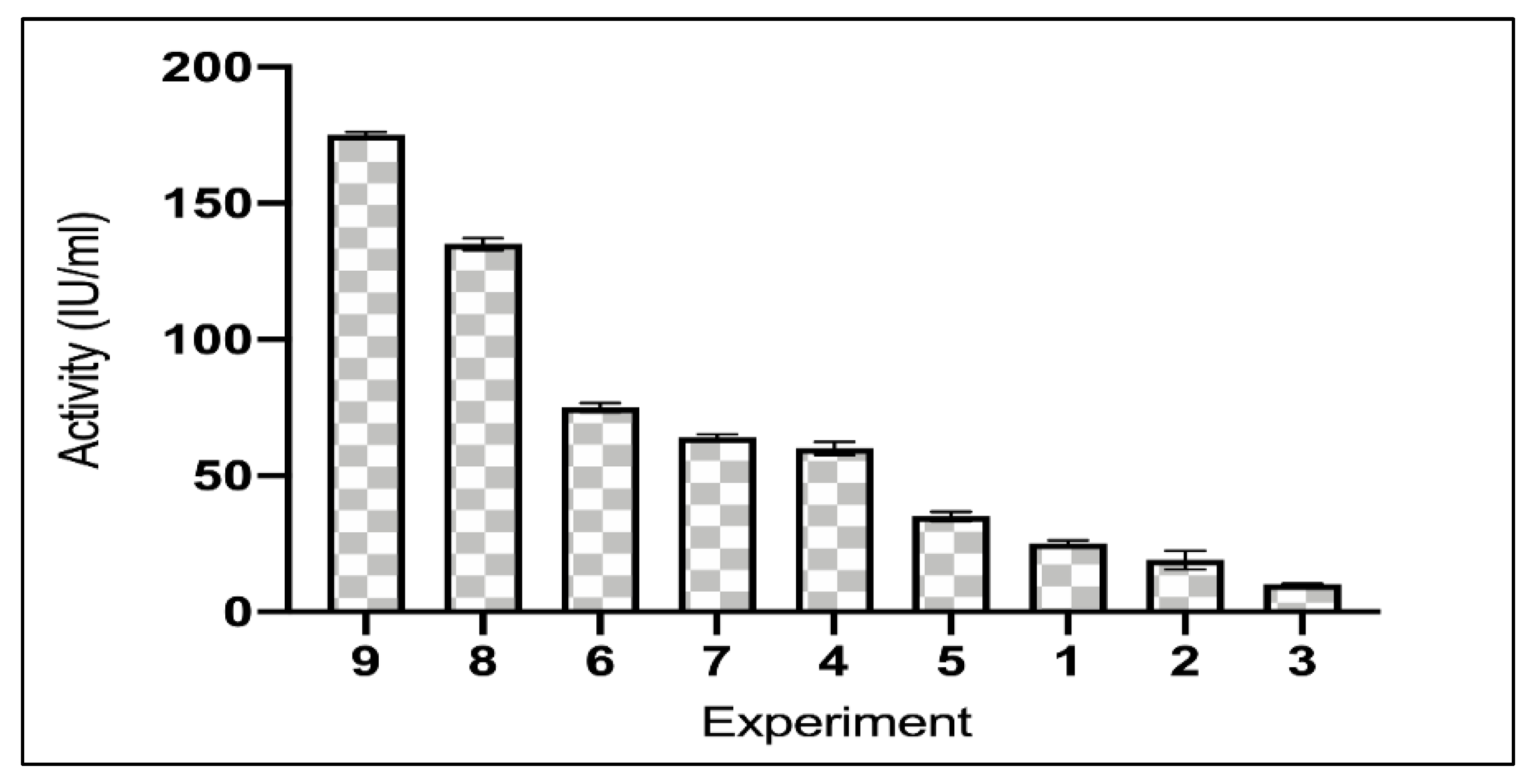
Figure 1. Asparaginase activities of Taguchi design experiments.
Ijerph 19 00680 g001
Figure 2. Purified asparaginase from L. theobromae on SDS-PAGE. (a) protein ladder, (b) purified asparaginase.
Ijerph 19 00680 g002
Figure 3. (a) The effect of pH on the purified asparaginase from L. theobromae. (b) The effect of temperature on the purified asparaginase from L. theobromae. (c) Thermal stability of purified asparaginase from L. theobromae at 37 °C. (d) Effect of inhibitors and activators on purified asparaginase from L. theobromae. (e) Purified asparaginase kinetics. 1. Michaelis Menten plot for the purified asparaginase enzyme from L. theobromae. 2. The different activities value of purified asparaginase from L. theobromae with increasing the substrate concentrations.
Ijerph 19 00680 g003
Figure 4. Viability response of M-NFS-60 cell line against serial two-fold dilutions of purified asparaginase from L. theobromae.
Ijerph 19 00680 g004
Figure 5. Viability response of WI-38 cell line against serial two-fold dilutions of purified asparaginase from L. theobromae.
Ijerph 19 00680 g005
Table 1. Summary of the steps involved in the purification of asparaginase from L. theobromae.
StepTotal Protein
(mg)
Activity *
(IU/mL)
Specific Activity
(IUmg−1)
Purification
Fold
Yield %
Induced enzyme9.21315.0034.201.00100.00
Acetone6.81250.0036.711.0779.37
Q-FF (0.3M NaCl)1.31138.91105.883.1044.10
Sephadex G-1000.1884.40468.0313.6826.79
* One unit of asparaginase (IU) is defined as the amount of enzyme that liberates 1 μmol of ammonia min−1 AT 37 °C.
Publisher’s Note: MDPI stays neutral with regard to jurisdictional claims in published maps and institutional affiliations.