Article

Correlation between MOVA3D, a Monocular Movement Analysis System, and Qualisys Track Manager (QTM) during Lower Limb Movements in Healthy Adults: A Preliminary Study

by
Liliane Pinho de Almeida
1,
Leandro Caetano Guenka
1,2,*,
Danielle de Oliveira Felipe
1,
Renato Porfirio Ishii
1,
Pedro Senna de Campos
1 and
Thomaz Nogueira Burke
1
1
Allied Health Institute, Federal University of Mato Grosso do Sul, Campo Grande 79070-900, Brazil
2
Medicine, State University of Mato Grosso do Sul, Campo Grande 79115-898, Brazil
*
Author to whom correspondence should be addressed.
Int. J. Environ. Res. Public Health 2023, 20(17), 6657; https://doi.org/10.3390/ijerph20176657
Submission received: 6 May 2023 / Revised: 21 August 2023 / Accepted: 24 August 2023 / Published: 26 August 2023
(This article belongs to the Special Issue New Advances in Physical Therapy and Rehabilitation)

Abstract

:
New technologies based on virtual reality and augmented reality offer promising perspectives in an attempt to increase the assessment of human kinematics. The aim of this work was to develop a markerless 3D motion analysis capture system (MOVA3D) and to test it versus Qualisys Track Manager (QTM). A digital camera was used to capture the data, and proprietary software capable of automatically inferring the joint centers in 3D and performing the angular kinematic calculations of interest was developed for such analysis. In the experiment, 10 subjects (22 to 50 years old), 5 men and 5 women, with a body mass index between 18.5 and 29.9 kg/m2, performed squatting, hip flexion, and abduction movements, and both systems measured the hip abduction/adduction angle and hip flexion/extension, simultaneously. The mean value of the difference between the QTM system and the MOVA3D system for all frames for each joint angle was analyzed with Pearson’s correlation coefficient (r). The MOVA3D system reached good (above 0.75) or excellent (above 0.90) correlations in 6 out of 8 variables. The average error remained below 12° in only 20 out of 24 variables analyzed. The MOVA3D system is therefore promising for use in telerehabilitation or other applications where this level of error is acceptable. Future studies should continue to validate the MOVA3D as updated versions of their software are developed.

1. Introduction

Telerehabilitation or e-rehabilitation is an integral part of e-medicine that allows physiotherapists and patients to control rehabilitation processes from a distance [1]. This form of rehabilitation was developed with the aim of providing the patient and the physiotherapist with greater control over the prescribed therapy so that it can be performed at home [2], reducing the time and cost of hospitalization [3], and allowing an increase in the coverage area of rehabilitation services, especially with respect to reaching patients living long distances from traditional rehabilitation services.
Several studies indicate that telerehabilitation can be useful and as effective as conventional treatments [4,5,6,7]. In addition, telerehabilitation increases adherence, motivation, and frequency of physiotherapy sessions [8]. Another advantage of telerehabilitation systems is the collection of quantitative data related to the therapy and the ease of access and manipulation of these data by health professionals [9,10,11]. Data collected through sensors can be further processed and used to develop more effective interventions [12,13].
There are several different possibilities for applications of motion capture technology. The majority of studies carried out in the field of rehabilitation aim to engage patients in an exercise regime proposed by the physiotherapist. These systems enable patients to receive real-time feedback on the prescribed activity, assist in the correction of joint positions during the execution of the movements, and aid in remote monitoring and adjustment of the exercise prescription between each clinical visit to the patient [14].
Telerehabilitation systems are composed of at least one camera that allows the physiotherapist to see and monitor the patient from a distance (videoconference). More complex systems include sensors capable of motion analysis, which can, in general, be classified into three main groups. The first group comprises systems in which users need to wear devices that capture their movements [15,16,17,18]. The second group includes motion capture systems, which allow users to carry out their activities without the need to wear sensors. Examples of this group are systems that use the Nintendo Wii Remote, Leap Motion, Kinect, or, more recently, depth cameras such as Intel’s RealSense. Finally, the third group seeks to use robotics as a tool for telerehabilitation. Among these systems, we can mention the MOTORE++ [19], HOMEHEAB [20], and the WAM robot [21].
Optical motion analysis techniques have been widely used in biomechanics for measuring large-scale motions, and kinematic data are computed using marker-based motion capture, such as the Qualisys Track Manager system (QTM—Qualisys AB, Gothenburg, Sweden), which is considered as a gold-standard measure for motion assessment [22], albeit expensive and, therefore, restricted to specialized centers, which limits its large-scale dissemination. On the other hand, 2D RGB cameras are widely used in various devices, being present in almost all smartphones sold to the general public; however, they present the ability to deliver variables in only two dimensions (there are no data on depth), thus requiring computational effort to infer 3D variables from 2D variables.
Single-camera markerless motion capture has the potential to facilitate at-home movement assessment due to the ease of setup, portability, and affordable cost of the technology [23]. A human motion capture system with an RGB-D camera or depth camera has a relevant application in research and industry due to its easy use [24]. The development of technology of this nature, if it is shown to be reliable, could represent an alternative for data collection by health professionals, or even for use in telerehabilitation systems, as the end user would need little training, since this technology is widely used.
Therefore, the aim of this work was to develop a monocular and markless 3D motion analysis capture system (MOVA3D) and to compare it against Qualisys Track Manager (QTM).

2. Materials and Methods

2.1. MOVA3D System

The system consists of an RGB digital camera to capture videos of the movements to be analyzed and software dedicated to the automatic identification of the individual, their joint centers, and body segments (MOVA3D). The computational technique employed is didactically divided into 3 independent stages: (1) automatic detection of human silhouettes in the image, (2) depth referencing between the camera and a reference point on the individual, and (3) calculation of the relative position between joints in the image.
The system generates a data set containing the X, Y, and Z variables of 18 joints or points of interest in the image: “Pelvis”, “R_Hip”, “R_Knee”, “R_Ankle”, “L_Hip”, “L_Knee”, “L_Ankle”, “Torso”, “Neck”, “Nose”, “Head”, “L_Shoulder”, “L_Elbow”, “L_Wrist”, “R_Shoulder”, “R_Elbow”, “R_Wrist”, and “Thorax”. Hip angles were derived in sagittal (flexion/extension) and frontal plane (abduction/adduction) using the 3D position of the “Knee”, “Hip”, and “Thorax” joint centers. “Hip”, Knee”, and “Ankle” were used for knee angle calculations. Range of motion (ROM) detection was performed by creating a motion stop or inversion identifier. The stop identifier algorithm detects, after the start of the movement, if the stop of the movement occurred. This happens by calculating the angular coefficient of the straight line, referring to the angles with variation in time. Thus, when identifying the point where the stability of the slope of the line occurs, the algorithm assumes a stop. In the current study, only the markers that delimit the lower limbs and hip were used in Qualisys analysis.

2.2. Subjects and Experimental Design

In the experiment, 10 individuals aged between 22 and 50 years (5 men and 5 women) with a body mass index between 18.5 and 29.9 kg/m2 were recruited. A wide range of age and BMI was included in the study to improve the external validity of the results. The exclusion criteria used were the presence of any physical, cognitive, or balance limitation, which could prevent or hinder the execution of the proposed activities during the capture of the movements. The experiment was conducted at the Movement Analysis Laboratory at the Associação de Pais e Amigos dos Excepcionais de Campo Grande (APAE/CG) in Campo Grande/MS, Brazil, from September 2020 to September 2021. Prior to capturing the movements, the individuals answered a questionnaire containing questions on socio-demographic data (email, contact, and age), anthropometric data (weight and height), and their physical condition. The study design was approved by the IRB-Universidade Federal de Mato Grosso do Sul (#2 9358720.7.0000.0021).
The experimental design comprised the concomitant evaluation of the subjects by two systems: Qualisys Track Manager (QTM—Qualisys AB, Sweden) and MOVA3D (own development). The Qualisys system used was configured with seven Oqus series 300, 1280, and 1024 (1.3 pixels) resolution cameras. Figure 1 shows the overall layout. Twenty-six anatomical markers were positioned on anatomical structures, as shown in Table 1. The volume to be collected was previously calibrated following the manufacturer’s recommendations. All assessments were performed by the same professional with experience in the QTM procedures.
The RGB camera of the MOVA3D system (Intel 435i with the depth function disabled) was positioned 1 m from the ground, coinciding with the frontal plane of the subjects. For better centralization of the evaluated individual, the camera was close to the right knee.

Experimental Setup and Data Collection

The experiment consisted of each participant performing three groups of activities, 5 repetitions each, while their movements were captured by both systems: squat, hip flexion, and hip abduction. The pace and amplitude of movement were not controlled so that there was naturalness in the execution. Individuals were asked to wear tight clothing (shorts or leggings), and women wore a sports bra, while men were bare-chested. The capture by the MOVA3D system was performed at a frequency of 30 Hz and by the Qualisys system at 100 Hz.

2.3. Data Processing

First, the trajectories of the markers with gaps greater than ten frames were interpolated in the data collection software itself (Qualisys Track Manager). The data collected by the Qualisys system were processed using a fourth-order zero-lag Butterworth low-pass filter with a cut-off frequency of 15 Hz, which was previously defined from the cut-off frequency calculation [25]. Since the order number of the filter is determined by the number of poles of its transfer function, the order in this study was then defined according to the complexity of the capacitors and their components [26,27].
With respect to the insertion of the markers, it was observed that each segment of the body needed to have at least 3 markers to ensure subsequent modeling in the 3D software and that, on the contrary, it may generate an overlap and displacement, leading to the need for a more detailed analysis [28]. Therefore, the determination of the points to fix the markers followed the recommendations and the kinematic model of Rocha et al. [29].
Kinematics of the lower limb including landmarks and joint angles assessed with the Qualisys system were calculated in the proximal segment, expressed in relation to Laboratory (global) coordinates with the same orientation. The sequence of rotations used the XYZ-axis, according to the International Society of Biomechanics (ISB) [30]. The range of motion (ROM) was defined as the maximum minus minimum angle during movements in degrees.
The MOVA3D data were processed using a Savitzky–Golay filter with a 17-frame window and a third-degree function. For the alignment of the signals between the two modes of data capture, a cubic interpolation was used, at the beginning of the peaks of the angles evaluated until the last peak, in the same period of time. The recorded data were manually synchronized by the start and end positions of the movement (distinct movement cues) without any interpolation of the data.
The synchronization between the systems was possible based on a specific common temporal event: the fall of an extra marker to the ground at the beginning of the collection of each movement. The following variables were calculated in each frame: knee flexion and extension angle, hip abduction angle, hip flexion, and extension angle.

2.4. Correlation of the MOVA3D System with Gold-Standard Measure

For each variable collected, the mean true error and Pearson’s correlation coefficient (r) were calculated. The mean true error was considered as the mean value of the difference between the Qualisys system and the MOVA3D system for all frames, for each joint angle in the analyzed movement. Pearson’s correlation coefficient demonstrates the strength and direction of the relationship between signals. Pearson’s correlation was interpreted according to the guidelines given by [31]: low (less than 0.5), moderate (between 0.5 and 0.75), good (between 0.75 and 0.9), and excellent (above 0.9). The entire data processing routine and statistical analyses were performed in a system developed in Python exclusively for this research.

3. Results

The participants, 5 males and 5 females, had a mean age of 30.2 years (95% CI 25.6–34.7), weight of 74.8 kg (95% CI 66.3–83.2), height of 172.4 cm (95% CI 167.2–177.6), and BMI of 25.0 kg/cm2 (95% CI 23.1–27.0). Table 2 presents the mean values of the maximum and minimum angles and range of motion (ROM), measured by the two systems in hip abduction, squat, and hip flexion movements. The results are the mean of the 10 individuals evaluated.
It is observed that the range of motion of hip flexion during the squat presented similar results in both assessment systems. Assessments showed no risk to individuals. Device acceptance was excellent.
Table 3 presents the mean error values of the MOVA3D system for the variables maximum angle, minimum angle, and ROM in the hip abduction, squat, and hip flexion exercises. Smaller errors were observed between the maximum and minimum values in hip flexion during the squat and in hip flexion.
Table 4 presents the results of the Pearson correlation index between the Qualysis and MOVA3D systems for the studied variables. Higher correlation coefficients were during right hip abduction and bilateral hip flexion during the squat.
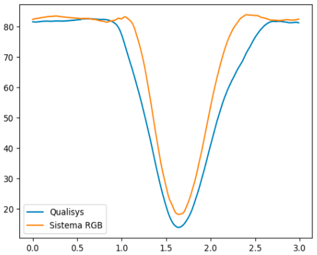
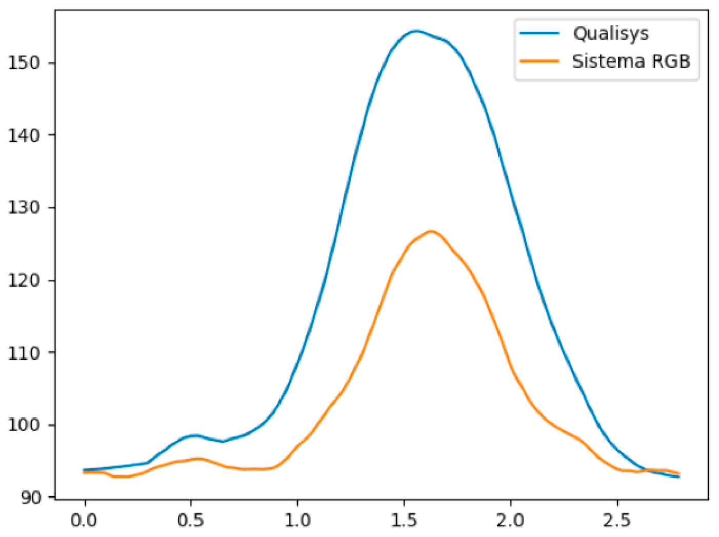
Figure 2 and Figure 3 illustrate, respectively, an example of a subject in the variation in the right hip flexion angle during the hip flexion movement, and the variation in the hip abduction angle during the right hip abduction movement, both in relation to time, measured by the Qualisys system and the MOVA3D system. Note that the mean error remains small throughout the range in Figure 2, and increases towards the end of the movement in Figure 3.

4. Discussion

The aim was to compare kinematic measurements of a three-dimensional motion capture and analysis system (MOVA3D), without markers and using a single RGB camera, for use in kinematic analysis. The correlation of this system with the Qualisys system is part of a larger objective of developing technological solutions for use in telerehabilitation systems that allow the physiotherapist to remotely manage and monitor the therapy prescribed at home, as well as increasing adherence to these exercises. This first stage of preliminary data analysis therefore represents an important step towards the construction of low-cost and easy-to-use systems that enable the dissemination of this type of technology in clinical practice. The pros and cons presented facilitate the improvement of these systems and also allow for greater transparency at the time of choice by the clinician.
In general, the MOVA3D system, when correlated to the Qualisys Track Manager (QTM) system, was able to adequately recognize the patterns of hip abduction, squat, and hip flexion movements, with a mean error ranging from 0.55° to 11.10°, except for the maximum angle in right hip abduction and minimum angle in right hip flexion (squat). Other investigations [32] found that most of the parameters have a mean error of 10°, which is comparable to the results obtained through the methodologies illustrated in this work.
As previously mentioned, our results show that the greatest mean true error was in the variable right hip abduction (hip abduction movement), when individuals reach the maximum angle of movement, reaching an error of 41.50°. Despite this, this same variable has a Pearson correlation index of 0.97, indicating that, despite having a high mean error, the variable behaved similarly to that observed by the QTM system (Figure 3).
We suppose that the estimate of the hip joint center made by the MOVA3D system suffers interference during the hip abduction movement, being displaced to the side of the body at the end of the movement, thus decreasing the calculated angle. The error was greater as hip abduction increased. This indicates a possibility of future improvement of the system, with adjustments in the prediction model of hip joint centers.
The variable right hip flexion (squat movement) also showed a high mean true error of 33° at the end of the hip flexion movement (minimum angle of movement). In this case, there was a moderate correlation (r = 0.55) between the MOVA3D and Qualisys measurements. This could have been due to occlusion of the hip by the knee during the end of the squat movement, since a single camera was used (monocular view), and this was positioned in the coronal plane in front of the subjects.
The occlusion of body parts is one of the biggest problems faced by systems without markers [33] that use monocular vision (use of only one camera) for the prediction of joint centers. Movements with large amplitudes performed in an orthogonal plane to the camera, such as hip flexion with the camera positioned in the coronal plane, can cause one joint, such as the knee, to overlap another (hip). In this case, the algorithm is required to predict the joint position without the visual information of the silhouette, which can increase the error.
The majority of studies on this theme in the literature validate systems for kinematic analysis based on depth cameras. These devices have, in addition to the common RGB camera, an infrared sensor, and an infrared point projector. The depth information is calculated through analysis of the projection distortion of the dot pattern by the software built into the camera itself, or by software developed for this purpose. These represent, therefore, real analyses, coming from the capture of direct data.
Our system, in comparison, uses only a standard RGB camera, as seen in cell phones or webcams, with monocular vision (only one lens). This represents almost all cameras built into smartphones sold around the world, which opens up possibilities for using our system on existing mobile devices, without the need to acquire or adapt external devices to capture movements.
Microsoft Kinect has been correlated in several studies for specific squat, hip abduction, and lunge movements, with correlations ranging from 0.18 to 0.83 when correlated to Vicon [32]. During the squat, knee flexion showed a high correlation between the systems (r = 0.88); for hip abduction, values ranged from 0.47 to 0.59; and for lunge, from 0.15 to 0.80 [30]. Schmitz et al. [34] found a correlation of 0.55 between Kinect V2 and Vicon for the evaluation of hip and knee angles during the squat, with results corroborated by Mentiplay et al. [35] for knee flexion during the single-leg squat exercise (r = 0.80). However, when assessing hip abduction, the authors found a low correlation and errors above 15° in the frontal plane. Kotsifaki et al. [36] found high agreement between systems for hip abduction and knee flexion movement during single-leg squat exercises.
Agustsson et al. [37] found a high correlation between the variables from a depth camera attached to an iPad and those collected by the QTM system to assess postural alterations. Vilas Boas et al. [38] found that both versions, V1 and V2, of Microsoft Kinect presented correlations ranging from moderate to poor for hip and upper limb angles during gait analysis, when compared to the Qualisys system. The best associations were observed for the knee joint, ranging from good to excellent [39].
RMS errors for hip and knee joint angles during gait have been reported, ranging from 4° to 10° [40] and 20.15° [38]. In another study, Bahadori et al. [41] found errors of 13.2% (SD 19.6) for knee angle and −3.9% for hip abduction, but with a standard deviation of 75, indicating that the Microsoft Kinect system, when compared to Vicon, is not suitable for clinical use. Tanaka et al. [42] found a moderate to high association between hip flexion and extension angles during gait when compared to the Vicon system.
Other systems such as Capture found a high correlation for heel height variables (r = 0.91) and a low correlation for knee varus (r = 0.29) when correlated to the Vicon system [43].
It can be observed, therefore, that currently, the most widely used systems for the evaluation of human movement are based on the tracking of reflective markers by infrared cameras, as is the case of the Qualisys, Vicon, Optotrak, and Motion Analysis systems. These systems have a high investment value and perform analyses with high precision and accuracy; however, they are difficult to use in clinical evaluations or inappropriate for application in telerehabilitation systems.
Systems without the use of markers (markerless) based on Microsoft Kinect have been developed with the aim of providing a quick and uncomplicated evaluation of human movement, and despite the loss in accuracy and precision, they are capable of evaluating movement patterns satisfactorily in several environments, which may enable their use in clinical and remote monitoring applications. However, despite these characteristics, Microsoft Kinect is not widely accessible to the end user (patients in physiotherapy clinics), and requires the use of specific software and cable connections to a desktop computer. This limits Its use in telerehabilitation systems.
Markerless systems still require refinement in their development, to allow data capture and feedback in real time for the user, with low energy and processing costs, enabling their use in smartphones or tablets. In the near future, the system would be part of telerehabilitation systems, allowing clinicians to assess and follow-up patients at a distance, reducing travels to clinics, and optimizing cost therapeutic performance.
In addition, and of utmost importance, future validation studies in comparison with the gold standard are required that allow more adequate use of these systems in telerehabilitation.

Limitation

This study has some limitations that affect its application. Firstly, the small number of volunteers did not represent the diversity of body pattern variations necessary to evaluate the capabilities of different anatomical characteristics. The low number of participants was due to COVID-19 restrictions on movement and social distancing.
Furthermore, the signals captured by Qualisys have more than three times the frequency of the RGB camera sensors (100 Hz/30 Hz). Therefore, signals need to be filtered and smoothed more effectively. To achieve this, it is necessary to ensure that the signals are of equal size through interpolation carried out simultaneously with the synchronization process from the beginning of the angular peak estimates to the last peak. A second-order polynomial interpolation function is recommended [44] instead of the cubic interpolation used in this study.
Despite these limitations, the study’s results are an essential step towards the rigorous validation of MOVA3D for clinical assessment of lower limb kinematics. It provides valuable insights into reliable and unreliable kinematic variables.

5. Conclusions

The MOVA3D system was shown to be superior to other markerless 3D motion analysis systems, reaching high correlations for six of the eight analyzed variables. The same occurred for the mean error, remaining below 12° in only 4 of the 24 variables analyzed. The MOVA3D system is therefore promising for use in kinematic evaluations with applications in telerehabilitation. The system presents room for improvement in hip abduction and flexion movements.

Author Contributions

Conceptualization, L.P.d.A. and T.N.B.; methodology, L.P.d.A., P.S.d.C. and R.P.I.; processing, T.N.B., P.S.d.C. and R.P.I.; formal analysis, L.P.d.A. and P.S.d.C.; investigation, L.P.d.A.; resources, T.N.B.; data curation, L.P.d.A.; writing—original draft preparation, L.P.d.A.; writing—review and editing, D.d.O.F.; visualization, L.C.G. and D.d.O.F.; supervision, L.C.G.; project administration, T.N.B. All authors have read and agreed to the published version of the manuscript.

Funding

This study was financed in part by the Coordenação de Aperfeiçoamento de Pessoal de Nível Superior—Brasil (CAPES)—Finance Code 001.

Institutional Review Board Statement

Study design was approved by the IRB-Universidade Federal de Mato Grosso do Sul (#2 9358720.7.0000.0021).

Informed Consent Statement

Informed consent was obtained from all subjects involved in the study.

Data Availability Statement

Not applicable.

Acknowledgments

The authors would like to thank the authorization to use the Movement Analysis Laboratory at the Associação de Pais e Amigos dos Excepcionais de Campo Grande (APAE/CG). This study was possible due to the support of the Federal University of Mato Grosso do Sul—UFMS/MEC—Brasil.

Conflicts of Interest

The authors declare no conflict of interest.

References

  1. Zampolini, M.; Todeschini, E.; Hermens, H.; Ilsbroukx, S.; Macellari, V.; Magni, R.; Rogante, M.; Scattareggia Marchese, S.; Vollenbroek, M.; Giacomozzi, C. Tele-rehabilitation: Present and future. Ann. Ist. Super. Sanita 2008, 44, 125–134. [Google Scholar]
  2. Brennan, D.M.; Mawson, S.; Brownsell, S. Telerehabilitation: Enabling the remote delivery of healthcare, rehabilitation, and self management. Stud. Health Technol. Inform. 2009, 145, 231–248. [Google Scholar] [PubMed]
  3. Dinesen, B.; Haesum, L.K.; Soerensen, N.; Nielsen, C.; Grann, O.; Hejlesen, O.; Toft, E.; Ehlers, L. Using preventive home monitoring to reduce hospital admission rates and reduce costs: A case study of telehealth among chronic obstructive pulmonary disease patients. J. Telemed. Telecare 2012, 18, 221–225. [Google Scholar] [CrossRef] [PubMed]
  4. Tousignant, M.; Moffet, H.; Boissy, P.; Corriveau, H.; Cabana, F.; Marquis, F. A randomized controlled trial of home telerehabilitation for post-knee arthroplasty. J. Telemed. Telecare 2011, 17, 195–198. [Google Scholar] [CrossRef]
  5. Cason, J. A pilot telerehabilitation program: Delivering early intervention services to rural families. Int. J. Telerehabil. 2009, 1, 29–38. [Google Scholar] [CrossRef] [PubMed]
  6. Weiss, P.L.; Sveistrup, H.; Rand, D.; Kizony, R. Video capture virtual reality: A decade of rehabilitation assessment and intervention. Phys. Ther. Rev. 2009, 14, 307–321. [Google Scholar] [CrossRef]
  7. Frederix, I.; Hansen, D.; Coninx, K.; Vandervoort, P.; Vandijck, D.; Hens, N.; Van Craenenbroeck, E.; Van Driessche, N.; Dendale, P. Effect of comprehensive cardiac telerehabilitation on one-year cardiovascular rehospitalization rate, medical costs and quality of life: A costeffectiveness analysis. Eur. J. Prev. Cardiol. 2016, 23, 674–682. [Google Scholar] [CrossRef]
  8. Pacheco, T.B.F.; Bezerra, D.A.; de Silva, J.P.; Cacho, Ê.W.A.; de Souza, C.G.; Cacho, R.O. The Implementation of Teleconsultations in a Physiotherapy Service During Covid-19 Pandemic in Brazil: A Case Report. Int. J. Telerehabil. 2021, 22, e6368. [Google Scholar] [CrossRef]
  9. Bidargaddi, N.; Sarela, A. Activity and heart rate-based measures for outpatient cardiac rehabilitation. Methods Inf. Med. 2008, 47, 208–216. [Google Scholar]
  10. Fan, Y.J.; Yin, Y.H.; Xu, L.D.; Zeng, Y.; Wu, F. IoT-Based Smart Rehabilitation System. IEEE Trans. Ind. Inform. 2014, 10, 1568–1577. [Google Scholar]
  11. Hamida, S.T.B.; Hamida, E.B.; Ahmed, B. A new mHealth communication framework for use in wearable WBANs and mobile technologies. Sensors 2015, 15, 3379–3408. [Google Scholar] [CrossRef] [PubMed]
  12. Rolim, C.O.; Koch, F.L.; Westphall, C.B.; Werner, J.; Fracalossi, A.; Salvador, G.S. A Cloud Computing Solution for Patient’s Data Collection in Health Care Institutions. In Proceedings of the 2010 Second International Conference on eHealth, Telemedicine, and Social Medicine, Saint Maarten, The Netherlands, 10–16 February 2010. [Google Scholar]
  13. Benharref, A.; Serhani, M.A. Novel Cloud and SOA-Based Framework for E-Health Monitoring Using Wireless Biosensors. IEEE J. Biomed. Health Inform. 2014, 18, 46–55. [Google Scholar] [CrossRef]
  14. Breedon, P.; Byrom, B.; Siena, L.; Muehlhausen, W. Enhancing the Measurement of Clinical Outcomes Using Microsoft Kinect. In Proceedings of the 2016 International Conference on Interactive Technologies and Games (ITAG), Notthingham, UK, 26–27 October 2016; pp. 61–69. [Google Scholar]
  15. Llorens, R.; Gil-Gomez, J.A.; Mesa-Gresa, P.; Alcaniz, M.; Colomer, C.; Noe, E. BioTrak: A comprehensive overview. In Proceedings of the 2011 International Conference on Virtual Rehabilitation (ICVR), Zurich, Switzerland, 27–29 June 2011. [Google Scholar]
  16. Spina, G.; Huang, G.; Vaes, A.; Spruit, M.; Amft, O. COPDTrainer: A smartphone-based motion rehabilitation training system with real-time acoustic feedback. In Proceedings of the 2013 ACM International Joint Conference on Pervasive and Ubiquitous Computing, Zurich, Switzerland, 9–12 September 2013. [Google Scholar]
  17. Giorgino, T.; Tormene, P.; Maggioni, G.; Pistarini, C.; Quaglini, S. Wireless Support to Poststroke Rehabilitation: MyHeart’s Neurological Rehabilitation Concept. IEEE Trans. Inf. Technol. Biomed. 2009, 13, 1012–1018. [Google Scholar] [CrossRef]
  18. Holden, M.K.; Dyar, T.A.; Dayan-Cimadoro, L. Telerehabilitation using a virtual environment improves upper extremity function in patients with stroke. IEEE Trans. Neural Syst. Rehabil. Eng. 2007, 15, 36–42. [Google Scholar] [CrossRef] [PubMed]
  19. Saracino, L.; Avizzano, C.A.; Ruffalde, E.; Cappiello, G.; Curto, Z.; Scoglio, A. Motore++ A portable haptic device for domestic rehabilitation. In Proceedings of the 42nd Annual Conference of the IEEE Industrial Electronics Society, Florence, Italy, 23–26 October 2016. [Google Scholar]
  20. Díaz, I.; Catalan, J.M.; Badesa, F.J.; Justo, X.; Lledo, L.D.; Ugartemendia, A.; Gil, J.J.; García-Aracil, N. Development of a robotic device for post-stroke home tele-rehabilitation. Adv. Mech. Eng. 2018, 10, 1687814017752302. [Google Scholar] [CrossRef]
  21. Bai, J.; Song, A.; Xu, B.; Nie, J.; Li, H. A Novel Human-Robot Cooperative Method for Upper Extremity Rehabilitation. Int. J. Soc. Robot. 2017, 9, 265–275. [Google Scholar] [CrossRef]
  22. Ganguly, A.; Rashidi, G.; Mombaur, K. Comparison of the Performance of the Leap Motion ControllerTM with a Standard Marker-Based Motion Capture System. Sensors 2021, 3, 1750. [Google Scholar] [CrossRef]
  23. Scott, B.; Seyres, M.; Philp, F.; Chadwick, E.K.; Blana, D. Healthcare applications of single camera markerless motion capture: A scoping review. PeerJ 2022, 26, 13517. [Google Scholar] [CrossRef]
  24. Regazzoni, D.; de Vecchi, G.; Rizzi, C. RGB cams vs RGB-D sensors: Low cost motion capture technologies performances and limitations. J. Manuf. Syst. 2014, 33, 719–728. [Google Scholar] [CrossRef]
  25. Costa, I.F. Uso de Ressoadores de Anéis Fendidos e Ressoadores de Anéis Fendidos Complementares Para o Melhoramento do Desempenho em Filtros Passa-Baixa em Microfita. Ph.D. Thesis, Federal University of Rio Grande do Norte, Natal, Brazil, 2018. [Google Scholar]
  26. Tomescu, S.S.; Bakker, R.; Beach, T.A.C.; Chandrashekar, N. The Effects of Filter Cutoff Frequency on Musculoskeletal Simulations of High-Impact Movements. J. Appl. Biomech. 2018, 34, 336–341. [Google Scholar] [CrossRef] [PubMed]
  27. Mai, P.; Willwacher, S. Effects of low-pass filter combinations on lower extremity joint moments in distance running. J. Biomech. 2019, 95, 109311. [Google Scholar] [CrossRef]
  28. Souza, I.C.M. Avaliação Biomecânica do Movimento Humano. Master’s Thesis, Universidade Católica Portuguesa, Porto, Portugal, 2020. [Google Scholar]
  29. Rocha, A.P.; Choupina, H.M.P.; Vilas-Boas, M.D.C.; Fernandes, J.M.; Cunha, J.P.S. System for automatic gait analysis based on a single RGB-D camera. PLoS ONE 2018, 13, 0201728. [Google Scholar] [CrossRef]
  30. Wu, G.; Siegler, S.; Allard, P.; Kirtley, C.; Leardini, A.; Rosenbaum, D.; Whittle, M.; D’Lima, D.D.; Cristofolini, L.; Witte, H.; et al. ISB recommendation on definitions of joint coordinate system of various joints for the reporting of human joint motion—Part I: Ankle, hip, and spine. J. Biomech. 2002, 35, 543–548. [Google Scholar] [CrossRef]
  31. Portney, L.G.; Watkins, M.P. Foundations of Clinical Research: Applications to Practice, 3rd ed.; Prentice Hall: Upper Saddle River, NJ, USA, 2009. [Google Scholar]
  32. Wochatz, M.; Tilgner, N.; Mueller, S.; Rabe, S.; Eichler, S.; John, M.; Völler, H.; Mayer, F. Reliability and validity of the Kinect V2 for the assessment of lower extremity rehabilitation exercises. Gait Posture 2019, 70, 330–335. [Google Scholar] [CrossRef]
  33. Geelen, J.E.; Branco, M.P.; Ramsey, N.F.; Van Der Helm, F.C.; Mugge, W.; Schouten, A.C. MarkerLess Motion Capture: ML-MoCap, a low-cost modular multi-camera setup. In Proceedings of the 2021 43rd Annual International Conference of the IEEE Engineering in Medicine & Biology Society (EMBC), Virtual Conference, 1–5 November 2021; pp. 4859–4862. [Google Scholar]
  34. Schmitz, A.; Ye, M.; Boggess, G.; Shapiro, R.; Yang, R.; Noehren, B. The measurement of in vivo joint angles during a squat using a single camera markerless motion capture system as compared to a marker-based system. Gait Posture 2015, 41, 694–698. [Google Scholar] [CrossRef] [PubMed]
  35. Mentiplay, B.F.; Hasanki, K.; Perraton, L.G.; Pua, Y.H.; Charlton, P.C.; Clark, R.A. Three-dimensional assessment of squats and drop jumps using the Microsoft Xbox One Kinect: Reliability and validity. J. Sports. Sci. 2018, 36, 2202–2209. [Google Scholar] [CrossRef] [PubMed]
  36. Kotsifaki, A.; Whiteley, R.; Hansen, C. Dual Kinect v2 system can capture lower limb kinematics reasonably well in a clinical setting: Concurrent validity of a dual camera markerless motion capture system in professional football players. BMJ Open Sport Exer. Med. 2018, 4, 000441. [Google Scholar] [CrossRef]
  37. Agustsson, A.; Gislason, M.K.; Ingvarsson, P.; Rodby-Bousquet, E.; Sveisson, T. Validity and reliability of an iPad with a three-dimensional camera for posture imaging. Gait Posture 2019, 68, 357–362. [Google Scholar] [CrossRef] [PubMed]
  38. Vilas-Boas, M.D.C.; Choupina, H.M.P.; Rocha, A.P.; Fernandes, J.M.; Cunha, J.P.S. Full-body motion assessment: Concurrent validation of two body tracking depth sensors versus a gold standard system durins gait. J. Biomech. 2019, 87, 189–196. [Google Scholar] [CrossRef]
  39. Chakraborty, S.; Nandy, A.; Yamaguchi, T.; Bonnet, V.; Venture, G. Accuracy of image data stream of a markerless motion capture system in determining the local dynamic stability and joint kinematics of human gait. J. Biomech. 2020, 104, 109718. [Google Scholar] [CrossRef]
  40. Xu, X.; McGorry, R.W.; Chou, L.S.; Lin, J.H.; Chang, C.C. Accuracy of the Microsoft Kinect for measuring gait parameters during treadmill walking. Gait Posture 2015, 42, 145–151. [Google Scholar] [CrossRef] [PubMed]
  41. Bahadori, S.; Davenport, P.; Immins, T.; Wainwright, T.W. Validation of joint angle measurements: Comparison of a novel low-cost marker-less system with an industry standard marker-based system. J. Med. Eng. Technol. 2019, 43, 19–24. [Google Scholar] [CrossRef] [PubMed]
  42. Tanaka, R.; Takimoto, H.; Yamasaki, T.; Higashi, A. Validity of time series kinematical data as measured by a markerless motion capture system on a flatland for gait assessment. J. Biomech. 2018, 71, 281–285. [Google Scholar] [CrossRef] [PubMed]
  43. Harsted, S.; Holsgaard-Larsen, A.; Hestbæk, L.; Boyle, E.; Lauridsen, H.H. Concurrent validity of lower extremity kinematics and jump characteristics captured in pre-school children by a markerless 3D motion capture system. Chiropr. Man. Ther. 2019, 11, 27–39. [Google Scholar] [CrossRef]
  44. Lafayette, T.B.d.G.; Kunst, V.H.d.L.; Melo, P.V.d.S.; Guedes, P.d.O.; Teixeira, J.M.X.N.; Vasconcelos, C.R.d.; Teichrieb, V.; da Gama, A.E.F. Validation of Angle Estimation Based on Body Tracking Data from RGB-D and RGB Cameras for Biomechanical Assessment. Sensors 2023, 23, 3. [Google Scholar] [CrossRef]
Figure 1. Experimental camera layout for RGB (those labeled in yellow) and QTM (those marked by the white triangles).
Ijerph 20 06657 g001
Figure 2. Variation of the right hip flexion angle during the hip flexion movement, measured by the Qualisys system (blue) and the MOVA3D system (orange). The X-axis represents time and the Y-axis the angular variation in degrees.
Ijerph 20 06657 g002
Figure 3. Variation of the right hip abduction angle during the hip abduction movement, measured by the Qualisys system (blue) and the MOVA3D system (orange). The X-axis represents time and the Y-axis the angular variation in degrees.
Ijerph 20 06657 g003
Table 1. Nomenclature of anatomical reference.
NomenclaturaAnatomical Reference
1R_ASISRight Anterior Superior Iliac Spine
2L_ASISLeft Anterior Superior Iliac Spine
3R_PSISRight Posterior Superior Iliac Spine
4L_PSISLeft Posterior Superior Iliac Spine
5R_TROCRight Trochanter
6L_TROCLeft Trochanter;
7R_EPILLateral Epicondyle of the Right Femur
8L_EPILLateral Epicondyle of the Left Femur
9R_MEPILMedial Epicondyle of the Right Femur
10L_MEPILMedial Epicondyle of the Left Femur
11R_FIBHRight Fibular Head
12L_FIBHLeft Fibular Head;
13R_TTUBRight Tibial Tuberosity
14L_TTUBLeft Tibial Tuberosity
15R_LMALRight Lateral Malleolus
16L_LMALLeft Lateral Malleolus
17R_MMALRight Medial Malleolus
18L_LMALLeft Medial Malleolus
19R_CALLeft Calcaneus
20L_CALRight Calcaneus
21R_1MET1st Right Metatarsal
22L_1MET1st Left Metatarsal
23R_2MET2nd Right Metatarsal
24L_2MET2nd Left Metatarsal
25R_5MET5th Right Metatarsal
26L-5MET5th Left Metatarsal
Description of acronyms and respective nomenclatures related to anatomical references.
Table 2. Mean values of the maximum and minimum angles.
Movement QualisysMova 3D
Maximum AngleMinimum AngleROMMaximum AngleMinimum AngleROM
Hip abductionABD_RH151.592.5591109020
ABD_LH115.992.223.7105.290.714.5
SquatFLX_RK65.27.557.754.16.947.2
FLX_LK67.37.459.966.810.556.5
FLX_RH7924.454.387.957.430.6
FLX_LH79.629.350.387.337.749.6
Hip flexionFLX_RH81.4418.44638621.5563.11
FLX_LH86.6675.3311.3386.1179.226.88
ROM: range of motion; ABD_RH: right hip abduction; ABD_LH: left hip abduction; FLX_RK: right knee flexion; FLX_LK: left knee flexion; FLX_RH: right hip flexion.
Table 3. Mean error of the maximum, minimum, and range of motion.
Movement Mean Error (Qualisys—Mova 3D)
Maximum AngleMinimum AngleROM
Hip abductionABD_RH41.502.5039.00
ABD_LH10.701.509.20
SquatFLX_RK11.100.6010.50
FLX_LK0.50−3.103.40
FLX_RH−8.90−33.0023.70
FLX_LH−7.70−8.400.70
Hip flexionFLX_RH−4.56−3.11−0.11
FLX_LH0.55−3.894.45
ROM: range of motion; ABD_RH: right hip abduction; ABD_LH: left hip abduction; FLX_RK: right knee flexion; FLX_LK: left knee flexion; FLX_RH: right hip flexion; FLX_LH: left hip flexion.
Table 4. Pearson’s correlation for maximum and minimum.
Pearson’s Correlation
rSD95% CIp
Hip abductionABD_RH0.970.040.03<0.001
ABD_LH0.840.120.07<0.001
SquatFLX_RK0.830.170.01<0.001
FLX_LK0.940.020.01<0.001
FLX_RH0.550.490.03<0.001
FLX_LH0.870.050.03<0.001
Hip flexionFLX_RH0.930.030.02<0.001
FLX_LH−0.180.650.42<0.001
r: Pearson correlation coefficient; SD standard deviation; 95% CI confidence interval; ABD_RH: right hip abduction; ABD_LH: left hip abduction; FLX_RK: right knee flexion; FLX_LK: left knee flexion; FLX_RH: right hip flexion; FLX_LH: left hip flexion.
Disclaimer/Publisher’s Note: The statements, opinions and data contained in all publications are solely those of the individual author(s) and contributor(s) and not of MDPI and/or the editor(s). MDPI and/or the editor(s) disclaim responsibility for any injury to people or property resulting from any ideas, methods, instructions or products referred to in the content.