Article

Facile Phase Control and Photocatalytic Performance of BiVO4 Crystals for Methylene Blue Degradation

1
Biochar Engineering Technology Research Center of Guangdong Province, School of Environmental and Chemical Engineering, Foshan University, Foshan 528000, China
2
School of Environmental Science and Engineering, Guangdong University of Petrochemical Technology, Maoming 525000, China
3
School of Life Science, Qufu Normal University, Qufu 273165, China
*
Author to whom correspondence should be addressed.
Int. J. Environ. Res. Public Health 2023, 20(4), 3093; https://doi.org/10.3390/ijerph20043093
Submission received: 13 December 2022 / Revised: 6 February 2023 / Accepted: 6 February 2023 / Published: 10 February 2023
(This article belongs to the Special Issue Research in Emerging Contaminants: Impact and Solutions)

Abstract

:
Emerging contaminants, which mainly exist as organic pollutants and pose adverse biological effects, could be removed using photocatalytic degradation, resulting in a low-cost and environmentally friendly solution. Herein, BiVO4 nanoparticles with different morphologies and photocatalytic performances were synthesized by hydrothermal treatment at different residence times. The XRD and SEM results indicate that the crystal phase of BiVO4 gradually transformed from a single tetragonal phase to a single monoclinic crystal phase as the hydrothermal time increased, and with the extension of the hydrothermal time, the morphology of BiVO4 nanoparticles gradually differentiated from a smooth spherical shape to flower-like shapes composed of polyhedrons; the size of the crystals also increased accordingly. Methylene blue (MB), used as a probe of organic pollutants, was degraded under visible light irradiation by all BiVO4 samples to investigate its photocatalytic activities. The experimental results show that the longer the hydrothermal time, the better the photocatalytic performance. The optimum hydrothermal time was 24 h, at which the sample showed the highest photocatalytic activity for MB degradation. This work shows a convenient strategy for control of the crystal phase of BiVO4-based photocatalysts based on the understanding of the crystal morphology evolution mechanism, which will benefit the researchers in designing new BiVO4-based photocatalysts with high efficiency for emerging contaminants’ degradation.

1. Introduction

The awareness of green eco-environmental protection has been enhanced due to the recent developments in social science and technology [1]. Photocatalytic technology has become increasingly important as a green and sustainable technology [2,3,4]. Emerging contaminants, such as microplastics, persistent organic pollutants, antibiotics, pesticides, and endocrine disruptor chemicals, could damage the reproduction of human beings and animals [5,6,7]. Most of the emerging contaminants exist as organic pollutants and could be removed by the adsorptive and photocatalytic approach, which has recently gained broad attention [8,9,10].
BiVO4 is a new type of visible light catalyst that has become popular due to its nontoxicity, high photostability, low cost of production, narrow band gap, response bands in visible light regions, and other advantages [3,11]. The crystallinity phase of BiVO4 can be easily controlled by adjusting preparation conditions, playing a vital role in its photocatalytic efficiency, which is a low-cost strategy for enhancing the photocatalytic performance for water treatment [12]. It has been found that there are three main crystal structures in the BiVO4: a tetrahedral scheelite structure; monoclinic scheelite structure; and tetrahedral zircon structure [13]. Among these, the band gap (Eg) of the BiVO4 with monoclinic and tetrahedral scheelite structures is approximately 2.4 eV [14], and that of the tetrahedral zircon structure is approximately 2.9 eV. Although monoclinic BiVO4 and tetrahedral BiVO4 have similar crystal structures [15], the BiVO4 with monoclinic scheelite structures has good visible light catalytic properties. At the same time, the BiVO4 with tetrahedral zircon structures has poor photocatalytic performances, indicating that the crystal structures have major influences on the photocatalytic properties of the BiVO4. The results of this study showed that the three crystal structures of the BiVO4 could be mutually converted under different conditions [16]. However, after becoming irradiated by a certain energy of the photon, the photoelectron generated by the BiVO4 was found to have a short separation time from the photo-hole, which led to minimal photon efficiency [17]. This factor was observed to largely limit the applications and development of the BiVO4.
In recent years, the studies regarding BiVO4 have mainly included the examination of the morphology [18] and the construction of a BiVO4-based heterojunction catalyst, as well as improvements in the preparation methods [19]. These improved methods were mainly determined in order to increase the separation times of the photogenic electron pairs and thereby increase the photocatalytic capacity of the BiVO4. However, many factors are known to influence BiVO4. For example, different preparation methods can affect the morphology, crystal types, and other properties of BiVO4 [20]. Additionally, the addition of additives was found to regulate the formation of its morphology [21]. It has also been observed that different materials have serious influences on the physical and chemical properties of BiVO4. In addition, a new BiVO4 composite has been synthesized by doping metal or other semiconducting materials [22], and this has become a major research focus in the field of photocatalytic materials.
The main preparation methods of BiVO4 include the following: high-temperature solid phase methods; hydrothermal methods; chemical precipitation methods [23]; sol-gel methods [24]; microwave synthesis methods [25], and so on. Among these methods, hydrothermal synthesis technology has been more widely studied. In the BiVO4 synthesis process using hydrothermal methods, the pH value of the precursor solution, reaction temperatures, reaction times, and precursor solution concentrations are the main factors affecting the nanoparticle sizes, crystal forms, morphology, and specific surface areas. In this research study, the variable hydrothermal times during the synthesis of the BiVO4 were controlled in order to explore the differences in the crystal structures, morphology, specific surface areas, and photocatalytic performances of the BiVO4 generated under different hydrothermal times. In this paper, methylene blue was used as a probe to investigate the photocatalytic performance of BiVO4 toward emerging organic contaminants [26]. Additionally, the generation processes of BiVO4 with different crystallinity phases and their photocatalytic performances were further studied to pave the way for the low-cost strategy of enhancing the photocatalytic performance of BiVO4 applied in water treatment and environmental protection.

2. Materials and Methods

2.1. Experimental Materials

In this study, the Bi(NO3)3·5H2O (AR) was obtained from Tianjin Kemiou Chemical Reagent Co., Ltd.; the NH4VO3 (AR) was provided by the Tianjin Fuchen Chemical Reagent Factory; the NaOH (AR) was obtained from Xilong Scientific Co., Ltd.; Tianjin Fuyu Fine Chemical Co., Ltd. provided the absolute ethyl alcohol (AR); and the HNO3 (AR) used in this study was obtained from Guangdong Guanghua Sci-tech Co., Ltd.

2.2. Preparation Methods

In this research study, under the conditions of no template and no surfactant, 2.45 g of Bi(NO3)3·5H2O and 0.6 g of NH4VO3 were dissolved in 40 mL of 4 mol/L HNO3 solution and NaOH solution, respectively. Then, after Bi(NO3)3·5H2O and NH4VO3 were fully dissolved, the two solutions were combined, followed by magnetic stirring for 20 min and sonification for 10 min. The NaOH and HNO3 solution was used to adjust the pH value to 6. Then, the obtained solution was placed in a static condition overnight and transferred to a reaction kettle with a Teflon-lined stainless-steel autoclave, which was followed by a reaction at 180 °C for 0, 3, 6, 12, 14, and 24 h periods. When they had been cooled to room temperature, the as-prepared samples were washed three times with deionized water and ethanol. Then, centrifugal filtering was performed, and a six-hour drying process at 80 °C was completed in order to obtain the purified products [27]. The preparetion illustration is presented as Figure 1. The hydrothermal times were 0, 3, 6, 12, 14, and 24 h, and the prepared products were, respectively, referred to as t = 0 BiVO4; t = 3 BiVO4; t = 6 BiVO4; t = 12 BiVO4; t = 14 BiVO4; and t = 24 BiVO4.

2.3. Characterizations

The BiVO4 structure was measured using X-ray powder diffraction (XRD, Smart Lab (3 KW)) between 10° and 75°, which used Cu K radiation (=0.15406 nm) produced at 40 kV and 40 mA. The JCPDS (Joint Committee on Powder Diffraction Standards) files were compared to identify the crystalline phases. The morphological examination used a field emission scanning electron microscope (SEM, FEI Quanta 400 FEG) with an energy dispersive X-ray analyzer system (EX-250). The specific surface area of the samples was characterized using a TriStar II 3020 surface area analyzer at 77 K. UV-Vis diffuse reflection spectra (DRS) were collected with a UV-Vis spectrophotometer (Lambda, 650).

2.4. Photocatalytic Activity Tests

30 mg of the BiVO4 prepared in the above-mentioned process was weighed and placed into 20 mg/L of methylene blue solution (30 mL), and then stirred under dark conditions for thirty minutes. The solution was then irradiated by visible light with a wavelength λ > 420 nm (550 W xenon lamp illumination, BL-GHX-V, Shanghai Bilang Instrument Manufacturing Co., Ltd.). The distance from the light source to the photocatalytic reactor was set as 10 cm. A shading system was equipped to ensure that the photocatalytic degradation was not affected by the light from outside. Sampling was conducted every 30 min, and the supernatant was collected in order to measure the absorbance after centrifugation.

3. Results and Discussion

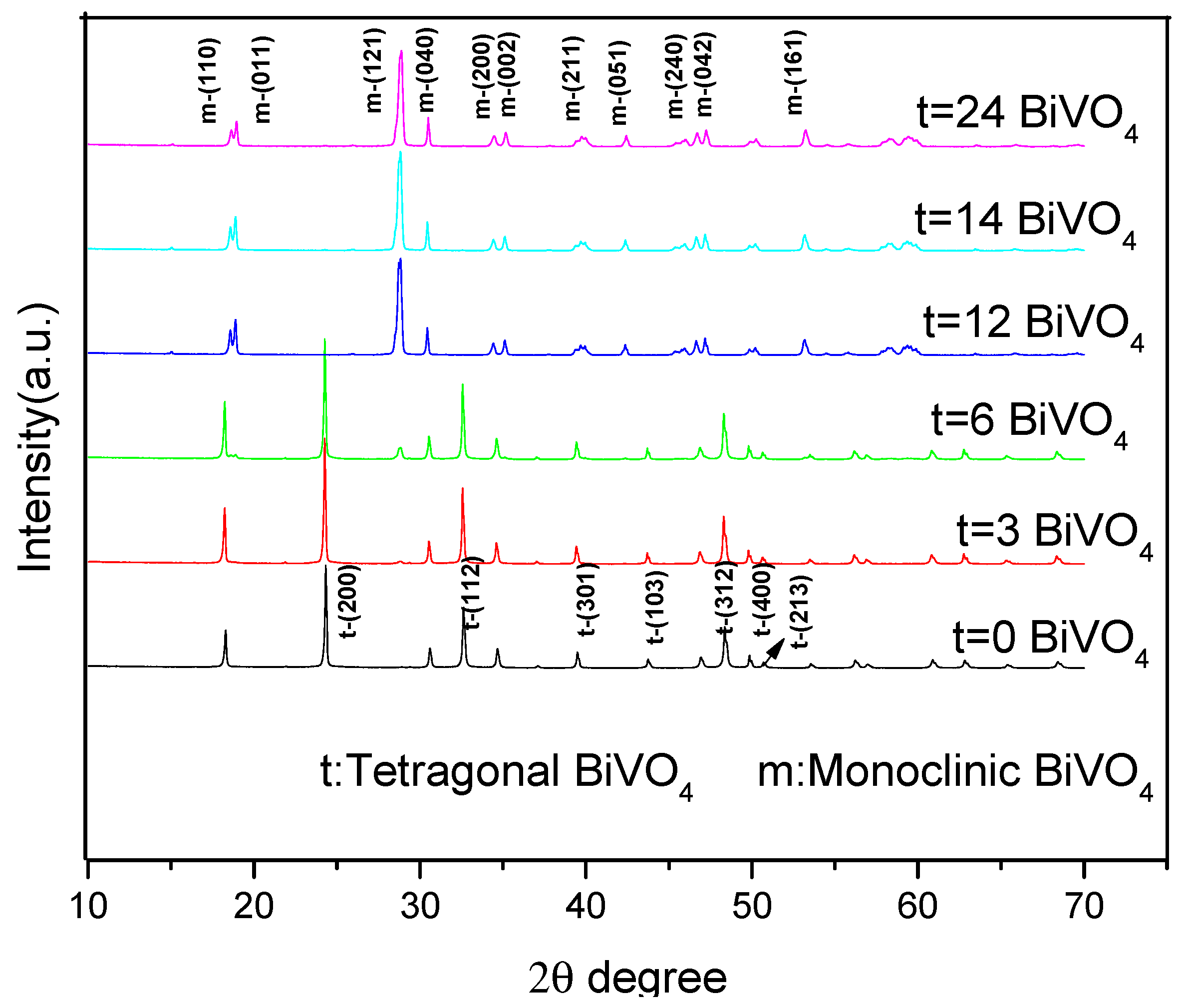
3.1. X-ray Diffraction (XRD)

Figure 2 details the X-ray diffraction patterns when the hydrothermal times were 0, 3, and 6 h, and 2 θ = 18.6°, 18.8°, 24.9°, 28.8°, 30.4°, 33.1°, 35.1°, 39.9°, 42.4°, 46.0°, 46.6°, 47.2°, 50.2°, 53.2°, 58.2°, and 59.4° with the diffraction peaks. When the hydrothermal times were zero and three hours, the obtained BiVO4 was found to display tetragonal diffraction peaks. When the hydrothermal time was six hours, a weak diffraction peak (121) in the monoclinic BiVO4 appeared. These findings indicated that when the hydrothermal times were zero and three hours, the tetragonal BiVO4 was obtained. However, when the hydrothermal time was six hours, the mixed crystals of the tetragonal and monoclinic phases were obtained. It was found that when the hydrothermal times were 12, 14, and 24 h, the characteristic peaks at 2 θ values of 18.6°, 18.8°, 28.8°, 30.4°, 35.1°, 39.9°, 42.4°, 46.0°, 46.6°, 47.2°, 50.2°, 53.2°, 58.2°, and 59.4° were consistent with the (110), (011), (120), (040), (200), (002), (211), (150), (024), (202), (161), (321), and (123) of the monoclinic BiVO4 (PDF NO. 75-1866). Therefore, the resulting product was determined to be a single monoclinic BiVO4 crystal [28]. It was also indicated that the times of the hydrothermal reactions were directly related to the crystal shape of the BiVO4. Furthermore, the longer hydrothermal times were found to be more conducive to the generation of the monoclinic BiVO4. The results of this study indicated that the generation process of the BiVO4 crystals was as follows: The tetragonal phase to mixed crystals of the tetragonal and monoclinic phases, and then to a single monoclinic phase.
In order to better clarify the influence degrees of the hydrothermal times on each crystal plan growth of the BiVO4, (011) was taken as the internal standard of the diffraction peak to normalize the XRD diffraction peak intensity. Table 1 lists the (121)/(011) and (040)/(011) intensity of each diffraction peak. It was observed that when t = 6 h, the (121) and (040) diffraction peaks were relatively weak, and the (121)/(011) and (040)/(011) values were 0.215 and 0.408. However, with the increases in the hydrothermal times, the (121) and (040) diffraction peaks were gradually strengthened, which indicated that certain hydrothermal times were conducive to the formations of the (121) and (040) diffraction peaks. In the m-BiVO4 standard PDF card (JCPDS No. 140688), the (040)/(121) intensity ratio is 0.25, which has been taken as the standard. When t = 6 h, the (040)/(121) had a value of 1.897, which indicated a weak (121) diffraction peak intensity. Then, as the hydrothermal times increased, the (040)/(121) values were determined to be 0.261, 0.296, and 0.307, which were close to the (040)/(121) values of 0.25 in the m-BiVO4 standard PDF card (JCPDS No. 140688). In this study, when t = 12 h, the value of 0.261 was almost approached, which confirmed that that particular hydrothermal time was favorable for the formation of m-BiVO4 diffraction peaks. However, when t was 14 and 24 h, it was observed that the (040)/(121) values displayed slightly increasing trends, indicating that when the hydrothermal time was 12 h, the value of the (040)/(121) was the most similar to the ratio in the m-BiVO4 standard PDF card (JCPDS No. 140688).

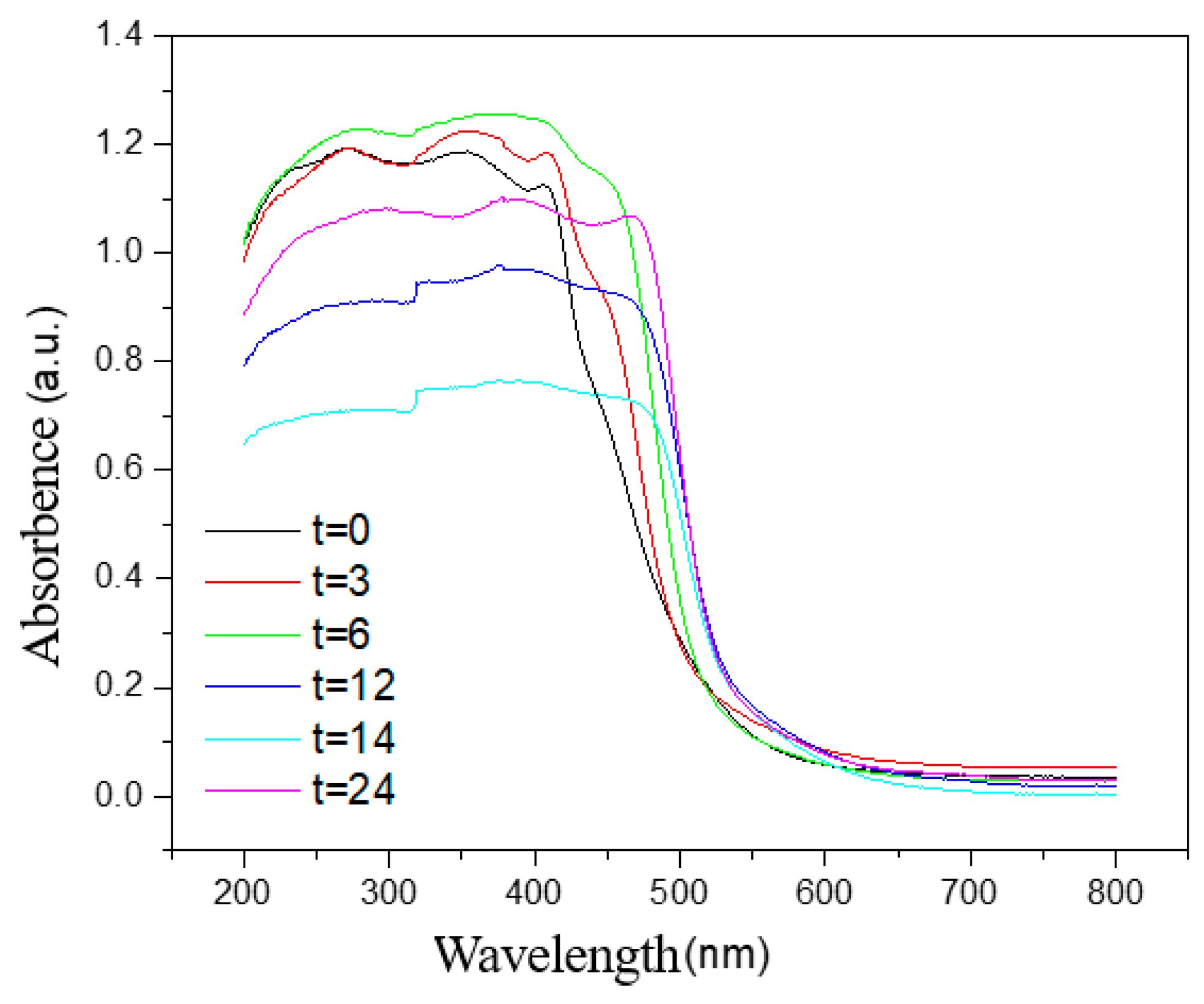
3.2. UV-Vis Test Analysis

Figure 3 details the UV-Vis spectra of BiVO4 under different hydrothermal time conditions. It can be seen that the samples in the range of 520 to 580 nm displayed strong absorption abilities. Additionally, the sharp drop in the regional absorbing boundary was due to the transition of the electrons in the semiconductor material during the absorption of certain energies [12]. It can be seen from the figure that when the hydrothermal time was 12 h, 14 h, and 24 h, the absorption band was more red-shifted than at 0 h, 3 h, and 6 h. This was found to be consistent with the conclusions reached regarding the BiVO4 XRD spectrum. Therefore, it can be seen that during the period ranging from 0 to 12 h, with the increases in the hydrothermal times, the absorption band of the BiVO4 generated the redshift, and its band gap also decreased. However, when the hydrothermal times were 14 and 24 h, its redshift was not obvious, which may be related to the agglomeration of the BiVO4 crystals, along with the diameter sizes of the crystals.

3.3. SEM Test Analysis

As can be seen from the SEM image displayed in Figure 4, as the hydrothermal time increased, the shape generally became spheroid. Additionally, when the hydrothermal times were 0 and 3 h, its spherical surfaces were relatively smooth. When the hydrothermal times were 6, 12, 14, and 24 h, the smooth spherical surfaces became gradually differentiated into many small polyhedron crystal pieces. Then, as the lengths of the hydrothermal times increased, the volumes of polyhedron crystals on the spherical surfaces were observed to gradually increase. Moreover, when the hydrothermal times were 14 and 24 h, the spheroids gradually showed hollow disintegration trends, and some differentiated polyhedron crystals appeared to be separated from the spheres. It was concluded that, according to the aforementioned process, the BiVO4 generation process could be speculated as follows: First, a smooth spheroid material was formed due to precipitation; then, the material was gradually differentiated and tiny particles were generated on the surfaces. These tiny particles gradually formed into polyhedron crystals. As the polyhedron crystals gradually increased, the agglomeration and clustering processes appeared to form chrysanthemum-like shapes (Table 2). Moreover, it was found that when the hydrothermal time was 24 h, the crystal particle sizes were significantly larger than those observed at the 14 h and 12 h stages, and serious agglomeration was evident. These findings indicated that the high-temperature and high-pressure conditions during the process of hydrothermal synthesis were favorable to the generation of monoclinic BiVO4 crystals. However, as the hydrothermal time increased, the agglomeration phenomenon of the BiVO4 crystals seemed to become more serious.
Figure 5 details this study’s SEM map with an observational diameter of 10 μm. It can be seen in the figure that when the hydrothermal times were 0, 3, and 6 h, circular spherical particles with uniform distributions and no agglomeration phenomena were evident. However, when the duration of the hydrothermal time was more than 12 h, it was observed that many small particles were present. These polyhedron crystals were the crystal particles that had been detached from the matrix. When the hydrothermal time was fourteen hours, many large concave parts were presented on the spherical surfaces (within the red circle highlighted in Figure 5). These parts were determined to be the traces of the BiVO4 crystals that had been detached from the matrix, which confirmed the speculation that the BiVO4 crystals were detached from the matrix.
The results of this study’s nitrogen adsorption experiment were carried out using a Tristar 3020 specific surface area analyzer which showed that when the hydrothermal times were 0, 3, 6, 12, 14, and 24 h, the respective specific surface areas were 2.5952 m2/g, 2.6115 m2/g, 2.7727 m2/g, 2.9271 m2/g, 3.0971 m2/g, and 2.0352 m2/g (Table 2). These results confirmed that with the increases in the durations of the hydrothermal times, the specific surface areas of the BiVO4 crystals displayed increasing trends. However, when the hydrothermal time was 24 h, it was determined that the causes of the sharp declines in specific surface areas were largely related to the agglomeration of the BiVO4 crystals [29].

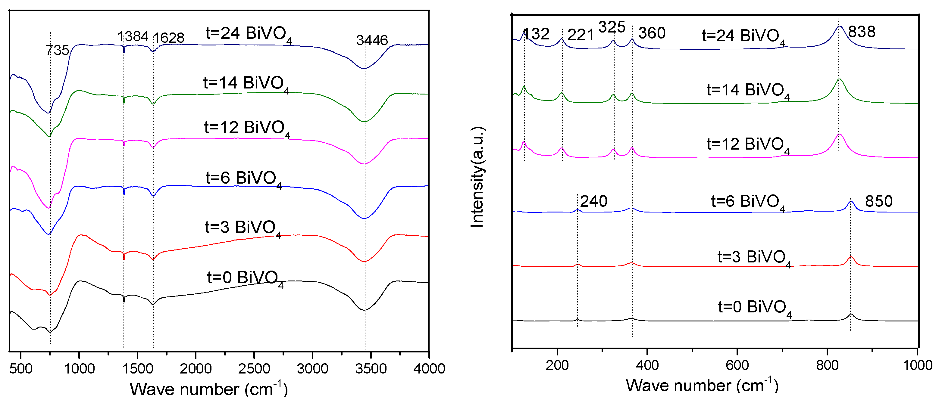
3.4. FT-IR and Raman Analyses

As can be seen from the infrared spectrum detailed in Figure 6, the absorption bands in the areas of 3446 cm−1, 1628 cm−1, and 1384 cm−1 were different forms of vibrations generated by the H-O-H bond in the water molecules. Meanwhile, in the area of 735 cm−1, a visible vibration peak had occurred, which had been generated by the VO43− tetrahedron in the BiVO4 crystals. As can be seen in the figure, when t = 0, 3, and 6 h, the Raman spectra displayed the same type of Raman peak. However, when t = 12, 14, and 24 h, another similar type of Raman peak was presented. Among these, the 838 cm−1 and 850 cm−1 peaks represented the Raman peaks of the V-O bonds in the monoclinic and tetragonal BiVO4, respectively, while the peaks at 360 cm−1 and 325 cm−1 were the V-O symmetric bending mode (Ag) and antisymmetric bending mode (Bg) of the VO43− group, respectively. The peak at 221 cm−1 was the outer mode generated by the spin and frequency shift of the V-O bond in the VO43− group. These results were found to be consistent with those of the XRD analysis. The internal structures of the BiVO4 with different crystal types were observed to be diversified, which resulted in different Raman peaks [30].

3.5. BiVO4 Generation Mechanism

As displayed in Figure 7, with the extension of the hydrothermal time, the morphological change of BiVO4 had four steps [12]:
Step 1 Oswald ripening process: During the process of the precursor solution preparation and overnight static placement, tiny particles were observed to have been deposited on the surfaces of the larger particles, which gradually formed into spherical particles with smooth surfaces [31].
Step 2 Particle differentiation process: It was observed that with the increases in the temperature and hydrothermal times, many fine crystal particles were gradually differentiated onto the smooth surfaces;
Step 3 Crystal detachment: It was found that with the further extension of the durations of the hydrothermal times, the spheroid matrix structure became damaged, and the surface differentiated crystals had gradually fallen off;
Step 4 Crystal agglomeration: It was further observed that with the continued extensions of hydrothermal times, the polyhedron crystals had gradually separated from the matrix and agglomeration occurred. Moreover, with the increases in the durations of the hydrothermal times, the BiVO4 crystals gradually transformed from single tetragonal and tetragonal-monoclinic mixed crystals to monoclinic crystals. These findings indicated that the tetragonal phase had converted to a monoclinic phase as the hydrothermal time increased. Meanwhile, the results of this study’s degradation experiment using methylene blue solution showed that the photocatalytic performances of the monoclinic BiVO4 crystals were superior to those of the tetragonal BiVO4 crystals. However, the agglomeration of the BiVO4 crystals tended to weaken their photocatalytic performances [32].

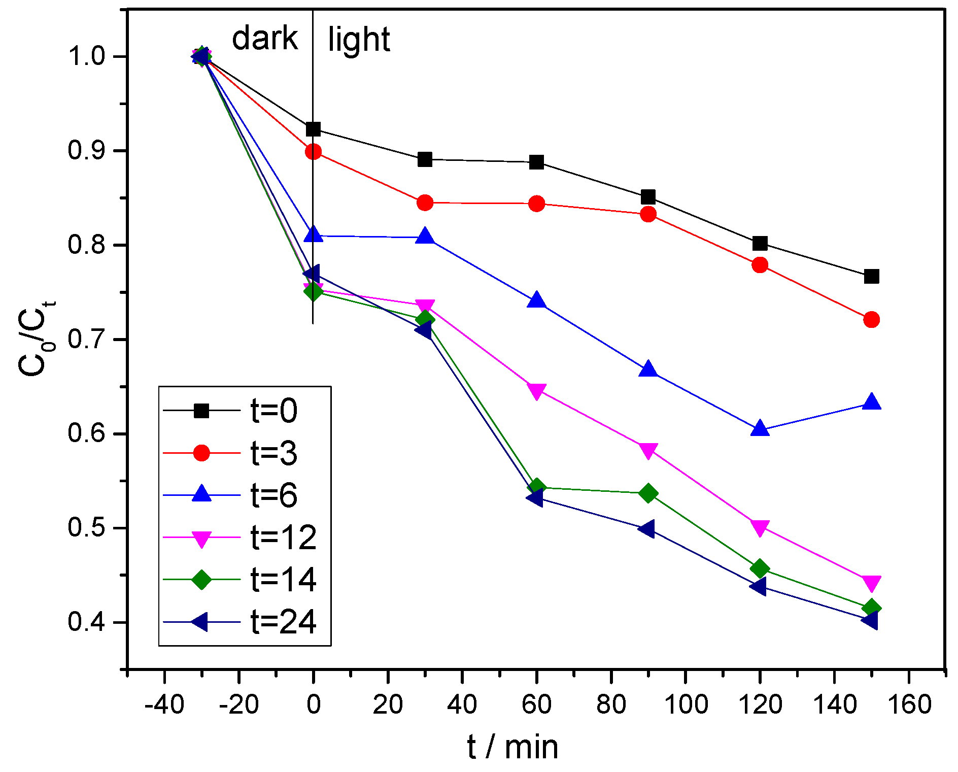
3.6. Photocatalytic Performance Analysis

It can be seen in Figure 8 that, when the hydrothermal time was 24 h, the BiVO4 displayed the best activity. Meanwhile, when hydrothermal times were zero and three hours, the degradation performances of the BiVO4 were observed to be somewhat less effective than when the hydrothermal times were 6, 12, 14, and 24 h. These results were found to be consistent with the absorption boundary values reflected by the solid ultraviolet. It was found that as the durations of the hydrothermal times increased, the photocatalytic performances of the prepared products, as well as the adsorption capacity of the methylene blue solution, were gradually and successively increased. However, when t = 24 h, the adsorption ability of the BiVO4 was found to be slightly weaker than when t = 14 h. This was determined to be related to the BiVO4 surface area. The degradation curve of the MB further confirmed that the photocatalytic performances of the monoclinic BiVO4 were superior to those of the tetragonal BiVO4 [25], which is consistent with the references that monoclinic BiVO4 possessed superior separation of the photo-induced charge carriers to that of tetragonal BiVO4 [12]. Additionally, the photocatalytic activity of BiVO4 in our study was compared with other references and is presented in Table 3. Furthermore, it was reported that the heterojunction structure between tetragonal BiVO4 and monoclinic BiVO4 could exhibit much higher photocatalytic activity [27], which is worth further exploration.

4. Conclusions

In this work, BiVO4 was successfully constructed through the hydrothermal method under different hydrothermal times. XRD results showed that the longer hydrothermal times were found to be more conducive to the generation of the monoclinic BiVO4. The UV-Vis absorption was more red-shifted when hydrothermal time increased from 3 h to 24 h. SEM results showed that the high temperature and high pressure conditions in the hydrothermal synthesis process are conducive to the formation of monoclinic BiVO4 crystals, but the agglomeration phenomenon of BiVO4 becomes more serious with the increase in hydrothermal time. The specific surface area of all the samples was in the range of 2.0~3.1 m2/g. Moreover, BiVO4 had excellent photocatalytic performance toward MB. Additionally, the photocatalytic activity shows that the crystal phase of BiVO4 plays a vital role in the photocatalytic performances: the removal efficiency of monoclinic BiVO4 (45% in 150 min), including adsorption and photocatalysis, was more than twice that of the tetragonal BiVO4 (20% in 150 min), which provides a facile guideline for the photocatalytic degradation of emerging organic contaminants using BiVO4.

Author Contributions

Conceptualization, Z.F.; methodology, Y.Z.; validation, H.C. (Heshan Cai); investigation, H.C. (Heshan Cai) and H.C. (Huacong Chen); resources, R.D.; data curation, L.C. and F.L.; supervision, J.C. All authors have read and agreed to the published version of the manuscript.

Funding

This work was supported by the National Natural Science Foundation of China (42107031, 22006024).

Institutional Review Board Statement

Not applicable.

Informed Consent Statement

Not applicable.

Data Availability Statement

Data available on request from the authors.

Conflicts of Interest

The authors declare no conflict of interest.

References

  1. Yurong, G.; Fang, Z.; Zwieten, L.V.; Bolan, N.; Dong, D.; Quin, B.F.; Meng, J.; Li, F.; Wu, F.; Wang, H.; et al. A critical review of biochar-based nitrogen fertilizers and their effects on crop production and the environment. Biochar 2022, 4, 36. [Google Scholar]
  2. Deng, F.; Shi, H.; Guo, Y.; Luo, X.; Zhou, J. Engineering paths of sustainable and green photocatalytic degradation technology for pharmaceuticals and organic contaminants of emerging concern. Curr. Opin. Green Sustain. Chem. 2021, 29, 100465. [Google Scholar] [CrossRef]
  3. Zhang, Y.; Zhou, B.; Chen, H.; Yuan, R. Heterogeneous photocatalytic oxidation for the removal of organophosphorus pollutants from aqueous solutions: A review. Sci. Total Environ. 2023, 856, 159048. [Google Scholar] [CrossRef] [PubMed]
  4. Weng, B.; Lu, K.Q.; Tang, Z.; Chen, H.M.; Xu, Y.J. Stabilizing ultrasmall Au clusters for enhanced photoredox catalysis. Nat. Commun. 2018, 9, 1543. [Google Scholar] [CrossRef] [PubMed]
  5. Zahmatkesh, S.; Bokhari, A.; Karimian, M.; Zahra, M.M.A.; Sillanpaa, M.; Panchal, H.; Alrubaie, A.J.; Rezakhani, Y. A comprehensive review of various approaches for treatment of tertiary wastewater with emerging contaminants: What do we know? Environ. Monit. Assess. 2022, 194, 884. [Google Scholar] [CrossRef]
  6. Tian, C.; Lv, J.; Zhang, W.; Wang, H.; Chao, J.; Chai, L.; Lin, Z. Accelerated Degradation of Microplastics at the Liquid Interface of Ice Crystals in Frozen Aqueous Solutions. Angew. Chem. Int. Ed. 2022, 61, e202206947. [Google Scholar] [CrossRef]
  7. Fang, Z.; Gao, Y.; Bolan, N.; Shaheen, S.M.; Xu, S.; Wu, X.; Xu, X.; Hu, H.; Lin, J.; Zhang, F.; et al. Conversion of biological solid waste to graphene-containing biochar for water remediation: A critical review. Chem. Eng. J. 2020, 390, 124611–124626. [Google Scholar] [CrossRef]
  8. Long, Z.; Song, H.; Zhang, G.; Gao, J.; Zhu, J. Fabrication of Bi-Bi3O4Cl plasmon photocatalysts for removal of aqueous emerging contaminants under visible light. J. Environ. Sci. 2022, 118, 87–100. [Google Scholar] [CrossRef]
  9. Fang, Z.; Gao, Y.; Zhang, F.; Zhu, K.; Shen, Z.; Liang, H.; Xie, Y.; Yu, C.; Bao, Y.; Feng, B.; et al. The adsorption mechanisms of oriental plane tree biochar toward bisphenol S: A combined thermodynamic evidence, spectroscopic analysis and theoretical calculations. Environ. Pollut. 2022, 310, 119819. [Google Scholar] [CrossRef]
  10. Gao, Y.; Fang, Z.; Lin, W.; Chen, H.; Bhatnagar, A.; Li, J.; Xie, Y.; Bao, Y.; Chen, J.; Zhao, H.; et al. Large-flake graphene-modified biochar for the removal of bisphenol S from water: Rapid oxygen escape mechanism for synthesis and improved adsorption performance. Environ. Pollut. 2023, 317, 120847. [Google Scholar] [CrossRef]
  11. Qin, N.; Zhang, S.; He, J.; Long, F.; Wang, L. In situ synthesis of BiVO4/BiOBr microsphere heterojunction with enhanced photocatalytic performance. J. Alloys Compd. 2022, 927, 166661. [Google Scholar] [CrossRef]
  12. Malathi, A.; Madhavan, J.; Ashokkumar, M.; Arunachalam, P. A review on BiVO4 photocatalyst: Activity enhancement methods for solar photocatalytic applications. Appl. Catal. A Gen. 2018, 555, 47–74. [Google Scholar] [CrossRef]
  13. Biswas, M.R.U.D.; Oh, W.-C. Synthesis of BiVO4-GO-PVDF nanocomposite: An excellent, newly designed material for high photocatalytic activity towards organic dye degradation by tuning band gap energies. Solid State Sci. 2018, 80, 22–30. [Google Scholar] [CrossRef]
  14. Ullah, H.; Tahir, A.A.; Mallick, T.K. Structural and electronic properties of oxygen defective and Se-doped p-type BiVO4(001) thin film for the applications of photocatalysis. Appl. Catal. B Environ. 2018, 224, 895–903. [Google Scholar] [CrossRef]
  15. Kshetri, Y.K.; Chaudhary, B.; Kamiyama, T.; Kim, T.-H.; Rosei, F.; Lee, S.W. Determination of ferroelastic phase transition temperature in BiVO4 by Raman spectroscopy. Mater. Lett. 2021, 291, 129519. [Google Scholar] [CrossRef]
  16. Samran, B.; Lunput, S.; Tonnonchiang, S.; Chaiwichian, S. BiFeO3/BiVO4 nanocomposite photocatalysts with highly enhanced photocatalytic activity for rhodamine B degradation under visible light irradiation. Phys. B Condens. Matter 2019, 561, 23–28. [Google Scholar] [CrossRef]
  17. Lv, C.; Chen, G.; Sun, J.; Zhou, Y.; Fan, S.; Zhang, C. Realizing nanosized interfacial contact via constructing BiVO4/Bi4V2O11 element-copied heterojunction nanofibres for superior photocatalytic properties. Appl. Catal. B Environ. 2015, 179, 54–60. [Google Scholar] [CrossRef]
  18. Samsudin, M.F.R.; Bashiri, R.; Mohamed, N.M.; Ng, Y.H.; Sufian, S. Tailoring the morphological structure of BiVO4 photocatalyst for enhanced photoelectrochemical solar hydrogen production from natural lake water. Appl. Surf. Sci. 2020, 504, 144417. [Google Scholar] [CrossRef]
  19. Guo, Z.; Wei, J.; Zhang, B.; Ruan, M.; Liu, Z. Construction and photoelectrocatalytic performance of TiO2/BiVO4 heterojunction modified with cobalt phosphate. J. Alloys Compd. 2020, 821, 153225. [Google Scholar] [CrossRef]
  20. Regmi, C.; Kshetri, Y.K.; Ray, S.K.; Pandey, R.P.; Lee, S.W. Utilization of visible to NIR light energy by Yb+3, Er+3 and Tm+3 doped BiVO4 for the photocatalytic degradation of methylene blue. Appl. Surf. Sci. 2017, 392, 61–70. [Google Scholar] [CrossRef]
  21. Tailor, N.K.; Abdi-Jalebi, M.; Gupta, V.; Hu, H.; Dar, M.I.; Li, G.; Satapathi, S. Recent progress in morphology optimization in perovskite solar cell. J. Mater. Chem. A 2020, 8, 21356–21386. [Google Scholar] [CrossRef]
  22. Wang, M.; Lu, W.; Chen, D.; Liu, J.; Hu, B.; Jin, L.; Lin, Y.; Yue, D.; Huang, J.; Wang, Z. Synthesis of dendritic-like BiVO4:Ag heterostructure for enhanced and fast photocatalytic degradation of RhB solution. Mater. Res. Bull. 2016, 84, 414–421. [Google Scholar] [CrossRef]
  23. Reli, M.; Troppová, I.; Šihor, M.; Pavlovský, J.; Praus, P.; Kočí, K. Photocatalytic decomposition of N2O over g-C3N4/BiVO4 composite. Appl. Surf. Sci. 2019, 469, 181–191. [Google Scholar] [CrossRef]
  24. Yu, X.; Wang, Z.; Li, E.; Li, X.; Cui, M.; Guo, C. Preparation of Different BiVO4 Catalysts and Their Photocatalytic Performance in the Coupling Reaction Between Alcohols and Amines. Catal. Lett. 2022, 152, 1244–1255. [Google Scholar] [CrossRef]
  25. Kshetri, Y.K.; Regmi, C.; Chaudhary, B.; Kim, H.-S.; Kim, T.-H.; Rosei, F.; Lee, S.W. BiVO4 ceramics for high-sensitivity and high-temperature optical thermometry. J. Lumin. 2021, 230, 117739. [Google Scholar] [CrossRef]
  26. Houas, A.; Lachheb, H.; Ksibi, M.; Elaloui, E.; Guillard, C.; Herrmann, J.-M. Photocatalytic degradation pathway of methylene blue in water. Appl. Catal. B Environ. 2001, 31, 145–157. [Google Scholar] [CrossRef]
  27. Yan, M.; Yan, Y.; Wu, Y.; Shi, W.; Hua, Y. Microwave-assisted synthesis of monoclinic–tetragonal BiVO4 heterojunctions with enhanced visible-light-driven photocatalytic degradation of tetracycline. RSC Adv. 2015, 5, 90255–90264. [Google Scholar] [CrossRef]
  28. Zhang, X.; Huang, Y.; Ma, F.; Zhang, Z.; Wei, X. Influences of vacancies on the structural, electronic and optical properties of monoclinic BiVO4. J. Phys. Chem. Solids 2018, 121, 85–92. [Google Scholar] [CrossRef]
  29. Teimori, H.; Balouchi, H.; Moradi, A.; Soltani, E. Quantifying seed germination response of deteriorated Trigonella foenum-graecum L. seed to temperatures and water potentials: Thermal time, hydrotime and hydrothermal time models. J. Appl. Res. Med. Aromat. Plants 2021, 20, 100276. [Google Scholar] [CrossRef]
  30. Sun, Z.; Yu, Z.; Liu, Y.; Shi, C.; Zhu, M.; Wang, A. Construction of 2D/2D BiVO4/g-C3N4 nanosheet heterostructures with improved photocatalytic activity. J. Colloid Interface Sci. 2019, 533, 251–258. [Google Scholar] [CrossRef]
  31. Suwanchawalit, C.; Buddee, S.; Wongnawa, S. Triton X-100 induced cuboid-like BiVO4 microsphere with high photocatalytic performance. J. Environ. Sci. 2017, 55, 257–265. [Google Scholar] [CrossRef]
  32. Kadam, A.N.; Babu, B.; Lee, S.-W.; Kim, J.; Yoo, K. Morphological guided sphere to dendrite BiVO4 for highly efficient organic pollutant removal and photoelectrochemical performance under solar light. Chemosphere 2022, 305, 135461. [Google Scholar] [CrossRef]
  33. Obregón, S.; Caballero, A.; Colón, G. Hydrothermal synthesis of BiVO4: Structural and morphological influence on the photocatalytic activity. Appl. Catal. B-Environ. 2012, 117–118, 59–66. [Google Scholar] [CrossRef]
  34. Ying, Y.; Tao, F.; Hong, T.; Wang, L. Controlled fabrication of bismuth vanadium oxide hierarchical microtubes with enhanced visible light photocatalytic activity. Mater. Sci. Semicond. Process. 2015, 32, 82–89. [Google Scholar] [CrossRef]
  35. Lu, Y.; Luo, Y.-S.; Xiao, H.-M.; Fu, S.-Y. Novel core–shell structured BiVO4 hollow spheres with an ultra-high surface area as visible-light-driven catalyst. CrystEngComm 2014, 16, 6059–6065. [Google Scholar] [CrossRef]
Figure 1. BiVO4 preparation flow chart.
Ijerph 20 03093 g001
Figure 2. XRD spectra of the BiVO4 prepared under different hydrothermal time conditions.
Ijerph 20 03093 g002
Figure 3. UV-Vis spectra of the BiVO4 under the different hydrothermal time conditions.
Ijerph 20 03093 g003
Figure 4. SEM maps of the BiVO4 prepared at different hydrothermal times with an observation diameter of 1 μm: (a) 0, (b) 3, (c) 6, (d) 12, (e) 14, (f) 24 h.
Ijerph 20 03093 g004
Figure 5. SEM maps of the BiVO4 prepared at different hydrothermal times with an observation diameter of 10 μm: (a) 0, (b) 3, (c) 6, (d) 12, (e) 14, (f) 24 h.
Ijerph 20 03093 g005
Figure 6. FTIR (left) and Raman spectra (right) of the BiVO4 prepared with different hydrothermal times.
Ijerph 20 03093 g006
Figure 7. Prediction diagram of the BiVO4 generation mechanism.
Ijerph 20 03093 g007
Figure 8. Degradation curves of the methylene blue solution under the different BiVO4 degradation conditions.
Ijerph 20 03093 g008
Table 1. XRD microstructure parameters of the BiVO4 products prepared under different hydrothermal conditions.
SampleTimeCrystal StructureDiffraction Intensity/CPSRelative Intensity Ratio
m-(121)m-(040)t-(200)t-(112)(121)/011(040)/(011)(040)/(121)
BiVO40 ht + m--590929,74417,675--0.537--
3 ht + m--666336,22921,987--0.410--
6 ht + m3610684834,80821,7940.2150.4081.897
12 hm27,9737297----3.7580.9420.261
14 hm28,6278476----2.8840.8540.296
24 hm27,7408516----3.7541.1520.307
Table 2. Related physical properties of the BiVO4 crystals.
Hydrothermal Time/hCrystal PhaseMorphologySpecific Surface Areas m2/gReasons for Changes in Specific Surface Area
0TetragonalSmooth spheroid2.5952 ± 0.1498Surface differentiation
3TetragonalRelatively smooth spheroid2.6115 ± 0.1642
6Tetragonal + minor monoclinicSurface-differentiated polyhedron2.7727 ± 0.1581
12MonoclinicClustered flower shape2.9271 ± 0.1085
14MonoclinicClustered flower shape3.0971 ± 0.1525
24MonoclinicPolyhedron agglomeration2.0352 ± 0.1276Crystal agglomeration
Table 3. Photocatalytic activity of BiVO4 toward different dyes under visible light irradiation.
MaterialsPhotocatalytic ParametersDegradation EfficiencyRefs.
BiVO4 peanut, 1.0 mg/mLMB, 10 mg/L, Xenon lamp with 35 W/m240% in 120 min[33]
BiVO4 microtube, 1.0 mg/mLMO, 20 mg/L, 250-W Xenon arc lamp 95% in 180 min[34]
BiVO4 spheres, 0.5 mg/mLRhB, 10−5 mol/L, 500 W Xenon lamp27% in 150 min[35]
BiVO4 biscuits, 0.5 mg/mLRhB, 10−5 mol/L, 500 W Xenon lamp44% in 150 min[35]
Tetragonal BiVO4, 1.0 mg/mLMB, 20 mg/L, 550 W Xenon lamp20% in 150 minThis study
Monoclinic BiVO4, 1.0 mg/mL MB, 20 mg/L, 550 W Xenon lamp45% in 150 minThis study
Disclaimer/Publisher’s Note: The statements, opinions and data contained in all publications are solely those of the individual author(s) and contributor(s) and not of MDPI and/or the editor(s). MDPI and/or the editor(s) disclaim responsibility for any injury to people or property resulting from any ideas, methods, instructions or products referred to in the content.

