
Characteristics of Pressure on the Apophysis in the Course of Paediatric Heel Pain—Preliminary Report

 ,
,  , , , , , and
Abstract
:1. Introduction
2. Aetiology
3. Diagnostics
4. Main Issue
4.1. Objective
4.2. Study Group
4.3. Methods
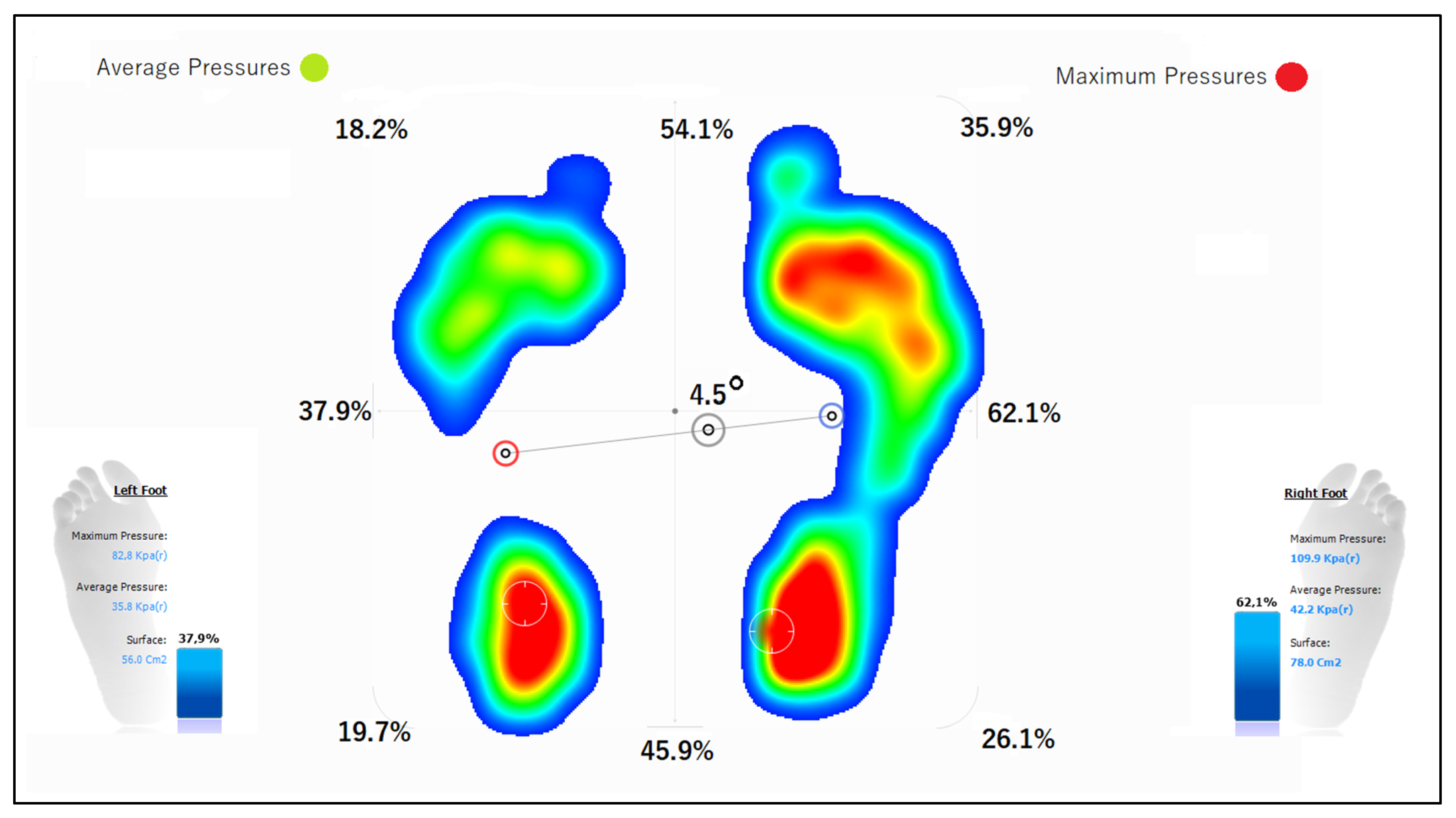
- Assessment of centre of gravity in anteroposterior and lateral view in a standing posture (Figure A1).
- Assessment of foot alignment—angle of abduction (Figure A2).
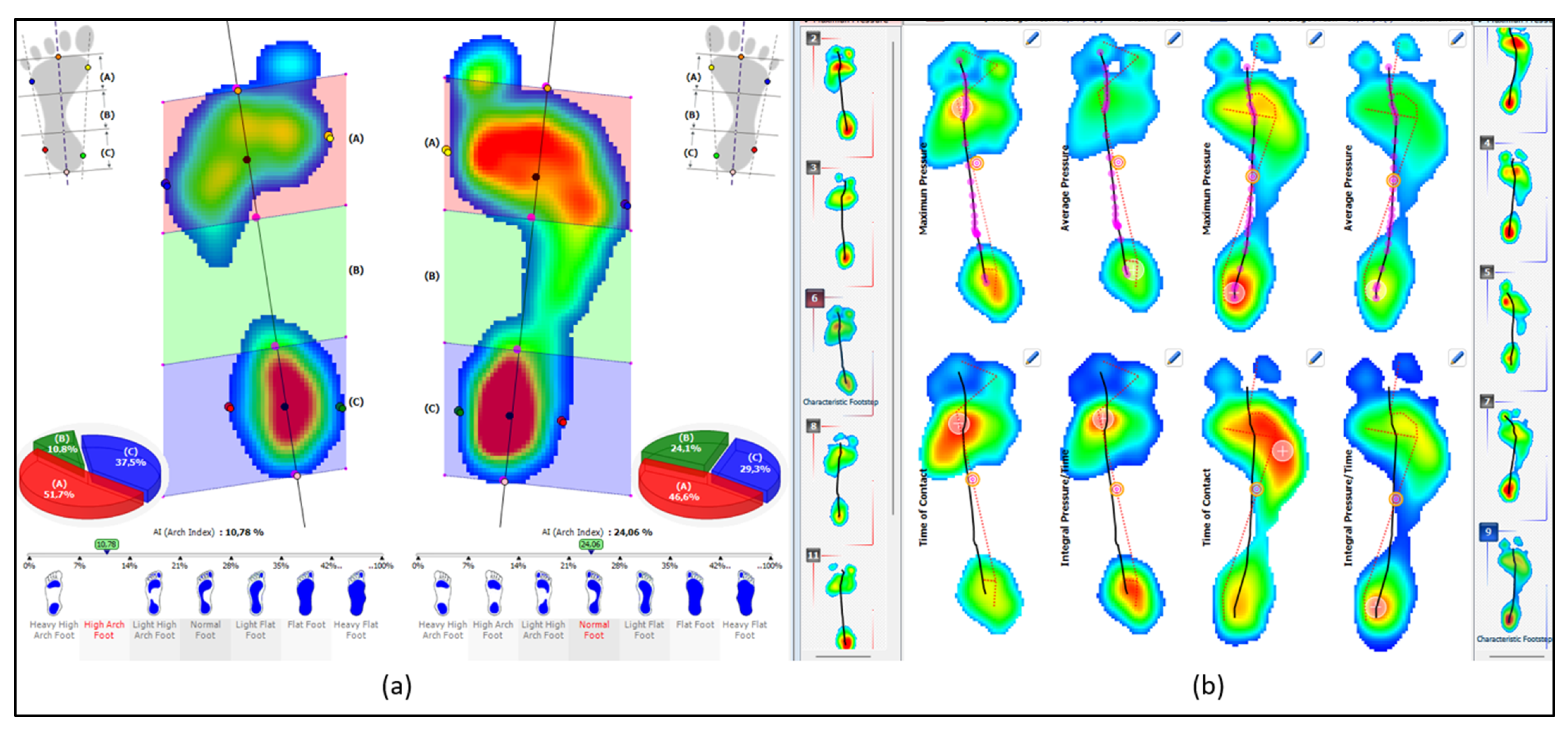
- Assessment of arches using the Arch Index, performed while standing and while walking (Figure A3).
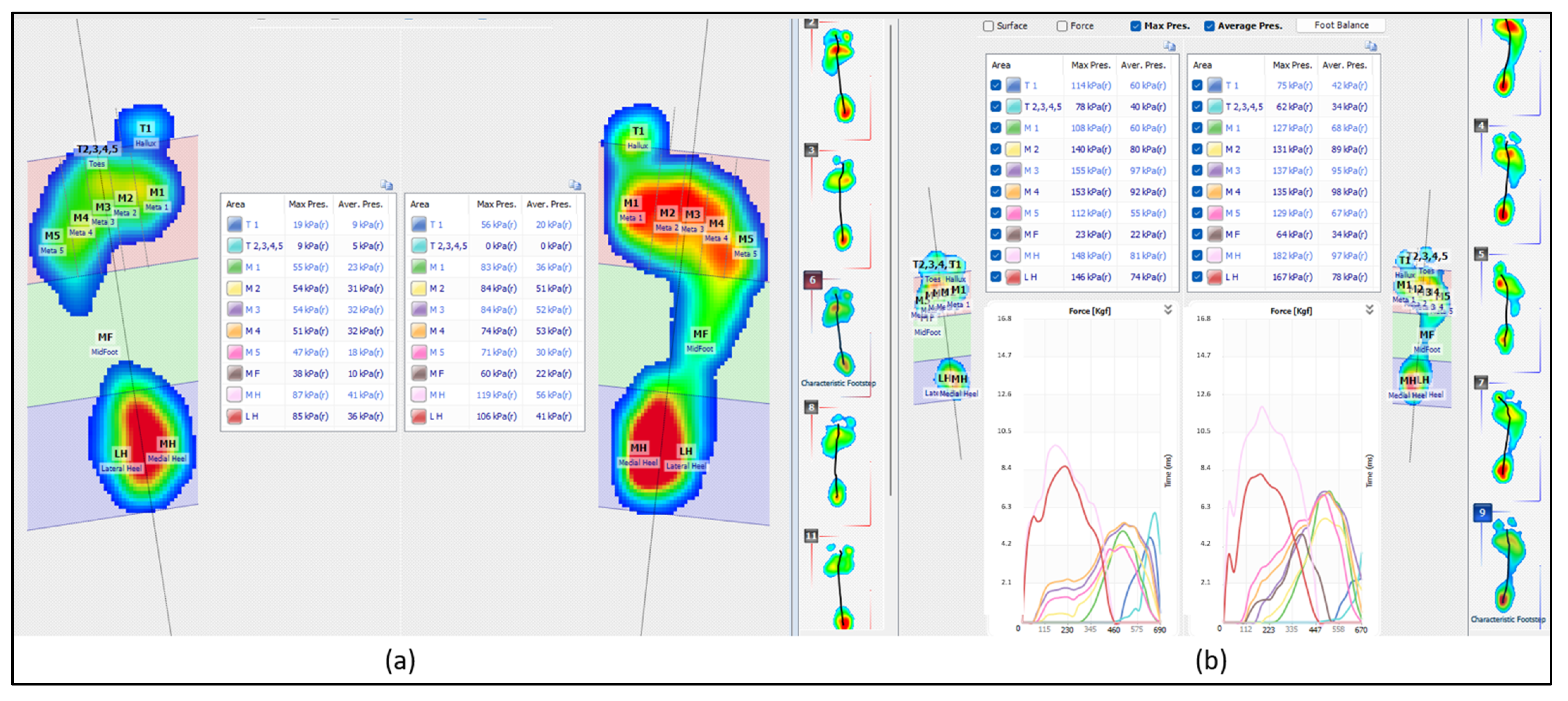
- Assessment of pressure distribution in individual regions of the foot while standing and while walking (Figure A4).
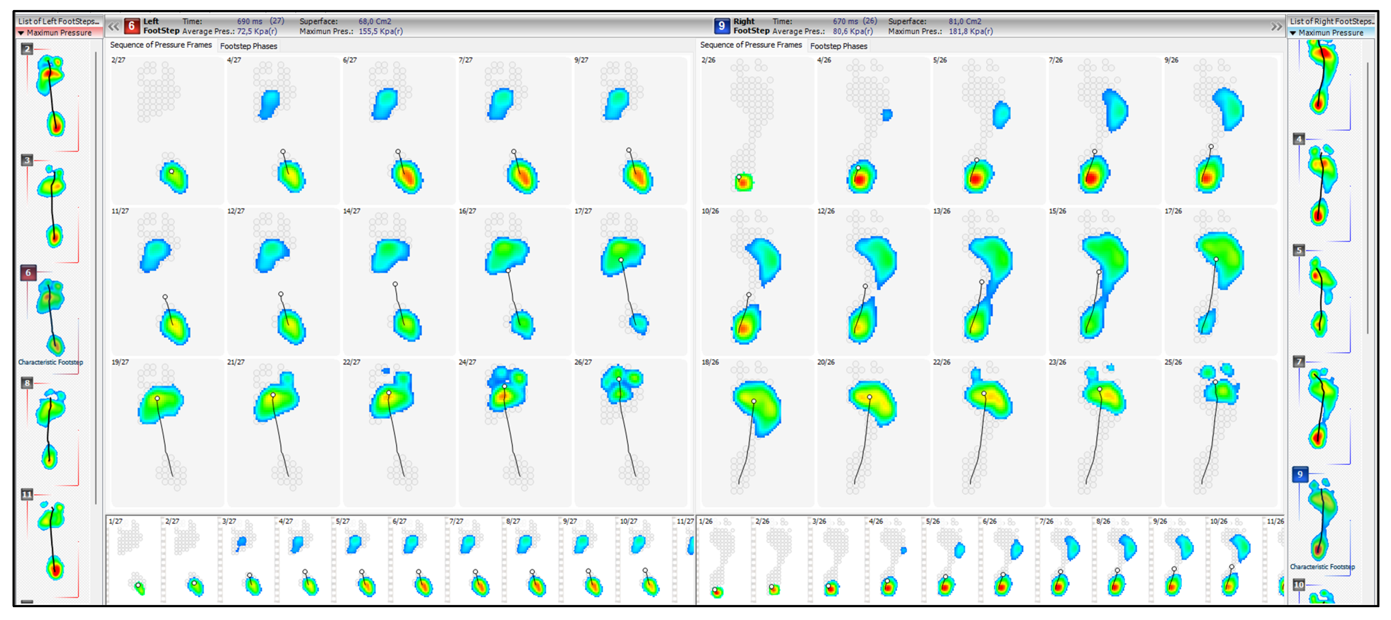
- Assessment of centre of pressure progression (gait line) to assess the stages of stance phase (including the involvement of the hindfoot, midfoot and forefoot, quality of the centre of pressure trajectory)—Figure A7.
5. Statistical Analysis
6. Results
7. Discussion
8. Conclusions
- In the clinical evaluation of people with paediatric heel pain, it is reasonable to study the distribution of pressures while standing and walking, including conducting a clinical observational evaluation of the detailed results obtained with pedobarography.
- Reducing the pressure on the heels in relation to the ground illustrates the shift of the centre of gravity to the front. It can also be the effect of bad posture. These factors are an important cause of the formation of traction of the apophysis of the heel and in the relationship of the Achilles–plantar fascia–plantar flexors of the foot, which may result in paediatric heel pain.
- Traction forces within the heel apophysis cause intracanal pressure on the calcaneal tuber and, at the same time, reduce the contact of the feet with the ground.
- Pedobarography should be combined with a physical assessment of the feet and body posture for defects that may predispose patients to increased tension of the structures attached to the calcaneal tubercle.
Author Contributions
Funding
Institutional Review Board Statement
Informed Consent Statement
Data Availability Statement
Conflicts of Interest
Appendix A


References
- Vergara Amador, E.; Erazo Acosta, L.M.; Moreno Gómez, L.Á. Document details—Calcaneus normal radiological patterns in children. Rev. Cuba. Ortop. Traumatol. 2017, 31, 1–11. [Google Scholar]
- Alison, M.; Joseph, D.P.; Mirene, K. Pediatric Heel Pain. Clin. Podiatr. Med. Surg. 2013, 30, 503–511. [Google Scholar]
- Ramponi, D.R.; Baker, C. Sever’s Disease (Calcaneal Apophysitis). Adv. Emerg. Nurs. J. 2019, 41, 10–14. [Google Scholar] [CrossRef] [PubMed]
- Chiodo, W.A.; Cook, K.D. Pediatric Heel Pain. Clin. Podiatr. Med. Surg. 2010, 27, 355–367. [Google Scholar] [CrossRef] [PubMed]
- Wiegerinck, J.I.; Yntema, C.; Brouwer, H.J.; Struijs, P.A.A. Incidence of calcaneal apophysitis in the general population. Eur. J. Pediatr. 2014, 173, 677–679. [Google Scholar] [CrossRef]
- De Jonge, J.S.; Van den Berg, C.; De Vos, R.J.; Van der Heide, H.J.; Weir, A.; Verhaar, J.A.; Bierma-Zeinstra, S.M.; Tol, J.L. Incidence of midportion Achilles tendinopathy in the general population. Br. J. Sports Med. 2011, 45, 1026–1028. [Google Scholar] [CrossRef] [Green Version]
- Volpe, R. Keys to diagnosing and treating calcaneal apophysitis. Podiatr. Today 2009, 22, 60–66. [Google Scholar]
- Howard, R. Diagnosing and treating Sever’s disease in children. Emerg. Nurse 2014, 22, 28–30. [Google Scholar] [CrossRef]
- Sever, J.W. Apophysitis of the os calcis. N. Y. Med. 1912, 95, 1025–1029. [Google Scholar]
- Kurtz, A.D. Apophysitis of the os calcis. Am. J. Orthop. Surg. 1917, 15, 659–663. [Google Scholar]
- Seder, J.I. Heel injuries incurred in running and jumping. Physician Sports Med. 1976, 4, 70–73. [Google Scholar]
- McKenzie, D.C.; Taunton, J.E.; Clement, D.B.; Smart, G.W.; McNicol, L.L. Calcaneal epiphysitis in adolescent athletes. Can. J. Appl. Sport Sci. 1981, 6, 123–125. [Google Scholar]
- International Classification of Diseases and Related Health Problems 10th Revision of World Health Organization. Available online: https://icd.who.int/browse10/2019 (accessed on 22 June 2022).
- Cassas, K.J.; Cassettari-Wayhs, A. Childhood and adolescent sports-related overuse injuries. Am. Fam. Physician 2006, 73, 1014–1022. [Google Scholar] [PubMed]
- James, A.M.; Williams, C.M.; Haines, T.P. Effectiveness of interventions in reducing pain and maintaining physical activity in children and adolescents with calcaneal apophysitis (Sever’s disease): A systematic review. J. Foot Ankle Res. 2013, 6, 16. [Google Scholar] [CrossRef] [Green Version]
- Madden, C.C.; Mellion, M.B. Sever’s disease and other causes of heel pain in adolescents. Am. Fam. Physician 1996, 54, 1995–2000. [Google Scholar] [PubMed]
- Micheli, L.J. The traction apophysitises. Clin. Sports Med. 1987, 6, 389–404. [Google Scholar] [CrossRef]
- Micheli, L.J.; Ireland, M.L. Prevention and management of calcaneal apophysitis in children: An overuse syndrome. J. Pediatr. Orthop. 1987, 7, 34–38. [Google Scholar] [CrossRef]
- Kannus, P. Etiology and pathophysiology of chronic tendon disorders in sports. Scand. J. Med. Sci. Sports 1997, 7, 78. [Google Scholar] [CrossRef] [PubMed]
- Micheli, L.J.; Fehlandt, A.F. Overuse injuries to tendons and apophyses in children and adolescents. Clin. Sports Med. 1992, 11, 713. [Google Scholar] [CrossRef]
- Kvist, M.; Heinonen, O. Calcaneal apophysitis (sever’s disease)—A common cause of heel pain in young athletes. Scand. J. Med. Sci. Sport. 1991, 1, 235–238. [Google Scholar] [CrossRef]
- Becerro de Bengoa Vallejo, R.; Losa Iglesias, M.E.; Rodriguez Sanz, D. Static and dynamic plantar pressures in children with and without sever disease: A case-control study. Phys. Ther. 2014, 94, 818–826. [Google Scholar] [CrossRef] [Green Version]
- Achar, S.; Yamanaka, J. Apophysitis and Osteochondrosis: Common Causes of Pain in Growing Bones. Am. Fam. Phys. 2019, 99, 10. [Google Scholar]
- De Inocencio, J. Musculoskeletal pain in primary paediatric care: Analysis of 1000 consecutive general paediatric clinic visits. Pediatrics 1998, 102, 63. [Google Scholar] [CrossRef] [Green Version]
- Krul, M.; Van der Wouden, J.C.; Schellevis, F.G.; Van Suijlekom Smit, L.W.; Koes, B.W. Foot problems in children presented to the family physician: A comparison between 1987 and 2001. Fam. Pract. 2009, 26, 174–179. [Google Scholar] [CrossRef] [Green Version]
- Krul, M.; Van der Wouden, J.C.; Schellevis, F.G.; Van Suijlekom Smit, L.W.; Koes, B.W. Musculoskeletal problems in overweight and obese pediatric patients. Ann. Fam. Med. 2009, 7, 352–356. [Google Scholar] [CrossRef]
- Arbab, D.; Wingenfeld, C.; Rath, B.; Lüring, C.; Quack, V.; Tingart, M. Osteochondrosen des kindlichen Fußes. Orthopäde 2013, 42, 20–29. [Google Scholar] [CrossRef] [PubMed]
- Fares, M.Y.; Salhab, M.A.; Khachfe, H.H.; Fares, J.; Haidar, R.; Musharrafieh, U. Sever’s Disease of the Pediatric Population: Clinical, Pathologic, and Therapeutic Considerations. Clin. Med. Res. 2021, 19, 132–137. [Google Scholar] [CrossRef] [PubMed]
- James, A.M.; Williams, C.M.; Luscombe, M.; Hunter, R.; Haines, T.P. Factors Associated with Pain Severity in Children with Calcaneal Apophysitis (Sever Disease). J. Pediatr. 2015, 167, 455–459. [Google Scholar] [CrossRef]
- Yamagiwa, H. Bone and joint diseases in children. Etiology and pathogenesis of osteochondral lesions in children. Osteochondritis dissecans and osteochondrosis. Clin. Calcium 2010, 20, 849–858. [Google Scholar]
- Skagen, P.S.; Horn, T.; Kruse, H.A.; Staergaard, B.; Rapport, M.M.; Nicolaisen, T. Osteochondritis dissecans (OCD), an endoplasmic reticulum storage disease? A morphological and molecular study of OCD fragments. Scand. J. Med. Sci. Sports 2011, 21, 7–33. [Google Scholar] [CrossRef]
- Yonetani, Y.; Nakamura, N.; Natsuume, T.; Shiozaki, Y.; Tanaka, Y.; Horibe, S. Histological evaluation of juvenile osteochondritis dissecans of the knee: A case series. Knee Surg. Sports Traumatol. Arthrosc. 2010, 18, 723–730. [Google Scholar] [CrossRef]
- Atanda, A., Jr.; Shah, S.A.; O’Brien, K. Osteochondrosis: Common causes of pain in growing bones. Am. Fam. Physician 2011, 83, 285–291. [Google Scholar] [PubMed]
- Hendrix, C.L. Calcaneal apophysitis (Sever disease). Clin. Podiatr. Med. Surg. 2005, 22, 55–62. [Google Scholar] [CrossRef]
- Rodríguez Sanz, D.; Becerro de Bengoa Vallejo, R.; López López, D.; Calvo Lobo, C.; Martínez Jiménez, E.M.; Perez Boal, E.; Losa Iglesias, M.E.; Palomo López, P. Slow velocity of the center of pressure and high heel pressures may increase the risk of Sever’s disease: A case-control study. BMC Pediatr. 2018, 18, 357. [Google Scholar] [CrossRef]
- Martinelli, N.; Spreafico, A.; Tramacere, I.; Marcolli, D.; Valli, F.; Curci, D. Prevalence and Associated Factors of Sever’s Disease in an Athletic Population. J. Am. Podiatr. Med. Assoc. 2019, 109, 351–356. [Google Scholar] [CrossRef]
- Belikan, P.; Farber, L.C.; Abel, F.; Nowak, T.E.; Drees, P.; Mattyasovszky, S.G. Incidence of calcaneal apophysitis (Sever’s disease) and return to play in adolescent athletes of a German youth soccer academy:a retrospective study of 10 years. J. Orthop. Surg. Res. 2022, 17, 83. [Google Scholar] [CrossRef] [PubMed]
- Santopietro, F.J. Foot and foot-related injuries in the young athlete. Clin. Sport Med. 1988, 7, 563–589. [Google Scholar] [CrossRef]
- James, A.M.; Williams, C.M.; Haines, T.P. Health related quality of life of children with calcaneal apophysitis: Child & parent perceptions. Health Qual. Life Outcomes 2016, 14, 95. [Google Scholar]
- Kim, H.K.; Mirjalili, S.A.; Fernandez, J. Gait kinetics, kinematics, spatiotemporal and foot plantar pressure alteration in response to long-distance running: Systematic review. Hum. Mov. Sci. 2018, 57, 342–356. [Google Scholar] [CrossRef] [PubMed]
- Hart, E.; Meehan, W.P.; Bae, D.S.; D’Hemecourt, P.; Stracciolini, A. The Young Injured Gymnast: A Literature Review and Discussion. Curr. Sports Med. Rep. 2018, 17, 366–375. [Google Scholar] [CrossRef] [PubMed]
- McGlamry, E.D.; Kitting, R.W. Aquinus foot: An analysis of the etiology, pathology and treatment techniques. J. Am. Podiatry Assoc. 1973, 63, 165–184. [Google Scholar]
- Knutzen, K.M.; Price, A. Lower extremity static and dynamic relationships with rearfoot motion in gait. J. Am. Podiatr. Med. Assoc. 1994, 84, 171–180. [Google Scholar] [PubMed]
- Perhamre, S.; Lundin, F.; Norlin, R.; Klässbo, M. Sever’s injury; treat it with a heel cup: A randomized, crossover study with two insole alternatives. Scand. J. Med. Sci. Sports 2011, 21, e42–e47. [Google Scholar] [CrossRef] [PubMed]
- Perhamre, S.; Lundin, F.; Norlin, R.; Klässbo, M. A heel cup improves the function of the heel pad in Sever’s injury: Effects on heel pad thickness, peak pressure and pain. Scand. J. Med. Sci. Sports. 2012, 22, 516–522. [Google Scholar] [CrossRef] [PubMed]
- Launay, F. Sports-related overuse injuries in children. Orthop. Traumatol. Surg. Res. 2015, 101, 139–147. [Google Scholar] [CrossRef] [Green Version]
- Orendurff, M.S.; Rohr, E.S.; Segal, A.D.; Medley, J.W.; Green, J.R.; Kadel, N.J. Regional foot pressure during running, cutting, jumping, and landing. Am. J. Sports Med. 2008, 36, 566–571. [Google Scholar] [CrossRef]
- Scharfbillig, R.W.; Jones, S.; Scutter, S.D. Sever’s disease: What does the literature really tell us? Am. Podiatr. Med. Assoc. 2008, 98, 212–223. [Google Scholar] [CrossRef] [PubMed] [Green Version]
- Kennedy, J.; Knowles, B.; Dolan, M.; Bohne, W. Foot and ankle injuries in the adolescent runner. Curr. Opin. Pediatr. 2005, 17, 34–42. [Google Scholar] [CrossRef]
- Ogden, J.A.; Ganey, T.M.; Hill, J.D.; Jaakkola, J.I. Sever’s injury: A stress fracture of the immature calcaneal metaphysis. J. Pediatr. Orthop. 2004, 24, 488–492. [Google Scholar] [CrossRef] [PubMed]
- Malanga, G.; Ramirez Del Toro, J. Common injuries of the foot and ankle in the child and adolescent athlete. Phys. Med. Rehabil. Clin. N. Am. 2008, 19, 347–371. [Google Scholar] [CrossRef]
- Lewin, P. Apophysitis of the os calcis. Surg. Gynecol. Obstet. 1926, 41, 578. [Google Scholar]
- Volpon, B.J.; de Carvalho Filho, G. Calcaneal apophysitis: A quantitative radiographic evaluation of the secondary ossification center. Arch. Orthop. Trauma Surg. 2002, 122, 338–341. [Google Scholar] [CrossRef]
- Hoang, Q.; Mortazavi, M. Pediatric overuse injuries in sports. Adv. Pediatr. 2012, 59, 359–383. [Google Scholar] [CrossRef]
- Klenerman, L. ABC of sports medicine: Musculoskeletal injuries in child athletes. BMJ 1994, 308, 1556. [Google Scholar] [CrossRef] [Green Version]
- Elengard, T.; Karlsson, J.; Silbernagel, K. Aspects of treatment for posterior heel pain in youth athletes. Open Access J. Sports Med. 2010, 1, 223–232. [Google Scholar] [PubMed] [Green Version]
- Caine, D.; DiFiori, J.; Maffulli, N. Physeal injuries in children’s and youth sports: Reasons for concern? Br. J. Sports Med. 2006, 40, 749–760. [Google Scholar] [CrossRef] [PubMed] [Green Version]
- Johnson, J. Overuse injuries in young athletes: Cause and prevention. Strength Cond. J. 2008, 30, 27–31. [Google Scholar] [CrossRef] [Green Version]
- Shanmugam, C.; Maffulli, N. Sports injuries in children. Br. Med. Bull. 2008, 86, 33–57. [Google Scholar] [CrossRef]
- Sothern, M.; Loftin, M.; Suskind, R.; Udall, J.; Blecker, U. The health benefits of physical activity in children and adolescents: Implication for chronic disease prevention. Eur. J. Pediatr. 1999, 158, 271–274. [Google Scholar] [CrossRef]
- Siffert, R.S. Classification of the osteochondroses. Clin. Orthop. Relat. Res. 1981, 158, 10–18. [Google Scholar] [CrossRef]
- Richie, L.B.; Sytsma, M.J. Matching osteochondritis dissecans lesions in identical twin brothers. Orthopedics 2013, 36, 1213–1216. [Google Scholar] [CrossRef] [Green Version]
- Juergen, B.; Mathias, W.; Christian, H. Osteochondritis Dissecans: Etiology, Pathology, and Imaging with a Special Focus on the Knee Joint. Cartilage 2018, 9, 346–362. [Google Scholar]
- Manusov, E.G.; Lillegard, W.A.; Raspa, R.F.; Epperly, T.D. Evaluation of pediatric foot problems: Part II. The hindfoot and the ankle. Am. Fam. Phys. 1996, 54, 1012–1026, 1031. [Google Scholar]
- Hogan, K.A.; Gross, R.H. Overuse injuries in pediatric athletes. Orthop. Clin. N. Am. 2003, 34, 405. [Google Scholar] [CrossRef]
- Lau, L.L.; Mahadev, A.; Hui, J.H. Common lower limb sport-related overuse injuries in young athletes. Ann. Acad. Med. Singap. 2008, 37, 315–319. [Google Scholar] [CrossRef]
- Toomey, P. Plantar heel pain. Foot Ankle Clin. 2009, 14, 229–245. [Google Scholar] [CrossRef]
- Kose, O. Do we really need radiographic assessment for the diagnosis of non-specific heel pain (calcaneal apophysitis) in children? Skeletal Radiol. 2010, 39, 359–361. [Google Scholar] [CrossRef] [PubMed]
- Hunt, G.; Stowell, T.; Alnwick, G.; Evans, S. Arch taping as a symptomatic treatment in patients with Sever’s disease: A multiple case series. Foot 2007, 17, 178–183. [Google Scholar] [CrossRef]
- Vaishya, R.; Agarwal, A.K.; Azizi, A.T.; Vijay, V. Haglund’s syndrome: A commonly seen mysterious condition. Cureus 2016, 8, e820. [Google Scholar] [CrossRef] [PubMed] [Green Version]
- Tu, P. Heel pain: Diagnosis and management. Am. Fam. Physician 2018, 97, 86–93. [Google Scholar]
- Christopher, N.C.; Congeni, J. Overuse injuries in the pediatric athlete: Evaluation, initial management, and strategies for prevention. Clin. Pediatr. Emerg. Med. 2002, 3, 118–128. [Google Scholar] [CrossRef]
- Chang, G.; Paz, D.; Dwek, J.; Chung, C. Lower extremity overuse injuries in pediatric athletes: Clinical presentation, imaging, findings and treatment. Clin. Imaging 2013, 37, 836–846. [Google Scholar] [CrossRef] [PubMed]
- Steinborn, M.; Glaser, C. Normal Variations and Pathologic Disorders of Chondrification and Ossification of the Foot and Related Diseases. Semin. Musculoskelet. Radiol. 2019, 23, 497–510. [Google Scholar] [CrossRef]
- Basra, S. Practical pointers ontreating Sever’s disease in young athletes. Podiatr. Today 2011, 24, 72–73. [Google Scholar]
- Chiroff, R.; Cooke, C.P. Osteochondritis dissecans: A histologic and microradiographic analysis of surgically excised lesions. J. Trauma 1975, 15, 689–696. [Google Scholar] [CrossRef] [PubMed]
- Clanton, T.; DeLee, J.C. Osteochondritis dissecans. History, pathophysiology and current treatment concepts. Clin. Orthop. Relat. Res. 1982, 167, 50–64. [Google Scholar] [CrossRef] [Green Version]
- Green, W.T.; Banks, H.H. Osteochondritis dissecans in children. Clin. Orthop. 1990, 255, 3–12. [Google Scholar] [CrossRef]
- Barrie, H.J. Hypertrophy and laminar calcification of cartilage in loose bodies as probable evidence of an ossification abnormality. J. Pathol. 1980, 132, 161–168. [Google Scholar] [CrossRef]
- Uozumi, H.; Sugita, T.; Aizawa, T.; Takahashi, A.; Ohnuma, M.; Itoi, E. Histologic findings and possible causes of osteochondritis dissecans of the knee. Am. J. Sports Med. 2009, 37, 2003–2008. [Google Scholar] [CrossRef]
- Rachel, J.N.; Williams, J.B.; Sawyer, J.R.; Warner, W.C.; Kelly, D.M. Is radiographic evaluation necessary in children with a clinical diagnosis of calcaneal apophysitis (sever disease)? Pediatr. Orthop. 2011, 31, 548–550. [Google Scholar] [CrossRef]
- Mustapić, M.; Borić, I.; Lepur, D.; Zadravec, D.; Visković, K. Sever’s disease complicated with osteomyelitis. Acta Clin. Croat. 2014, 53, 252–255. [Google Scholar] [PubMed]
- Kumar, S.; Jain, N.; Karpe, P.; Limaye, R. Osteomyelitis complicating Sever’s disease: A report of two cases. J. Clin. Orthop. Trauma 2020, 11, 310–313. [Google Scholar] [CrossRef] [PubMed]
- Chorley, J.; Powers, C.R. Clinical Features and Management of Heel Pain in the Young Athlete. Available online: http://www.uptodate.com/online/content/topic.do?topicKey=ped_trau/13559 (accessed on 25 October 2010).
- Griffin, L.Y. Common sports injuries of the foot and ankle seen in children and adolescents. Orthop. Clin. N. Am. 1994, 25, 83–93. [Google Scholar] [CrossRef]
- Krantz, M.K. Calcaneal apophysitis: A clinical and roentgenologic study. J. Am. Podiatr. Assoc. 1965, 55, 801–807. [Google Scholar]
- Heneghan, M.A.; Wallace, T. Heel pain due to retrocalcaneal bursitis: Radiographic diagnosis (with an historical footnote on Sever’s disease). Pediatr. Radiol. 1985, 15, 119–122. [Google Scholar] [CrossRef]
- Nery, C.A.S.; Prado, I.; Cho, Y.J.; Oliveira, A.C.; Pereira, S.E.M. Osteocondrite de Sever: Importância do radiodiagnóstico. Acta Ortop. Bras. 1996, 4, 104–108. [Google Scholar]
- Gao, Y.; Liu, J.; Li, Y.; Liu, Q.; Xu, S. Radiographic study of Sever’s disease. Exp. Ther. Med. 2020, 20, 933–937. [Google Scholar] [CrossRef]
- Tóth, F.; Nissi, M.J.; Ellermann, J.M.; Wang, L.; Shea, K.G.; Polousky, J. Novel application of magnetic resonance imaging demonstrates characteristic differences in vasculature at predilection sites of osteochondritis dissecans. Am. J. Sports Med. 2015, 43, 2522–2527. [Google Scholar] [CrossRef] [Green Version]
- Martel, G.; Kiss, S.; Gilbert, G.; Anne-Archard, N.; Richard, H.; Moser, T. Differences in the vascular tree of the femoral trochlear growth cartilage at osteochondrosis-susceptible sites in foals revealed by SWI 3T MRI. J. Orthop. Res. 2016, 34, 1539–1546. [Google Scholar] [CrossRef] [Green Version]
- Singh, R.; Rohilla, R.; Siwach, R.C.; Magu, N.K.; Sangwan, S.S.; Sharma, A. Diagnostic significance of radiologic measurements in posterior heel pain. Foot 2008, 18, 91–98. [Google Scholar] [CrossRef]
- Van Sterkenburg, M.N.; Muller, B.; Maas, M.; Sierevelt, I.N.; Van Dijk, C.N. Appearance of the wheight-bearing lateral radiograph in retrocalcaneal bursitis. Acta Orthop. 2010, 81, 387–390. [Google Scholar] [CrossRef] [Green Version]
- Nakamae, A.; Engebretsen, L.; Bahr, R.; Krosshaug, T.; Ochi, M. Natural history of bone bruises after acute knee injury: Clinical outcome and histopathological findings. Knee Surg. Sports Traumatol. Arthrosc. 2006, 14, 1252–1258. [Google Scholar] [CrossRef] [PubMed]
- Redmond, A.C.; Crosbie, J.; Ouvrier, R.A. Development and validation of a novel rating system for scoring standing foot posture: The Foot Posture Index. Clin. Biomech. 2006, 21, 89–98. [Google Scholar] [CrossRef] [PubMed]
- Redmond, A.C.; Crane, Y.Z.; Menz, H.B. Normative values for the Foot Posture Index. J. Foot Ankle Res. 2008, 1, 6. [Google Scholar] [CrossRef] [Green Version]
- Evans, A.M.; Copper, A.W.; Scharfbillig, R.W.; Scutter, S.D.; Williams, M.T. Reliability of the foot posture index and traditional measures of foot position. J. Am. Podiatr. Med. Assoc. 2003, 93, 203–213. [Google Scholar] [CrossRef]
- Sobel, E.; Levitz, S.J.; Caselli, M.A.; Tran, M.; Lepore, F.; Lilja, E.; Sinaie, M.; Wain, E. Reevaluation of the relaxed calcaneal stance position. Reliability and normal values in children and adults. J. Am. Podiatr. Med. Assoc. 1999, 89, 258–264. [Google Scholar] [CrossRef]
- Najdi, H.; Mouarbes, D.; Makhour, F.; Dimassi, A.; Jawish, R. Forefoot adduction in children. Management and Treatment. Leban. Med. J. 2016, 64, 134–141. [Google Scholar] [CrossRef] [PubMed] [Green Version]
- Kaufmann, G.; Dammerer, D.; Heyenbrock, F.; Braito, M.; Moertlbauer, L.; Liebensteiner, M. Minimally invasive versus open chevron osteotomy for hallux valgus correction: A randomized controlled trial. Int. Orthop. 2019, 43, 343–350. [Google Scholar] [CrossRef] [Green Version]
- Czaprowski, D.; Stoliński, Ł.; Tyrakowski, M.; Kozinoga, M.; Kotwicki, T. Non-structural misalignments of body posture in the sagittal plane. Scoliosis Spinal Disord. 2018, 13, 6. [Google Scholar] [CrossRef] [Green Version]
- Mooney, J.F. Lower extremity rotational and angular issues in children. Pediatr. Clin. N. Am. 2014, 61, 1175–1183. [Google Scholar] [CrossRef]
- Güven, M.; Akman, B.; Ünay, K.; Özturan, E.K.; Çakıcı, H.; Eren, A. A new radiographic measurement method for evaluation of tibial torsion: A pilot study in adults. Clin. Orthop. Relat. Res. 2009, 467, 1807–1812. [Google Scholar] [CrossRef] [PubMed] [Green Version]
- Staheli, L.T.; Corbett, M.; Wyss, C.; King, H. Lower-extremity rotational problems in children: Normal values to guide management. J. Bone Jt. Surg. 1985, 67, 39–47. [Google Scholar] [CrossRef]
- Staheli, L.T. Rotational problems in children. J. Bone Jt. Surg. 1993, 75, 939–949. [Google Scholar] [CrossRef]
- McPoil, T.; Cornwal, M.W. Relationship between neutral subtalar joint position and pattern of rearfoot motion during walking. Foot Ankle Int. 1994, 15, 141–145. [Google Scholar] [CrossRef] [PubMed]
- Becerro de Bengoa Vallejo, R.; Iglesias, M.E.L.; Rodríguez Sanz, D.; Prados Frutos, J.C.; Salvadores Fuentes, P.; López Chicharro, J. Plantar pressures in children with and without sever’s disease. J. Am. Podiatr. Med. Assoc. 2011, 101, 17–24. [Google Scholar] [CrossRef]
- Scharfbillig, R.W.; Jones, S.; Scutter, S. Sever’s Disease a Prospective Study of Risk Factors. J. Am. Podiatr. Med. Assoc. 2011, 101, 133–145. [Google Scholar] [CrossRef]
- Young, R.; Nix, S.; Wholohan, A.; Bradhurst, R.; Reed, L. Interventions for increasing ankle joint dorsiflexion: A systematic review and meta-analysis. J. Foot Ankle Res. 2013, 6, 46. [Google Scholar] [CrossRef] [Green Version]
- Downey, M.S.; Schwartz, J.M. Ankle equinus. In McGlamry’s Comprehensive Textbook of Foot and Ankle Surgery; Wolters Kluwer Health/Lippincott Williams & Wilkins: Philadelphia, PA, USA, 2013; pp. 541–584. [Google Scholar]
- James, A.M.; Williams, C.M.; Haines, T.P. Heel raises versus prefabricated orthoses in the treatment of posterior heel pain associated with calcaneal apophysitis (Sever’s Disease): A randomised control trial. J. Foot Ankle Res. 2010, 3, 3. [Google Scholar] [CrossRef] [Green Version]
- Frisch, A.; Croisier, J.; Urhausen, A.; Seil, R.; Thelsen, D. Injuries risk factors and prevention initiatives in youth sport. Br. Med. Bull. 2009, 92, 95–121. [Google Scholar] [CrossRef] [PubMed] [Green Version]
- Lieberman, D. What we can learn about running from barefoot running: An evolutionary medical perspective. Exerc. Sports Sci. Rev. 2012, 4, 63–72. [Google Scholar] [CrossRef]
- Aronow, M.S.; Diaz-Doran, V.; Sullivan, R.J.; Adams, D.J. The effect of triceps surae contracture force on plantar foot pressure distribution. Foot Ankle Int. 2006, 27, 43–45. [Google Scholar] [CrossRef] [PubMed]
- Kose, O.; Celiktas, M.; Yigit, S.; Kisin, B. Can we make a diagnosis with radiographic examination alone in calcaneal apophysitis (Sever’s disease)? J. Pediatr. Orthop. B 2010, 19, 396–398. [Google Scholar] [CrossRef] [PubMed]
- Perry, J. Chapter 11: Ankle and Foot Gait Deviations. In Gait Analysis: Normal and Pathological Function, 2nd ed.; Slack Inc.: San Francisco, CA, USA, 2010. [Google Scholar]
- Naaktgeboren, K.; Dorgo, S.; Boyle, J.B. Growth Plate Injuries in Children in Sports: A Review of Sever’s Disease. Strength Condit. J. 2017, 39, 59–68. [Google Scholar] [CrossRef]
- Johnson, C.H.; Christensen, J.C. Biomechanics of the first ray part V: The effect of equinus deformity. A 3-dimensional kinematic study on a cadaver model. J. Foot Ankle Surg. 2005, 44, 114–120. [Google Scholar] [CrossRef]
- Zhu, G.; Wang, Z.; Yuan, C.; Geng, X.; Zhang, C.; Huang, J.; Wang, X.; Ma, X. A Radiographic Study of Biomechanical Relationship between the Achilles Tendon and Plantar Fascia. Biomed. Res. Int. 2020, 2020, 5319640. [Google Scholar] [CrossRef] [Green Version]
- Zwirner, J.; Zhang, M.; Ondruschka, B.; Keichi, A.; Niels, H. An ossifying bridge—On the structural continuity between theAchilles tendon and the plantar fascia. Sci. Rep. 2020, 10, 14523. [Google Scholar] [CrossRef] [PubMed]
- Cavanagh, P.R.; Hewitt, F.G.; Perry, J.E. In-shoe plantar pressure measurement. Foot 1998, 2, 185–194. [Google Scholar] [CrossRef]
- Wejsflog, G. Numerical characteristics of the plantogram of the child’s foot. Chir. Narz. Ruchu Ortop. Pol. 1963, 28, 301–307. [Google Scholar]
- Opanowska, M.; Ptretkiewicz-Abacjew, E. An assessment of shape of the longitudinal and the transverse foot arch in male and female pole vaulters. Balt. J. Health Phys. Activ. 2015, 7, 5. [Google Scholar] [CrossRef]
- Puszczałkowska-Lizis, E.; Kozioł, K.; Omorczyk, J. Perception of footwear comfort and its relationship with the foot structure among youngest-old women and men. PeerJ 2021, 9, e12385. [Google Scholar] [CrossRef]
- Cavanagh, P.R.; Rodgers, M.M. The arch index: A useful measure from footprints. J. Biomech. 1987, 20, 547–551. [Google Scholar] [CrossRef] [PubMed]
- Menz, H.B. Alternative techniquesforthe clinical assessment of foot pronation. J. Am. Podiatr. Med. Assoc. 1998, 88, 119–129. [Google Scholar] [CrossRef] [PubMed]
- Lavery, L.A.; Armstrong, D.G.; Wunderlich, R.P.; Tredwell, J.; Boulton, A.J.M. Predictive value of foot pressure assessment as part of a population-based diabetes disease management program. Diabetes Care 2003, 26, 1069–1073. [Google Scholar] [CrossRef] [Green Version]
- Goswami, A. A new gait parameterization technique by means of cyclogram moments: Application to human slope walking. Gait Posture 1998, 8, 15–36. [Google Scholar] [CrossRef] [PubMed]
- Chong, A.; Al-Baghadadi, J.A.A.; Milcurn, P.D. Human Plantar Pressure Image and Foot Shape Matching. J. Biosci. Med. 2015, 3, 36. [Google Scholar] [CrossRef] [Green Version]
- Zinger, A.; Schmitt, D.; Hughes-Oliver, C.; Queen, R.M. The effect of ankle osteoarthritis and total ankle arthroplasty on center of pressure position. J. Orthop. Res. 2021, 39, 1245–1252. [Google Scholar] [CrossRef] [PubMed]


| Postural Defect | Side (L-Left/R-Right) | n | No Posture Defect | Occurrences of Defects | Type of Postural Defects |
|---|---|---|---|---|---|
| Hindfoot | L | 32 | 28 (87.5%) | cv: 4 (12.5%) cvr: 0 (0%) | cv—valgus cvr—varus |
| R | 32 | 28 (87.5%) | cv: 5 (15.62%) cvr: 0 (0%) | ||
| Arch defects | L | 32 | 24 (75.0%) | p: 8 (25%) c: 0 (0%) | p—pes planus c—pes cavus |
| P | 32 | 23 (71.87%) | p: 9 (28.12%) c: 0 (0%) | ||
| Complex midfoot defects | L | 32 | 1 (3.12%) | pv: 31 (96.87%) e/ev: 0 (0%) | pv—planovalgus e—equinus ev—equinovarus |
| R | 32 | 1 (3.12%) | pv: 31 (96.87%) e/ev: 0 (0%) | ||
| L | 32 | 16 (50.0%) | 16 (50%) | metatarsus adductus (forefoot adduction) | |
| R | 32 | 17 (53.12%) | 15 (46.87%) | ||
| L | 32 | 28 (87.5%) | 4 (12.12%) | z-foot (skew foot) | |
| R | 32 | 28 (87.5%) | 4 (12.12%) | ||
| L | 28 | 2 (7.14%) | 26 (78.79%) | 1st ray instability | |
| R | 28 | 2 (7.14%) | 26 (78.79%) | ||
| Forward tilt of body | 24 | 6 (25.0%) | fp: 18 (75%) | fp—forward tilt of body |
| Standing Pressure (p) | |||||||
|---|---|---|---|---|---|---|---|
| Examined Region | Variable | n | min | max | mean | st. dev. | 95% CI |
| Centre of gravity projection | Pressure—left | 33 | 0.379 | 0.757 | 0.5145 | 0.0798 | (0.4863; 0.5428) |
| Pressure—right | 33 | 0.243 | 0.621 | 0.4885 | 0.07658 | (0.4613; 0.5156) | |
| Pressure—front | 33 | 0.039 | 0.76 | 0.4695 | 0.1407 | (0.4196; 0.5194) | |
| Pressure—back | 33 | 0.24 | 0.961 | 0.5305 | 0.1407 | (0.4804; 0.5801) | |
| Variable | n | min | max | mean | st. dev. | 95% CI |
|---|---|---|---|---|---|---|
| Pressure—front | 29 | 0.039 | 0.76 | 0.4792 | 0.1407 | (0.4196; 0.5194) |
| Pressure—back | 29 | 0.24 | 0.961 | 0.5208 | 0.1407 | (0.4804; 0.5801) |
| Variable | n | max | min | mean | st. dev. | 95% CI |
|---|---|---|---|---|---|---|
| Pressure—front | 4 | 0.303 | 0.478 | 0.399 | 0.0854 | (0.2633, 0.5352) |
| Pressure—back | 4 | 0.522 | 0.697 | 0.601 | 0.0854 | (0.4648, 0.7367) |
| Arch Index (AI) | n | Standing | Walking | ||||
|---|---|---|---|---|---|---|---|
| <21 [%] | 21–28 [%] | >28 [%] | <21 [%] | 21–28 [%] | >28 [%] | ||
| Left foot | 33 | 19 | 7 | 7 | 21 | 6 | 6 |
| AI<% = 57.57% | AI% = 21.21% | AI>% = 21.21% | AI<% = 63.63% | AI% = 18.18% | AI% = 18.18% | ||
| Right foot | 33 | 14 | 8 | 11 | 19 | 4 | 10 |
| AI<% = 42.42% | AI% = 24.24% | AI>% = 33.33% | AI<% = 57.57% | AI% = 12.12% | AI>% = 30.30% | ||
| AI While Standing | AI While Walking | ||||||||||
|---|---|---|---|---|---|---|---|---|---|---|---|
| Variable | n | min | max [%] | mean [%] | st. dev. [%] | 95% CI | min | max [%] | mean [%] | st. dev. [%] | 95% CI |
| Left | 33 | 0 | 38.63 | 18.41 | 10.24 | (14.78; 22.04) | 0 | 33.84 | 16.98 | 9.987 | (13.44; 20.52) |
| Right | 33 | 0 | 37.71 | 21.32 | 10.33 | (17.65; 24.98) | 0 | 44.35 | 19.71 | 11.29 | (15.71; 23.71) |
| n | min (%) | max (%) | mean (%) | SD (%) | 95% Conf. Interval | |
|---|---|---|---|---|---|---|
| MHoverLH_L standing—left foot | 33 | −17.24 | 39.13 | 11.3 | 13.03 | (6.677, 15.92) |
| MHoverLH_R standing—right foot | 33 | −29.63 | 42.11 | 12.18 | 15.46 | (6.702, 17.66) |
| MHoverLH_L_walking—left foot | 33 | −4.11 | 30.85 | 8.006 | 8.696 | (4.923, 11.09) |
| MHoverLH_R_walking—right foot | 33 | −5.208 | 27.27 | 8.653 | 8.285 | (5.715, 11.59) |
Disclaimer/Publisher’s Note: The statements, opinions and data contained in all publications are solely those of the individual author(s) and contributor(s) and not of MDPI and/or the editor(s). MDPI and/or the editor(s) disclaim responsibility for any injury to people or property resulting from any ideas, methods, instructions or products referred to in the content. |
© 2023 by the authors. Licensee MDPI, Basel, Switzerland. This article is an open access article distributed under the terms and conditions of the Creative Commons Attribution (CC BY) license (https://creativecommons.org/licenses/by/4.0/).
Share and Cite
Bitenc-Jasiejko, A.; Walińska, A.; Konior, K.; Gonta, K.; Skomro, P.; Kijak, K.; Kowacka, M.; Lietz-Kijak, D. Characteristics of Pressure on the Apophysis in the Course of Paediatric Heel Pain—Preliminary Report. Int. J. Environ. Res. Public Health 2023, 20, 5403. https://doi.org/10.3390/ijerph20075403
Bitenc-Jasiejko A, Walińska A, Konior K, Gonta K, Skomro P, Kijak K, Kowacka M, Lietz-Kijak D. Characteristics of Pressure on the Apophysis in the Course of Paediatric Heel Pain—Preliminary Report. International Journal of Environmental Research and Public Health. 2023; 20(7):5403. https://doi.org/10.3390/ijerph20075403
Chicago/Turabian StyleBitenc-Jasiejko, Aleksandra, Anna Walińska, Krzysztof Konior, Kinga Gonta, Piotr Skomro, Konrad Kijak, Małgorzata Kowacka, and Danuta Lietz-Kijak. 2023. "Characteristics of Pressure on the Apophysis in the Course of Paediatric Heel Pain—Preliminary Report" International Journal of Environmental Research and Public Health 20, no. 7: 5403. https://doi.org/10.3390/ijerph20075403
APA StyleBitenc-Jasiejko, A., Walińska, A., Konior, K., Gonta, K., Skomro, P., Kijak, K., Kowacka, M., & Lietz-Kijak, D. (2023). Characteristics of Pressure on the Apophysis in the Course of Paediatric Heel Pain—Preliminary Report. International Journal of Environmental Research and Public Health, 20(7), 5403. https://doi.org/10.3390/ijerph20075403