Article

Prevalence of Antimicrobial Resistant Escherichia coli from Sinking Creek in Northeast Tennessee

1
Department of Biostatistics & Epidemiology, College of Public Health, East Tennessee State University, Johnson City, TN 37614, USA
2
Department of Environmental and Occupational Health and Safety Sciences, College of Public Health, East Tennessee State University, Johnson City, TN 37614, USA
3
Department of Geosciences, College of Arts and Sciences, East Tennessee State University, Johnson City, TN 37614, USA
*
Author to whom correspondence should be addressed.
Int. J. Environ. Res. Public Health 2024, 21(10), 1285; https://doi.org/10.3390/ijerph21101285
Submission received: 14 August 2024 / Revised: 23 September 2024 / Accepted: 25 September 2024 / Published: 26 September 2024

Abstract

:
Antibiotic resistance (AR) is a critical global health threat exacerbated by complex human–animal–environment interactions. Aquatic environments, particularly surface water systems, can serve as reservoirs and transmission routes for AR bacteria. This study investigated the prevalence of AR E. coli in Sinking Creek, a pathogen-impacted creek in Northeast Tennessee. Water samples were collected monthly from four sites along the creek over a 6-month period. E. coli isolates were cultured, identified, and tested for susceptibility to eight antibiotics using the Kirby–Bauer disk diffusion method and broth disk elution method for colistin. Data were analyzed to determine the prevalence of AR and multidrug resistance (MDR) among isolates. Of the 122 water samples, 89.3% contained E. coli. Among the 177 isolates tested, resistance was highest to ciprofloxacin (64.2%) and nitrofurantoin (62.7%), and lowest to fosfomycin (14.1%) and colistin (6.0%). Significant differences in resistance to ceftriaxone and amoxicillin/clavulanic acid were observed between sampling sites. MDR was prevalent in 47.5% of isolates, with 5.1% resistant to seven antibiotics. The most frequent MDR patterns (6.8%) included three antibiotics: ceftriaxone, ciprofloxacin, and nitrofurantoin. The high prevalence of AR E. coli in Sinking Creek poses a significant public health risk, highlighting the need for ongoing surveillance and intervention strategies to prevent the spread of AR bacteria.

1. Introduction

Antibiotic resistance (AR) poses a significant global health threat and has been recognized by the WHO as one of the top ten threats to human health [1,2]. The epidemiology of AR bacteria is quite complex due to the human–animal–environmental interaction involved in developing and transmitting AR organisms [3]. Aquatic environments are increasingly recognized as reservoirs and pathways for the transmission of AR [4,5,6,7]. Aquatic ecosystems, particularly surface water systems, are exposed to pollutants from various anthropogenic and agricultural sources [8,9]. Surface water continuously receives both non-pathogenic and pathogenic bacteria from various sources such as septic systems, wastewater treatment plants, agriculture, and wildlife [10,11]. Antibiotic-resistant bacteria in aquatic environment can present a health risk to humans due to drinking contaminated water, ingested contaminated water during recreational activities, or/and the consumption of produce irrigated with contaminated water [4,12,13,14,15].
Escherichia coli, which are abundant in the gastrointestinal tract of humans and animals, can act as carriers or reservoirs of AR genes [16,17]. They are also capable of exchanging AR genetic material with bacteria of the same or different bacterial species [18,19]. Studies have demonstrated that E. coli is a very adaptable organism capable of thriving in external environments, including surface waters [20,21]. Specifically, research has reported the ubiquitous presence of AR E. coli in various water sources and the emergence of multi-drug resistant E. coli [22,23]. A previous study conducted in Upper Oconee Creek, a mixed-use watershed in Athens, GA, USA, revealed that 6.9% (n = 496 E. coli isolates from 458 water samples) exhibited resistance to at least 1 of the 14 tested antibiotics, while 3% of the isolates were resistant to multiple antibiotics [24].
Antibiotic resistant E. coli has been detected in various water sources, including surface water, drinking water, recreational water, and wastewater. For instance, in the Chesapeake Bay watershed, E. coli isolates, detected in streams and rivers, particularly in areas with heavy livestock farming, were pan-susceptible (67%), resistant to at least one antibiotic (33%), resistant to ampicillin (24%), resistant to cefazolin (13%), and 8% were multi-drug-resistant [25]. In another study conducted in Colorado, the authors showed that the prevalence of E. coli isolates exhibiting ESBL resistance increased by 1.70% in receiving surface water compared to the wastewater effluent [26]. In Iowa, authors reported that E. coli isolates were resistant to ESBL (44%), cephalosporin (42%), and carbapenems (6%) in the water samples obtained from 24 stream locations across the state [27].
Despite these studies on E. coli from diverse aquatic ecosystems, those focused on AR E. coli and in anthropogenic-impacted aquatic ecosystems remain scarce, particularly in developed countries. A study conducted on an anthropogenic-impacted stream in Indiana [28] showed that 31.3% of E. coli O157:H7 were resistant to tetracycline, and 3.1% were resistant to ciprofloxacin. In developing countries, a review article from South America highlighted the health and environmental issues with antibiotic resistance in anthropogenic-impacted water systems [29]. Moreover, studies from Asia, such as in Vietnam and Malaysia, reported various AR diversities in anthropogenic-impacted aquatic systems [30,31].
Previous research on Sinking Creek (an anthropogenic-impacted aquatic ecosystem in Northeast Tennessee) has shown high levels of fecal contamination using coliforms including E. coli as water quality indicators [32,33,34,35]. These research studies revealed the relationship between several environmental factors, such as the chemical and microbial parameters of water and E. coli concentrations in Sinking Creek. Additionally, McCurdy et al. [36] indicated that sources of E. coli in Sinking Creek might be contamination from residential wastewater. However, none of these studies reported AR levels in this creek. Therefore, this study aimed to establish a baseline prevalence of AR E. coli in water samples from Sinking Creek.

2. Materials and Methods

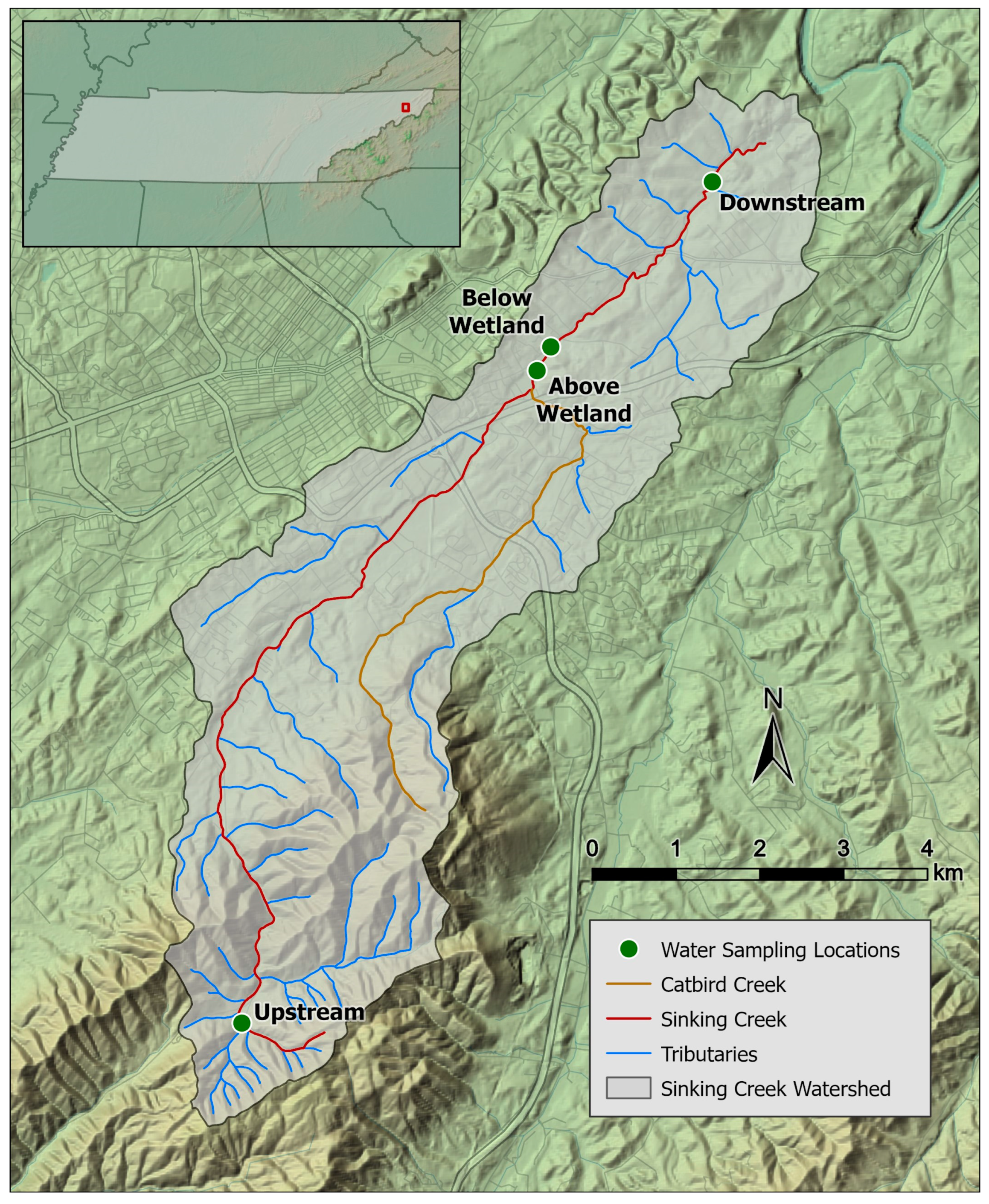
2.1. Study Area and Sample Collection

Sinking Creek was our study site. This creek is part of the Watauga River watershed (HUC 06010103), which is located in multiple counties in Eastern Tennessee. Sinking Creek land use is a mixture of forest (79.8%), agricultural (9.8%), and urban (5%), but is continually changing due to rapid economic and housing development [37]. This creek has 10 stream miles that do not meet the USEPA standard for E. coli [37]. Four sampling points were selected along the creek (Figure 1) based on previous E. coli concentration data [32,34,35]. Sinking Creek flows southwest to northeast, starting in the mountainous area south of Johnson City, TN, USA, and submerges underground northeast of Johnson City before entering the Watauga River. One sampling point was upstream near the headwaters, one was upstream of a wetland (i.e., above wetland) in Jacob’s Nature Park (Johnson City, TN, USA), one was downstream of the wetland (i.e., below wetland), and one was further downstream just above where the creek submerges underground (Figure 1). Sinking Creek flows through the wetland, and during the development of Jacob’s Nature Park, the wetland was expanded to reduce the E. coli in Sinking Creek. Ongoing monitoring studies have shown minimal reduction in E. coli [32,33,34,35].
At each sampling point, 3–4 water samples (15 mL each) were collected from the middle of the creek using a sterile 15-mL Falcon tube (Fisher Scientific, Pittsburgh, PA, USA). Samples were collected monthly over 6 months (June–December 2023). All samples were transported to the ETSU-Environmental Health Science Laboratory (EHSL) on ice, and the analyses were initiated within 3 h of arrival.

2.2. Microbiological Analyses

2.2.1. E. coli Isolation

Upon arrival at the EHSL, the water samples were cultured to isolate E. coli. The samples were vortexed and then filtered using 25 mm, 0.45 µm pore polycarbonate nucleopore filters (Osmonics, Inc., Minnetonka, MN, USA) using a vacuum pump according to the USEPA method 1603 [38]. After filtration, the filters were placed on CHROMagar™ E. coli (Difco, Becton Dickenson, Sparks, MD, USA) plates and incubated for 16–18 h at 35 °C. After incubation, up to three typical colonies (blueish color with smooth surface) were subsequently streaked onto CHROMagar plates and incubated 16–18 h at 35 °C, to isolate pure colonies. The colonies were screened for lactose fermentation using the Indole spot test (Remel, Thermoscientific, Lenexa, KS, USA). Biochemical test strips (RapID ONE test; Remel, Lenexa, KS, USA) were used as quality control on every 25th isolate to confirm that the isolated bacteria were E. coli. The isolates were transferred into Cryovial tubes (Fisher Scientific, Pittsburgh, PA, USA) and stored at −80 °C until further analysis.

2.2.2. Antibiotic Susceptibility Testing

Antibiotic susceptibility testing was carried out using the Kirby–Bauer disk diffusion method. About 3–4 E. coli colonies were picked from a CHROMagar™ E. coli plate, inoculated into 2 mL phosphate buffered saline (PBS) solution, then vortexed to achieve an inoculum equivalent to the 0.5 McFarland standard. One millimeter aliquot from the E. coli-PBS suspension was transferred into 4 mL of Muller Hinton (MH) (Fisher Scientific, Pittsburgh, PA, USA) soft agar and vortexed. This solution was poured over an MH agar plate to form a thin overlay agar. The plates were left for 10 min to solidify before an antibiotic disk placement. The disks were placed on the MH agar plates using an antibiotic disk dispenser (Fisher Scientific, Pittsburgh, PA, USA). The seven antibiotic disks used in this study included the following: amoxicillin/clavulanic acid, tetracycline, trimethoprim-sulfamethoxazole, ceftriaxone, ciprofloxacin, nitrofurantoin, and fosfomycin (Table 1). The plates were placed agar-side up in the incubator for 16–18 h at 35 °C. Thereafter, the diameters of zones of inhibition for each antibiotic were recorded in mm using an electronic caliper. These measurements were interpreted as susceptible, intermediate, or resistant according to the Clinical and Laboratory Standards Institute (CLSI) guidelines [39]. E. coli ATCC 25922 (American Type Culture Collection, Manassas, VA, USA) strain was used as a reference strain. The seven antibiotics used to test for E. coli isolates antibiotic susceptibility and their breakpoints are shown in Table 1. For data analyses, intermediate antibiotic susceptibility test results were reclassified as susceptible.

2.2.3. Polymyxin (Colistin) Susceptibility Testing

Antimicrobial susceptibility testing of colistin was performed using the broth disk elution method as per CLSI recommendations [39]. Briefly, frozen E. coli isolates were revived from the Cryovials on blood agar plates, then 3–5 E. coli isolates were inoculated into sterile saline (5 mL) and vortexed to attain an inoculum corresponding to 0.5 McFarland standard. Colistin disks at three different concentrations 1, 2, and 4 µg, were added to three Cation-Adjusted Mueller–Hinton Broth (CAMHB) tubes, then gently vortexed and left for at least 30 min at room temperature. An additional CAMHB without colistin was used as a negative control. An inoculum of 50 µL from the E. coli suspension was added to each CAMHB tube, vortexed, and incubated at 35 °C for 16–20 h. For inoculum purity check, a 10 µL loop from the E. coli suspension was subcultured onto blood agar plates and incubated similarly to the CAMHB tubes. Thereafter, the MIC was read as the lowest concentration where the growth of the tested isolate was completely inhibited. The results were interpreted as susceptible or intermediate if ≤2 µg/mL or resistant if ≥4 if µg/mL. A positive control ATCC E. coli 25922 was used as a colistin reference strain. We were able to revive 50 frozen isolates out of 177 for colistin testing.

2.3. Data Analysis

The antibiotic resistance outcomes (resistant or susceptible) and multidrug-resistant (MDR) isolates (defined as resistant to at least an antibiotic in three different antimicrobial classes) were cross-tabulated by sampling the location. A Fisher’s exact test or likelihood ratio chi-square test, as appropriate, was used for the analysis, conducted in STATA version 17.0 software (Stata Corp., College Station, TX, USA).

3. Results

Of the 122 water samples tested, 89.3% had at least one E. coli-positive isolate. There was no significant difference in E. coli isolation percentages between the sampling sites. A total of 177 isolates were tested for antibiotic susceptibility. The highest prevalence of AR E. coli to individual antibiotics was to ciprofloxacin (64.2%) and nitrofurantoin (62.7%) and the lowest was to fosfomycin (14.1%) and colistin (6.0%) (Table 2). There were significant (p < 0.05) differences between sampling sites for E. coli resistance to ceftriaxone and amoxicillin/clavulanic acid, with the highest prevalence in wetland locations. The overall individual AR to colistin was 6% out of 50 isolates tested. Three out of 50 isolates had growth at 4 µg/mL colistin concentration.
Multidrug resistance was common among E. coli isolates (i.e., 47.5%) with 5.1% of the 177 isolates being resistant to seven antibiotics except for colistin (Table 3). One of the most common MDR patterns was ceftriaxone, ciprofloxacin, nitrofurantoin (7.5%), ceftriaxone, tetracycline, ciprofloxacin, nitrofurantoin (6.8%), and ceftriaxone, tetracycline, ciprofloxacin, amoxicillin/clavulanic acid, fosfomycin, trimethoprim-sulfamethoxazole, nitrofurantoin (6.1%). Multi-drug-resistant isolates that included colistin also included nitrofurantoin, amoxicillin/clavulanic acid, tetracycline, and/or ciprofloxacin.
The frequency distribution of the antibiotic-resistant phenotypes to the eight antibiotics is shown in Figure 2. Nearly 23 isolates (13%) were resistant to three antibiotics as well as 23 (13%) to four antibiotics. Furthermore, 20 isolates (11.3%), 9 isolates (5.1%), and 9 isolates (5.1%) were resistant to five, six, and seven antibiotics, respectively. The distribution of the prevalence and phenotype of MDR E. coli by sampling site is shown in Table 4. The highest MDR AR E. coli prevalence occurred in samples collected from the wetland sampling sites. The most frequent MDR patterns (6.8%) included three antibiotics: ceftriaxone, ciprofloxacin, and nitrofurantoin, regardless of the site. While the highest MDR prevalence (17.9%) was in isolates from the downstream site (ceftriaxone, ciprofloxacin, and nitrofurantoin), the MDR in isolates from the upstream site had four different phenotypes.

4. Discussion

Our study established a baseline prevalence of AR E. coli from a pathogen-impacted creek in Northeast Tennessee. While previous research conducted on the same stream showed that it exceeded USEPA standards for coliform bacteria including E. coli [32,33,34,35,36], the prevalence of AR was not quantified. Our study demonstrates a high prevalence of MDR E. coli present in this creek, which is a significant public health concern because of the potential dissemination of these isolates to humans through drinking water and recreational activities [40]. Sinking Creek flows through multiple recreation areas such as Jacob’s Nature Park (Johnson City, TN, USA), residential areas in two counties (Carter and Washington), and discharges in Watauga River, which is used for recreational activities. Previous studies have reported the possible human exposure to AR E. coli through recreational activities [41,42].
Land use in the Sinking Creek watershed includes a mixture of agriculture and urban, which could contribute to AR E. coli in the water [32,35]. Previous reports showed that defective septic tanks from residential areas can overflow or leak into nearby water bodies [43,44,45]. The high prevalence of resistance to ciprofloxacin (64.2%), nitrofurantoin (62.7%), and ceftriaxone (40.1%) in this study, which are antibiotics used exclusively in human medicine [46], might indicate human contamination sources. A previous study conducted in a mixed-use watershed in Georgia revealed that 6.5% of E. coli isolates were resistant to at least one antibiotic such as tetracycline (4.1%), and 3% were resistant to multiple antibiotics [24]. These data are lower than 30.5% of E. coli in this study exhibiting tetracycline resistance. This difference might be due to differences in land use in this region and other environmental exposure factors. The mixture of land use around Sinking Creek makes it a valuable representative for other watersheds of a similar size.
Multidrug resistance was common among E. coli isolates (i.e., 47.5%) with most frequent MDR patterns (6.8%) including ceftriaxone, ciprofloxacin, and nitrofurantoin. Ciprofloxacin and nitrofurantoin are frequently used to treat urinary tract infections (UTIs) [46]. Several reports have shown that E. coli isolates resistant to ciprofloxacin and nitrofurantoin were common causes of UTIs [47,48]. The highest MDR AR E. coli prevalence was in samples collected from the wetland sampling sites. These sites are located within Jacob’s Nature Park. Furthermore, we observed that water flows slower in these sites than upstream and downstream sites. The increased input of organic matter from the wetland in the middle section of Sinking Creek may also influence bacterial growth [32,33,34,35,49]. Moreover, agriculture pollution, such as water runoff, can also be a source of AR E. coli contamination, as has been shown in a previous study [50]. Previous studies on surface water (streams, rivers, and lakes) showed variability in AR E. coli prevalence. For instance, a high AR E. coli prevalence was reported from urban rivers in Milwaukee, Wisconsin, where all isolates were determined to be multi-drug-resistant [51]. Resistance to tetracycline (35%), ciprofloxacin (65%), trimethoprim (50%), sulfamethoxazole (88%), and fosfomycin (55%) was also reported, and is higher than what is reported in this study. The authors explained that their findings were due to river contamination with wastewater effluent containing a greater incidence of AR E. coli, history of contamination from human activities, and historical high abundance of antibiotics in the river [51]. A study conducted at rivers in South Carolina [52] revealed that 47% of E. coli isolates were resistant to multiple antibiotics, whereas a study conducted at a pathogen-impacted stream in Indiana [28] that tested 21 E. coli O157:H7 isolates showed that 31.3% were resistant to tetracycline (similar to our findings), but 3.1% were resistant to ciprofloxacin compared to 64.2% in this study. Another study reported a high prevalence (i.e., 63%) of multi-drug-resistant E. coli isolates from multiple water sources including rivers and streams in Ghana which might be due to human and agriculture non-point source contamination [53]. Moreover, resistance to ciprofloxacin (74.2%) and tetracycline (21.5%) was relatively high in their study, in agreement with our data. This variability in study findings could be attributed to factors such as historically pathogen-impacted water, land use and potential sources of contamination, the number of samples/isolates tested, and choices of antibiotics used for susceptibility testing.
Our study revealed that among 50 E. coli isolates tested for colistin resistance, 3 (6%) were resistant. The detection of colistin resistance in E. coli isolates from surface water that is pathogen-impaired is a significant finding which may explain the detection of this resistance phenotype. Few studies in the U.S. have reported colistin resistance in E. coli originating from surface water. One study conducted at a watershed in Indiana reported colistin resistance in one E. coli isolate out of 70 tested [54]. The authors did not provide an explanation for colistin resistance. At another watershed in Indiana, the authors did not detect colistin resistance among 97 E. coli isolates [55]. Nonetheless, colistin resistance in studies conducted in developing countries such as Thailand and China was not uncommon [56,57]. Colistin resistance has been identified in humans as well as in animals and their products in the U.S. Specifically, E. coli resistance to colistin was previously detected at a low level; 0.1% in animals at slaughter and 0.02% from animal meat [58,59] and in a few patients in the U.S. [60,61]. A recent report from Tennessee revealed that raw milk contained four E. coli isolates with the pmrB gene, which is commonly associated with colistin resistance [62]. There are global concerns over the rise in bacterial resistance to colistin [63]. This antibiotic is often considered a “last-resort” drug for treating infections with Gram-negative bacteria that exhibit resistance to multiple antibiotics, such as those caused by AR E. coli. When bacteria resist colistin, treatment options for severe infections become extremely limited, increasing the risk of treatment failure and mortality.
There are limitations to this study. First, it was conducted on a single pathogen-impacted stream in Northeast Tennessee, which may not be representative of other rivers in the region. However, this is the first antibiotic resistance study conducted on a stream in this region and demonstrates the potential negative impact on human health. Additionally, the study could be a representative for other watersheds of similar size and environment. Second, the study was conducted over a 6-month period, so seasonality could not be assessed. Our primary goal was to determine the prevalence of AR E. coli, and we plan to follow up with a longitudinal study to assess seasonality among other risk factors. Third, factors such as water chemistry (pH, total dissolved solids), stream depth, width, and velocity were not consistently measured during each sampling visit and thus were not included in the analysis. Since we aimed to establish a baseline prevalence of AR E. coli in water samples, these factors might have had a limited impact on AR phenotypes per E. coli isolated from the samples.

5. Conclusions

In conclusion, our study has established a baseline prevalence of AR E. coli in Sinking Creek, a pathogen-impacted river in Northeast Tennessee. The high prevalence (i.e., 47.5%) of MDR E. coli, particularly those resistant to ciprofloxacin (64.2%), nitrofurantoin (62.7%), and ceftriaxone (40.1%), drugs used in treatment of a variety of infections, underscores a significant public health concern.
The land use in the Sinking Creek watershed includes a mixture of agriculture and urban and could be a contributing source of AR E. coli in the water. The detection of colistin-resistant E. coli (6%) is a concern to human and environmental health given the prior low detection levels in the U.S. in humans, animals, and the environment.
This study emphasizes the need for continuous monitoring of environmental dissemination and sources of AR bacteria to be able to mitigate the spread of resistant bacteria from aquatic ecosystems to humans. Further longitudinal studies are warranted to explore seasonal variations and other risk factors influencing the prevalence of AR E. coli in this region.

Author Contributions

Conceptualization, W.Q.A. and P.S.; methodology, W.Q.A., P.S. and J.S.; formal analysis, J.S., C.M., A.G., P.O.-M. and T.A.J.; investigation, W.Q.A., P.S., J.S., C.M., A.G. and P.O.-M.; writing—original draft preparation, W.Q.A.; writing—review and editing, P.S, J.S., C.M., A.G., P.O.-M. and T.A.J. All authors have read and agreed to the published version of the manuscript.

Funding

This research received no external funding.

Institutional Review Board Statement

Not applicable.

Informed Consent Statement

Not applicable.

Data Availability Statement

The original data presented in the study are openly available in Harvard Dataverse. https://doi.org/10.7910/DVN/DEHMCT (accessed on 20 September 2024).

Acknowledgments

We extend our appreciation to the following students: Emmanuel Mensah, Averi Sills, Hemanta Neupane, and Mohammed Oladele Suleiman, who assisted in water sampling and laboratory analysis.

Conflicts of Interest

The authors declare no conflicts of interest.

References

  1. WHO (World Health Organization). Antibiotic Resistance. Available online: https://www.who.int/news-room/fact-sheets/detail/antimicrobial-resistance (accessed on 11 August 2024).
  2. EclinicalMedicine. Antimicrobial resistance: A top ten global public health threat. eClinicalMedicine 2021, 41, 101221. [Google Scholar] [CrossRef] [PubMed]
  3. Acar, J.F.; Moulin, G. Antimicrobial resistance: A complex issue. Rev. Sci. Tech. 2012, 31, 23–31. [Google Scholar] [CrossRef]
  4. Wellington, E.M.; Boxall, A.B.; Cross, P.; Feil, E.J.; Gaze, W.H.; Hawkey, P.M.; Johnson-Rollings, A.S.; Jones, D.L.; Lee, N.M.; Otten, W.; et al. The role of the natural environment in the emergence of antibiotic resistance in gram-negative bacteria. Lancet Infect. Dis. 2013, 13, 155–165. [Google Scholar] [CrossRef]
  5. Kusi, J.; Ojewole, C.O.; Ojewole, A.E.; Nwi-Mozu, I. Antimicrobial Resistance Development Pathways in Surface Waters and Public Health Implications. Antibiotics 2022, 11, 821. [Google Scholar] [CrossRef] [PubMed]
  6. Singh, A.K.; Kaur, R.; Verma, S.; Singh, S. Antimicrobials and Antibiotic Resistance Genes in Water Bodies: Pollution, Risk, and Control. Front. Environ. Sci. 2022, 10, 830861. [Google Scholar] [CrossRef]
  7. Cho, S.; Jackson, C.R.; Frye, J.G. Freshwater environment as a reservoir of extended-spectrum β-lactamase-producing Enterobacteriaceae. J. Appl. Microbiol. 2023, 134, lxad034. [Google Scholar] [CrossRef]
  8. Bashir, I.; Lone, F.A.; Bhat, R.A.; Mir, S.A.; Dar, Z.A.; Dar, S.A. Concerns and Threats of Contamination on Aquatic Ecosystems. In Bioremediation and Biotechnology; Springer Nature: Cham, Switzerland, 2020; pp. 1–26. [Google Scholar]
  9. Nappier, S.P.; Liguori, K.; Ichida, A.M.; Stewart, J.R.; Jones, K.R. Antibiotic Resistance in Recreational Waters: State of the Science. Int. J. Environ. Res. Public Health 2020, 17, 8034. [Google Scholar] [CrossRef]
  10. Pruden, A.; Pei, R.; Storteboom, H.; Carlson, K.H. Antibiotic resistance genes as emerging contaminants: Studies in northern Colorado. Environ. Sci. Technol. 2006, 40, 7445–7450. [Google Scholar] [CrossRef]
  11. Pereira, A.; Santos, A.; Tacão, M.; Alves, A.; Henriques, I.; Correia, A. Genetic diversity and antimicrobial resistance of Escherichia coli from Tagus estuary (Portugal). Sci. Total Environ. 2013, 461–462, 65–71. [Google Scholar] [CrossRef]
  12. Karkman, A.; Do, T.T.; Walsh, F.; Virta, M.P.J. Antibiotic-Resistance Genes in Waste Water. Trends Microbiol. 2018, 26, 220–228. [Google Scholar] [CrossRef]
  13. Baquero, F.; Martínez, J.L.; Cantón, R. Antibiotics and antibiotic resistance in water environments. Curr. Opin. Biotechnol. 2008, 19, 260–265. [Google Scholar] [CrossRef] [PubMed]
  14. Suzuki, S.; Pruden, A.; Virta, M.; Zhang, T. Editorial: Antibiotic Resistance in Aquatic Systems. Front. Microbiol. 2017, 8, 14. [Google Scholar] [CrossRef] [PubMed]
  15. Leonard, A.F.C.; Zhang, L.; Balfour, A.J.; Garside, R.; Hawkey, P.M.; Murray, A.K.; Ukoumunne, O.C.; Gaze, W.H. Exposure to and colonisation by antibiotic-resistant E. coli in UK coastal water users: Environmental surveillance, exposure assessment, and epidemiological study (Beach Bum Survey). Environ. Int. 2018, 114, 326–333. [Google Scholar] [CrossRef] [PubMed]
  16. Martinez, J.L. Environmental pollution by antibiotics and by antibiotic resistance determinants. Environ. Pollut. 2009, 157, 2893–2902. [Google Scholar] [CrossRef]
  17. Partridge, S.R. Analysis of antibiotic resistance regions in Gram-negative bacteria. FEMS Microbiol. Rev. 2011, 35, 820–855. [Google Scholar] [CrossRef]
  18. Bennett, P.M. Plasmid encoded antibiotic resistance: Acquisition and transfer of antibiotic resistance genes in bacteria. Br. J. Pharmacol. 2008, 153 (Suppl. S1), S347–S357. [Google Scholar] [CrossRef]
  19. Huddleston, J.R. Horizontal gene transfer in the human gastrointestinal tract: Potential spread of antibiotic resistance genes. Infect. Drug Resist. 2014, 7, 167–176. [Google Scholar] [CrossRef]
  20. Ishii, S.; Ksoll, W.B.; Hicks, R.E.; Sadowsky, M.J. Presence and growth of naturalized Escherichia coli in temperate soils from Lake Superior watersheds. Appl. Environ. Microbiol. 2006, 72, 612–621. [Google Scholar] [CrossRef]
  21. Berthe, T.; Ratajczak, M.; Clermont, O.; Denamur, E.; Petit, F. Evidence for coexistence of distinct Escherichia coli populations in various aquatic environments and their survival in estuary water. Appl. Environ. Microbiol. 2013, 79, 4684–4693. [Google Scholar] [CrossRef]
  22. Djordjevic, S.P.; Stokes, H.W.; Roy Chowdhury, P. Mobile elements, zoonotic pathogens and commensal bacteria: Conduits for the delivery of resistance genes into humans, production animals and soil microbiota. Front. Microbiol. 2013, 4, 86. [Google Scholar] [CrossRef]
  23. Harwood, V.J.; Staley, C.; Badgley, B.D.; Borges, K.; Korajkic, A. Microbial source tracking markers for detection of fecal contamination in environmental waters: Relationships between pathogens and human health outcomes. FEMS Microbiol. Rev. 2014, 38, 1–40. [Google Scholar] [CrossRef] [PubMed]
  24. Cho, S.; Hiott, L.M.; Barrett, J.B.; McMillan, E.A.; House, S.L.; Humayoun, S.B.; Adams, E.S.; Jackson, C.R.; Frye, J.G. Prevalence and characterization of Escherichia coli isolated from the Upper Oconee Watershed in Northeast Georgia. PLoS ONE 2018, 13, e0197005. [Google Scholar] [CrossRef] [PubMed]
  25. Amato, H.K.; Wong, N.M.; Pelc, C.; Taylor, K.; Price, L.B.; Altabet, M.; Jordan, T.E.; Graham, J.P. Effects of concentrated poultry operations and cropland manure application on antibiotic resistant Escherichia coli and nutrient pollution in Chesapeake Bay watersheds. Sci. Total Environ. 2020, 735, 139401. [Google Scholar] [CrossRef]
  26. Haberecht, H.B.; Nealon, N.J.; Gilliland, J.R.; Holder, A.V.; Runyan, C.; Oppel, R.C.; Ibrahim, H.M.; Mueller, L.; Schrupp, F.; Vilchez, S.; et al. Antimicrobial-Resistant Escherichia coli from Environmental Waters in Northern Colorado. J. Environ. Public Health 2019, 2019, 3862949. [Google Scholar] [CrossRef] [PubMed]
  27. Givens, C.E.; Kolpin, D.W.; Hubbard, L.E.; Meppelink, S.M.; Cwiertny, D.M.; Thompson, D.A.; Lane, R.F.; Wilson, M.C. Simultaneous stream assessment of antibiotics, bacteria, antibiotic resistant bacteria, and antibiotic resistance genes in an agricultural region of the United States. Sci. Total Environ. 2023, 904, 166753. [Google Scholar] [CrossRef]
  28. Fincher, L.M.; Parker, C.D.; Chauret, C.P. Occurrence and Antibiotic Resistance of Escherichia coli O157:H7 in a Watershed in North-Central Indiana. J. Environ. Qual. 2009, 38, 997–1004. [Google Scholar] [CrossRef] [PubMed]
  29. Domínguez, D.C.; Chacón, L.M.; Wallace, D.J. Anthropogenic Activities and the Problem of Antibiotic Resistance in Latin America: A Water Issue. Water 2021, 13, 2693. [Google Scholar] [CrossRef]
  30. Bong, C.W.; Low, K.Y.; Chai, L.C.; Lee, C.W. Prevalence and Diversity of Antibiotic Resistant Escherichia coli from Anthropogenic-Impacted Larut River. Front. Public Health 2022, 10, 794513. [Google Scholar] [CrossRef]
  31. Truong, T.; Hoang, T.L.; Tran, L.T.; Pham, T.P.T.; Le, T.-H. Prevalence of Antibiotic Resistance Genes in the Saigon River Impacted by Anthropogenic Activities. Water 2021, 13, 2234. [Google Scholar] [CrossRef]
  32. Gilfillan, D.; Joyner, T.A.; Scheuerman, P. Maxent estimation of aquatic Escherichia coli stream impairment. PeerJ 2018, 6, e5610. [Google Scholar] [CrossRef]
  33. Hall, K.K.; Evanshen, B.G.; Maier, K.J.; Scheuerman, P.R. Application of multivariate statistical methodology to model factors influencing fate and transport of fecal pollution in surface waters. J. Environ. Qual. 2014, 43, 358–370. [Google Scholar] [CrossRef] [PubMed]
  34. Hall, K.K.; Scheuerman, P.R. Development of Multiple Regression Models to Predict Sources of Fecal Pollution. Water Environ. Res. 2017, 89, 1961–1969. [Google Scholar] [CrossRef] [PubMed]
  35. Gilfillan, D.; Hall, K.; Joyner, T.A.; Scheuerman, P. Canonical Variable Selection for Ecological Modeling of Fecal Indicators. J. Environ. Qual. 2018, 47, 974–984. [Google Scholar] [CrossRef] [PubMed]
  36. McCurdy, P.; Luffman, I.; Joyner, T.A.; Maier, K. Storm sampling to assess inclement weather impacts on water quality in a karst watershed: Sinking Creek, Watauga watershed, East Tennessee. J. Environ. Qual. 2021, 50, 429–440. [Google Scholar] [CrossRef] [PubMed]
  37. TDEC (Tennessee Department of Environment and Conservation). Water Quality Rules, Reports & Publications. Available online: https://www.tn.gov/environment/program-areas/wr-water-resources/water-quality/water-quality-reports---publications.html (accessed on 29 July 2024).
  38. USEPA. Method 1603 Escherichia coli (E. coli) in Water by Membrane Filtration Using Modified Membrane-Thermotolerant Escherichia coli Agar (Modified m-TEC), EPA 821-R-02-023; Environmental Protection Agency, Office of Research and Development: Cincinnati, OH, USA, 2002. Available online: https://nepis.epa.gov/Exe/ZyNET.exe/P1008MZV.txt?ZyActionD=ZyDocument&Client=EPA&Index=2000%20Thru%202005&Docs=&Query=&Time=&EndTime=&SearchMethod=1&TocRestrict=n&Toc=&TocEntry=&QField=&QFieldYear=&QFieldMonth=&QFieldDay=&UseQField=&IntQFieldOp=0&ExtQFie (accessed on 10 March 2024).
  39. CLSI. Performance Standards for Antimicrobial Susceptibility Testing, 31st ed.; CLSI Supplement M100; Clinical and Laboratory Standards Institute: Wayne, PA, USA, 2021. [Google Scholar]
  40. Coleman, B.L.; Salvadori, M.I.; McGeer, A.J.; Sibley, K.A.; Neumann, N.F.; Bondy, S.J.; Gutmanis, I.A.; McEwen, S.A.; Lavoie, M.; Strong, D.; et al. The role of drinking water in the transmission of antimicrobial-resistant E. coli. Epidemiol. Infect. 2012, 140, 633–642. [Google Scholar] [CrossRef]
  41. O‘Flaherty, E.; Solimini, A.; Pantanella, F.; Cummins, E. The potential human exposure to antibiotic resistant-Escherichia coli through recreational water. Sci. Total Environ. 2019, 650, 786–795. [Google Scholar] [CrossRef]
  42. Mughini-Gras, L.; Dorado-García, A.; van Duijkeren, E.; van den Bunt, G.; Dierikx, C.M.; Bonten, M.J.M.; Bootsma, M.C.J.; Schmitt, H.; Hald, T.; Evers, E.G.; et al. Attributable sources of community-acquired carriage of Escherichia coli containing β-lactam antibiotic resistance genes: A population-based modelling study. Lancet Planet. Health 2019, 3, e357–e369. [Google Scholar] [CrossRef]
  43. Masters, N.; Wiegand, A.; Ahmed, W.; Katouli, M. Escherichia coli virulence genes profile of surface waters as an indicator of water quality. Water Res. 2011, 45, 6321–6333. [Google Scholar] [CrossRef]
  44. Iverson, G.; Humphrey, C.P.; Postma, M.H.; O’Driscoll, M.A.; Manda, A.K.; Finley, A. Influence of Sewered versus Septic Systems on Watershed Exports of E. coli. Water Air Soil Pollut. 2017, 228, 237. [Google Scholar] [CrossRef]
  45. USEPA. Septic System Impacts on Water Sources. Available online: https://www.epa.gov/septic/septic-system-impacts-water-sources (accessed on 10 June 2024).
  46. CDC. All Antibiotic Classes|A.R. & Patient Safety Portal. Available online: https://arpsp.cdc.gov/profile/antibiotic-use/all-classes (accessed on 10 June 2024).
  47. Karlowsky James, A.; Kelly Laurie, J.; Thornsberry, C.; Jones Mark, E.; Sahm Daniel, F. Trends in Antimicrobial Resistance among Urinary Tract Infection Isolates of Escherichia coli from Female Outpatients in the United States. Antimicrob. Agents Chemother. 2002, 46, 2540–2545. [Google Scholar] [CrossRef]
  48. Karlowsky, J.A.; Thornsberry, C.; Jones, M.E.; Sahm, D.F. Susceptibility of Antimicrobial-Resistant Urinary Escherichia coli Isolates to Fluoroquinolones and Nitrofurantoin. Clin. Infect. Dis. 2003, 36, 183–187. [Google Scholar] [CrossRef]
  49. Gregory, L.F.; Karthikeyan, R.; Aitkenhead-Peterson, J.A.; Gentry, T.J.; Wagner, K.L.; Harmel, R.D. Nutrient loading impacts on culturable E. coli and other heterotrophic bacteria fate in simulated stream mesocosms. Water Res. 2017, 126, 442–449. [Google Scholar] [CrossRef]
  50. Ibekwe, A.M.; Murinda, S.E.; Graves, A.K. Genetic diversity and antimicrobial resistance of Escherichia coli from human and animal sources uncovers multiple resistances from human sources. PLoS ONE 2011, 6, e20819. [Google Scholar] [CrossRef] [PubMed]
  51. Kappell, A.D.; DeNies, M.S.; Ahuja, N.H.; Ledeboer, N.A.; Newton, R.J.; Hristova, K.R. Detection of multi-drug resistant Escherichia coli in the urban waterways of Milwaukee, WI. Front. Microbiol. 2015, 6, 336. [Google Scholar] [CrossRef]
  52. Webster, L.F.; Thompson, B.C.; Fulton, M.H.; Chestnut, D.E.; Van Dolah, R.F.; Leight, A.K.; Scott, G.I. Identification of sources of Escherichia coli in South Carolina estuaries using antibiotic resistance analysis. J. Exp. Mar. Biol. Ecol. 2004, 298, 179–195. [Google Scholar] [CrossRef]
  53. Odonkor, S.T.; Addo, K.K. Prevalence of Multidrug-Resistant Escherichia coli Isolated from Drinking Water Sources. Int. J. Microbiol. 2018, 2018, 7204013. [Google Scholar] [CrossRef]
  54. Thornton, C.N.; Tanner, W.D.; VanDerslice, J.A.; Brazelton, W.J. Localized effect of treated wastewater effluent on the resistome of an urban watershed. GigaScience 2020, 9, giaa125. [Google Scholar] [CrossRef] [PubMed]
  55. Sidhu, A.S.; Mikolajczyk, F.N.; Fisher, J.C. Antimicrobial Resistance Linked to Septic System Contamination in the Indiana Lake Michigan Watershed. Antibiotics 2023, 12, 569. [Google Scholar] [CrossRef]
  56. Tabut, P.; Yongyod, R.; Ungcharoen, R.; Kerdsin, A. The Distribution of Mobile Colistin-Resistant Genes, Carbapenemase-Encoding Genes, and Fluoroquinolone-Resistant Genes in Escherichia coli Isolated from Natural Water Sources in Upper Northeast Thailand. Antibiotics 2022, 11, 1760. [Google Scholar] [CrossRef]
  57. Snyman, Y.; Whitelaw, A.C.; Barnes, J.M.; Maloba, M.R.B.; Newton-Foot, M. Characterisation of mobile colistin resistance genes (mcr-3 and mcr-5) in river and storm water in regions of the Western Cape of South Africa. Antimicrob. Resist. Infect. Control 2021, 10, 96. [Google Scholar] [CrossRef]
  58. Meinersmann, R.J.; Ladely, S.R.; Plumblee, J.R.; Cook, K.L.; Thacker, E. Prevalence of mcr-1 in the Cecal Contents of Food Animals in the United States. Antimicrob. Agents Chemother. 2017, 61, e02244-16. [Google Scholar] [CrossRef] [PubMed]
  59. Wang, Y.; Hou, N.; Johnston, J.; Sarreal, C.; Jarosh, J.; Hughes, A.C.; Gu, Y.; He, X. Low prevalence of mobile colistin-resistance in U.S. meat, catfish, poultry and genomic characterization of a mcr-1 positive Escherichia coli strain. Food Control 2020, 118, 107434. [Google Scholar] [CrossRef]
  60. Henig, O.; Rojas, L.J.; Bachman, M.A.; Rudin, S.D.; Brennan, B.M.; Soehnlen, M.K.; Jones, K.L.; Mills, J.P.; Dombecki, C.R.; Valyko, A.M.; et al. Identification of four patients with colistin-resistant Escherichia coli containing the mobile colistin resistance mcr-1 gene from a single health system in Michigan. Infect. Control Hosp. Epidemiol. 2019, 40, 1059–1062. [Google Scholar] [CrossRef] [PubMed]
  61. Monte, D.F.; Nelson, V.; Cerdeira, L.; Keelara, S.; Greene, S.; Griffin, D.; Rath, S.; Hall, R.; Page, N.; Lawson, T.; et al. Multidrug- and colistin-resistant Salmonella enterica 4,[5],12:i:- sequence type 34 carrying the mcr-3.1 gene on the IncHI2 plasmid recovered from a human. J. Med. Microbiol. 2019, 68, 986–990. [Google Scholar] [CrossRef] [PubMed]
  62. Gelalcha, B.D.; Mohammed, R.I.; Gelgie, A.E.; Kerro Dego, O. Molecular epidemiology and pathogenomics of extended-spectrum beta-lactamase producing- Escherichia coli and - Klebsiella pneumoniae isolates from bulk tank milk in Tennessee, USA. Front. Microbiol. 2023, 14, 1283165. [Google Scholar] [CrossRef]
  63. Mondal, A.H.; Khare, K.; Saxena, P.; Debnath, P.; Mukhopadhyay, K.; Yadav, D. A Review on Colistin Resistance: An Antibiotic of Last Resort. Microorganisms 2024, 12, 772. [Google Scholar] [CrossRef]
Figure 1. Water sampling site along Sinking Creek in Northeast Tennessee. Red box indicates the location of Sinking Creek in Northeast Tennessee.
Ijerph 21 01285 g001
Figure 2. Bar chart showing the frequency distribution of phenotypic antibiotic resistance to up to eight antibiotics among E. coli isolates (N = 177) from water samples originating from Sinking Creek in Northeast Tennessee.
Ijerph 21 01285 g002
Table 1. The disk diffusion zone of inhibition interpretation for antibiotics used to test susceptibility of E. coli isolates (N = 177) from water samples of Sinking Creek (Johnson City, TN, USA).
Drug Class/Antibiotic 1Concentration (μg)Zones of Inhibition Diameter in mm Interpretation 2
SIR
Sulfonamides
Trimethoprim & Sulfamethoxazole1.25/23.75≥1645,245≤10
Penicillin β-lactamase inhibitor combinations
Amoxicillin/
Clavulanic acid
20/10≥1814–17≤13
Tetracyclines
Tetracycline30≥1545,274≤11
Cephalosporins
Ceftriaxone30≥2320–22≤19
Fluoroquinolone
Ciprofloxacin5≥3121–30≤20
Urinary anti-infectives
Nitrofurantoin300≥1715–16≤14
Phosphonic antibiotics
Fosfomycin200≥1613–15≤12
1 Colistin susceptibility testing was performed according to the Clinical and Laboratory Standards Institute (CLSI) broth disk elution method using three concentrations (1, 2, and 4 µg). The results were interpreted as susceptible or intermediate ≤ 2 µg/mL or resistant if ≥4 if µg/mL. 2 The zones of inhibition values interpretation into sensitive (S), intermediate (I), and resistant (R) was according to the CLSI guidelines.
Table 2. Prevalence of antibiotic-resistant E. coli isolates (N = 177) recovered from water samples from Sinking Creek, Johnson City, TN, USA.
No. (%) of Isolates
Antibiotics/Sampling SiteUpstream
(n = 63)
Above Wetland
(n = 43)
Below Wetland
(n = 29)
Downstream
(n = 42)
p-Value 1Overall Prevalence
(n = 177)
Ceftriaxone23 (36.5)23 (53.5)16 (55.2)9 (21.4)0.00571 (40.1)
Tetracycline17 (27.0)14 (32.6)12 (41.4)11 (26.2)0.48254 (30.5)
Ciprofloxacin43 (68.3)28 (65.1)15 (51.7)27 (64.3)0.488113 (63.8)
Amoxicillin/
Clavulanic acid
18 (28.6)19 (44.2)10 (34.5)5 (11.9)0.00852 (29.4)
Fosfomycin12 (19.1)9 (20.9)2 (6.9)2 (4.8)0.06425 (14.1)
Trimethoprim-Sulfamethoxazole14 (22.2)12 (27.9)8 (27.6)4 (9.5)0.12638 (21.5)
Nitrofurantoin34 (54.0)30 (69.8)20 (69.0)27 (64.3)0.333111 (62.7)
Colistin 21 (7.1)0 (0)2 (18.1)0 (0.0)0.2433 (6.0)
1 p-value < 0.05 indicates that percentages of antibiotic-resistant E. coli isolates were significantly different by sampling sites based on chi-square test in STATA 17.0 software; 2 For colistin, the number of isolates tested from upstream, above wetland, below wetland, and downstream were 14, 11, 11, and 14, respectively.
Table 3. Antibiotic-resistant phenotypes of E. coli isolates (N = 177) from water samples collected from Sinking Creek, Johnson City, TN, USA.
Resistant Phenotype 1FrequencyPercentage
No resistance3017.00
Resistant to only one antibiotic3117.50
CIP_NIF2013.61
CIP138.84
NIF_117.48
CRO_CIP_NIF106.80
CRO_TET_CIP_NIF106.80
CRO_TET_CIP_AMC_FOS_SXT_NIF96.12
COL36.00
TET_CIP64.08
CRO_CIP_AMC_NIF53.40
CRO_TET_CIP_AMC_NIF53.40
CRO_CIP_AMC_FOS_SXT_NIF42.72
CRO_CIP_AMC_SXT_NIF42.72
CRO_TET_CIP_SXT_NIF42.72
TET42.72
CRO_AMC_FOS_SXT_NIF32.04
CRO_TET_CIP_AMC_SXT_NIF32.04
CIP_AMC_NIF21.36
CIP_SXT21.36
CRO_CIP21.36
CRO_TET_AMC_SXT_NIF21.36
TET_CIP_NIF21.36
AMC10.68
AMC_NIF10.68
AMC_NIF_COL10.68
CIP_AMC10.68
CIP_AMC_FOS_SXT_NIF10.68
CIP_AMC_SXT_NIF10.68
CRO_AMC_NIF10.68
CRO_AMC_SXT_NIF10.68
CRO_CIP_AMC_FOS_NIF10.68
CRO_CIP_FOS_NIF10.68
CRO_CIP_FOS_SXT_NIF10.68
CRO_FOS_NIF10.68
CRO_TET_AMC_FOS_SXT_NIF10.68
CRO_TET_CIP10.68
CRO_TET_CIP_AMC_FOS_NIF10.68
CRO_TET_SXT_NIF10.68
FOS_NIF10.68
TET_AMC_NIF10.68
TET_AMC_NIF_COL10.68
TET_CIP_AMC10.68
TET_CIP_AMC_FOS10.68
TET_CIP_COL10.68
TET_CIP_SXT_NIF10.68
1 T CIP, Ciprofloxacin; NIF, Nitrofurantoin; CRO, Ceftriaxone; TET, Tetracycline; AMC, Amoxicillin/Clavulanic acid; COL, Colistin; SXT, Trimethoprim-Sulfamethoxazole; FOS, Fosfomycin.
Table 4. Distribution of antibiotic-resistant E. coli isolates from water samples by sampling site (Sinking creek, Johnson City, TN, USA) 1.
Sampling Site
(No. Isolates)
MDR
No. (%)
Top Most Frequent MDR (>5%) Phenotype and %Highest MDR Profile and %
Upstream
(n = 63)
27 (42.9)CRO_TET_CIP_AMC_FOS_SXT_NIF (5.7%)
CRO_CIP_AMC_FOS_SXT_NIF (5.7%)
CRO_AMC_FOS_SXT_NIF (5.7%)
CRO_CIP_NIF (5.7%)
CRO_TET_CIP_AMC_FOS_SXT_NIF (5.7%)
Above Wetland
(n = 43)
26 (60.7)CRO_CIP_AMC_FOS_SXT_NIF (16.7%)CRO_CIP_AMC_FOS_SXT_NIF (16.7%)
Below Wetland
(n = 29)
15 (51.7)CRO_TET_CIP_NIF (18.8%)CRO_TET_CIP_AMC_SXT_NIF (9.1%)
Downstream
(n = 42)
15 (35.7)CRO_CIP_NIF (17.9%)CRO_CIP_AMC_FOS_SXT_NIF (2.8%)
1 CIP, Ciprofloxacin; NIF, Nitrofurantoin; CRO, Ceftriaxone; TET, Tetracycline; AMC, Amoxicillin/Clavulanic acid; COL, Colistin; SXT, Trimethoprim-Sulfamethoxazole; FOS, Fosfomycin.
Disclaimer/Publisher’s Note: The statements, opinions and data contained in all publications are solely those of the individual author(s) and contributor(s) and not of MDPI and/or the editor(s). MDPI and/or the editor(s) disclaim responsibility for any injury to people or property resulting from any ideas, methods, instructions or products referred to in the content.

