Prognostic Implications of Intratumoral and Peritumoral Infiltrating Lymphocytes in Pancreatic Ductal Adenocarcinoma


Abstract
:1. Introduction
2. Materials and Methods
2.1. Published Study Search and Selection Criteria
2.2. Data Extraction
2.3. Statistical Analysis
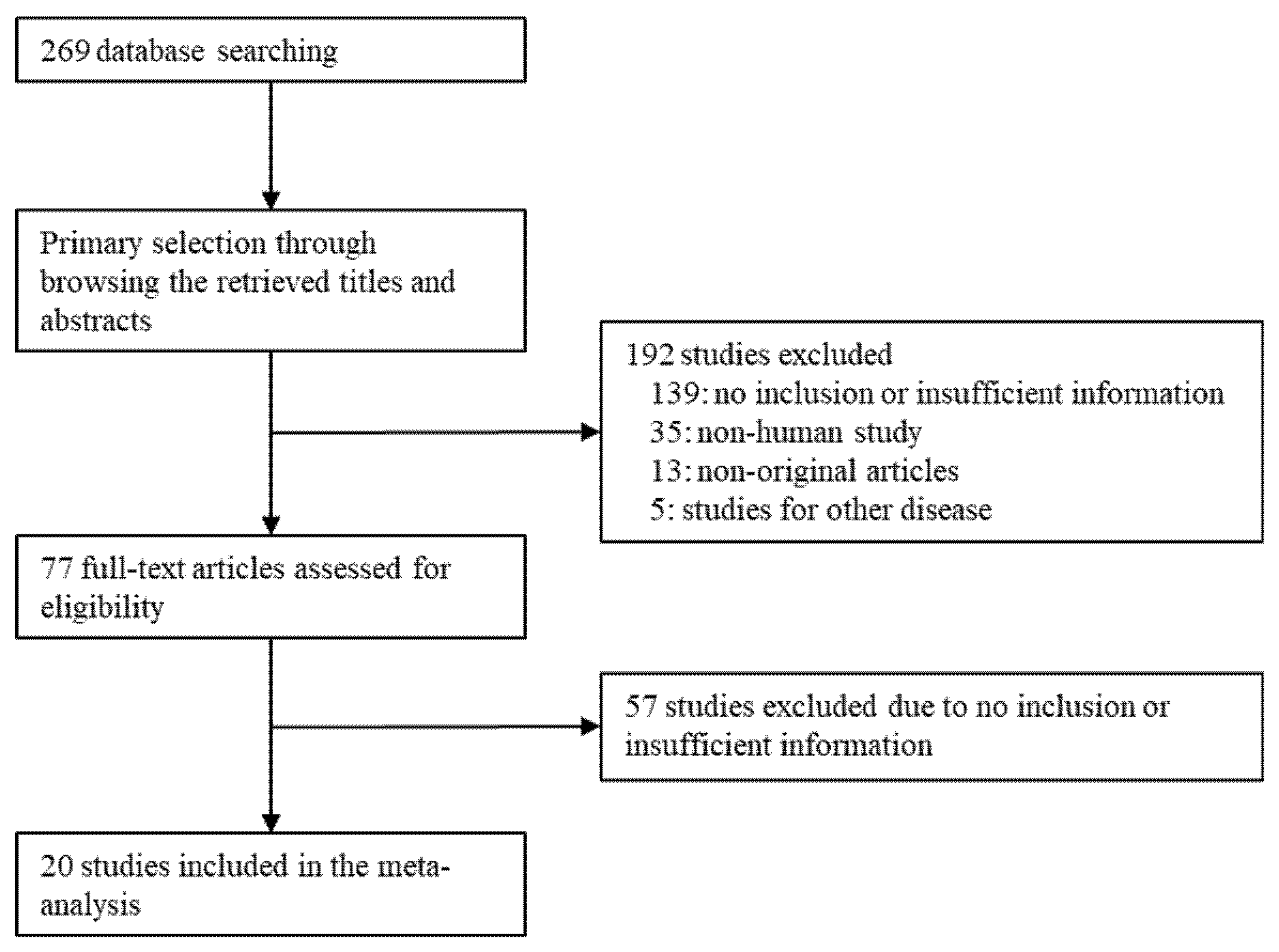
3. Results
3.1. Selection and Characteristics of the Studies
3.2. Correlation between Intra- or Peritumoral Infiltrating T-lymphocytes and Overall Survival in Pancreatic Ductal Adenocarcinoma
3.3. Correlation between Intra- or Peritumoral Infiltrating T-lymphocytes and Disease-Free Survival in Pancreatic Ductal Adenocarcinoma
4. Discussion
5. Conclusions
Author Contributions
Funding
Institutional Review Board Statement
Informed Consent Statement
Data Availability Statement
Conflicts of Interest
References
- WHO Classification of Tumours Editorial Board. WHO Classification of Tumours of the Digestive System, 5th ed.; International Agency for Research on Cancer: Lyon, France, 2018. [Google Scholar]
- Siegel, R.; Ward, E.; Brawley, O.; Jemal, A. Cancer statistics, 2011: The impact of eliminating socioeconomic and racial disparities on premature cancer deaths. CA Cancer J. Clin. 2020, 61, 212–236. [Google Scholar] [CrossRef]
- Philip, P.A.; Mooney, M.; Jaffe, D.; Eckhardt, G.; Moore, M.; Meropol, N.; Emens, L.; O’Reilly, E.; Korc, M.; Ellis, L.; et al. Consensus report of the national cancer institute clinical trials planning meeting on pancreas cancer treatment. J. Clin. Oncol. 2009, 27, 5660–5669. [Google Scholar] [CrossRef]
- Ino, Y.; Yamazaki-Itoh, R.; Shimada, K.; Iwasaki, M.; Kosuge, T.; Kanai, Y.; Hiraoka, N. Immune cell infiltration as an indicator of the immune microenvironment of pancreatic cancer. Br. J. Cancer 2013, 108, 914–923. [Google Scholar] [CrossRef]
- Biswas, S.K.; Mantovani, A. Macrophage plasticity and interaction with lymphocyte subsets: Cancer as a paradigm. Nat. Immunol. 2010, 11, 889–896. [Google Scholar] [CrossRef]
- Dunn, G.P.; Old, L.J.; Schreiber, R.D. The three Es of cancer immunoediting. Annu. Rev. Immunol. 2004, 22, 329–360. [Google Scholar] [CrossRef]
- Ferrone, C.; Dranoff, G. Dual roles for immunity in gastrointestinal cancers. J. Clin. Oncol. 2010, 28, 4045–4051. [Google Scholar] [CrossRef] [Green Version]
- Grivennikov, S.I.; Greten, F.R.; Karin, M. Immunity, inflammation, and cancer. Cell 2010, 140, 883–899. [Google Scholar] [CrossRef] [Green Version]
- Hanahan, D.; Weinberg, R.A. Hallmarks of cancer: The next generation. Cell 2021, 144, 646–674. [Google Scholar] [CrossRef] [Green Version]
- Hiraoka, N.; Yamazaki-Itoh, R.; Ino, Y.; Mizuguchi, Y.; Yamada, T.; Hirohashi, S.; Kanai, Y. CXCL17 and ICAM2 are associated with a potential anti-tumor immune response in early intraepithelial stages of human pancreatic carcinogenesis. Gastroenterology 2011, 140, 310–321. [Google Scholar] [CrossRef]
- Sica, A.; Bronte, V. Altered macrophage differentiation and immune dysfunction in tumor development. J. Clin. Invest. 2007, 117, 1155–1166. [Google Scholar] [CrossRef]
- Pages, F.; Berger, A.; Camus, M.; Sanchez-Cabo, F.; Costes, A.; Molidor, R.; Mlecnik, B.; Kirilovsky, A.; Nilsson, M.; Damotte, D.; et al. Effector memory T cells, early metastasis, and survival in colorectal cancer. N. Engl. J. Med. 2005, 353, 2654–2666. [Google Scholar] [CrossRef] [PubMed]
- Zhang, L.; Conejo-Garcia, J.R.; Katsaros, D.; Gimotty, P.A.; Massobrio, M.; Regnani, G.; Makrigiannakis, A.; Gray, H.; Schlienger, K.; Liebman, M.N.; et al. Intratumoral T cells, recurrence, and survival in epithelial ovarian cancer. N. Engl. J. Med. 2003, 348, 203–213. [Google Scholar] [CrossRef] [PubMed] [Green Version]
- Diana, A.; Wang, L.M.; D’Costa, Z.; Allen, P.; Azad, A.; Silva, M.A.; Soonawalla, Z.; Liu, S.; McKenna, W.G.; Muschel, R.J.; et al. Prognostic value, localization and correlation of PD-1/PD-L1, CD8 and FOXP3 with the desmoplastic stroma in pancreatic ductal adenocarcinoma. Oncotarget 2016, 7, 40992–41004. [Google Scholar] [CrossRef] [PubMed] [Green Version]
- Fukunaga, A.; Miyamoto, M.; Cho, Y.; Murakami, S.; Kawarada, Y.; Oshikiri, T.; Kato, K.; Kurokawa, T.; Suzuoki, M.; Nakakubo, Y.; et al. CD8+ tumor-infiltrating lymphocytes together with CD4+ tumor-infiltrating lymphocytes and dendritic cells improve the prognosis of patients with pancreatic adenocarcinoma. Pancreas 2004, 28, e26–e31. [Google Scholar] [CrossRef]
- Homma, Y.; Taniguchi, K.; Murakami, T.; Nakagawa, K.; Nakazawa, M.; Matsuyama, R.; Mori, R.; Takeda, K.; Ueda, M.; Ichikawa, Y.; et al. Immunological impact of neoadjuvant chemoradiotherapy in patients with borderline resectable pancreatic ductal adenocarcinoma. Ann. Surg. Oncol. 2014, 21, 670–676. [Google Scholar] [CrossRef]
- Hwang, H.K.; Kim, H.I.; Kim, S.H.; Choi, J.; Kang, C.M.; Kim, K.S.; Lee, W.J. Prognostic impact of the tumor-infiltrating regulatory T-cell (Foxp3+)/activated cytotoxic T lymphocyte (granzyme B+) ratio on resected left-sided pancreatic cancer. Oncol. Lett. 2016, 12, 4477–4484. [Google Scholar] [CrossRef] [Green Version]
- Liu, H.L.; Guan, C.J.; Wu, Y.J.; Hu, M.G.; Zhao, Z.M.; Liu, R. Clinical Significance of Preoperative CD8+ Central Memory T Cells for Operable Pancreatic Adenocarcinoma. Dig. Surg. 2015, 32, 433–438. [Google Scholar] [CrossRef]
- Liu, L.; Zhao, G.; Wu, W.; Rong, Y.; Jin, D.; Wang, D.; Lou, W.; Qin, X. Low intratumoral regulatory T cells and high peritumoral CD8(+) T cells relate to long-term survival in patients with pancreatic ductal adenocarcinoma after pancreatectomy. Cancer Immunol. Immunother. 2016, 65, 73–82. [Google Scholar] [CrossRef]
- Michelakos, T.; Cai, L.; Villani, V.; Sabbatino, F.; Kontos, F.; Fernández-Del Castillo, C.; Yamada, T.; Neyaz, A.; Taylor, M.S.; Deshpande, V.; et al. Tumor microenvironment immune response in pancreatic ductal adenocarcinoma patients treated with neoadjuvant therapy. J. Natl. Cancer Inst. 2020, 113, 182–191. [Google Scholar] [CrossRef]
- Mota Reyes, C.; Teller, S.; Muckenhuber, A.; Konukiewitz, B.; Safak, O.; Weichert, W.; Friess, H.; Ceyhan, G.O.; Demir, I.E. Neoadjuvant Therapy Remodels the Pancreatic Cancer Microenvironment via Depletion of Protumorigenic Immune Cells. Clin. Cancer Res. 2020, 26, 220–231. [Google Scholar] [CrossRef] [Green Version]
- Nejati, R.; Goldstein, J.B.; Halperin, D.M.; Wang, H.; Hejazi, N.; Rashid, A.; Katz, M.H.; Lee, J.E.; Fleming, J.B.; Rodriguez-Canales, J.; et al. Prognostic Significance of Tumor-Infiltrating Lymphocytes in Patients with Pancreatic Ductal Adenocarcinoma Treated with Neoadjuvant Chemotherapy. Pancreas 2017, 46, 1180–1187. [Google Scholar] [CrossRef] [PubMed]
- Sadozai, H.; Acharjee, A.; Eppenberger-Castori, S.; Gloor, B.; Gruber, T.; Schenk, M.; Karamitopoulou, E. Distinct Stromal and Immune Features Collectively Contribute to Long-Term Survival in Pancreatic Cancer. Front. Immunol. 2021, 12, 643529. [Google Scholar] [CrossRef]
- Seifert, L.; Plesca, I.; Müller, L.; Sommer, U.; Heiduk, M.; von Renesse, J.; Digomann, D.; Glück, J.; Klimova, A.; Weitz, J.; et al. LAG-3-Expressing Tumor-Infiltrating T Cells Are Associated with Reduced Disease-Free Survival in Pancreatic Cancer. Cancers 2021, 13, 1297. [Google Scholar] [CrossRef]
- Tahkola, K.; Mecklin, J.P.; Wirta, E.V.; Ahtiainen, M.; Helminen, O.; Böhm, J.; Kellokumpu, I. High immune cell score predicts improved survival in pancreatic cancer. Virchows Arch. 2018, 472, 653–665. [Google Scholar] [CrossRef] [PubMed]
- Tahkola, K.; Leppänen, J.; Ahtiainen, M.; Väyrynen, J.; Haapasaari, K.M.; Karttunen, T.; Kellokumpu, I.; Helminen, O.; Böhm, J. Immune cell score in pancreatic cancer-comparison of hotspot and whole-section techniques. Virchows Arch. 2019, 474, 691–699. [Google Scholar] [CrossRef] [Green Version]
- Tang, Y.; Xu, X.; Guo, S.; Zhang, C.; Tang, Y.; Tian, Y.; Ni, B.; Lu, B.; Wang, H. An increased abundance of tumor-infiltrating regulatory T cells is correlated with the progression and prognosis of pancreatic ductal adenocarcinoma. PLoS ONE 2014, 9, e91551. [Google Scholar] [CrossRef] [PubMed] [Green Version]
- Tewari, N.; Zaitoun, A.M.; Arora, A.; Madhusudan, S.; Ilyas, M.; Lobo, D.N. The presence of tumour-associated lymphocytes confers a good prognosis in pancreatic ductal adenocarcinoma: An immunohistochemical study of tissue microarrays. BMC Cancer 2013, 13, 436. [Google Scholar] [CrossRef]
- Tsukamoto, M.; Imai, K.; Ishimoto, T.; Komohara, Y.; Yamashita, Y.I.; Nakagawa, S.; Umezaki, N.; Yamao, T.; Kitano, Y.; Miyata, T.; et al. PD-L1 expression enhancement by infiltrating macrophage-derived tumor necrosis factor-α leads to poor pancreatic cancer prognosis. Cancer Sci. 2019, 110, 310–320. [Google Scholar] [CrossRef] [PubMed]
- Wang, X.; Lang, M.; Zhao, T.; Feng, X.; Zheng, C.; Huang, C.; Hao, J.; Dong, J.; Luo, L.; Li, X.; et al. Cancer-FOXP3 directly activated CCL5 to recruit FOXP3+Treg cells in pancreatic ductal adenocarcinoma. Oncogene 2017, 36, 3048–3058. [Google Scholar] [CrossRef]
- Wartenberg, M.; Zlobec, I.; Perren, A.; Koelzer, V.H.; Gloor, B.; Lugli, A.; Karamitopoulou, E. Accumulation of FOXP3+T-cells in the tumor microenvironment is associated with an epithelial-mesenchymal-transition-type tumor budding phenotype and is an independent prognostic factor in surgically resected pancreatic ductal adenocarcinoma. Oncotarget 2015, 6, 4190–4201. [Google Scholar] [CrossRef] [Green Version]
- Zhang, Y.; Xu, J.; Hua, J.; Liu, J.; Liang, C.; Meng, Q.; Wei, M.; Zhang, B.; Yu, X.; Shi, S. A PD-L2-based immune marker signature helps to predict survival in resected pancreatic ductal adenocarcinoma. J. Immunother. Cancer 2019, 7, 233. [Google Scholar] [CrossRef] [Green Version]
- Parmar, M.K.; Torri, V.; Stewart, L. Extracting summary statistics to perform meta-analyses of the published literature for survival endpoints. Stat. Med. 1998, 17, 2815–2834. [Google Scholar] [CrossRef]
- Yusuf, S.; Peto, R.; Lewis, J.; Collins, R.; Sleightet, P. Beta blockade during and after myocardial infarction: An overview of the randomized trials. Prog. Cardiovasc. Dis. 1985, 27, 335–371. [Google Scholar] [CrossRef]
- Siegel, R.L.; Miller, K.D.; Jemal, A. Cancer statistics, 2016. CA Cancer J. Clin. 2016, 66, 7–30. [Google Scholar] [CrossRef] [PubMed] [Green Version]
- Hartwig, W.; Büchler, M.W. Pancreatic Cancer: Current Options for Diagnosis, Staging and Therapeutic Management. Gastrointest. Tumors 2013, 1, 41–52. [Google Scholar] [CrossRef] [PubMed] [Green Version]
- Wandmacher, A.M.; Letsch, A.; Sebens, S. Challenges and Future Perspectives of Immunotherapy in Pancreatic Cancer. Cancers 2021, 13, 4235. [Google Scholar] [CrossRef] [PubMed]
- Karpathiou, G.; Vieville, M.; Gavid, M.; Camy, F.; Dumollard, J.M.; Magné, N.; Froudarakis, M.; Prades, J.M.; Peoc’h, M. Prognostic significance of tumor budding, tumor-stroma ratio, cell nests size, and stroma type in laryngeal and pharyngeal squamous cell carcinomas. Head Neck 2019, 41, 1918–1927. [Google Scholar] [CrossRef]
- Mahajan, U.M.; Langhoff, E.; Goni, E.; Costello, E.; Greenhalf, W.; Halloran, C.; Ormanns, S.; Kruger, S.; Boeck, S.; Ribback, S.; et al. Immune Cell and Stromal Signature Associated with Progression-Free Survival of Patients with Resected Pancreatic Ductal Adenocarcinoma. Gastroenterology 2018, 155, 1625–1639.e2. [Google Scholar] [CrossRef] [Green Version]
- Yamanaka, T.; Matsumoto, S.; Teramukai, S.; Ishiwata, R.; Nagai, Y.; Fukushimaet, M. The baseline ratio of neutrophils to lymphocytes is associated with patient prognosis in advanced gastric cancer. Oncology 2007, 73, 215–220. [Google Scholar] [CrossRef]
- Watt, J.; Kocher, H.M. The desmoplastic stroma of pancreatic cancer is a barrier to immune cell infiltration. OncoImmunology 2013, 2, e26788. [Google Scholar] [CrossRef] [Green Version]
- Munigala, S.; Kanwal, F.; Xian, H.; Agarwal, B. New diagnosis of chronic pancreatitis: Risk of missing an underlying pancreatic cancer. Am. J. Gastroenterol. 2014, 109, 1824–1830. [Google Scholar] [CrossRef] [PubMed]
- Foucher, E.D.; Ghigo, C.; Chouaib, S.; Galon, J.; Iovanna, J.; Olive, D. Pancreatic ductal adenocarcinoma: A strong imbalance of good and bad immunological cops in the tumor microenvironment. Front. Immunol. 2018, 9, 1044. [Google Scholar] [CrossRef] [PubMed]
- Vonderheide, R.H. The immune revolution: A case for priming, not checkpoint. Cancer Cell 2018, 33, 563–569. [Google Scholar] [CrossRef] [Green Version]
- Karakhanova, S.; Ryschich, E.; Mosl, B.; Harig, S.; Jäger, D.; Schmidt, J.; Hartwig, W.; Werner, J.; Bazhin, A.V. Prognostic and predictive value of immunological parameters for chemoradioimmunotherapy in patients with pancreatic adenocarcinoma. Br. J. Cancer 2015, 112, 1027. [Google Scholar] [CrossRef] [Green Version]
- Masugi, Y.; Abe, T.; Ueno, A.; Fujii-Nishimura, Y.; Ojima, H.; Endo, Y.; Fujita, Y.; Kitago, M.; Shinoda, M.; Kitagawa, Y.; et al. Characterization of spatial distribution of tumor-infiltrating CD8(+) T cells refines their prognostic utility for pancreatic cancer survival. Mod. Pathol. 2019, 32, 1495–1507. [Google Scholar] [CrossRef]
- Hou, Y.C.; Chao, Y.J.; Hsieh, M.H.; Tung, H.L.; Wang, H.C.; Shan, Y.S. Low CD8+ T Cell Infiltration and High PD-L1 Expression Are Associated with Level of CD44+/CD133+ Cancer Stem Cells and Predict an Unfavorable Prognosis in Pancreatic Cancer. Cancers 2019, 11, 541. [Google Scholar] [CrossRef] [Green Version]
- Tanaka, A.; Sakaguchi, S. Regulatory T cells in cancer immunotherapy. Cell Res. 2017, 27, 109–118. [Google Scholar] [CrossRef] [Green Version]
- Fridman, W.H.; Pages, F.; Sautes-Fridman, C.; Galon, J. The immune contexture in human tumours: Impact on clinical outcome. Nat. Rev. Cancer 2012, 12, 298–306. [Google Scholar] [CrossRef] [PubMed]
- Álvaro, T.; Lejeune, M.; Salvadó, M.T.; Banham, A.H.; Roncador, G.; Montalba, C. Outcome in Hodgkin’s lymphoma can be predicted from the presence of accompanying cytotoxic and regulatory T cells. Clin. Cancer Res. 2005, 11, 1467–1473. [Google Scholar] [CrossRef] [Green Version]
- Wilke, C.M.; Wu, K.; Zhao, E.; Wang, G.; Zou, W. Prognostic significance of regulatory T cells in tumor. Int. J. Cancer 2010, 127, 748–758. [Google Scholar] [CrossRef] [PubMed]
- Ko, Y.S.; Pyo, J.S. Clinicopathological significance and prognostic role of tumor-infiltrating lymphocytes in colorectal cancer. Int. J. Biol. Markers 2019, 34, 132–138. [Google Scholar] [CrossRef] [PubMed] [Green Version]
- Galon, J.; Pagès, F.; Marincola, F.M.; Angell, H.K.; Thurin, M.; Lugli, A.; Zlobec, I.; Berger, A.; Bifulco, C.; Botti, G.; et al. Cancer classification using the immunoscore: A worldwide task force. J. Transl. Med. 2012, 10, 205. [Google Scholar] [CrossRef] [PubMed]

| References | Location | Number of Patients | Tumor Stage | No of Chemo-Radiotherapy | Analyzed Parameters |
|---|---|---|---|---|---|
| Diana 2016 [14] | Canada | 145 | I-III | 126 | CD8, FOXP3 |
| Fukunaga 2004 [15] | Japan | 80 | I-IV | 0 | CD4, CD8 |
| Homma 2014 [16] | Japan | 22 | I-III | 17 | CD8 |
| Hwang 2016 [17] | Korea | 30 | I-III | 0 | CD4 |
| Ino 2013 [4] | Japan | 212 | I-IV | 94 | CD4, CD8, FOXP3 |
| Liu 2015 [18] | China | 72 | I-III | ND | CD8 |
| Liu 2016 [19] | China | 92 | I-III | 92 | CD8 |
| Michelakos 2020 [20] | USA | 133 | I-II | 63 | CD8 |
| Mota Reyes 2019 [21] | Germany | 74 | I-IV | 37 | CD4 |
| Nejati 2017 [22] | USA | 136 | I-IV | 136 | CD4 |
| Sadozai 2021 [23] | Switzerland | 112 | I-III | ND | CD3 |
| Seifert 2021 [24] | Germany | 69 | I-IV | 17 | CD3, CD4, CD8 |
| Tahkola 2018 [25] | Finland | 108 | I-II | 0 | CD3, CD8, Immune cell score |
| Tahkola 2019 [26] | Finland | 79 | I-III | 0 | CD3, CD8, Immune cell score |
| Tang 2014 [27] | USA | 228 | I-IV | 0 | CD4, CD8 |
| Tewari 2013 [28] | UK | 81 | I-III | ND | CD3, CD8 |
| Tsukamoto 2019 [29] | Japan | 235 | I-IV | ND | CD8 |
| Wang 2017 [30] | China | 120 | II | 120 | FOXP3 |
| Wartenberg 2015 [31] | Switzerland | 120 | I-IV | 120 | CD8 |
| Zhang 2019 [32] | China | 305 | I-III | 0 | CD3, CD8, FOXP3 |
| Tumor-Infiltrating Lymphocytes | Number of Subsets | Fixed Effect (95% CI) | Heterogeneity Test (p-Value) | Random Effect (95% CI) | Egger’s Test (p-Value) |
|---|---|---|---|---|---|
| Intratumoral CD3 | 5 | 0.747 (0.620, 0.900) | 0.714 | 0.747 (0.620, 0.900) | 0.138 |
| Stage I to III | 4 | 0.721 (0.590–0.880) | 0.778 | 0.721 (0.590–0.880) | 0.303 |
| Western population | 4 | 0.826 (0.648, 1.052) | 0.917 | 0.826 (0.648, 1.052) | 0.945 |
| Eastern population | 1 | 0.646 (0.482, 0.865) | 1.000 | 0.646 (0.482, 0.865) | NA |
| Intratumoral CD4 | 6 | 0.755 (0.632, 0.902) | 0.481 | 0.755 (0.632, 0.902) | 0.424 |
| Stage I to III | 1 | 0.618 (0.261, 1.463) | 1.000 | 0.618 (0.261, 1.463) | NA |
| Western population | 3 | 0.774 (0.602, 0.994) | 0.127 | 0.745 (0.515, 1.079) | 0.361 |
| Eastern population | 3 | 0.736 (0.572, 0.949) | 0.865 | 0.736 (0.572, 0.949) | 0.736 |
| Intratumoral CD8 | 12 | 0.804 (0.711, 0.910) | 0.003 | 0.754 (0.611, 0.930) | 0.053 |
| Stage I to III | 8 | 0.833 (0.699, 0.994) | <0.001 | 0.695 (0.478, 1.012) | 0.035 |
| Western population | 5 | 0.786 (0.648, 0.953) | 0.530 | 0.786 (0.648, 0.953) | 0.452 |
| Eastern population | 7 | 0.817 (0.696, 0.959) | <0.001 | 0.709 (0.496, 1.013) | 0.151 |
| Intratumoral FOXP3 | 4 | 1.363 (1.133, 1.640) | 0.338 | 1.358 (1.115, 1.655) | 0.307 |
| Stage I to III | 3 | 1.397 (1.112, 1.755) | 0.198 | 1.361 (1.010, 1.835) | 0.238 |
| Western population | 1 | 0.965 (0.561. 1.660) | 1.000 | 0.965 (0.561. 1.660) | NA |
| Eastern population | 3 | 1.426 (1.172, 1.737) | 0.448 | 1.426 (1.172, 1.737) | 0.973 |
| Immune cell score | 2 | 0.776 (0.566, 1.065) | <0.001 | 0.776 (0.566, 1.065) | NA |
| Stage I to III | 2 | 0.776 (0.566, 1.065) | <0.001 | 0.776 (0.566, 1.065) | NA |
| Western population | 2 | 0.776 (0.566, 1.065) | <0.001 | 0.776 (0.566, 1.065) | NA |
| Tumor-Infiltrating Lymphocytes | Number of Subsets | Fixed Effect (95% CI) | Heterogeneity Test (p-Value) | Random Effect (95% CI) | Egger’s Test (p-Value) |
|---|---|---|---|---|---|
| Peritumoral CD3 | 4 | 1.001 (1.000, 1.002) | 0.105 | 1.029 (0.847, 1.251) | 0.912 |
| Peritumoral CD4 | 1 | 0.998 (0.997, 1.000) | 1.000 | 0.998 (0.997, 1.000) | NA |
| Peritumoral CD8 | 6 | 0.998 (0.997, 0.999) | < 0.001 | 0.824 (0.549, 1.239) | 0.584 |
| Peritumoral FOXP3 | 2 | 1.004 (1.001, 1.007) | 0.135 | 1.151 (0.746, 1.775) | -NA |
| Tumor-Infiltrating Lymphocytes | Number of Subsets | Fixed Effect (95% CI) | Heterogeneity Test (p-Value) | Random Effect (95% CI) | Egger’s Test (p-Value) |
|---|---|---|---|---|---|
| CD3 | |||||
| Intratumoral, high vs. low | 3 | 0.796 (0.595, 1.066) | 0.799 | 0.796 (0.595, 1.066) | 0.699 |
| Peritumoral, high vs. low | 1 | 0.560 (0.290, 1.081) | 1.000 | 0.560 (0.290, 1.081) | NA |
| CD4 | |||||
| Intratumoral, high vs. low | 2 | 0.525 (0.341, 0.810) | 0.815 | 0.525 (0.341, 0.810) | NA |
| CD8 | |||||
| Intratumoral, high vs. low | 3 | 0.854 (0.644, 1.134) | 0.955 | 0.854 (0.644, 1.134) | 0.460 |
| Peritumoral, high vs. low | 2 | 0.810 (0.580, 1.131) | 0.959 | 0.810 (0.580, 1.131) | NA |
| Immune cell score | |||||
| High vs. low | 2 | 0.766 (0.558, 1.052) | 0.915 | 0.766 (0.558, 1.052) | NA |
Publisher’s Note: MDPI stays neutral with regard to jurisdictional claims in published maps and institutional affiliations. |
© 2021 by the authors. Licensee MDPI, Basel, Switzerland. This article is an open access article distributed under the terms and conditions of the Creative Commons Attribution (CC BY) license (https://creativecommons.org/licenses/by/4.0/).
Share and Cite
Pyo, J.-S.; Son, B.K.; Lee, H.Y.; Oh, I.H.; Chung, K.H. Prognostic Implications of Intratumoral and Peritumoral Infiltrating Lymphocytes in Pancreatic Ductal Adenocarcinoma. Curr. Oncol. 2021, 28, 4367-4376. https://doi.org/10.3390/curroncol28060371
Pyo J-S, Son BK, Lee HY, Oh IH, Chung KH. Prognostic Implications of Intratumoral and Peritumoral Infiltrating Lymphocytes in Pancreatic Ductal Adenocarcinoma. Current Oncology. 2021; 28(6):4367-4376. https://doi.org/10.3390/curroncol28060371
Chicago/Turabian StylePyo, Jung-Soo, Byoung Kwan Son, Hyo Young Lee, Il Hwan Oh, and Kwang Hyun Chung. 2021. "Prognostic Implications of Intratumoral and Peritumoral Infiltrating Lymphocytes in Pancreatic Ductal Adenocarcinoma" Current Oncology 28, no. 6: 4367-4376. https://doi.org/10.3390/curroncol28060371
APA StylePyo, J.-S., Son, B. K., Lee, H. Y., Oh, I. H., & Chung, K. H. (2021). Prognostic Implications of Intratumoral and Peritumoral Infiltrating Lymphocytes in Pancreatic Ductal Adenocarcinoma. Current Oncology, 28(6), 4367-4376. https://doi.org/10.3390/curroncol28060371