Application of Ultrasound Radiomics in Differentiating Benign from Malignant Breast Nodules in Women with Post-Silicone Breast Augmentation

Abstract
:1. Introduction
2. Materials and Methods
2.1. Data Source
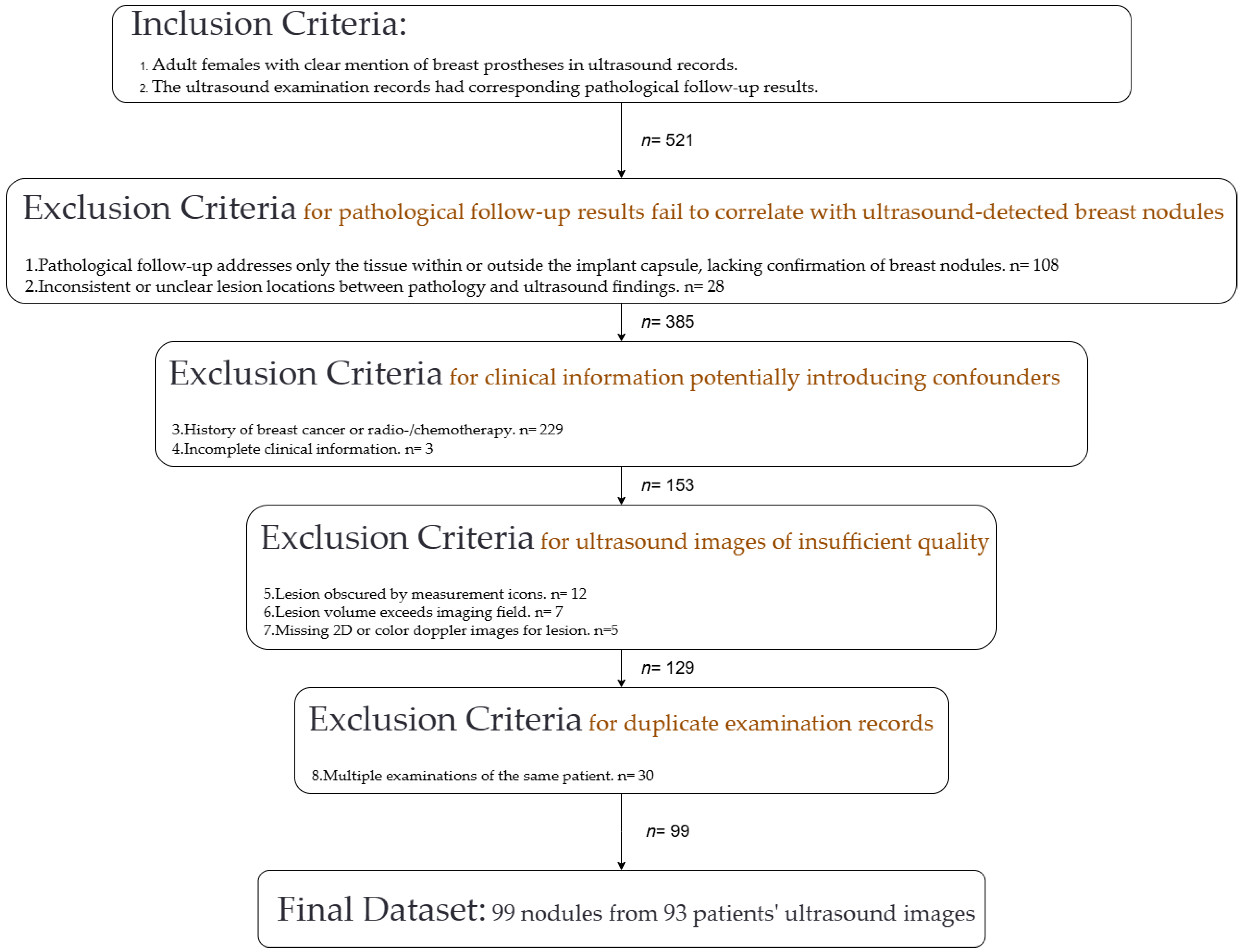
2.2. Study Subjects
2.3. Outcome Measures
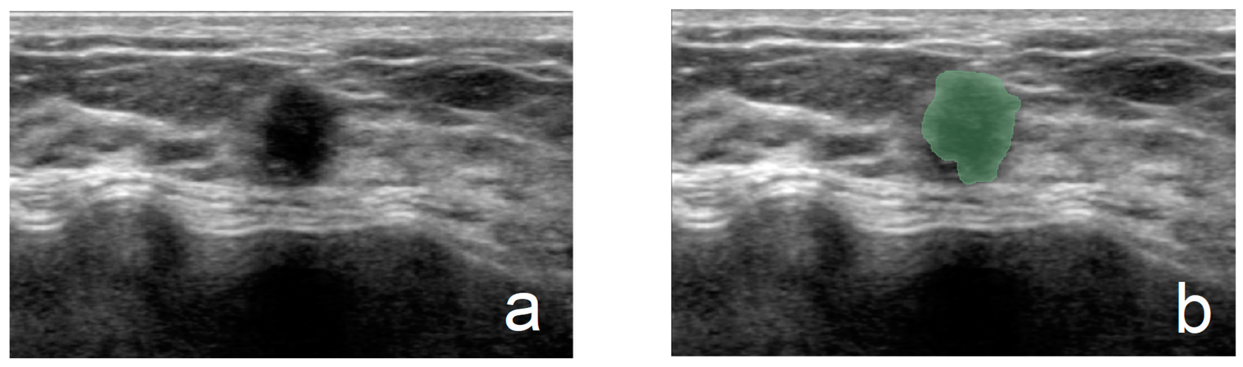
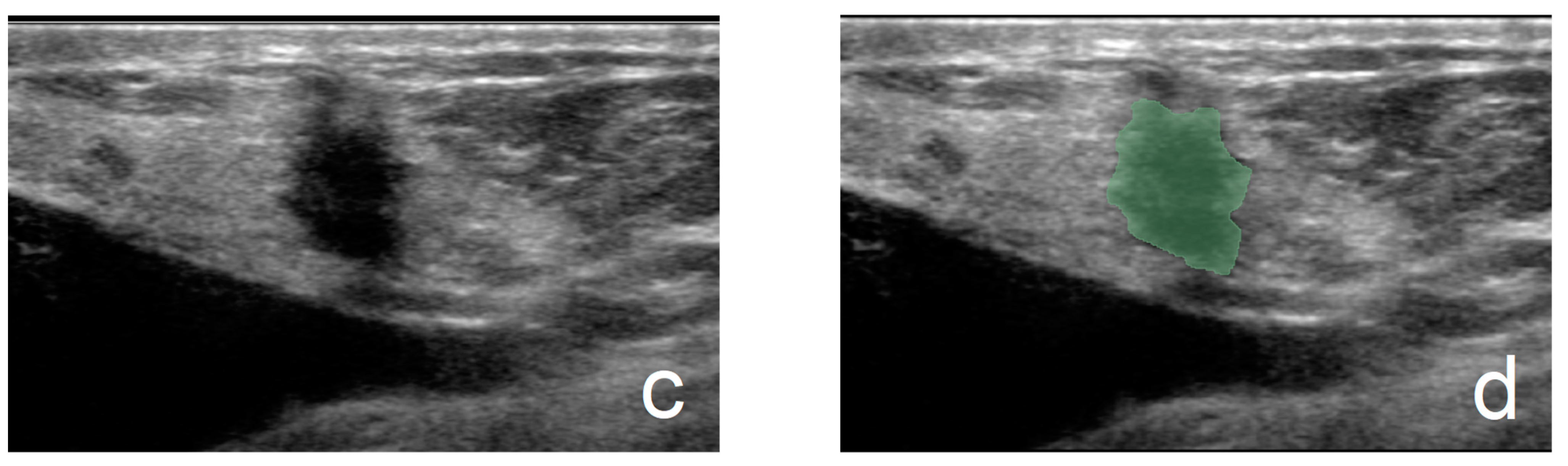
2.4. Acquisition of Ultrasound Pictures, Preprocessing, and ROI Delineation
2.5. Dataset Construction
2.6. Feature Extraction
2.7. Feature Screening
2.8. Model Establishment and Evaluation
2.9. Statistical Analysis
3. Results
3.1. Clinical and Pathological Information
3.2. Feature Extraction and Selection
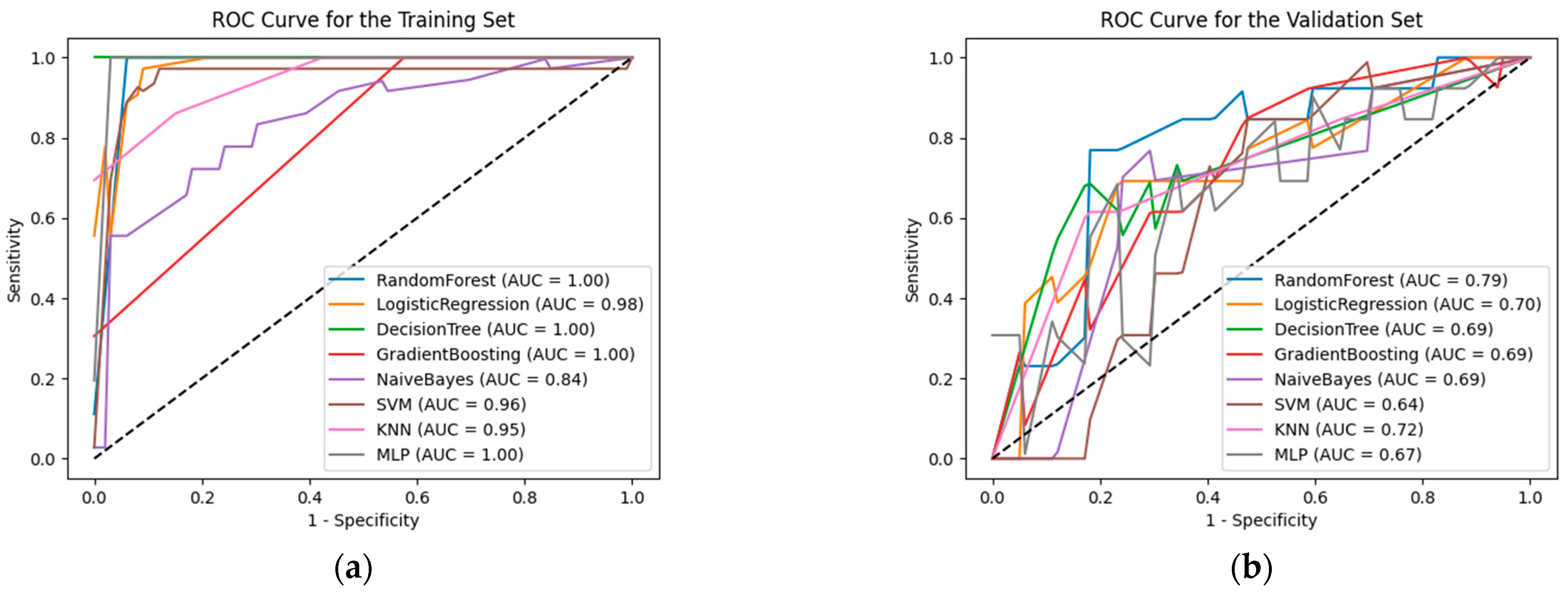
3.3. Model Evaluation
4. Discussion
- Conduct multi-center, prospective studies to increase the sample size and diversity, thereby enhancing the generalizability of the research findings;
- Explore deep learning studies combining multimodal ultrasound images with clinical information and other imaging modalities (such as MRI and CT) to construct more comprehensive diagnostic models;
- Investigate the temporal trends of ultrasound radiomics features to assess their role in monitoring complications associated with breast prostheses;
- Gain a deeper understanding of the relationship between ultrasound radiomics features and the type and material of breast prostheses to guide prosthesis selection and long-term management.
5. Conclusions
Author Contributions
Funding
Institutional Review Board Statement
Informed Consent Statement
Data Availability Statement
Conflicts of Interest
References
- Beekman, W.H.; Hage, J.J.; Jorna, L.B.; Mulder, J.W. Augmentation mammaplasty: The story before the silicone bag prosthesis. Ann. Plast. Surg. 1999, 43, 446–451. [Google Scholar] [CrossRef] [PubMed]
- Bizjak, M.; Selmi, C.; Praprotnik, S.; Bruck, O.; Perricone, C.; Ehrenfeld, M.; Shoenfeld, Y. Silicone implants and lymphoma: The role of inflammation. J. Autoimmun. 2015, 65, 64–73. [Google Scholar] [CrossRef] [PubMed]
- Chetlen, A.; Niell, B.L.; Brown, A.; Baskies, A.M.; Battaglia, T.; Chen, A.; Jochelson, M.S.; Klein, K.A.; Malak, S.F.; Mehta, T.S.; et al. ACR Appropriateness Criteria® Breast Implant Evaluation: 2023 Update. J. Am. Coll. Radiol. 2023, 20, S329–S350. [Google Scholar] [CrossRef] [PubMed]
- Longo, B.; Di Napoli, A.; Curigliano, G.; Veronesi, P.; Pileri, S.; Martelli, M.; De Vita, R.; Felici, N.; Cirillo, P.; Bernardi, C.; et al. Clinical recommendations for diagnosis and treatment according to current updated knowledge on BIA-ALCL. Breast 2022, 66, 332–341. [Google Scholar] [CrossRef]
- Lambin, P.; Leijenaar, R.T.H.; Deist, T.M.; Peerlings, J.; de Jong, E.E.C.; van Timmeren, J.; Sanduleanu, S.; Larue, R.; Even, A.J.G.; Jochems, A.; et al. Radiomics: The bridge between medical imaging and personalized medicine. Nat. Rev. Clin. Oncol. 2017, 14, 749–762. [Google Scholar] [CrossRef] [PubMed]
- Conti, A.; Duggento, A.; Indovina, I.; Guerrisi, M.; Toschi, N. Radiomics in breast cancer classification and prediction. Semin. Cancer Biol. 2021, 72, 238–250. [Google Scholar] [CrossRef]
- van Griethuysen, J.J.M.; Fedorov, A.; Parmar, C.; Hosny, A.; Aucoin, N.; Narayan, V.; Beets-Tan, R.G.H.; Fillion-Robin, J.C.; Pieper, S.; Aerts, H. Computational Radiomics System to Decode the Radiographic Phenotype. Cancer Res. 2017, 77, e104–e107. [Google Scholar] [CrossRef]
- Rocco, N.; Rispoli, C.; Moja, L.; Amato, B.; Iannone, L.; Testa, S.; Spano, A.; Catanuto, G.; Accurso, A.; Nava, M.B. Different types of implants for reconstructive breast surgery. Cochrane Database Syst. Rev. 2016, 2016, Cd010895. [Google Scholar] [CrossRef] [PubMed]
- Shin, B.H.; Kim, B.H.; Kim, S.; Lee, K.; Choy, Y.B.; Heo, C.Y. Silicone breast implant modification review: Overcoming capsular contracture. Biomater. Res. 2018, 22, 37. [Google Scholar] [CrossRef]
- Hillard, C.; Fowler, J.D.; Barta, R.; Cunningham, B. Silicone breast implant rupture: A review. Gland Surg. 2017, 6, 163–168. [Google Scholar] [CrossRef]
- Balk, E.M.; Earley, A.; Avendano, E.A.; Raman, G. Long-Term Health Outcomes in Women with Silicone Gel Breast Implants: A Systematic Review. Ann. Intern. Med. 2016, 164, 164–175. [Google Scholar] [CrossRef] [PubMed]
- Chao, A.H.; Garza, R., 3rd; Povoski, S.P. A review of the use of silicone implants in breast surgery. Expert Rev. Med. Devices 2016, 13, 143–156. [Google Scholar] [CrossRef] [PubMed]
- Coroneos, C.J.; Selber, J.C.; Offodile, A.C., 2nd; Butler, C.E.; Clemens, M.W. US FDA Breast Implant Postapproval Studies: Long-term Outcomes in 99,993 Patients. Ann. Surg. 2019, 269, 30–36. [Google Scholar] [CrossRef] [PubMed]
- Cohen Tervaert, J.W.; Mohazab, N.; Redmond, D.; van Eeden, C.; Osman, M. Breast implant illness: Scientific evidence of its existence. Expert Rev. Clin. Immunol. 2022, 18, 15–29. [Google Scholar] [CrossRef] [PubMed]
- Suh, L.J.; Khan, I.; Kelley-Patteson, C.; Mohan, G.; Hassanein, A.H.; Sinha, M. Breast Implant-Associated Immunological Disorders. J. Immunol. Res. 2022, 2022, 8536149. [Google Scholar] [CrossRef] [PubMed]
- Watad, A.; Rosenberg, V.; Tiosano, S.; Cohen Tervaert, J.W.; Yavne, Y.; Shoenfeld, Y.; Shalev, V.; Chodick, G.; Amital, H. Silicone breast implants and the risk of autoimmune/rheumatic disorders: A real-world analysis. Int. J. Epidemiol. 2018, 47, 1846–1854. [Google Scholar] [CrossRef] [PubMed]
- Sudarshan, V.K.; Mookiah, M.R.; Acharya, U.R.; Chandran, V.; Molinari, F.; Fujita, H.; Ng, K.H. Application of wavelet techniques for cancer diagnosis using ultrasound images: A Review. Comput. Biol. Med. 2016, 69, 97–111. [Google Scholar] [CrossRef]
- Lepola, A.; Arponen, O.; Okuma, H.; Holli-Helenius, K.; Junkkari, H.; Könönen, M.; Auvinen, P.; Sudah, M.; Sutela, A.; Vanninen, R. Association between breast cancer’s prognostic factors and 3D textural features of non-contrast-enhanced T(1) weighted breast MRI. Br. J. Radiol. 2022, 95, 20210702. [Google Scholar] [CrossRef] [PubMed]
- Chitalia, R.D.; Kontos, D. Role of texture analysis in breast MRI as a cancer biomarker: A review. J. Magn. Reson. Imaging 2019, 49, 927–938. [Google Scholar] [CrossRef] [PubMed]
- Bismeijer, T.; van der Velden, B.H.M.; Canisius, S.; Lips, E.H.; Loo, C.E.; Viergever, M.A.; Wesseling, J.; Gilhuijs, K.G.A.; Wessels, L.F.A. Radiogenomic Analysis of Breast Cancer by Linking MRI Phenotypes with Tumor Gene Expression. Radiology 2020, 296, 277–287. [Google Scholar] [CrossRef] [PubMed]
- Gillies, R.J.; Kinahan, P.E.; Hricak, H. Radiomics: Images Are More than Pictures, They Are Data. Radiology 2016, 278, 563–577. [Google Scholar] [CrossRef]
- Fujioka, T.; Kubota, K.; Mori, M.; Kikuchi, Y.; Katsuta, L.; Kasahara, M.; Oda, G.; Ishiba, T.; Nakagawa, T.; Tateishi, U. Distinction between benign and malignant breast masses at breast ultrasound using deep learning method with convolutional neural network. Jpn. J. Radiol. 2019, 37, 466–472. [Google Scholar] [CrossRef] [PubMed]
- Shi, S.; An, X.; Li, Y. Ultrasound Radiomics-Based Logistic Regression Model to Differentiate Between Benign and Malignant Breast Nodules. J. Ultrasound Med. 2023, 42, 869–879. [Google Scholar] [CrossRef] [PubMed]
- Romeo, V.; Cuocolo, R.; Apolito, R.; Stanzione, A.; Ventimiglia, A.; Vitale, A.; Verde, F.; Accurso, A.; Amitrano, M.; Insabato, L.; et al. Clinical value of radiomics and machine learning in breast ultrasound: A multicenter study for differential diagnosis of benign and malignant lesions. Eur. Radiol. 2021, 31, 9511–9519. [Google Scholar] [CrossRef] [PubMed]
- Shen, Y.; Shamout, F.E.; Oliver, J.R.; Witowski, J.; Kannan, K.; Park, J.; Wu, N.; Huddleston, C.; Wolfson, S.; Millet, A.; et al. Artificial intelligence system reduces false-positive findings in the interpretation of breast ultrasound exams. Nat. Commun. 2021, 12, 5645. [Google Scholar] [CrossRef] [PubMed]
- Gu, J.; Jiang, T. Ultrasound radiomics in personalized breast management: Current status and future prospects. Front. Oncol. 2022, 12, 963612. [Google Scholar] [CrossRef] [PubMed]


| Pathological Type | Number of Lesions | |
|---|---|---|
| Malignant | Invasive ductal carcinoma | 44 |
| Ductal carcinoma in situ | 3 | |
| Mucinous carcinoma | 2 | |
| Invasive lobular carcinoma | 1 | |
| Benign | Adenosis | 16 |
| Fibroadenoma | 10 | |
| Granuloma | 7 | |
| Fibrocystic | 6 | |
| Blue gel-like material | 5 | |
| Inflammation | 4 | |
| Phyllodes tumor, benign | 1 | |
| Total | 99 |
| Years Since Surgery | Cases, n |
|---|---|
| ≤5 | 14 |
| 6–10 | 18 |
| 11–15 | 20 |
| 16–20 | 18 |
| >20 | 10 |
| Information Missing | 13 |
| Total | 93 |
| Statistical Test | Training Set (69) | Validation Set (30) | p Value | |||||||||||
|---|---|---|---|---|---|---|---|---|---|---|---|---|---|---|
| Malignant (M) or Benign (B), n | Chi-square test | (M) 32 | (B) 37 | (M) 18 | (B) 12 | 0.213 (2-sided) | ||||||||
| Left (L) or Right (R), n | Chi-square test | (L) 36 | (R) 33 | (L) 17 | (R) 13 | 0.827 (2-sided) | ||||||||
| Quadrant, n | Fisher’s exact test | c.a. 3 | l.i. 9 | l.o. 15 | u.i. 19 | u.o. 23 | c.a. 2 | l.i. 2 | l.o. 5 | u.i. 6 | u.o. 15 | 0.554 (2-sided) | ||
| Maximum diameter, median ± SD, mm | Mann-Whitney U test | 18.77 ± 10.73 | 16.8 ± 10.08 | 0.393 | ||||||||||
| Model | Group | Sensitivity | Specificity | AUC (95%CI) | Accuracy | Brier Score | Log Loss Score |
|---|---|---|---|---|---|---|---|
| Random Forest | TS | 1.000 | 1.000 | 1.000 (1.000–1.000) | 1.000 | 0.017 | 0.104 |
| VS | 0.765 | 0.838 | 0.787 (0.561–0.960) | 0.796 | 0.197 | 0.599 | |
| Logistic Regression | TS | 0.909 | 0.972 | 0.977 (0.947–0.998) | 0.942 | 0.058 | 0.208 |
| VS | 0.529 | 0.692 | 0.701 (0.448–0.886) | 0.600 | 0.260 | 0.830 | |
| Decision Tree | TS | 1.000 | 1.000 | 1.000 (1.000–1.000) | 1.000 | 0.000 | 0.000 |
| VS | 0.778 | 0.618 | 0.698 (0.533–0.871) | 0.708 | 0.292 | 10.074 | |
| Gradient Boosting | TS | 1.000 | 1.000 | 1.000 (1.000–1.000) | 1.000 | 0.000 | 0.000 |
| VS | 0.418 | 0.846 | 0.692 (0.456–0.900) | 0.604 | 0.355 | 3.965 | |
| Naïve Bayes | TS | 0.697 | 0.806 | 0.843 (0.754–0.931) | 0.754 | 0.222 | 0.693 |
| VS | 0.412 | 0.769 | 0.692 (0.472–0.919) | 0.567 | 0.248 | 0.713 | |
| Support Vector Machine (SVM) | TS | 0.909 | 0.944 | 0.959 (0.904–0.998) | 0.928 | 0.080 | 0.391 |
| VS | 0.529 | 0.846 | 0.638 (0.429–0.823) | 0.667 | 0.268 | 0.948 | |
| K-Nearest Neighbor (KNN) | TS | 0.849 | 0.861 | 0.947 (0.902–0.983) | 0.855 | 0.089 | 0.247 |
| VS | 0.765 | 0.615 | 0.717 (0.493–0.889) | 0.700 | 0.252 | 5.998 | |
| Multilayer Perceptron (MLP) | TS | 0.992 | 0.997 | 0.998 (0.993–0.999) | 0.995 | 0.004 | 0.017 |
| VS | 0.581 | 0.732 | 0.665 (0.448–0.840) | 0.647 | 0.348 | 9.833 |
Disclaimer/Publisher’s Note: The statements, opinions and data contained in all publications are solely those of the individual author(s) and contributor(s) and not of MDPI and/or the editor(s). MDPI and/or the editor(s) disclaim responsibility for any injury to people or property resulting from any ideas, methods, instructions or products referred to in the content. |
© 2025 by the authors. Licensee MDPI, Basel, Switzerland. This article is an open access article distributed under the terms and conditions of the Creative Commons Attribution (CC BY) license (https://creativecommons.org/licenses/by/4.0/).
Share and Cite
Hao, L.; Chen, Y.; Su, X.; Ma, B. Application of Ultrasound Radiomics in Differentiating Benign from Malignant Breast Nodules in Women with Post-Silicone Breast Augmentation. Curr. Oncol. 2025, 32, 29. https://doi.org/10.3390/curroncol32010029
Hao L, Chen Y, Su X, Ma B. Application of Ultrasound Radiomics in Differentiating Benign from Malignant Breast Nodules in Women with Post-Silicone Breast Augmentation. Current Oncology. 2025; 32(1):29. https://doi.org/10.3390/curroncol32010029
Chicago/Turabian StyleHao, Ling, Yang Chen, Xuejiao Su, and Buyun Ma. 2025. "Application of Ultrasound Radiomics in Differentiating Benign from Malignant Breast Nodules in Women with Post-Silicone Breast Augmentation" Current Oncology 32, no. 1: 29. https://doi.org/10.3390/curroncol32010029
APA StyleHao, L., Chen, Y., Su, X., & Ma, B. (2025). Application of Ultrasound Radiomics in Differentiating Benign from Malignant Breast Nodules in Women with Post-Silicone Breast Augmentation. Current Oncology, 32(1), 29. https://doi.org/10.3390/curroncol32010029