Time-Resolved Photoluminescence Microscopy for the Analysis of Semiconductor-Based Paint Layers

 , ,
, ,  ,
,
Abstract
:
1. Introduction
2. Materials and Methods
2.1. The Micro-Samples
2.2. TRPL Microscopy
- ◾
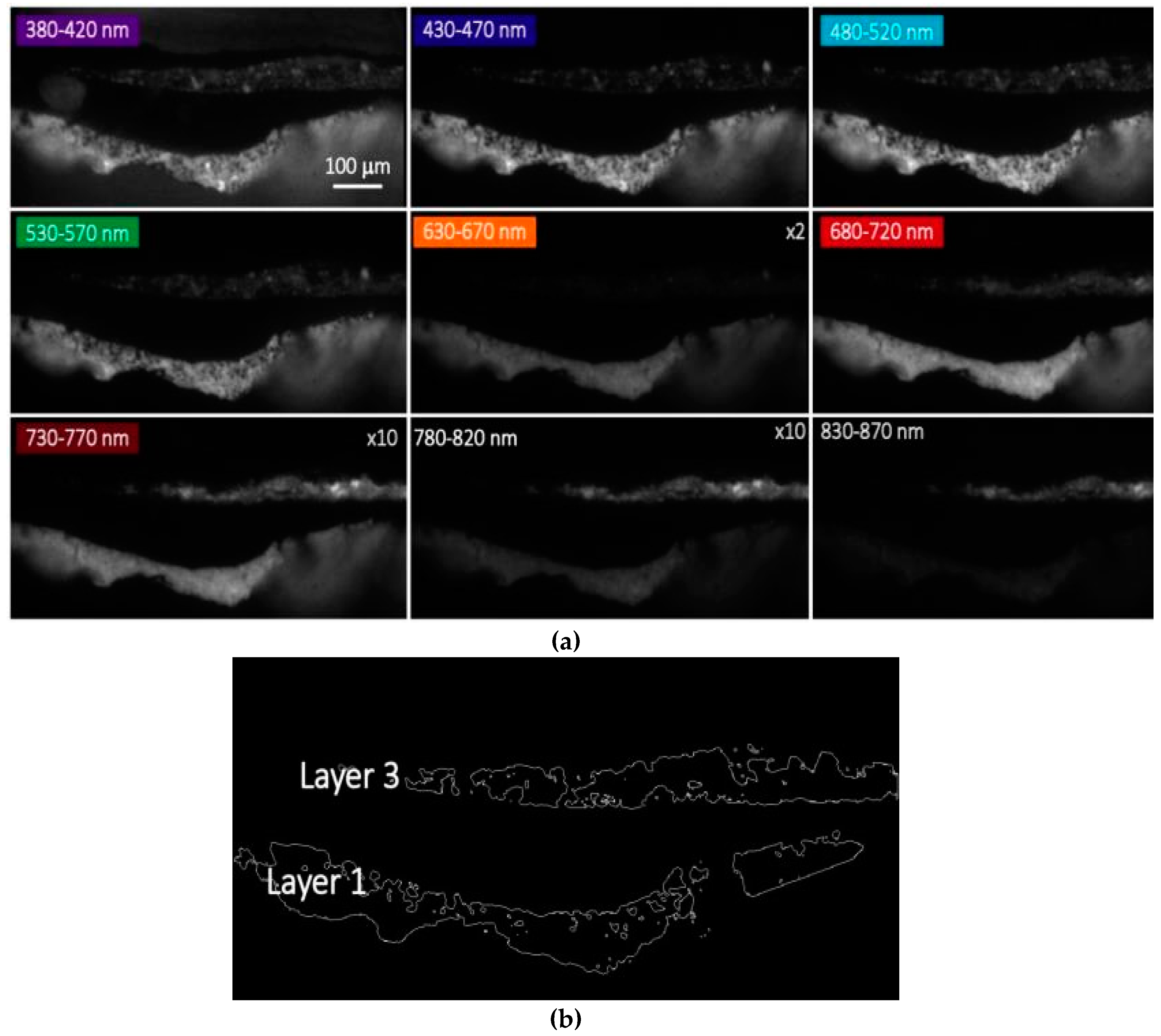
- Spectral measurements at the nanosecond (ns) and microsecond (μs) timescale: a sequence of PL gated images at a fixed delay is recorded in different spectral bands. In the present case study, analysis of ns and μs emissions are achieved by employing a gates with a temporal width of W = 10 ns synchronous with laser pulse (delay D = 0 ns) and a gate with a temporal width of W = 10 μs set at a delay D = 0.2 μs after the pulsed excitation, respectively.
- ◾
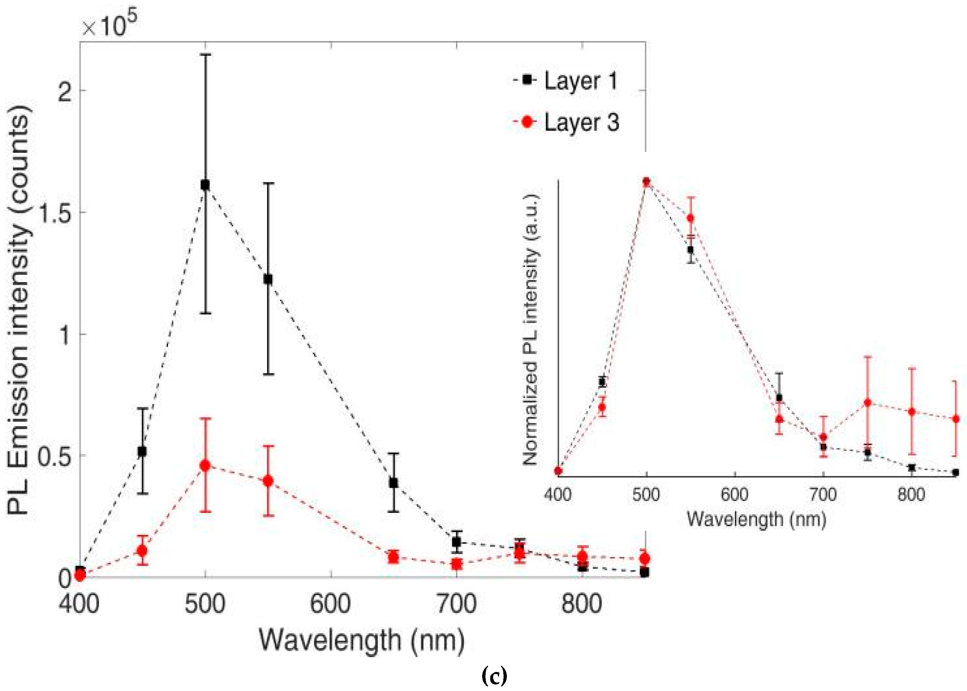
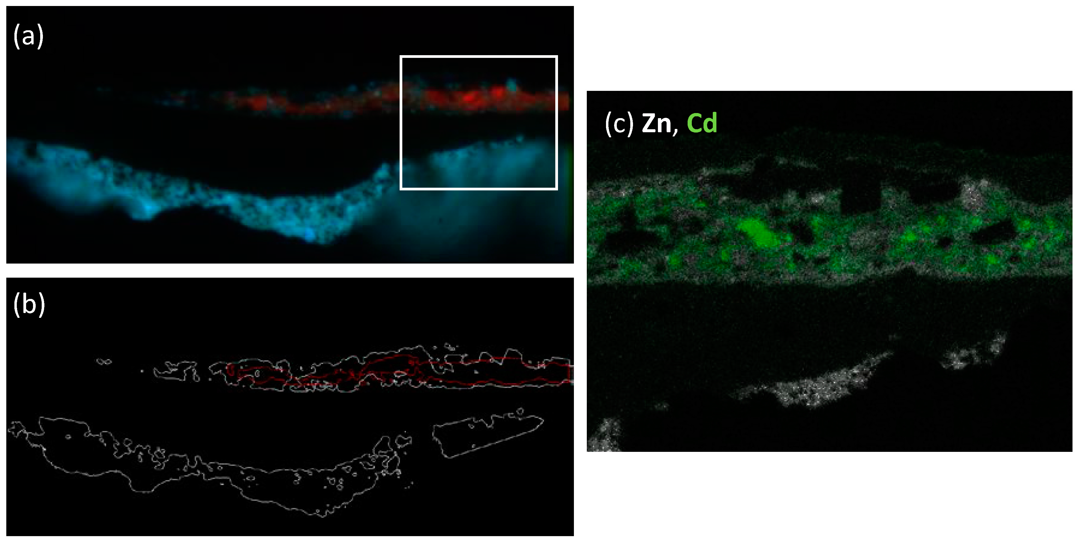
- Spectral data analysis: following correction for the detector efficiency, it is possible to reconstruct the PL spectrum in selected regions of interest (ROIs) of the analyzed sample. In this reconstruction procedure, for the sake of simplicity, each bandpass filter is modeled as a Dirac delta function centered at the filter central wavelength and the spectral transmission of filters are accounted for in the overall spectral detection efficiency (Figure S3 in Supplementary Materials). ROIs are selected on the basis of intensity thresholds on a selected spectral image. Following selection, the PL spectrum in each ROI is shown as the mean of intensity values within the ROI with error bars reporting the ROI standard deviation.
- ◾
- Decay kinetic measurements at the ns and μs timescale: a sequence of PL gated images at a fixed spectral band is recorded at different delays with respect to laser pulses. For this work, analysis of ns emission decay kinetics is achieved by employing a gate with a temporal width of W = 10 ns and temporally sampling the emission decay kinetic from 0 to 60 ns. Emission decay kinetics for the µs timescale are analyzed by employing a gate with temporal width of W = 1 μs and temporally sampling the emission decay kinetic from 0.1 to 10 μs.
- ◾
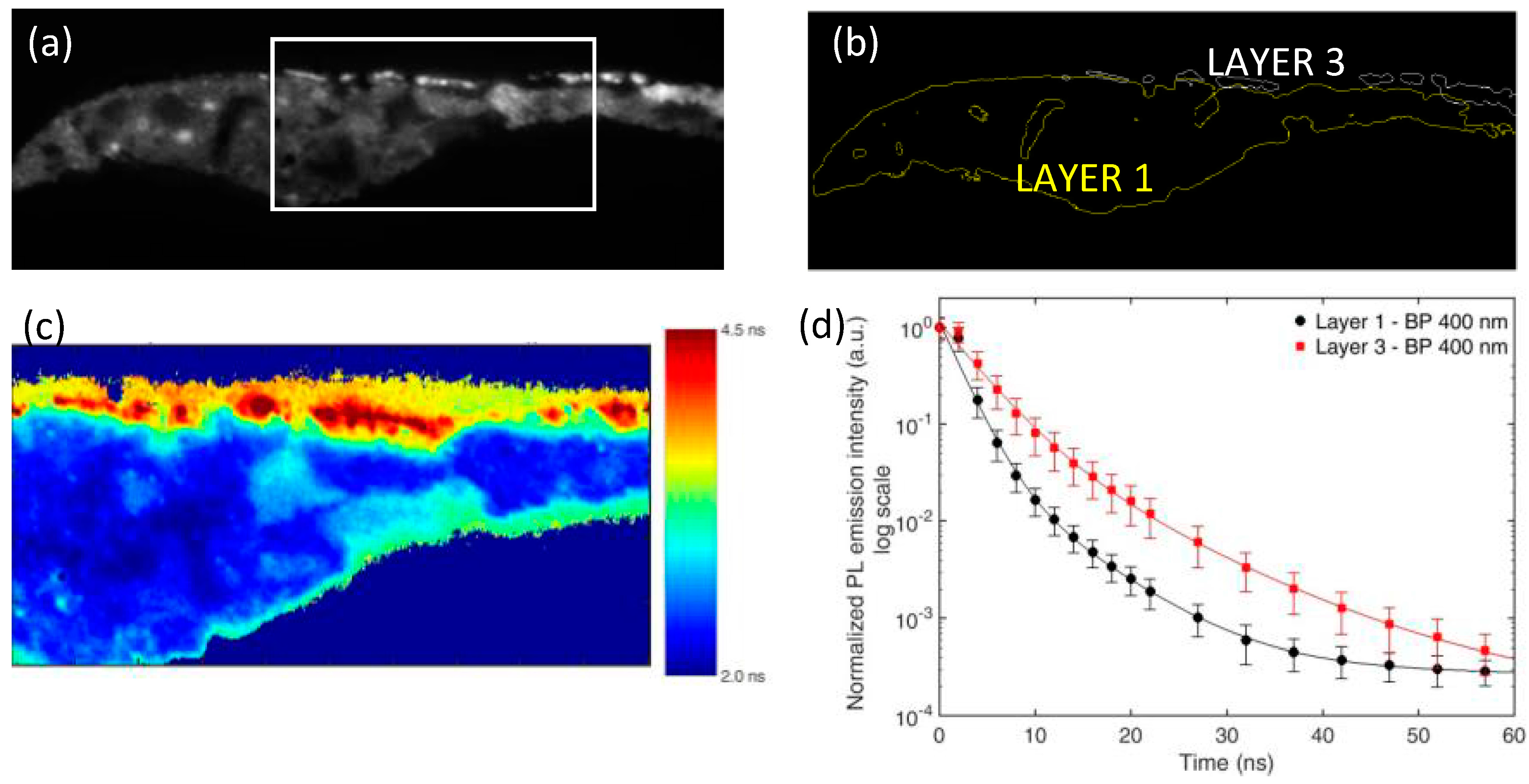
- Decay kinetic analysis: it is possible to reconstruct the emission decay kinetic in each point of the analyzed sample. A qualitative estimate of lifetime heterogeneities in the field of view is first provided by the lifetime map, calculated by fitting the data with a simple mono-exponential decay model on a pixel-by-pixel basis [28]. Following this, the emission decay kinetics of selected areas (ROIs) of the specimen are extracted and analyzed through non-linear fitting of a multi-exponential decay model with a maximum of three components. In the employed decay model, the intensifier gate has been considered as a rectangular function of width W, and the temporal width of laser pulses (~1 ns) has been neglected [31]. As for spectral analysis, ROIs are selected on the basis of intensity thresholds. Following selection, the PL decay kinetic in each ROI is shown as the mean of intensity values within the ROI with error bars reporting the ROI standard deviation.
2.3. Optical Microscopy and Scanning Electron Microscopy with Energy-Dispersive X-ray Spectroscopy (SEM-EDX)
3. Results
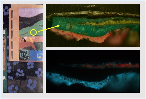
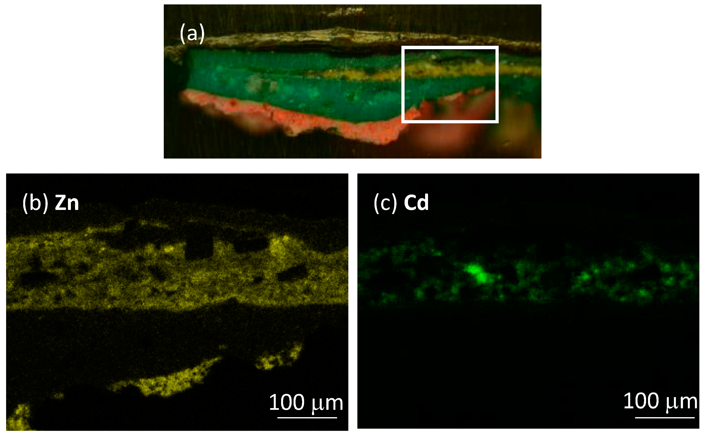
3.1. Sample 2105
3.2. Sample 2107
4. Discussion
- (i)
- The cadmium yellow paint, produced with the imperfect synthesis processes available at the beginning of the 20th century, is characterized by a high density of crystal defects, which can give rise to the formation of high density of states within the forbidden CdS bandgap. As a consequence, electron trapping of excited electrons is favored with respect to direct recombination, giving rise to a strong quenching of the radiative BE emission.
- (ii)
- The cadmium yellow paint has suffered severe chemical degradation that has altered the original CdS pigment into novel reaction products. In this vision, the detected PL could be ascribed to TS optical emission of novel compounds. This hypothesis is supported by the recent research on altered cadmium yellow paints [18,38,39,40], where pigment degradation has been associated with the formation of cadmium carbonates, sulphates, oxalates and hydroxides.
5. Conclusions
Supplementary Materials
Acknowledgments
Author Contributions
Conflicts of Interest
References
- Tachikawa, T.; Karimata, I.; Kobori, Y. Surface Charge Trapping in Organolead Halide Perovskites Explored by Single-Particle Photoluminescence Imaging. J. Phys. Chem. Lett. 2015, 6, 3195–3201. [Google Scholar] [CrossRef]
- Lefebvre, J.; Austing, D.G.; Bond, J.; Finnie, P. Photoluminescence Imaging of Suspended Single-Walled Carbon Nanotubes. Nano Lett. 2006, 6, 1603–1608. [Google Scholar] [CrossRef] [PubMed]
- Layek, A.; De, S.; Thorat, R.; Chowdhury, A. Spectrally Resolved Photoluminescence Imaging of ZnO Nanocrystals at Single-Particle Levels. J. Phys. Chem. Lett. 2011, 2, 1241–1247. [Google Scholar] [CrossRef] [PubMed]
- Crochet, J.J.; Duque, J.G.; Werner, J.H.; Doorn, S.K. Photoluminescence imaging of electronic-impurity-induced exciton quenching in single-walled carbon nanotubes. Nat. Nanotechnol. 2012, 7, 126–132. [Google Scholar] [CrossRef] [PubMed]
- Alberi, K.; Fluegel, B.; Moutinho, H.; Dhere, R.G.; Li, J.V.; Mascarenhas, A. Measuring long-range carrier diffusion across multiple grains in polycrystalline semiconductors by photoluminescence imaging. Nat. Commun. 2013, 4, 2699. [Google Scholar] [CrossRef] [PubMed]
- Consuegra, S.; Díaz-del-Río, P.; Hunt Ortiz, M.A.; Hurtado, V.; Montero Ruiz, I. Neolithic and Chalcolithic—VI to III millennia BC—Use of cinnabar (HgS) in the Iberian Peninsula: Analytical identification and lead isotope data for an early mineral exploitation of the Almadén (Ciudad Real, Spain) mining district. In History of Research in Mineral Resources; Ortiz, J.E., Puche, O., Rabano, I., Mazadiego, L.F., Eds.; Instituto Geológico y Minero de España: Madrid, Spain, 2011; p. 13. [Google Scholar]
- Clementi, C.; Rosi, F.; Romani, A.; Vivani, R.; Brunetti, B.G.; Miliani, C. Photoluminescence Properties of Zinc Oxide in Paints: A Study of the Effect of Self-Absorption and Passivation. Appl. Spectrosc. 2012, 66, 1233. [Google Scholar] [CrossRef] [PubMed]
- Artesani, A.; Bellei, S.; Capogrosso, V.; Cesaratto, A.; Mosca, S.; Nevin, A.; Valentini, G.; Comelli, D. Photoluminescence properties of zinc white: An insight into its emission mechanisms through the study of historical artist materials. Appl. Phys. A 2016, 122. [Google Scholar] [CrossRef]
- Rosi, F.; Grazia, C.; Gabrieli, F.; Romani, A.; Paolantoni, M.; Vivani, R.; Brunetti, B.G.; Colomban, P.; Miliani, C. UV–Vis-NIR and micro Raman spectroscopies for the non destructive identification of Cd1−xZnxS solid solutions in cadmium yellow pigments. Microchem. J. 2016, 124, 856. [Google Scholar] [CrossRef]
- Grazia, C.; Rosi, F.; Gabrieli, F.; Romani, A.; Paolantoni, M.; Vivani, R.; Brunetti, B.G.; Colomban, P.; Miliani, C. UV–Vis-NIR and microRaman spectroscopies for investigating the composition of ternary CdS1−xSex solid solutions employed as artists’ pigments. Microchem. J. 2016, 125, 279. [Google Scholar] [CrossRef]
- Cesaratto, A.; D’Andrea, C.; Nevin, A.; Valentini, G.; Tassone, F.; Alberti, R.; Frizzi, T.; Comelli, D. Analysis of cadmium-based pigments with time-resolved photoluminescence. Anal. Methods 2014, 6, 130–138. [Google Scholar] [CrossRef]
- Bellei, S.; Nevin, A.; Cesaratto, A.; Capogrosso, V.; Vezin, H.; Tokarski, C.; Valentini, G.; Comelli, D. Multianalytical Study of Historical Luminescent Lithopone for the Detection of Impurities and Trace Metal Ions. Anal. Chem. 2015, 87, 6049–6056. [Google Scholar] [CrossRef] [PubMed]
- Capogrosso, V.; Gabrieli, F.; Bellei, S.; Cartechini, L.; Cesaratto, A.; Trcera, N.; Rosi, F.; Valentini, G.; Comelli, D.; Nevin, A. An integrated approach based on micro-mapping analytical techniques for the detection of impurities in historical Zn-based white pigments. J. Anal. At. Spectrom. 2015, 30. [Google Scholar] [CrossRef]
- Francesca, C.; Volker, R. High-resolution fluorescence mapping of impurities in historical zinc oxide pigments: Hard X-ray nanoprobe applications to the paints of Pablo Picasso. Appl. Phys. A 2013, 111, 1–8. [Google Scholar] [CrossRef]
- Hermans, J.J.; Keune, K.; van Loon, A.; Iedema, P.D. The crystallization of metal soaps and fatty acids in oil paint model systems. Phys. Chem. Chem. Phys. 2016, 18, 10896–10905. [Google Scholar] [CrossRef] [PubMed]
- Chen-Wiegart, Y.K.; Catalano, J.; Williams, G.J.; Murphy, A.; Yao, Y.; Zumbulyadis, N.; Centeno, S.A.; Dybowski, C.; Thieme, J. Elemental and Molecular Segregation in Oil Paintings due to Lead Soap Degradation. Sci. Rep. 2017, 7, 11656. [Google Scholar] [CrossRef] [PubMed]
- Monico, L.; van der Snickt, G.; Janssens, K.; de Nolf, W.; Miliani, C.; Verbeeck, J.; Tian, H.; Tan, H.; Dik, J.; Radepont, M.; et al. Degradation Process of Lead Chromate in Paintings by Vincent van Gogh Studied by Means of Synchrotron X-ray Spectromicroscopy and Related Methods. 1. Artificially Aged Model Samples. Anal. Chem. 2011, 83, 1214–1223. [Google Scholar] [CrossRef] [PubMed]
- Van der Snickt, G.; Dik, J.; Cotte, M.; Janssens, K.; Jaroszewicz, J.; de Nolf, W.; Groenewegen, J.; van der Loeff, L. Characterization of a Degraded Cadmium Yellow (CdS) Pigment in an Oil Painting by Means of Synchrotron Radiation Based X-ray Techniques. Anal. Chem. 2009, 81, 2600–2610. [Google Scholar] [CrossRef] [PubMed]
- Latour, G.; Robinet, L.; Dazzi, A.; Portier, F.; Deniset-Besseau, A.; Schanne-Klein, M. Correlative nonlinear optical microscopy and infrared nanoscopy reveals collagen degradation in altered parchments. Sci. Rep. 2016, 6, 26344. [Google Scholar] [CrossRef] [PubMed] [Green Version]
- Bertrand, L.; Réfrégiers, M.; Berrie, B.; Échard, J.P.; Thoury, M. A multiscalar photoluminescence approach to discriminate among semiconducting historical zinc white pigments. Analyst 2013, 138, 4463–4469. [Google Scholar] [CrossRef] [PubMed]
- Thoury, M.; Mille, B.; Séverin-Fabiani, T.; Robbiola, L.; Réfrégiers, M.; Jarrige, J.; Bertrand, L. High spatial dynamics-photoluminescence imaging reveals the metallurgy of the earliest lost-wax cast object. Nat. Commun. 2016, 7, 13356. [Google Scholar] [CrossRef] [PubMed]
- Becker, W. Fluorescence lifetime imaging–techniques and applications. J. Microsc. 2012, 247, 119–136. [Google Scholar] [CrossRef] [PubMed]
- Auksorius, E.; Boruah, B.R.; Dunsby, C.; Lanigan, P.M.P.; Kennedy, G.; Neil, M.A.A.; French, P.M.W. Stimulated emission depletion microscopy with a supercontinuum source and fluorescence lifetime imaging. Opt. Lett. 2008, 33, 113–115. [Google Scholar] [CrossRef] [PubMed]
- Bastiaens, P.I.H.; Squire, A. Fluorescence lifetime imaging microscopy: Spatial resolution of biochemical processes in the cell. Trends Cell Biol. 1999, 9, 48–52. [Google Scholar] [CrossRef]
- Savateeva, D.; Melnikau, D.; Lesnyak, V.; Gaponik, N.; Rakovich, Y.P. Hybrid organic/inorganic semiconductor nanostructures with highly efficient energy transfer. J. Mater. Chem. 2012, 22, 10816–10820. [Google Scholar] [CrossRef]
- Jahn, K.; Buschmann, V.; Hille, C. Simultaneous fluorescence and phosphorescence lifetime imaging, microscopy in living cells. Sci. Rep. 2015, 5, 14334. [Google Scholar] [CrossRef] [PubMed]
- Van Munster, E.B.; Gadella, T.W. Fluorescence Lifetime Imaging Microscopy (FLIM). Adv. Biochem. Eng. Biotechnol. 2005, 95, 143–175. [Google Scholar]
- C2RMF Report n. 977; C2RMF: Paris, France, 1977.
- Comelli, D.; Valentini, G.; Cubeddu, R.; Toniolo, L. Fluorescence lifetime imaging and Fourier transform infrared spectroscopy of Michelangelo’s David. Appl. Spectrosc. 2005, 59, 1174–1181. [Google Scholar] [CrossRef] [PubMed]
- Comelli, D.; Capogrosso, V.; Orsenigo, C.; Nevin, A. Dual wavelength excitation for the time-resolved photoluminescence imaging of painted ancient Egyptian objects. Heritage Sci. 2016, 4, 21. [Google Scholar] [CrossRef]
- Nevin, A.; Cesaratto, A.; Bellei, S.; D’Andrea, C.; Toniolo, L.; Valentini, G.; Comelli, D. Time-resolved photoluminescence spectroscopy and imaging: New approaches to the analysis of cultural heritage and its degradation. Sensors 2014, 14, 6338–6355. [Google Scholar] [CrossRef] [PubMed] [Green Version]
- Comelli, D.; Nevin, A.; Brambilla, A.; Osticioli, I.; Valentini, G.; Toniolo, L.; Fratelli, M.; Cubeddu, R. On the discovery of an unusual luminescent pigment in Van Gogh’s painting “les bretonnes et le pardon de pont Aven”. Appl. Phys. A: Mater. Sci. Process. 2012, 106, 25–34. [Google Scholar] [CrossRef]
- Artesani, A.; Gherardi, F.; Nevin, A.; Valentini, G.; Comelli, D. A photoluminescence study of the changes induced in the zinc white pigment by formation of zinc complexes. Materials 2017, 10, 340. [Google Scholar] [CrossRef] [PubMed]
- Janotti, A.; Van De Walle, C.G. Fundamentals of zinc oxide as a semiconductor. Rep. Prog. Phys. 2009, 72. [Google Scholar] [CrossRef]
- Verri, G.; Clementi, C.; Comelli, D.; Cather, S.; Piqué, F. Correction of ultraviolet-induced fluorescence spectra for the examination of polychromy. Appl. Spectrosc. 2008, 62, 1295–1302. [Google Scholar] [CrossRef] [PubMed]
- Thoury, M.; Delaney, J.K.; De La Rie, E.R.; Palmer, M.; Morales, K.; Krueger, J. Near-infrared luminescence of cadmium pigments: In situ identification and mapping in paintings. Appl. Spectrosc. 2011, 65, 939–951. [Google Scholar] [CrossRef] [PubMed]
- Anglos, D.; Solomidou, M.; Zergioti, I.; Zafiropulos, V.; Papazoglou, T.G.; Fotakis, C. Laser-induced fluorescence in artwork diagnostics: An application in pigment analysis. Appl. Spectrosc. 1996, 50, 1331–1334. [Google Scholar] [CrossRef]
- Leone, B.; Burnstock, A.; Jones, C.; Hallebeek, P.; Keune, K.; Boon, J. The deterioration of cadmium sulphide yellow artists’ pigments. In Proceedings of the 14th Triennial Meeting, The Hague, The Netherlands, 12–16 September 2005; Verger, I., Ed.; ICOM Committee for Conservation; James & James: London, UK, 2005; Volume 2. [Google Scholar]
- Pouyet, E.; Cotte, M.; Fayard, B.; Salomé, M.; Meirer, F.; Mehta, A.; Uffelman, E.S.; Hull, A.; Vanmeert, F.; Kieffer, J.; et al. 2D X-ray and FTIR micro-analysis of the degradation of cadmium yellow pigment in paintings of Henri Matisse. Appl. Phys. A: Mater. Sci. Process. 2015, 121, 967–980. [Google Scholar] [CrossRef]
- Mass, J.; Sedlmair, J.; Patterson, C.S.; Carson, D.; Buckley, B.; Hirschmugl, C. SR-FTIR imaging of the altered cadmium sulfide yellow paints in Henri Matisse’s Le Bonheur de vivre 1905-6—Examination of visually distinct degradation regions. Analyst 2013, 138, 6032–6043. [Google Scholar] [CrossRef] [PubMed]
- Di Bartolo, B.; Godberg, V.; Pacheco, D. Luminescence of Inorganic Solids; Springer: New York, NY, USA, 1966. [Google Scholar]
- Ummartyotin, S.; Infahsaeng, Y. A comprehensive review on ZnS: From synthesis to an approach on solar cell. Renew. Sustain. Energy Rev. 2016, 55, 17–24. [Google Scholar] [CrossRef]
- Tiwari, A.; Dhoble, S.J. Stabilization of ZnS nanoparticles by polymeric matrices: Syntheses, optical properties and recent applications. RSC Adv. 2016, 6, 64400–64420. [Google Scholar] [CrossRef]
- Prathap, P.; Subbaiah, Y.P.V.; Ramakrishna Reddy, K.T.; Miles, R.W. Influence of growth rate on microstructure and optoelectronic behaviour of ZnS films. J. Phys. D Appl. Phys. 2007, 40, 5275–5282. [Google Scholar] [CrossRef]
- Gonzalez, V.; Gourier, D.; Calligaro, T.; Toussaint, K.; Wallez, G.; Menu, M. Revealing the origin and history of lead-white pigments by their Photoluminescence properties. Anal. Chem. 2017, 89, 2909–2918. [Google Scholar] [CrossRef] [PubMed]


| Sample/Layer | Main PL Emissions (Reported As: Spectral Band of Maximum Intensity/Decay Kinetic Timescale) | Elemental Composition (Main Components) | Proposed Pigment Identification |
|---|---|---|---|
| 2105/Layer 1 | 400 nm/ns timescale (BE); 550 nm/μs timescale (TS) | Zn, O, S, Ba, Hg | zinc white (ZnO); cinnabar (HgS) |
| 2105/Layer 3 | 400 nm/ns timescale (BE); 550 nm/μs timescale (TS) | Zn, O, S, Ba, Cd, Cr (trace) | zinc white (ZnO); barium white (BaSO4); cadmium yellow (ZnxCd1−xS)—(altered or unperfectly synthetized) |
| 750 nm/μs timescale (TS) | |||
| 2107/Layer 1 | 400 nm/ns timescale (BE); 550 nm/μs timescale (TS) | Zn, O, Ba, S, Al, Co | zinc white (ZnO); barium white (BaSO4); cobalt blue (CoO·Al2O3) |
| 2107/Layer3 | 400 nm/ns timescale (BE or shallow TS); 400 nm/μs timescale (TS) | Zn, S | zinc sulphide (ZnS) |
© 2017 by the authors. Licensee MDPI, Basel, Switzerland. This article is an open access article distributed under the terms and conditions of the Creative Commons Attribution (CC BY) license (http://creativecommons.org/licenses/by/4.0/).
Share and Cite
Comelli, D.; Artesani, A.; Nevin, A.; Mosca, S.; Gonzalez, V.; Eveno, M.; Valentini, G. Time-Resolved Photoluminescence Microscopy for the Analysis of Semiconductor-Based Paint Layers. Materials 2017, 10, 1335. https://doi.org/10.3390/ma10111335
Comelli D, Artesani A, Nevin A, Mosca S, Gonzalez V, Eveno M, Valentini G. Time-Resolved Photoluminescence Microscopy for the Analysis of Semiconductor-Based Paint Layers. Materials. 2017; 10(11):1335. https://doi.org/10.3390/ma10111335
Chicago/Turabian StyleComelli, Daniela, Alessia Artesani, Austin Nevin, Sara Mosca, Victor Gonzalez, Myriam Eveno, and Gianluca Valentini. 2017. "Time-Resolved Photoluminescence Microscopy for the Analysis of Semiconductor-Based Paint Layers" Materials 10, no. 11: 1335. https://doi.org/10.3390/ma10111335
APA StyleComelli, D., Artesani, A., Nevin, A., Mosca, S., Gonzalez, V., Eveno, M., & Valentini, G. (2017). Time-Resolved Photoluminescence Microscopy for the Analysis of Semiconductor-Based Paint Layers. Materials, 10(11), 1335. https://doi.org/10.3390/ma10111335