
Chitosan Biomaterials for Current and Potential Dental Applications

 ,
,  , ,
, ,  and
and
Abstract
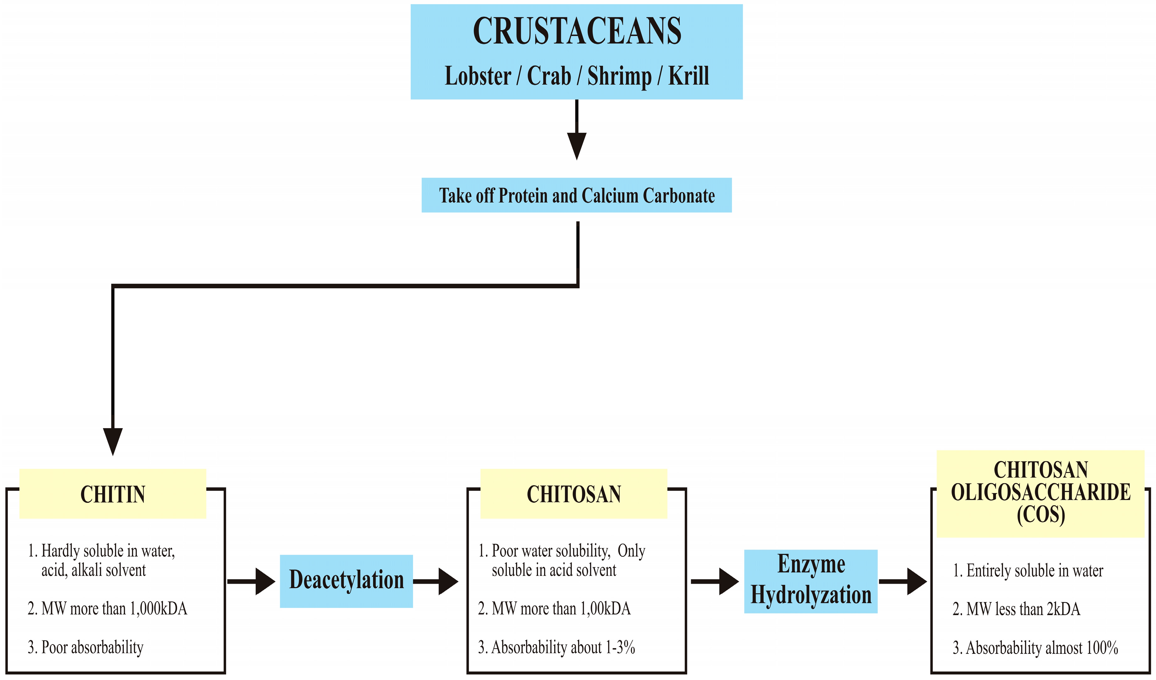
:1. Introduction
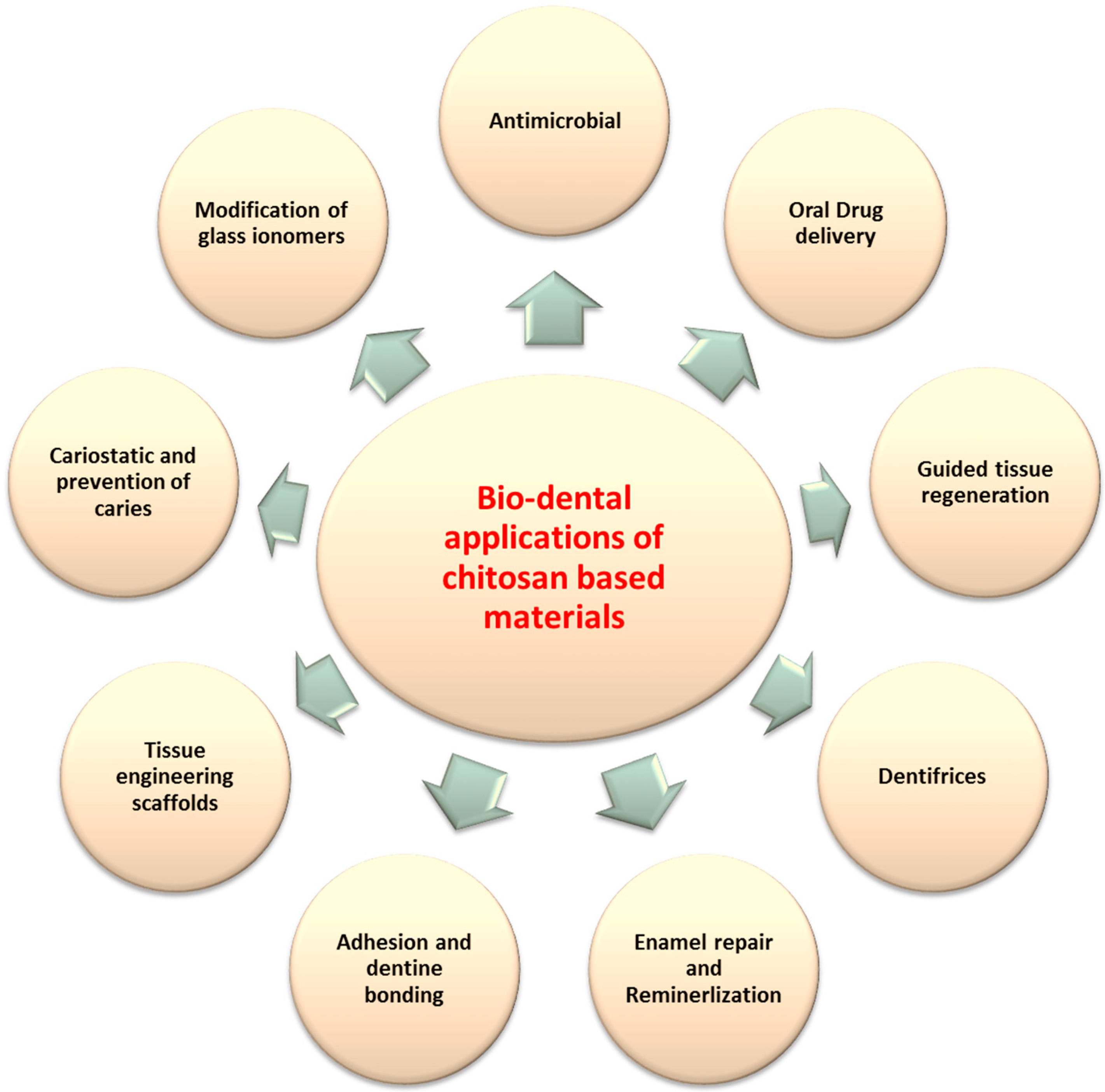
2. Applications of Chitosan Materials in Dentistry
2.1. Oral Drug Delivery
2.2. Guided Tissue Regeneration (GTR)
2.3. Modifications of Dentifrices
2.4. Enamel Repair
2.5. Adhesion and Dentine Bonding
2.6. Modification of Dental Restorative Materials
2.7. Chitosan for Coating Dental Implants
2.8. Stem-Based Regenerative Therapeutics
3. Conclusions
Author Contributions
Conflicts of Interest
References
- Sionkowska, A. Current research on the blends of natural and synthetic polymers as new biomaterials: Review. Prog. Polym. Sci. 2011, 36, 1254–1276. [Google Scholar] [CrossRef]
- Helary, C.; Bataille, I.; Abed, A.; Illoul, C.; Anglo, A.; Louedec, L.; Letourneur, D.; Meddahi-Pellé, A.; Giraud-Guille, M.M. Concentrated collagen hydrogels as dermal substitutes. Biomaterials 2010, 31, 481–490. [Google Scholar] [CrossRef] [PubMed]
- Becker, J.; Al-Nawas, B.; Klein, M.O.; Schliephake, H.; Terheyden, H.; Schwarz, F. Use of a new cross-linked collagen membrane for the treatment of dehiscence-type defects at titanium implants: A prospective, randomized-controlled double-blinded clinical multicenter study. Clin. Oral Implants Res. 2009, 20, 742–749. [Google Scholar] [CrossRef] [PubMed]
- Chen, J.; Chang, G.; Chen, J. Electrospun collagen/chitosan nanofibrous membrane as wound dressing. Coll. Surf. Physicochem. Eng. Asp. 2008, 313, 183–188. [Google Scholar] [CrossRef]
- Demol, J.; Lambrechts, D.; Geris, L.; Schrooten, J.; Van Oosterwyck, H. Towards a quantitative understanding of oxygen tension and cell density evolution in fibrin hydrogels. Biomaterials 2011, 32, 107–118. [Google Scholar] [CrossRef] [PubMed]
- Des Rieux, A.; Shikanov, A.; Shea, L.D. Fibrin hydrogels for non-viral vector delivery in vitro. J. Control. Release 2009, 136, 148–154. [Google Scholar] [CrossRef] [PubMed]
- Hatakeyama, I.; Marukawa, E.; Takahashi, Y.; Omura, K. Effects of platelet-poor plasma, platelet-rich plasma, and platelet-rich fibrin on healing of extraction sockets with buccal dehiscence in dogs. Tissue Eng. Part A 2013, 20, 874–882. [Google Scholar] [CrossRef] [PubMed]
- Elliott, W.H.; Bonani, W.; Maniglio, D.; Motta, A.; Tan, W.; Migliaresi, C. Silk Hydrogels of Tunable Structure and Viscoelastic Properties using Different Chronological Orders of Genipin and Physical Crosslinking. ACS Appl. Mater. Interfaces 2015, 7, 12099–12108. [Google Scholar] [CrossRef] [PubMed]
- Wang, H.; Zhang, Y. Processing silk hydrogel and its applications in biomedical materials. Biotechnol. Prog. 2015, 31, 630–640. [Google Scholar] [CrossRef] [PubMed]
- Zafar, M.S.; Al-Samadani, K.H. Potential use of natural silk for bio-dental applications. J. Taibah Univ. Med. Sci. 2014, 9, 171–177. [Google Scholar] [CrossRef]
- Deng, J.; She, R.; Huang, W.; Dong, Z.; Mo, G.; Liu, B. A silk fibroin/chitosan scaffold in combination with bone marrow-derived mesenchymal stem cells to repair cartilage defects in the rabbit knee. J. Mater. Sci. Mater. Med. 2013, 24, 2037–2046. [Google Scholar] [CrossRef] [PubMed]
- Qasim, S.B.; Najeeb, S.; Delaine-Smith, R.M.; Rawlinson, A.; Rehman, I.U. Potential of electrospun chitosan fibers as a surface layer in functionally graded GTR membrane for periodontal regeneration. Dent. Mater. 2017, 33, 71–83. [Google Scholar] [CrossRef] [PubMed]
- Huang, G.; Zhai, J.; Cheng, S.; Wang, Y.; Yang, L.; Liu, H.; Ran, R. The application of chitosan and its derivatives as nanosized carriers for the delivery of chemical drugs and genes or proteins. Curr. Drug Targets 2016, 17, 811–816. [Google Scholar]
- Norowski, P.A.; Fujiwara, T.; Clem, W.C.; Adatrow, P.C.; Eckstein, E.C.; Haggard, W.O.; Bumgardner, J.D. Novel naturally crosslinked electrospun nanofibrous chitosan mats for guided bone regeneration membranes: Material characterization and cytocompatibility. J. Tissue Eng. Regen. Med. 2015, 9, 577–583. [Google Scholar] [CrossRef] [PubMed]
- Paul, W.; Sharma, C.P. Chitosan and alginate wound dressings: A short review. Trends Biomater. Artif. Organs 2004, 18, 18–23. [Google Scholar]
- Younes, I.; Rinaudo, M. Chitin and chitosan preparation from marine sources. Structure, properties and applications. Mar. Drugs 2015, 13, 1133–1174. [Google Scholar] [CrossRef] [PubMed]
- Zhu, K.Y.; Merzendorfer, H.; Zhang, W.; Zhang, J.; Muthukrishnan, S. Biosynthesis, turnover, and functions of chitin in insects. Annu. Rev. Entomol. 2016, 61, 177–196. [Google Scholar] [CrossRef] [PubMed]
- Merzendorfer, H. The cellular basis of chitin synthesis in fungi and insects: Common principles and differences. Eur. J. Cell Biol. 2011, 90, 759–769. [Google Scholar] [CrossRef] [PubMed]
- Finke, M.D. Estimate of chitin in raw whole insects. Zoo Biol. 2007, 26, 105–115. [Google Scholar] [CrossRef] [PubMed]
- Blumenthal, H.J.; Roseman, S. Quantitative estimation of chitin in fungi. J. Bacteriol. 1957, 74, 222–224. [Google Scholar] [PubMed]
- Ifuku, S.; Nomura, R.; Morimoto, M.; Saimoto, H. Preparation of chitin nanofibers from mushrooms. Materials 2011, 4, 1417–1425. [Google Scholar]
- Nitschke, J.; Altenbach, H.; Malolepszy, T.; Mölleken, H. A new method for the quantification of chitin and chitosan in edible mushrooms. Carbohydr. Res. 2011, 346, 1307–1310. [Google Scholar] [CrossRef] [PubMed]
- Vetter, J. Chitin content of cultivated mushrooms Agaricus bisporus, Pleurotus ostreatus and Lentinula edodes. Food Chem. 2007, 102, 6–9. [Google Scholar]
- No, H.K.; Meyers, S.P. Preparation and characterization of chitin and chitosan—A review. J. Aquat. Food Prod. Technol. 1995, 4, 27–52. [Google Scholar]
- Knorr, D. Functional properties of chitin and chitosan. J. Food Sci. 1982, 47, 593–595. [Google Scholar] [CrossRef]
- Kumar, M.N.R. A review of chitin and chitosan applications. React. Funct. Polym. 2000, 46, 1–27. [Google Scholar] [CrossRef]
- Sarasam, A.R.; Brown, P.; Khajotia, S.S.; Dmytryk, J.J.; Madihally, S.V. Antibacterial activity of chitosan-based matrices on oral pathogens. J. Mater. Sci. Mater. Med. 2008, 19, 1083–1090. [Google Scholar] [CrossRef] [PubMed]
- Hayashi, Y.; Ohara, N.; Ganno, T.; Yamaguchi, K.; Ishizaki, T.; Nakamura, T.; Sato, M. Chewing chitosan-containing gum effectively inhibits the growth of cariogenic bacteria. Arch. Oral Biol. 2007, 52, 290–294. [Google Scholar] [CrossRef] [PubMed]
- Ignatova, M.; Manolova, N.; Rashkov, I. Novel antibacterial fibers of quaternized chitosan and poly(vinyl pyrrolidone) prepared by electrospinning. Eur. Polym. J. 2007, 43, 1112–1122. [Google Scholar]
- Guo, T.; Zhao, J.N.; Chang, J.B.; Ding, Z.; Hong, H.; Chen, J.N.; Zhang, J.F. Porous chitosan-gelatin scaffold containing plasmid DNA encoding transforming growth factor-beta 1 for chondrocytes proliferation. Biomaterials 2006, 27, 1095–1103. [Google Scholar] [PubMed]
- Bumgardner, J.D.; Wiser, R.; Gerard, P.D.; Bergin, P.; Chestnutt, B.; Marini, M.; Ramsey, V.; Elder, S.H.; Gilbert, J.A. Chitosan: Potential use as a bioactive coating for orthopaedic and craniofacial/dental implants. J. Biomater. Sci. Polym. Ed. 2003, 14, 423–438. [Google Scholar] [CrossRef] [PubMed]
- De la Torre, P.M.; Torrado, G.; Torrado, S. Poly (acrylic acid) chitosan interpolymer complexes for stomach controlled antibiotic delivery. J. Biomed. Mater. Res. Part B Appl. Biomater. 2005, 72, 191–197. [Google Scholar] [CrossRef] [PubMed]
- Aksungur, P.; Sungur, A.; Ãœnal, S.; Iskit, A.B.; Squier, C.A.; Åženel, S. Chitosan delivery systems for the treatment of oral mucositis: In vitro and in vivo studies. J. Control. Release 2004, 98, 269–279. [Google Scholar] [CrossRef] [PubMed]
- Chen, S.C.; Wu, Y.C.; Mi, F.L.; Lin, Y.H.; Yu, L.C.; Sung, H.W. A novel pH-sensitive hydrogel composed of N,O-carboxymethyl chitosan and alginate cross-linked by genipin for protein drug delivery. J. Control. Release 2004, 96, 285–300. [Google Scholar] [CrossRef] [PubMed]
- Lee, J.E.; Kim, S.E.; Kwon, I.C.; Ahn, H.J.; Cho, H.; Lee, S.H.; Kim, H.J.; Seong, S.C.; Lee, M.C. Effects of a chitosan scaffold containing TGF-beta1 encapsulated chitosan microspheres on in vitro chondrocyte culture. Artif. Organs 2004, 28, 829–839. [Google Scholar] [CrossRef] [PubMed]
- Subramanian, A.; Lin, H.Y.; Vu, D.; Larsen, G. Synthesis and evaluation of scaffolds prepared from chitosan fibers for potential use in cartilage tissue engineering. Biomed. Sci. Instrum. 2004, 40, 117–122. [Google Scholar] [PubMed]
- Drury, J.L.; Mooney, D.J. Hydrogels for tissue engineering: Scaffold design variables and applications. Biomaterials 2003, 24, 4337–4351. [Google Scholar] [CrossRef]
- Qasim, S.B.; Husain, S.; Huang, Y.; Pogorielov, M.; Deineka, V.; Lyndin, M.; Rawlinson, A.; Rehman, I.U. In Vitro and in vivo degradation studies of freeze gelated porous chitosan composite scaffolds for tissue engineering applications. Polym. Degrad. Stab. 2017, 136, 31–38. [Google Scholar] [CrossRef]
- Li, W.; Long, Y.; Liu, Y.; Long, K.; Liu, S.; Wang, Z.; Wang, Y.; Ren, L. Fabrication and characterization of chitosan–collagen crosslinked membranes for corneal tissue engineering. J. Biomater. Sci. Polym. Ed. 2014, 25, 1962–1972. [Google Scholar] [CrossRef] [PubMed]
- Pichayakorn, W.; Boonme, P. Evaluation of cross-linked chitosan microparticles containing metronidazole for periodontitis treatment. Mater. Sci. Eng. C 2013, 33, 1197–1202. [Google Scholar] [CrossRef] [PubMed]
- De Carvalho, M.; Stamford, T.; Pereira, E.; Dos Santos, P.; Sampaio, F. Chitosan as an oral antimicrobial agent. Formatex 2011, 2012, 13. [Google Scholar]
- Dilamian, M.; Montazer, M.; Masoumi, J. Antimicrobial electrospun membranes of chitosan/poly(ethylene oxide) incorporating poly(hexamethylene biguanide) hydrochloride. Carbohydr. Polym. 2013, 94, 364–371. [Google Scholar] [CrossRef] [PubMed]
- Yadav, A.; Bhise, S. Chitosan: A potential biomaterial effective against typhoid. Curr. Sci. 2004, 87, 1176–1178. [Google Scholar]
- VandeVord, P.J.; Matthew, H.W.T.; DeSilva, S.P.; Mayton, L.; Wu, B.; Wooley, P.H. Evaluation of the biocompatibility of a chitosan scaffold in mice. J. Biomed. Mater. Res. 2002, 59, 585–590. [Google Scholar] [CrossRef] [PubMed]
- Konovalova, M.V.; Markov, P.A.; Durnev, E.A.; Kurek, D.V.; Popov, S.V.; Varlamov, V.P. Preparation and biocompatibility evaluation of pectin and chitosan cryogels for biomedical application. J. Biomed. Mater. Res. Part A 2017, 105, 547–556. [Google Scholar] [CrossRef] [PubMed]
- Yhee, J.Y.; Koo, H.; Lee, D.E.; Choi, K.; Kwon, I.C.; Kim, K. Multifunctional chitosan nanoparticles for tumor imaging and therapy. In Chitosan for Biomaterials I; Springer: Berlin/Heidelberg, Germany, 2011; pp. 139–161. [Google Scholar]
- Hong, Y.; Song, H.; Gong, Y.; Mao, Z.; Gao, C.; Shen, J. Covalently crosslinked chitosan hydrogel: Properties of in vitro degradation and chondrocyte encapsulation. Acta Biomater. 2007, 3, 23–31. [Google Scholar] [CrossRef] [PubMed]
- Chen, M.; Mi, F.; Liao, Z.; Sung, H. Chitosan: Its applications in drug-eluting devices. In Chitosan for Biomaterials I; Springer: Berlin/Heidelberg, Germany, 2011; pp. 185–230. [Google Scholar]
- Brostow, W.; Lobland, H.E.H. Materials: Introduction and Applications; John Wiley & Sons: Hoboken, NJ, USA, 2016. [Google Scholar]
- Ainola, M.; Tomaszewski, W.; Ostrowska, B.; Wesolowska, E.; Wagner, H.D.; Swieszkowski, W.; Sillat, T.; Peltola, E.; Konttinen, Y.T. A bioactive hybrid three-dimensional tissue-engineering construct for cartilage repair. J. Biomater. Appl. 2016, 30, 873–885. [Google Scholar] [CrossRef] [PubMed]
- Grobler, S.R.; Perchyonok, V.T.; Mulder, R.; Moodley, D. Towards Bioactive Dental Restorative Materials with Chitosan and Na-nodiamonds: Evaluation and Application. Int. J. Dent. Oral Sci. 2015, 2, 147–154. [Google Scholar]
- Hamilton, M.F.; Otte, A.D.; Gregory, R.L.; Pinal, R.; Ferreira-Zandona, A.; Bottino, M.C. Physicomechanical and antibacterial properties of experimental resin-based dental sealants modified with nylon-6 and chitosan nanofibers. J. Biomed. Mater. Res. B Appl. Biomater. 2015, 103, 1560–1568. [Google Scholar] [CrossRef] [PubMed]
- Croisier, F.; Jerome, C. Chitosan-based biomaterials for tissue engineering. Eur. Polym. J. 2013, 49, 780–792. [Google Scholar] [CrossRef]
- Chen, X.; Liu, C.; Liu, C.; Meng, X.; Lee, C.M.; Park, H. Preparation and biocompatibility of chitosan microcarriers as biomaterial. Biochem. Eng. J. 2006, 27, 269–274. [Google Scholar] [CrossRef]
- Dhandayuthapani, B.; Krishnan, U.M.; Sethuraman, S. Fabrication and characterization of chitosan-gelatin blend nanofibers for skin tissue engineering. J. Biomed. Mater. Res. Part B Appl. Biomater. 2010, 94B, 264–272. [Google Scholar] [CrossRef] [PubMed]
- Zivanovic, S.; Li, J.J.; Davidson, P.M.; Kit, K. Physical, mechanical, and antibacterial properties of chitosan/PEO blend films. Biomacromolecules 2007, 8, 1505–1510. [Google Scholar] [CrossRef] [PubMed]
- Saboktakin, M.R.; Tabatabaie, R.; Maharramov, A.; Ramazanov, M.A. Synthesis and characterization of superparamagnetic chitosan—Dextran sulfate hydrogels as nano carriers for colon-specific drug delivery. Carbohydr. Polym. 2010, 81, 372–376. [Google Scholar] [CrossRef]
- Abdel Mouez, M.; Zaki, N.M.; Mansour, S.; Geneidi, A.S. Bioavailability enhancement of verapamil HCl via intranasal chitosan microspheres. Eur. J. Pharm. Sci. 2014, 51, 59–66. [Google Scholar] [CrossRef] [PubMed]
- Soran, Z.; Aydin, R.S.; Gumusderelioglu, M. Chitosan scaffolds with BMP-6 loaded alginate microspheres for periodontal tissue engineering. J. Microencapsul. 2012, 29, 770–780. [Google Scholar] [CrossRef] [PubMed]
- Zhang, Y.; Wei, W.; Lv, P.; Wang, L.; Ma, G. Preparation and evaluation of alginate—Chitosan microspheres for oral delivery of insulin. Eur. J. Pharm. Biopharm. 2011, 77, 11–19. [Google Scholar] [CrossRef] [PubMed]
- Kumari, S.; Singh, R.P. Glycolic acid-functionalized chitosan-Co3O4-Fe3O4 hybrid magnetic nanoparticles-based nanohybrid scaffolds for drug-delivery and tissue engineering. J. Mater. Sci. 2013, 48, 1524–1532. [Google Scholar] [CrossRef]
- Singh, K.; Tiwary, A.K.; Rana, V. Spray dried chitosan-EDTA superior microparticles as solid substrate for the oral delivery of amphotericin B. Int. J. Biol. Macromol. 2013, 58, 310–319. [Google Scholar] [CrossRef] [PubMed]
- Wang, Y.; Liu, P.; Du, J.; Sun, Y.; Li, F.; Duan, Y. Targeted siRNA delivery by anti-HER2 antibody-modified nanoparticles of mPEG-chitosan diblock copolymer. J. Biomater. Sci. Polym. Ed. 2013, 24, 1219–1232. [Google Scholar] [CrossRef] [PubMed]
- Keegan, G.M.; Smart, J.D.; Ingram, M.J.; Barnes, L.; Burnett, G.R.; Rees, G.D. Chitosan microparticles for the controlled delivery of fluoride. J. Dent. 2012, 40, 229–240. [Google Scholar] [CrossRef] [PubMed]
- Struszczyk, M.H.; Struszczyk, K.J. Medical Application of Chitin and Its Derivatives. Pol. Chitin Soc. 2007, XII, 139–147. [Google Scholar]
- Al-Bayaty, F.H.; Kamaruddin, A.A.; Ismail, M.A.; Abdulla, M.A. Formulation and evaluation of a new biodegradable periodontal chip containing thymoquinone in a chitosan base for the management of chronic periodontitis. J. Nanomater. 2013, 2013, 16. [Google Scholar] [CrossRef]
- Albasarah, Y.Y.; Somavarapu, S.; Stapleton, P.; Taylor, K.M. Chitosan-coated antifungal formulations for nebulisation. J. Pharm. Pharmacol. 2010, 62, 821–828. [Google Scholar] [CrossRef] [PubMed]
- Paul, D.R.; Robeson, L.M. Polymer nanotechnology: Nanocomposites. Polymer 2008, 49, 3187–3204. [Google Scholar] [CrossRef]
- Khurshid, Z.; Zafar, M.; Qasim, S.; Shahab, S.; Naseem, M.; AbuReqaiba, A. Advances in Nanotechnology for Restorative Dentistry. Materials 2015, 8, 717–731. [Google Scholar] [CrossRef]
- Samprasit, W.; Kaomongkolgit, R.; Sukma, M.; Rojanarata, T.; Ngawhirunpat, T.; Opanasopit, P. Mucoadhesive electrospun chitosan-based nanofibre mats for dental caries prevention. Carbohydr. Polym. 2015, 117, 933–940. [Google Scholar] [CrossRef] [PubMed]
- Li, W.; Mauck, R.L.; Tuan, R.S. Electrospun Nanofibrous Scaffolds: Production, Characterization, and Applications for Tissue Engineering and Drug Delivery. J. Biomed. Nanotechnol. 2005, 1, 259–275. [Google Scholar] [CrossRef]
- Zafar, M.; Najeeb, S.; Khurshid, Z.; Vazirzadeh, M.; Zohaib, S.; Najeeb, B.; Sefat, F. Potential of electrospun nanofibers for biomedical and dental applications. Materials 2016, 9, 73. [Google Scholar] [CrossRef]
- Naseri, N.; Algan, C.; Jacobs, V.; John, M.; Oksman, K.; Mathew, A.P. Electrospun chitosan-based nanocomposite mats reinforced with chitin nanocrystals for wound dressing. Carbohydr. Polym. 2014, 109, 7–15. [Google Scholar] [CrossRef] [PubMed]
- Almas, K.; Al-Sanawi, E.; Al-Shahrani, B. The effect of tongue scraper on mutans streptococci and lactobacilli in patients with caries and periodontal disease. Odontostomatol. Trop. 2005, 28, 5–10. [Google Scholar] [PubMed]
- Hamada, S.; Slade, H.D. Biology, immunology, and cariogenicity of Streptococcus mutans. Microbiol. Rev. 1980, 44, 331–384. [Google Scholar] [PubMed]
- Sakanaka, S.; Kim, M.; Taniguchi, M.; Yamamoto, T. Antibacterial substances in Japanese green tea extract against Streptococcus mutans, a cariogenic bacterium. Agric. Biol. Chem. 1989, 53, 2307–2311. [Google Scholar] [CrossRef]
- Johansson, A.; Buhlin, K.; Koski, R.; Gustafsson, A. The immunoreactivity of systemic antibodies to Actinobacillus actinomycetemcomitans and Porphyromonas gingivalis in adult periodontitis. Eur. J. Oral Sci. 2005, 113, 197–202. [Google Scholar] [CrossRef] [PubMed]
- Park, E.; Na, H.S.; Kim, S.M.; Wallet, S.; Cha, S.; Chung, J. Xylitol, an anticaries agent, exhibits potent inhibition of inflammatory responses in human thp-1-derived macrophages infected with Porphyromonas gingivalis. J. Periodontol. 2014, 85, e212–e223. [Google Scholar] [CrossRef] [PubMed]
- Tarsi, R.; Corbin, B.; Pruzzo, C.; Muzzarelli, R. Effect of low molecular weight chitosans on the adhesive properties of oral streptococci. Mol. Oral Microbiol. 1998, 13, 217–224. [Google Scholar] [CrossRef]
- Shuai, H.; Yang, C.; Hans, I.; Harn, C.; York, R.L.; Liao, T.; Chen, W.; Yeh, J.A.; Cheng, C. Using surfaces to modulate the morphology and structure of attached cells—A case of cancer cells on chitosan membranes. Chem. Sci. 2013, 4, 3058–3067. [Google Scholar] [CrossRef]
- Kong, M.; Chen, X.G.; Xing, K.; Park, H.J. Antimicrobial properties of chitosan and mode of action: A state of the art review. Int. J. Food Microbiol. 2010, 144, 51–63. [Google Scholar] [CrossRef] [PubMed]
- Feng, Y.; Xia, W. Preparation, characterization and antibacterial activity of water-soluble O-fumaryl-chitosan. Carbohydr. Polym. 2011, 83, 1169–1173. [Google Scholar] [CrossRef]
- Goy, R.C.; Britto, D.D.; Assis, O.B. A review of the antimicrobial activity of chitosan. Polímeros 2009, 19, 241–247. [Google Scholar] [CrossRef]
- Martínez-Camacho, A.; Cortez-Rocha, M.; Ezquerra-Brauer, J.; Graciano-Verdugo, A.; Rodriguez-Félix, F.; Castillo-Ortega, M.; Yépiz-Gómez, M.; Plascencia-Jatomea, M. Chitosan composite films: Thermal, structural, mechanical and antifungal properties. Carbohydr. Polym. 2010, 82, 305–315. [Google Scholar] [CrossRef]
- Pihlstrom, B.L.; Michalowicz, B.S.; Johnson, N.W. Periodontal diseases. Lancet 2005, 366, 1809–1820. [Google Scholar] [CrossRef]
- Albandar, J.M.; Rams, T.E. Global epidemiology of periodontal diseases: An overview. Periodontology 2000 2002, 29, 7–10. [Google Scholar] [CrossRef] [PubMed]
- Dye, B.A. Global periodontal disease epidemiology. Periodontology 2000 2012, 58, 10–25. [Google Scholar] [CrossRef] [PubMed]
- Niazi, F.; Naseem, M.; Khurshid, Z.; Zafar, M.S.; Almas, K. Role of Salvadora persica chewing stick (miswak): A natural toothbrush for holistic oral health. Eur. J. Dent. 2016, 10, 301–308. [Google Scholar] [PubMed]
- Malik, A.S.; Shaukat, M.S.; Qureshi, A.A.; Abdur, R. Comparative effectiveness of chewing stick and toothbrush: A randomized clinical trial. N. Am. J. Med. Sci. 2014, 6, 333. [Google Scholar] [CrossRef] [PubMed]
- Deas, D.E.; Moritz, A.J.; Sagun, R.S.; Gruwell, S.F.; Powell, C.A. Scaling and root planing vs. conservative surgery in the treatment of chronic periodontitis. Periodontology 2000 2016, 71, 128–139. [Google Scholar] [CrossRef] [PubMed]
- Zafar, M. Comparing the effects of manual and ultrasonic instrumentation on root surface mechanical properties. Eur. J. Dent. 2016, 10, 517–521. [Google Scholar] [CrossRef] [PubMed]
- Stockmann, P.; Park, J.; von Wilmowsky, C.; Nkenke, E.; Felszeghy, E.; Dehner, J.; Schmitt, C.; Tudor, C.; Schlegel, K.A. Guided bone regeneration in pig calvarial bone defects using autologous mesenchymal stem/progenitor cells—A comparison of different tissue sources. J. Cranio-Maxillofac. Surg. 2012, 40, 310–320. [Google Scholar] [CrossRef] [PubMed]
- Zafar, M.; Khurshid, Z.; Almas, K. Oral tissue engineering progress and challenges. Tissue Eng. Regen. Med. 2015, 12, 387–397. [Google Scholar] [CrossRef]
- Villar, C.C.; Cochran, D.L. Regeneration of Periodontal Tissues: Guided Tissue Regeneration. Dent. Clin. N. Am. 2010, 54, 73–92. [Google Scholar] [CrossRef] [PubMed]
- Jung, R.E.; Haelg, G.A.; Thoma, D.S.; Haemmerle, C.H.F. A randomized, controlled clinical trial to evaluate a new membrane for guided bone regeneration around dental implants. Clin. Oral Implants Res. 2009, 20, 162–168. [Google Scholar] [CrossRef] [PubMed]
- Ramel, C.F.; Wismeijer, D.A.; Hammerle, C.H.F.; Jung, R.E. A Randomized, Controlled Clinical Evaluation of a Synthetic Gel Membrane for Guided Bone Regeneration Around Dental Implants: Clinical and Radiologic 1-and 3-Year Results. Int. J. Oral Maxillofac. Implants 2012, 27, 435–441. [Google Scholar] [PubMed]
- Bottino, M.C.; Thomas, V.; Schmidt, G.; Vohra, Y.K.; Chu, T.G.; Kowolik, M.J.; Janowski, G.M. Recent advances in the development of GTR/GBR membranes for periodontal regeneration—A materials perspective. Dent. Mater. 2012, 28, 703–721. [Google Scholar] [CrossRef] [PubMed]
- Scantlebury, T.; Ambruster, J. The development of guided regeneration: Making the impossible possible and the unpredictable predictable. J. Evid. Based Dent. Pract. 2012, 12, 101–117. [Google Scholar] [CrossRef]
- Lotfi, G.; Shokrgozar, M.A.; Mofid, R.; Abbas, F.M.; Ghanavati, F.; Baghban, A.A.; Yavari, S.K.; Pajoumshariati, S. Biological Evaluation (In Vitro and In Vivo) of Bilayered Collagenous Coated (Nano Electrospun and Solid Wall) Chitosan Membrane for Periodontal Guided Bone Regeneration. Ann. Biomed. Eng. 2016, 44, 2132–2144. [Google Scholar] [CrossRef] [PubMed]
- Miranda, D.G.; Malmonge, S.M.; Campos, D.M.; Attik, N.G.; Grosgogeat, B.; Gritsch, K. A chitosan-hyaluronic acid hydrogel scaffold for periodontal tissue engineering. J. Biomed. Mater. Res. Part B Appl. Biomater. 2016, 104, 1691–1702. [Google Scholar] [CrossRef] [PubMed]
- Fraga, A.F.; de Almeida Filho, E.; da Silva Rigo, E.C.; Boschi, A.O. Synthesis of chitosan/hydroxyapatite membranes coated with hydroxycarbonate apatite for guided tissue regeneration purposes. Appl. Surf. Sci. 2011, 257, 3888–3892. [Google Scholar] [CrossRef]
- Ma, S.; Chen, Z.; Qiao, F.; Sun, Y.; Yang, X.; Deng, X.; Cen, L.; Cai, Q.; Wu, M.; Zhang, X. Guided bone regeneration with tripolyphosphate cross-linked asymmetric chitosan membrane. J. Dent. 2014, 42, 1603–1612. [Google Scholar] [CrossRef] [PubMed]
- Muzzarelli, R.A.A. Genipin-crosslinked chitosan hydrogels as biomedical and pharmaceutical aids. Carbohydr. Polym. 2009, 77, 1–9. [Google Scholar] [CrossRef]
- Xu, J.; Strandman, S.; Zhu, J.X.X.; Barralet, J.; Cerruti, M. Genipin-crosslinked catechol-chitosan mucoadhesive hydrogels for buccal drug delivery. Biomaterials 2015, 37, 395–404. [Google Scholar] [CrossRef] [PubMed]
- Ang, T.; Sultana, F.; Hutmacher, D.; Wong, Y.S.; Fuh, J.; Mo, X.; Loh, H.T.; Burdet, E.; Teoh, S. Fabrication of 3D chitosan–hydroxyapatite scaffolds using a robotic dispensing system. Mater. Sci. Eng. C 2002, 20, 35–42. [Google Scholar] [CrossRef]
- Han, J.; Zhou, Z.; Yin, R.; Yang, D.; Nie, J. Alginate—Chitosan/hydroxyapatite polyelectrolyte complex porous scaffolds: Preparation and characterization. Int. J. Biol. Macromol. 2010, 46, 199–205. [Google Scholar] [CrossRef] [PubMed]
- Oliveira, J.M.; Rodrigues, M.T.; Silva, S.S.; Malafaya, P.B.; Gomes, M.E.; Viegas, C.A.; Dias, I.R.; Azevedo, J.T.; Mano, J.F.; Reis, R.L. Novel hydroxyapatite/chitosan bilayered scaffold for osteochondral tissue-engineering applications: Scaffold design and its performance when seeded with goat bone marrow stromal cells. Biomaterials 2006, 27, 6123–6137. [Google Scholar] [CrossRef] [PubMed]
- Chavanne, P.; Stevanovic, S.; Wüthrich, A.; Braissant, O.; Pieles, U.; Gruner, P.; Schumacher, R. 3D printed chitosan/hydroxyapatite scaffolds for potential use in regenera-tive medicine. Biomed. Tech. 2013, 58, 1. [Google Scholar]
- Jiang, T.; Zhang, Z.; Zhou, Y.; Liu, Y.; Wang, Z.; Tong, H.; Shen, X.; Wang, Y. Surface Functionalization of Titanium with Chitosan/Gelatin via Electrophoretic Deposition: Characterization and Cell Behavior. Biomacromolecules 2010, 11, 1254–1260. [Google Scholar] [CrossRef] [PubMed]
- Bottino, M.C.; Thomas, V.; Janowski, G.M. A novel spatially designed and functionally graded electrospun membrane for periodontal regeneration. Acta Biomater. 2011, 7, 216–224. [Google Scholar] [CrossRef] [PubMed]
- Bottino, M.C.; Arthur, R.A.; Waeiss, R.A.; Kamocki, K.; Gregson, K.S.; Gregory, R.L. Biodegradable nanofibrous drug delivery systems: Effects of metronidazole and ciprofloxacin on periodontopathogens and commensal oral bacteria. Clin. Oral Investig. 2014, 18, 2151–2158. [Google Scholar] [CrossRef] [PubMed]
- Liao, S.; Wang, W.; Uo, M.; Ohkawa, S.; Akasaka, T.; Tamura, K.; Cui, F.Z.; Watari, F. A three-layered nano-carbonated hydroxyapatite/collagen/PLGA composite membrane for guided tissue regeneration. Biomaterials 2005, 26, 7564–7571. [Google Scholar] [CrossRef] [PubMed]
- Qasim, S.B.; Delaine-Smith, R.M.; Fey, T.; Rawlinson, A.; Rehman, I.U. Freeze gelated porous membranes for periodontal tissue regeneration. Acta Biomater. 2015, 23, 317–328. [Google Scholar] [CrossRef] [PubMed]
- Leong, K.; Chua, C.; Sudarmadji, N.; Yeong, W. Engineering functionally graded tissue engineering scaffolds. J. Mech. Behav. Biomed. Mater. 2008, 1, 140–152. [Google Scholar] [CrossRef] [PubMed]
- Maggio, B.; Guibert, R.G.; Mason, S.C.; Karwal, R.; Rees, G.D.; Kelly, S.; Zero, D.T. Evaluation of mouthrinse and dentifrice regimens in an in situ erosion remineralisation model. J. Dent. 2010, 38, S37–S44. [Google Scholar] [CrossRef]
- Zero, D.T.; Hara, A.T.; Kelly, S.A.; Gonzalez-Cabezas, C.; Eckert, G.J.; Barlow, A.P.; Mason, S.C. Evaluation of a desensitizing test dentifrice using an in situ erosion remineralization model. J. Clin. Dent. 2006, 17, 112–116. [Google Scholar] [PubMed]
- Hara, A.T.; Kelly, S.A.; Gonzalez-Cabezas, C.; Eckert, G.J.; Barlow, A.P.; Mason, S.C.; Zero, D.T. Influence of fluoride availability of dentifrices on eroded enamel remineralization in situ. Caries Res. 2009, 43, 57–63. [Google Scholar] [CrossRef] [PubMed]
- Young, A.; Thrane, P.S.; Saxegaard, E.; Jonski, G.; Rölla, G. Effect of stannous fluoride toothpaste on erosion-like lesions: An in vivo study. Eur. J. Oral Sci. 2006, 114, 180–183. [Google Scholar] [CrossRef] [PubMed]
- Hooper, S.; Newcombe, R.G.; Faller, R.; Eversole, S.; Addy, M.; West, N. The protective effects of toothpaste against erosion by orange juice: Studies in situ and in vitro. J. Dent. 2007, 35, 476–481. [Google Scholar] [CrossRef] [PubMed]
- Ganss, C.; Lussi, A.; Grunau, O.; Klimek, J.; Schlueter, N. Conventional and anti-erosion fluoride toothpastes: Effect on enamel erosion and erosion-abrasion. Caries Res. 2011, 45, 581–589. [Google Scholar] [CrossRef] [PubMed]
- Ganss, C.; Von Hinckeldey, J.; Tolle, A.; Schulze, K.; Klimek, J.; Schlueter, N. Efficacy of the stannous ion and a biopolymer in toothpastes on enamel erosion/abrasion. J. Dent. 2012, 40, 1036–1043. [Google Scholar] [CrossRef] [PubMed]
- Schlüter, N.; Klimek, J.; Ganss, C. Effect of a chitosan additive to a Sn2 -containing toothpaste on its anti-erosive/anti-abrasive efficacy—A controlled randomised in situ trial. Clin. Oral Investig. 2014, 18, 107–115. [Google Scholar] [CrossRef] [PubMed]
- Ozalp, S.; Tulunoglu, O. SEM–EDX analysis of brushing abrasion of chitosan and propolis based toothpastes on sound and artificial carious primary enamel surfaces. Int. J. Paediatr. Dent. 2014, 24, 349–357. [Google Scholar] [CrossRef] [PubMed]
- Ganss, C.; Klimek, J.; Schlueter, N. Erosion/abrasion-preventing potential of NaF and F/Sn/chitosan toothpastes in dentine and impact of the organic matrix. Caries Res. 2014, 48, 163–169. [Google Scholar] [CrossRef] [PubMed]
- Carvalho, T.; Lussi, A. Combined effect of a fluoride-, stannous-and chitosan-containing toothpaste and stannous-containing rinse on the prevention of initial enamel erosion–abrasion. J. Dent. 2014, 42, 450–459. [Google Scholar] [CrossRef] [PubMed]
- Aykut-Yetkiner, A.; Attin, T.; Wiegand, A. Prevention of dentine erosion by brushing with anti-erosive toothpastes. J. Dent. 2014, 42, 856–861. [Google Scholar] [CrossRef] [PubMed]
- Young, A.; Smistad, G.; Karlsen, J.; Rolla, G.; Rykke, M. Zeta potentials of human enamel and hydroxyapatite as measured by the Coulter DELSA 440. Adv. Dent. Res. 1997, 11, 560–565. [Google Scholar] [CrossRef] [PubMed]
- Van Der Mei, H.C.; Henny, C.; Engels, E.; De Vries, J.; Dijkstra, R.J.; Busscher, H.J. Chitosan adsorption to salivary pellicles. Eur. J. Oral Sci. 2007, 115, 303–307. [Google Scholar] [CrossRef] [PubMed]
- Schlueter, N.; Klimek, J.; Ganss, C. Randomised in situ study on the efficacy of a tin/chitosan toothpaste on erosive-abrasive enamel loss. Caries Res. 2013, 47, 574–581. [Google Scholar] [CrossRef] [PubMed]
- Zafar, M.S.; Ahmed, N. Nanomechanical characterization of exfoliated and retained deciduous incisors. Technol. Health Care 2014, 22, 785–793. [Google Scholar] [PubMed]
- Zafar, M.S.; Ahmed, N. The effects of acid etching time on surface mechanical properties of dental hard tissues. Dent. Mater. J. 2015, 34, 315–320. [Google Scholar] [CrossRef] [PubMed]
- Ruan, Q.; Siddiqah, N.; Li, X.; Nutt, S.; Moradian-Oldak, J. Amelogenin—Chitosan matrix for human enamel regrowth: Effects of viscosity and supersaturation degree. Connect. Tissue Res. 2014, 55, 150–154. [Google Scholar] [CrossRef] [PubMed]
- Choi, B.K.; Kim, K.Y.; Yoo, Y.J.; Oh, S.J.; Choi, J.H.; Kim, C.Y. In vitro antimicrobial activity of a chitooligosaccharide mixture against Actinobacillus actinomycetemcomitans and Streptococcus mutans. Int. J. Antimicrob. Agents 2001, 18, 553–557. [Google Scholar] [CrossRef]
- Murray, P.; Windsor, L.; Hafez, A.; Stevenson, R.; Cox, C. Comparison of pulp responses to resin composites. Oper. Dent. 2003, 28, 242–250. [Google Scholar] [PubMed]
- Chen, R.; Liu, C.; Tseng, W.; Jeng, J.; Lin, C. Cytotoxicity of three dentin bonding agents on human dental pulp cells. J. Dent. 2003, 31, 223–229. [Google Scholar] [CrossRef]
- Perchyonok, V.T.; Grobler, S.; Zhang, S.; Olivier, A.; Oberholzer, T. Insights into chitosan hydrogels on dentine bond strength and cytotoxicity. Open J. Stomatol. 2013, 3, 75–82. [Google Scholar] [CrossRef]
- Perchyonok, V.T.; Zhang, S.; Grobler, S.R.; Oberholzer, T.G. Insights into and relative effect of chitosan-H, chitosan-H-propolis, chitosan-H-propolis-nystatin and chitosan-H-nystatin on dentine bond strength. Eur. J. Dent. 2013, 7, 412–418. [Google Scholar] [CrossRef] [PubMed]
- Saoud, T.M.A.; Sigurdsson, A.; Rosenberg, P.A.; Lin, L.M.; Ricucci, D. Treatment of a large cystlike inflammatory periapical lesion associated with mature necrotic teeth using regenerative endodontic therapy. J. Endod. 2014, 40, 2081–2086. [Google Scholar] [CrossRef] [PubMed]
- Cohenca, N.; Paranjpe, A.; Berg, J. Vital pulp therapy. Dent. Clin. N. Am. 2013, 57, 59–73. [Google Scholar] [CrossRef] [PubMed]
- Tziafas, D.; Belibasakis, G.; Veis, A.; Papadimitriou, S. Dentin Regeneration in Vital Pulp Therapy: Design Principales. Adv. Dent. Res. 2001, 15, 96–100. [Google Scholar] [CrossRef] [PubMed]
- Drummond, J.L. Degradation, fatigue, and failure of resin dental composite materials. J. Dent. Res. 2008, 87, 710–719. [Google Scholar] [CrossRef] [PubMed]
- Ho, T.F.T.; Smales, R.J.; Fang, D.T.S. A 2-year clinical study of two glass ionomer cements used in the atraumatic restorative treatment (ART) technique. Community Dent. Oral Epidemiol. 1999, 27, 195–201. [Google Scholar] [PubMed]
- Bentley, C.; Drake, C. Longevity of restorations in a dental school clinic. J. Dent. Educ. 1986, 50, 594–600. [Google Scholar] [PubMed]
- Önal, B.; Pamir, T. The two-year clinical performance of esthetic restorative materials in noncarious cervical lesions. J. Am. Dent. Assoc. 2005, 136, 1547–1555. [Google Scholar] [CrossRef] [PubMed]
- Wilson, A.D.; Kent, B.E. The glass-ionomer cement, a new translucent dental filling material. J. Appl. Chem. Biotechnol. 1971, 21, 313. [Google Scholar] [CrossRef]
- Najeeb, S.; Khurshid, Z.; Zafar, M.S.; Khan, A.S.; Zohaib, S.; Martí, J.M.N.; Sauro, S.; Matinlinna, J.P.; Rehman, I.U. Modifications in Glass Ionomer Cements: Nano-Sized Fillers and Bioactive Nanoceramics. Int. J. Mol. Sci. 2016, 17, 1134. [Google Scholar] [CrossRef] [PubMed]
- Pameijer, C.H.; Garcia-Godoy, F.; Morrow, B.R.; Jefferies, S.R. Flexural strength and flexural fatigue properties of resin-modified glass ionomers. J. Clin. Dent. 2015, 26, 23–27. [Google Scholar] [PubMed]
- Babannavar, R.; Shenoy, A. Evaluation of shear bond strength of silorane resin to conventional, resin-modified glass ionomers and nano-ionomer cements. J. Investig. Clin. Dent. 2014, 5, 295–300. [Google Scholar] [CrossRef] [PubMed]
- Zafar, M.S.; Ahmed, N. Therapeutic roles of fluoride released from restorative dental materials. Fluoride 2015, 48, 184–194. [Google Scholar]
- Shah, F.A. Fluoride-containing bioactive glasses: Glass design, structure, bioactivity, cellular interactions, and recent developments. Mater. Sci. Eng. C 2016, 58, 1279–1289. [Google Scholar] [CrossRef] [PubMed]
- Mount, G.J. Glass ionomers: A review of their current status. Oper. Dent. 1999, 24, 115–124. [Google Scholar] [PubMed]
- Hewlett, E.R.; Mount, G.J. Glass ionomers in contemporary restorative dentistry—A clinical update. J. Calif. Dent. Assoc. 2003, 31, 483–492. [Google Scholar] [PubMed]
- Zafar, M.S. Effects of Surface Pre-Reacted Glass Particles on Fluoride Release of Dental Restorative Materials. World Appl. Sci. J. 2013, 28, 457–462. [Google Scholar]
- Moshaverinia, A.; Roohpour, N.; Chee, W.W.L.; Schricker, S.R. A review of polyelectrolyte modifications in conventional glass-ionomer dental cements. J. Mater. Chem. 2012, 22, 2824–2833. [Google Scholar] [CrossRef]
- Nicholson, J.W. Chemistry of glass-ionomer cements: A review. Biomaterials 1998, 19, 485–494. [Google Scholar] [CrossRef]
- Petri, D.F.S.; Donegá, J.; Benassi, A.M.; Bocangel, J.A. Preliminary study on chitosan modified glass ionomer restoratives. Dent. Mater. 2007, 23, 1004–1010. [Google Scholar] [CrossRef] [PubMed]
- Abraham, D.; Thomas, A.M.; Chopra, S.; Koshy, S. A Comparative Evaluation of Microleakage of Glass Ionomer Cement and Chitosan-modified Glass Ionomer Cement: An in vitro Study. Int. J. Clin. Pediatr. Dent. 2014, 7, 6–10. [Google Scholar] [CrossRef] [PubMed]
- Peniche, C.; Argüelles-Monal, W.; Davidenko, N.; Sastre, R.; Gallardo, A.; San Román, J. Self-curing membranes of chitosan/PAA IPNs obtained by radical polymerization: Preparation, characterization and interpolymer complexation. Biomaterials 1999, 20, 1869–1878. [Google Scholar] [CrossRef]
- Borchard, G.; Junginger, H.E. Modern drug delivery applications of chitosan. Adv. Drug Deliv. Rev. 2001, 52, 103. [Google Scholar] [PubMed]
- Goldberg, M.; Lacerda-Pinheiro, S.; Priam, F.; Jegat, N.; Six, N.; Bonnefoix, M.; Septier, D.; Chaussain-Miller, C.; Veis, A.; Denbesten, P. Matricellular molecules and odontoblast progenitors as tools for dentin repair and regeneration. Clin. Oral Investig. 2008, 12, 109–112. [Google Scholar] [CrossRef] [PubMed]
- Limapornvanich, A.; Jitpukdeebodintra, S.; Hengtrakool, C.; Kedjarune-Leggat, U. Bovine serum albumin release from novel chitosan-fluoro-aluminosilicate glass ionomer cement: Stability and cytotoxicity studies. J. Dent. 2009, 37, 686–690. [Google Scholar] [CrossRef] [PubMed]
- Kumar, M.R.; Muzzarelli, R.; Muzzarelli, C.; Sashiwa, H.; Domb, A. Chitosan chemistry and pharmaceutical perspectives. Chem. Rev. 2004, 104, 6017–6084. [Google Scholar] [CrossRef] [PubMed]
- Wanachottrakul, N.; Chotigeat, W.; Kedjarune-Leggat, U. Effect of novel chitosan-fluoroaluminosilicate resin modified glass ionomer cement supplemented with translationally controlled tumor protein on pulp cells. J. Mater. Sci. Mater. Med. 2014, 25, 1077–1085. [Google Scholar] [CrossRef] [PubMed]
- Rakkiettiwong, N.; Hengtrakool, C.; Thammasitboon, K.; Kedjarune-Leggat, U. Effect of novel chitosan-fluoroaluminosilicate glass ionomer cement with added transforming growth factor beta-1 on pulp cells. J. Endod. 2011, 37, 367–371. [Google Scholar] [CrossRef] [PubMed]
- Li, J.; Beetzen, M.V.; Sundström, F. Strength and setting behavior of resin-modified glass ionomer cements. Acta Odontol. Scand. 1995, 53, 311–317. [Google Scholar] [CrossRef] [PubMed]
- Pawlowska, E.; Poplawski, T.; Ksiazek, D.; Szczepanska, J.; Blasiak, J. Genotoxicity and cytotoxicity of 2-hydroxyethyl methacrylate. Mutat. Res./Genet. Toxicol. Environ. Mutagen. 2010, 696, 122–129. [Google Scholar] [CrossRef] [PubMed]
- Rathke, A.; Alt, A.; Gambin, N.; Haller, B. Dentin diffusion of HEMA released from etch-and-rinse and self-etch bonding systems. Eur. J. Oral Sci. 2007, 115, 510–516. [Google Scholar] [CrossRef] [PubMed]
- Imazato, S.; Horikawa, D.; Nishida, M.; Ebisu, S. Effects of monomers eluted from dental resin restoratives on osteoblast-like cells. J Biomed. Mater. Res. B Appl. Biomater. 2009, 88, 378–386. [Google Scholar] [CrossRef] [PubMed]
- Javed, F.; Vohra, F.; Zafar, S.; Almas, K. Significance of Osteogenic Surface Coatings on Implants to Enhance Osseointegration Under Osteoporotic-like Conditions. Implant Dent. 2014, 23, 679–686. [Google Scholar] [CrossRef] [PubMed]
- Albrektsson, T.; Brånemark, P.; Hansson, H.; Lindström, J. Osseointegrated titanium implants: Requirements for ensuring a long-lasting, direct bone-to-implant anchorage in man. Acta Orthop. 1981, 52, 155–170. [Google Scholar] [CrossRef]
- Mori, H.; Manabe, M.; Kurachi, Y.; Nagumo, M. Osseointegration of dental implants in rabbit bone with low mineral density. J. Oral Maxillofac. Surg. 1997, 55, 351–361. [Google Scholar] [CrossRef]
- Najeeb, S.; Khurshid, Z.; Zohaib, S.; Zafar, M.S. Bioactivity and osseointegration of PEEK are inferior to those of titanium-A systematic review. J. Oral Implantol. 2016, 42, 512–516. [Google Scholar] [CrossRef] [PubMed]
- Abtahi, J.; Tengvall, P.; Aspenberg, P. A bisphosphonate-coating improves the fixation of metal implants in human bone. A randomized trial of dental implants. Bone 2012, 50, 1148–1151. [Google Scholar] [CrossRef] [PubMed]
- Pang, X.; Huang, Y. Physical Properties of Nano-HAs/ZrO2 Coating on Surface of Titanium Materials Used in Dental-Implants and Its Biological Compatibility. J. Nanosci. Nanotechnol. 2012, 12, 902–910. [Google Scholar] [CrossRef] [PubMed]
- Najeeb, S.; Zafar, M.S.; Khurshid, Z.; Siddiqui, F. Applications of polyetheretherketone (PEEK) in oral implantology and prosthodontics. J. Prosthodont. Res. 2016, 60, 12–19. [Google Scholar] [CrossRef] [PubMed]
- Najeeb, S.; Khurshid, Z.; Siddiqui, F.; Zohaib, S.; Zafar, M.S. Outcomes of dental implant therapy in patients with down syndrome: A systematic review. J. Evid. Based Dent. Pract. 2017. [Google Scholar] [CrossRef]
- Alghamdi, H.S.; Cuijpers, V.M.J.I.; Wolke, J.G.C.; van den Beucken, J.J.J.P.; Jansen, J.A. Calcium-phosphate-coated Oral Implants Promote Osseointegration in Osteoporosis. J. Dent. Res. 2013, 92, 982–988. [Google Scholar] [CrossRef] [PubMed]
- Luo, Y.; Teng, Z.; Li, Y.; Wang, Q. Solid lipid nanoparticles for oral drug delivery: Chitosan coating improves stability, controlled delivery, mucoadhesion and cellular uptake. Carbohydr. Polym. 2015, 122, 221–229. [Google Scholar] [CrossRef] [PubMed]
- Bumgardner, J.D.; Chesnutt, B.M.; Yuan, Y.; Yang, Y.; Appleford, M.; Oh, S.; McLaughlin, R.; Elder, S.H.; Ong, J.L. The integration of chitosan-coated titanium in bone: An in vivo study in rabbits. Implant Dent. 2007, 16, 66–79. [Google Scholar] [CrossRef] [PubMed]
- Norowski, P.A.; Courtney, H.S.; Babu, J.; Haggard, W.O.; Bumgardner, J.D. Chitosan coatings deliver antimicrobials from titanium implants: A preliminary study. Implant Dent. 2011, 20, 56–67. [Google Scholar] [CrossRef] [PubMed]
- Wan, M.; Du, W.; Zhou, X.; Xu, X.; Zheng, L. Stem cell-based tooth engineering and their potential in dental medicine. Curr. Stem Cell Res. Ther. 2015, 10, 443–449. [Google Scholar] [CrossRef] [PubMed]
- Nakashima, M.; Iohara, K.; Murakami, M.; Nakamura, H.; Sato, Y.; Ariji, Y.; Matsushita, K. Pulp regeneration by transplantation of dental pulp stem cells in pulpitis: A pilot clinical study. Stem Cell Res. Ther. 2017, 8, 61. [Google Scholar] [CrossRef] [PubMed]
- Nakashima, M.; Iohara, K. Regeneration of dental pulp by stem cells. Adv. Dent. Res. 2011, 23, 313–319. [Google Scholar] [CrossRef] [PubMed]
- Yang, X.; Han, G.; Pang, X.; Fan, M. Chitosan/collagen scaffold containing bone morphogenetic protein-7 DNA supports dental pulp stem cell differentiation in vitro and in vivo. J. Biomed. Mater. Res. Part A 2012. [Google Scholar] [CrossRef] [PubMed]
- Egusa, H.; Sonoyama, W.; Nishimura, M.; Atsuta, I.; Akiyama, K. Stem cells in dentistry–Part II: Clinical applications. J. Prosthodont. Res. 2012, 56, 229–248. [Google Scholar] [CrossRef] [PubMed]
- D’aquino, R.; De Rosa, A.; Laino, G.; Caruso, F.; Guida, L.; Rullo, R.; Checchi, V.; Laino, L.; Tirino, V.; Papaccio, G. Human dental pulp stem cells: From biology to clinical applications. J. Exp. Zool. Part B Mol. Dev. Evol. 2009, 312, 408–415. [Google Scholar] [CrossRef] [PubMed]


| Study | Type of Study | Active Ingredients of Tested Dentifrice | Controls | Erosive Solution (s) | Methodology | Results |
|---|---|---|---|---|---|---|
| Ganss et al. [120] | In vitro | Chitosan, NaF, KNO3/NaF, HA/NaF, ZnCO3-HA, SnF2 | F-free mouthwash, F-containing mouthwash | 0.05 M citric acid | Profilometric analysis of extracted teeth; immersion only and brushing | Slurry only: SnF2 most effective (p ≤ 0.005); Toothbrush simulation: KNO3 most effective (p ≤ 0.005). |
| Ganss et al. [121] | In vitro | NaF, NaF/SnCl2, AmF/NaF/SnCl2, AmF/NaF/SnCl2/chitosan, AmF/SnF2, | SnF2, placebo toothpaste | 0.05 wt. % citric acid | Profilometric analysis of extracted teeth; brushing | AmF/NaF/SnCl2/chitosan was most effective in preventing tissue loss (p ≤ 0.01). |
| Schlueter et al. [122] | Random-ised in situ trial (double blinded) | F/Sn, F/Sn/chitosan | Placebo toothpaste, SnF2 gel | 0.5% citric acid | Profilometric analysis of enamel specimens in situ; slurry (3 weeks) without/with brushing | No significant difference among Sn-containing pastes after only immersion and immersion and brushing. |
| Ozalp et al. [123] | In vitro | Chitosan, propolis, AmF | No treatment | Demineralization solution | SEM-EDX analysis of sound and demineralized brushed enamel | No significant differences between the tested pastes on sound lesions. |
| Ganss et al. [124] | In vitro | NaF, AmF/NaF/SnCl2/chitosan | Placebo, SnF2 gel | Citric acid (1%), citric acid (1%) + collagenase | Profilometric analysis of dentine sections; slurry only, slurry + brushing | AmF/NaF/SnCl2/chitosan significantly reduced erosion with organic tissue loss when brushed (p ≤ 0.05). No differences with slurries only. |
| Carvalho and Lussi [125] | In vitro | NaF (with and without NaF rinse), F/Sn/chitosan (with and without Sn rinse) | Placebo toothpaste | Artificial saliva, 1% citric acid | SEM/EDX of enamel specimens brushed with tested toothpastes Surface micro-hardness, tooth structure loss | F/Sn/chitosan followed by Sn rinse showed the least reduction in surface hardness (p < 0.001) and the least substance loss (p < 0.05). |
| Aykut-Yetkiner et al. [126] | In vitro | AmF, NaF/Nano-HA, ZnCO3-HA, NaF/AmF/SnCl2/Chitosan, NaF/HA, NaF/KNO3 | No treatment | Citric acid, HCl/pepsin | Profilometry of bovine dentine specimens brushed with tested toothpastes | All toothpastes reduced significantly but AmF toothpaste had the most significant effect. |
| Chitosan in GICs (wt. %) | Flexural Strength (MPa) | Fluoride Release (µg/cm2) | |
|---|---|---|---|
| After 21 h | After 1 Month | ||
| 0 | 14.27 ± 2.60 | ~100 | ~500 |
| 0.004 | 18.41 ± 3.26 | ~1500 | ~3700 |
| 0.012 | 17.00 ± 3.98 | ~400 | ~1000 |
| 0.025 | 15.07 ± 4.34 | NR | NR |
| 0.045 | 6.88 ± 1.63 | NR | NR |
© 2017 by the authors. Licensee MDPI, Basel, Switzerland. This article is an open access article distributed under the terms and conditions of the Creative Commons Attribution (CC BY) license (http://creativecommons.org/licenses/by/4.0/).
Share and Cite
Husain, S.; Al-Samadani, K.H.; Najeeb, S.; Zafar, M.S.; Khurshid, Z.; Zohaib, S.; Qasim, S.B. Chitosan Biomaterials for Current and Potential Dental Applications. Materials 2017, 10, 602. https://doi.org/10.3390/ma10060602
Husain S, Al-Samadani KH, Najeeb S, Zafar MS, Khurshid Z, Zohaib S, Qasim SB. Chitosan Biomaterials for Current and Potential Dental Applications. Materials. 2017; 10(6):602. https://doi.org/10.3390/ma10060602
Chicago/Turabian StyleHusain, Shehriar, Khalid H. Al-Samadani, Shariq Najeeb, Muhammad S. Zafar, Zohaib Khurshid, Sana Zohaib, and Saad B. Qasim. 2017. "Chitosan Biomaterials for Current and Potential Dental Applications" Materials 10, no. 6: 602. https://doi.org/10.3390/ma10060602
APA StyleHusain, S., Al-Samadani, K. H., Najeeb, S., Zafar, M. S., Khurshid, Z., Zohaib, S., & Qasim, S. B. (2017). Chitosan Biomaterials for Current and Potential Dental Applications. Materials, 10(6), 602. https://doi.org/10.3390/ma10060602