1. Introduction
The doping of III-nitride semiconductors with rare earth ions, largely motivated by the promise of high-efficiency red GaN-based LEDs, has a fairly recent history [
1]. By contrast, the
p-type activation of GaN is well established, and underpins all commercial activities associated with the blue LED, although the nature of the Mg acceptor remains controversial.
In recent experiments, first described in [
2], we implanted Eu as a ‘spectator ion’ to probe the lattice location of Mg in GaN(Mg):Eu [
3]. We observed photochromic switching between different configurations of an Eu-Mg defect. By first cooling the sample in the dark and then using photoluminescence to monitor the process at a range of fixed temperatures, excitation densities and wavelengths, we managed to tune the characteristic switching time over many orders of magnitude. Linking the distinct Eu-Mg defect configurations with the shallow transient and deep ground states (STS and DGS) of the Mg acceptor, in the Lany–Zunger (L–Z) model [
4], we determined an energy barrier of 27.7 meV for transitions between STS and DGS in agreement with theory. The experimental results suggest that at low temperatures holes are localized in deep ground states on N atoms axially bonded to Mg, as predicted by Lyons et al. [
5].
The unexpected observation of transitions from excited
5D
1 levels of Eu at lower temperatures completes our spectroscopic survey and explains an apparent anomaly in the temperature dependences of
5D
0→
7F
J transitions [
6]. Prolonged exposure of state-of the-art Eu-implanted HVPE GaN leads to photodissociation of the Eu-Mg centres when migrating Mg atoms fail to return, eventually leading to emergence of emission from un-associated Eu2 defects [
7]. The spectroscopic effects of triple doping GaN(Mg):Eu with O or N are briefly described.
2. Materials and Methods
In the GaN lattice, Eu substitutes for Ga and takes the charge state of 3+ with 6 electrons in the
4f shell. Hund’s rules predict a multi-electron ground state, labelled
7F
0 in Russells–Saunders atomic term notation (
2S+1L
J), with spin and orbital components of 3ħ opposed to produce a total angular momentum
J = 0. The states
7F
0,1,2,3 form a close-lying ground state manifold connected to the
5D
0,1,2 excited states by optical transitions of various types. (States lying higher than
5D
1 are not considered here.) The atomic-like states are split further in the symmetric environment of the GaN crystal field, providing a valuable clue to the actual defect configurations, which may be reduced from the expected quasi-
C3v symmetry of a substitutional site by the presence of intrinsic or extrinsic defects, resulting in what is called ‘site multiplicity’ [
8]. For example, singlet levels with
J = 0 do not split in any crystal field, while the
J = 1 triplets split into doublet and singlet in an axial crystal field and into three separate levels in a non-axial field [
9].
Different sites can sometimes be distinguished also by selective excitation [
10,
11]. For example, Eu2 defects, consisting of unassociated Eu atoms on Ga sites [
11], can be excited only by light with photon energy greater than the GaN bandgap; Eu1 defects, associated with a vacancy [
12], can be excited in addition by a broad band of photon energies just below the gap [
10].
Doubly doping GaN to produce GaN(Mg):Eu samples proceeds in two stages. GaN doped with ~10
18 to 10
20 cm
−3 Mg is grown at Cambridge by metal organic vapour phase epitaxy (MOVPE) as a 300 nm layer on an undoped 3 μm GaN template on 50 mm sapphire wafers. 1 × 1 cm
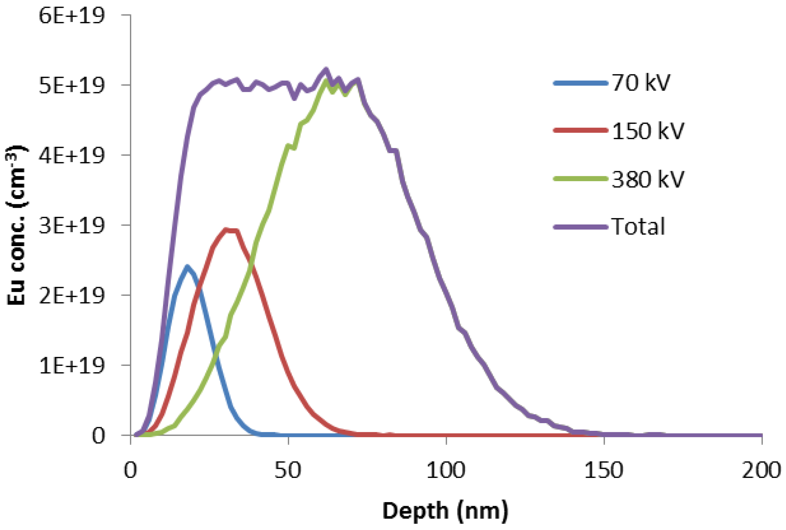
2 offcuts are implanted by Eu ions in Lisbon to produce an approximately uniform depth profile, as shown in
Figure 1. The lattice damage that results from ion implantation is annealed out under high pressure at high temperature in Warsaw (1400 °C, 1 GPa of N
2). Details of the sample preparation can be found in Ref. [
13]. One sample was grown and Mg-doped by HVPE prior to implantation/annealing.
Photoluminescence/excitation (PL/E) spectroscopy at Strathclyde is the main analytical technique used to characterize samples and examine HPS. Samples cooled in a helium cryorefrigerator are excited by monochromated light from a 1 kW Xe arc lamp, a 20 mW 355 nm laser or a pulsed nitrogen laser, in order of increasing achievable excitation density, analysed by a 2/3 m monochromator with a 1800 L/mm holographic grating and detected with a 16 bit 1064 × 128 pixel CCD camera.
3. Results
This section will be divided into the following subheadings. In
Section 3.1, we present the spectrum of as-received GaN(Mg):Eu samples at room temperature, featuring mainly Eu0 luminescence, and introduce the 1-3-5 rule for Eu
3+ defects in any host; in
Section 3.2, we describe Hysteretic Photochromic Switching, as revealed in the temperature dependence of PL during a cooling-warming cycle; in
Section 3.3, we describe the determination of the energy barrier that separates the photochromic defect configurations and link these observations to the Lany–Zunger model of the Mg acceptor; subsection
Section 3.4 considers transitions from excited
5D
1 states;
Section 3.5 reveals the effects of irreversible photo-dissociation of Eu-Mg defects in GaN material of exceptional quality.
3.1. Eu0 Spectrum
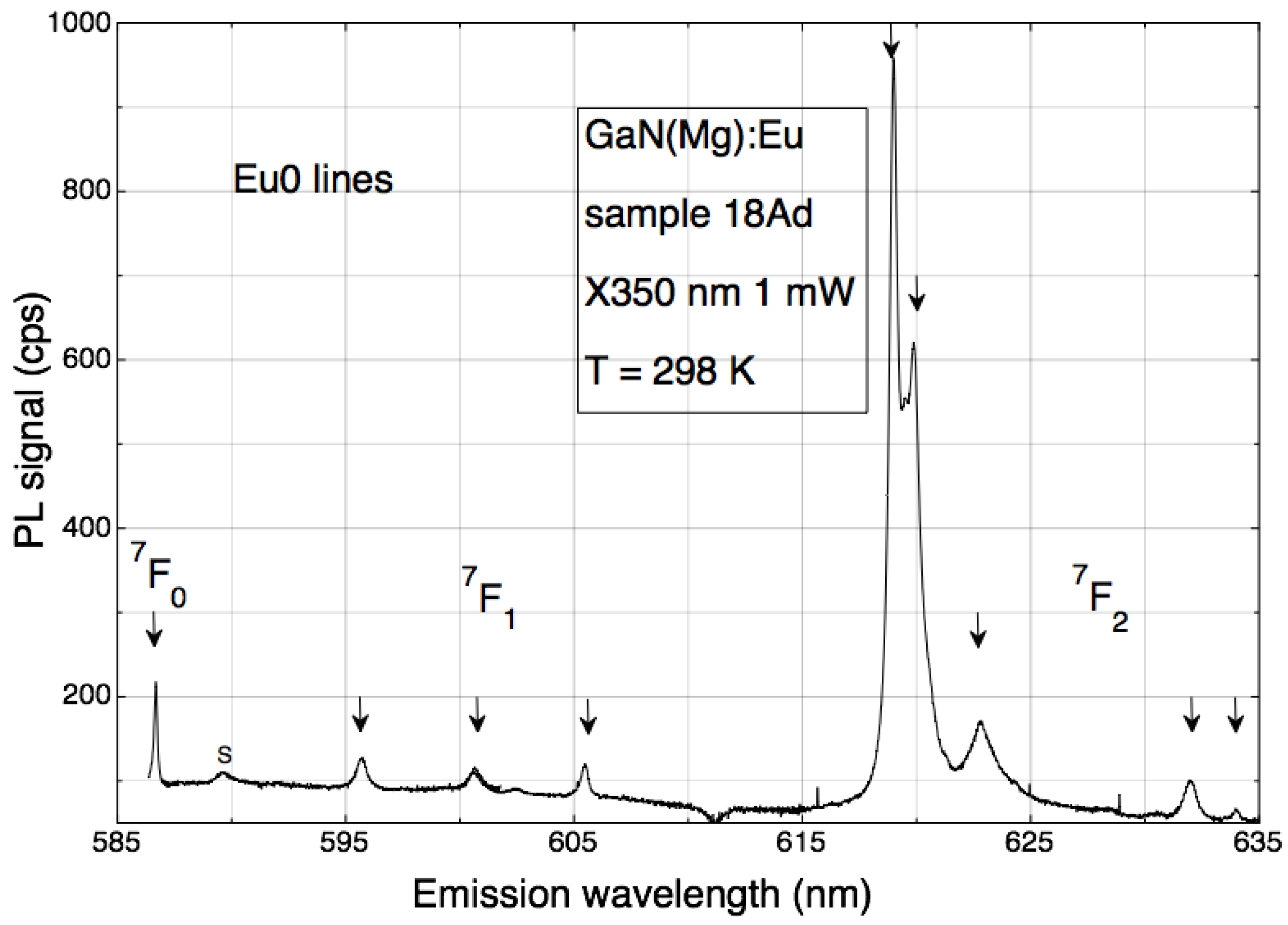
At room temperature, the typical Eu0 PL spectrum clearly exemplifies the 1-3-5 rule. Transitions from the
5D
0 excited state are identified in
Figure 2 by the label of the terminal state, with degeneracies of 1 (
7F
0), 3 (
7F
1) and 5 (
7F
2), respectively. All degeneracies are lifted by a crystal field with lower than axial symmetry, to produce a set of well separated lines indicated by arrows.
The Eu0 spectrum appears only in Eu- and Mg- (double) doped GaN [
1,
2] and careful doping comparisons show that its maximum intensity is achieved when the concentrations of Eu and Mg are approximately equal. This leads us to a model of the Eu0 defect comprising a single Eu atom in close association with a Mg atom, both substituting on the Ga sub-lattice.
3.2. HPS
When samples are cooled below about 50 K, we encounter (with much surprise) the photochromic switching of the Eu0 spectrum, in all of its components, to a completely different one.
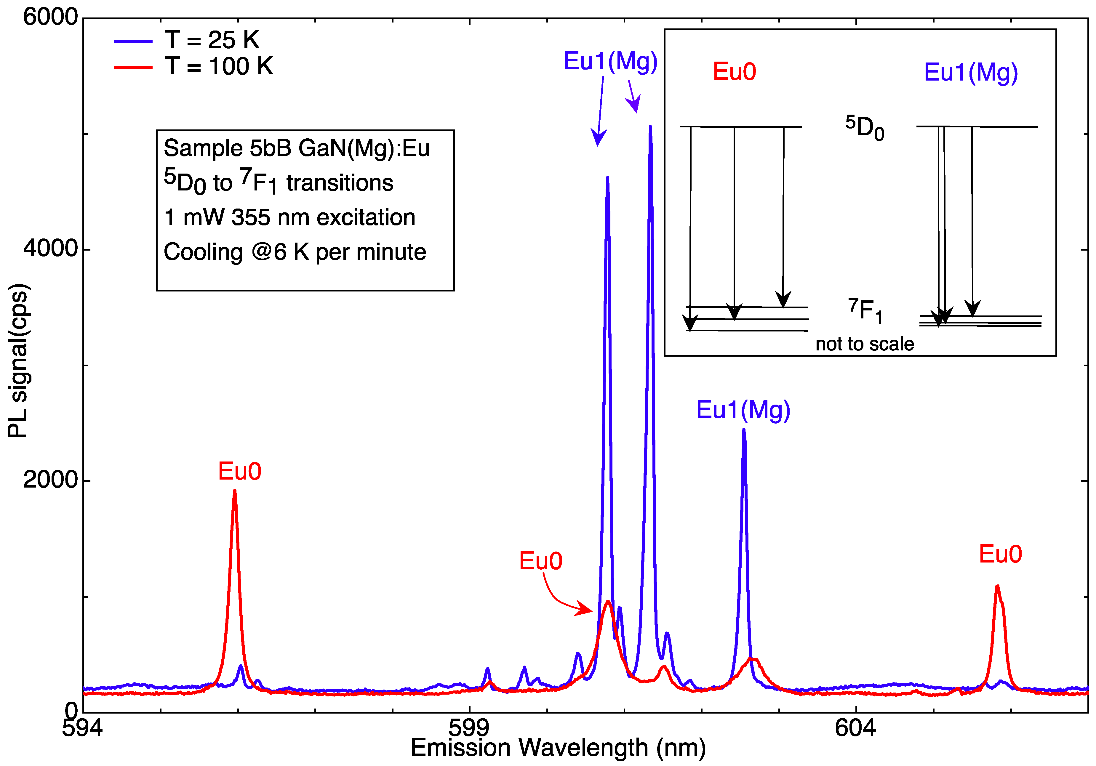
We assigned the low-temperature spectrum to Eu1(Mg), due to its superficial spectral similarities with the Eu1 centre observed in undoped GaN:Eu. Eu1(Mg) is much more symmetrical than Eu0, with, most notably, the
7F
1 state splitting as 3 = 2 + 1, that is to say into a (close-lying) doublet and a singlet [
8]. The profound alteration of the spectrum between 100 K and 25 K is shown in
Figure 3.
The switching of Eu0 to Eu1(Mg) is soon found to be hysteretic (hence
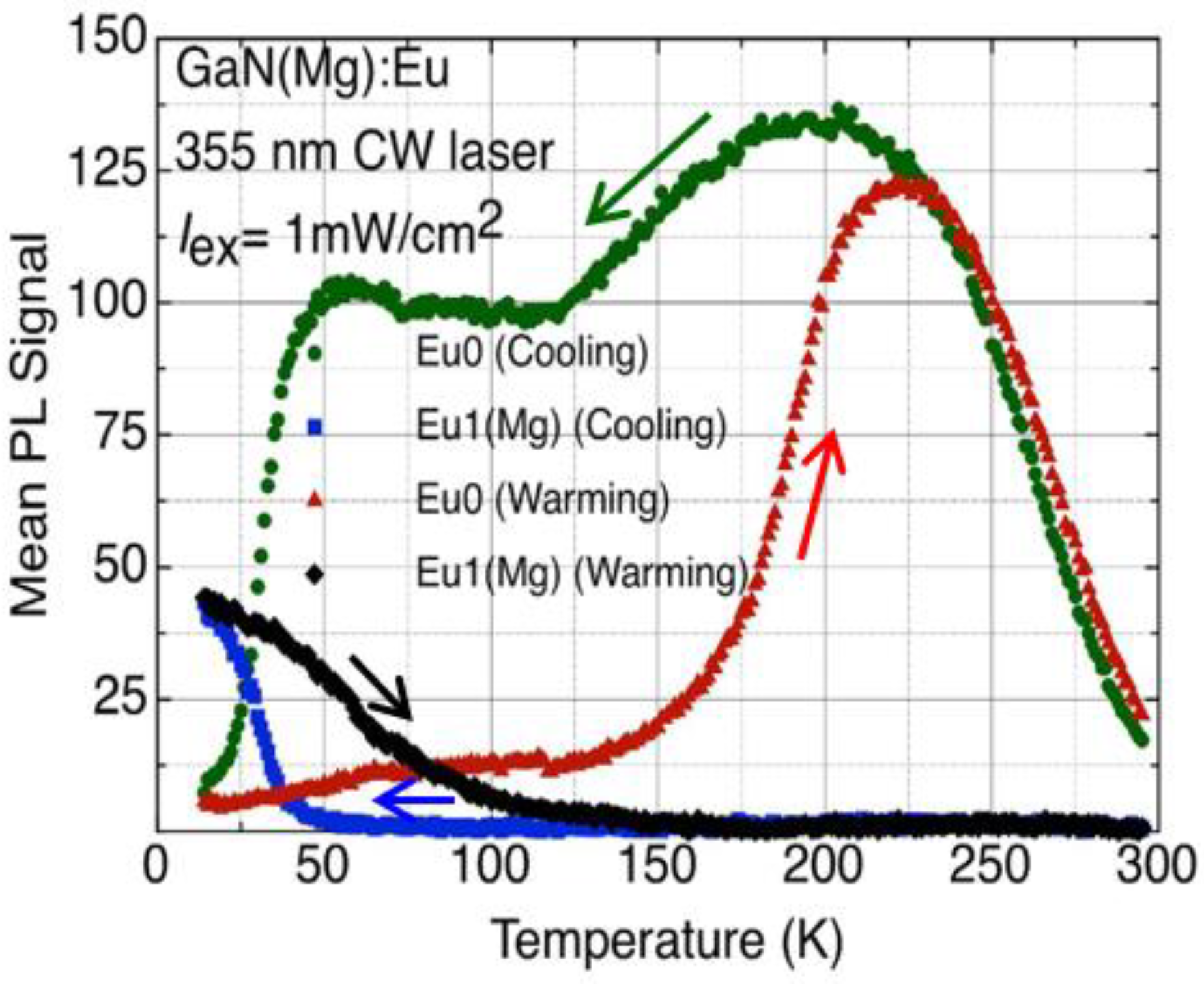
HPS): the reverse process occurs at a much higher temperature. The different hysteretic behaviours of Eu0 and Eu1(Mg) are shown in
Figure 4 for a typical experimental cycle.
Switching upon cooling (switchdown) offers the clearest perspective: the intensity of Eu0 drops to zero over a small temperature range, while that of Eu1(Mg) increases to a maximum value at the base temperature of our cryostat, ~10 K.
Switchback from Eu1(Mg) to Eu0 upon sample warming is more complicated and not fully understood. The maximum Eu1(Mg) signal at 10 K does not equal the maximum Eu0, but we ascribe this difference to one of transition probability between the more symmetric centre (less probable for intra-
f shell transitions) and the less symmetric one. We conclude that the switching is one-to-one with respect to defect numbers. The anomalous decrease in intensity of the Eu0 signal between its peak at 200 K and the start of the plateau at 125 K, explained only recently [
6], will feature in
Section 3.4.
3.3. Quantifying Photochromism
Without a mathematical model of the temperature dependence, it is difficult to extract physical parameters from the experiments described in
Section 3.2. With the realisation that the observation of photochromism requires light both to
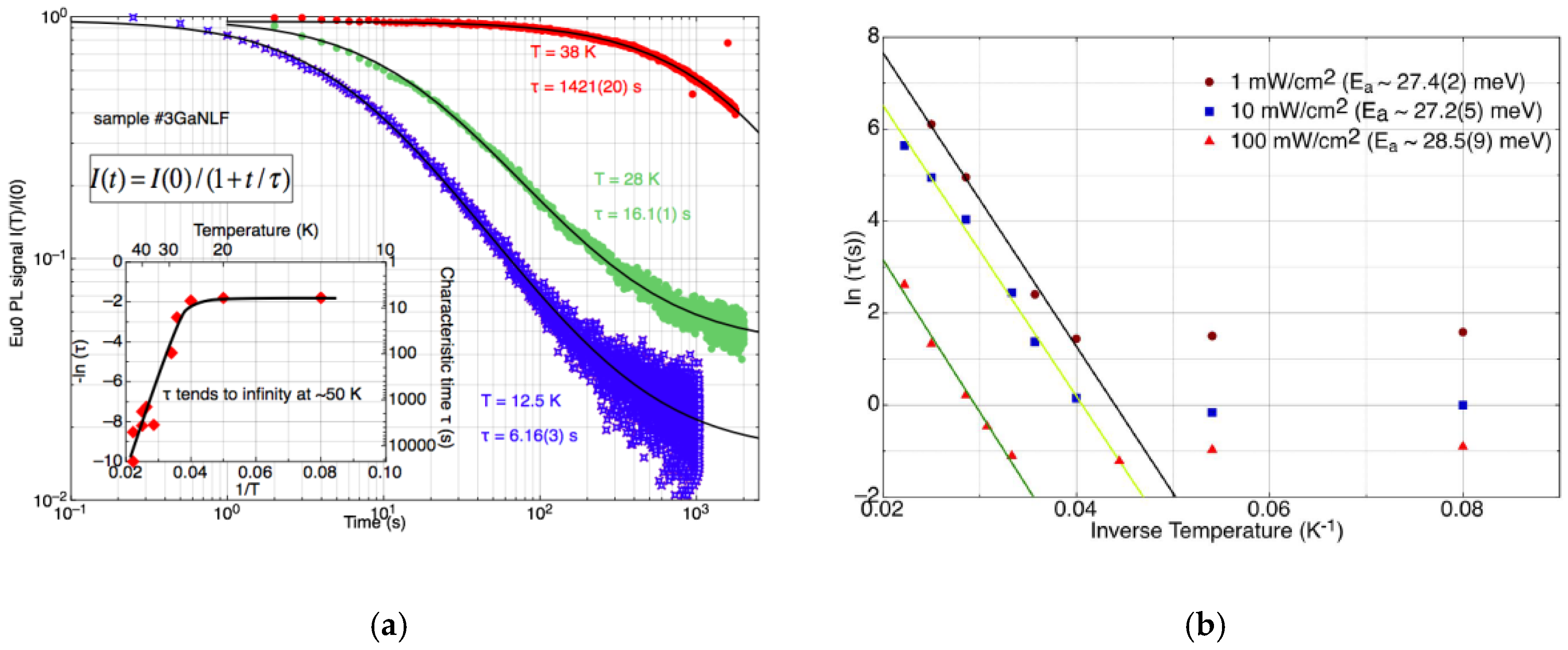
observe the effect, through PL, and to cause it, we devised a measurement protocol in which the sample was cooled to a fixed temperature in the dark and then subjected to a fixed wavelength and intensity of light for periods long enough that switching could be called essentially complete, while measuring the spectrum at 0.1 or 0.25 s intervals. (For brevity—and to reflect the situation in which the Mg-Eu complex is ‘surprised’ out of its non-equilibrium state by the sudden illumination—we familiarly refer to this as the ‘Ninja!’ protocol.)
Some general results can be cited for these experiments. For fixed excitation power and wavelength, the switching profile is hyperbolic in time, with a characteristic time constant
τ that can be extracted from the temporal dependence of switching by fitting:
to the dataset, where
I(
t) is the (mean) PL intensity of the unswitched (Eu0) component at time
t. The switching rate slows with increasing temperature, in an anti-Arrhenius relationship that reflects the fact that colder samples are further from equilibrium; it is the return to equilibrium that is captured in the Ninja! runs. Increasing the incident power decreases the switching time, as might be expected.
Some example results for a particular sample are illustrated in
Figure 5a,b. By fitting the temperature dependence of the switching time, the barrier between the photochromic states is estimated to be 27.7 meV, more or less independent of the excitation power density.
3.4. Transitions from 5D1 Levels
In conventional PL of semiconductors, it is expected that thermalisation between excited states should decrease the relative intensity of transitions originating on states lying higher in energy as the temperature decreases. The intensity should mirror a decreasing population of the higher lying levels through the operation of Boltzmann statistics.
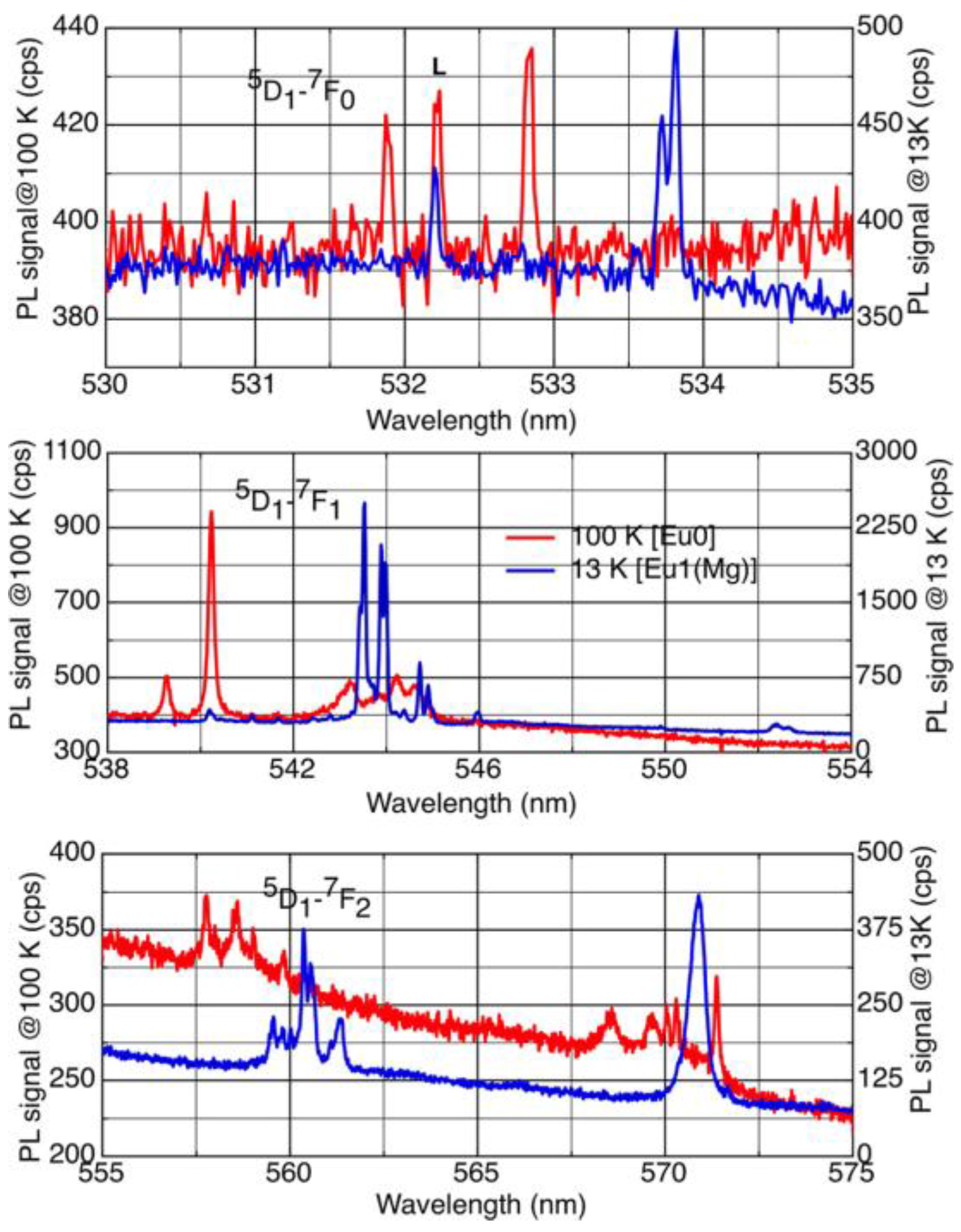
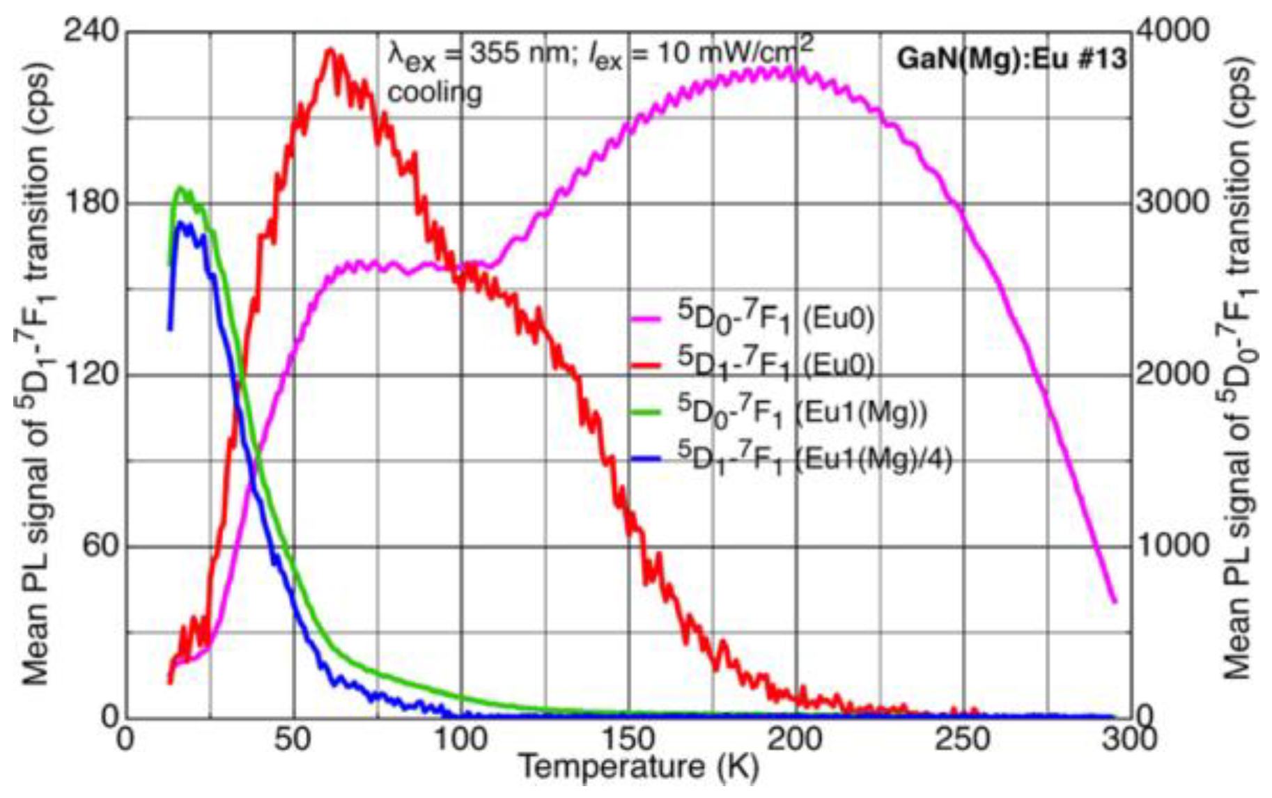
On the contrary, transitions from the
5D
1 levels in GaN(Mg):Eu are stronger at lower temperatures. Such transitions for both configurations of Eu-Mg defects are shown in
Figure 6, and their temperature dependences in
Figure 7.
The
7F
0,1,2 level splittings derived from an analysis of the spectra of
Figure 6 confirm those found previously for the stronger transitions from the
5D
0 states to these levels. This allows us to determine the
5D
1 to
5D
0 energy separation as well as the (relatively small) splittings within the
5D
1 levels caused by the crystal field [
6].
3.5. Photodissociation
When samples are subjected to prolonged exposure to above-bandgap light at low temperatures, further spectral changes occur, which we refer to as ‘second switching’ or ‘blitching’: bleaching of the total emission is accompanied by switching from Eu1(Mg) to a set of ever more symmetrical centres, labelled Eu1(Mg2), Eu1(Mg3) etc. [
14,
15,
16].
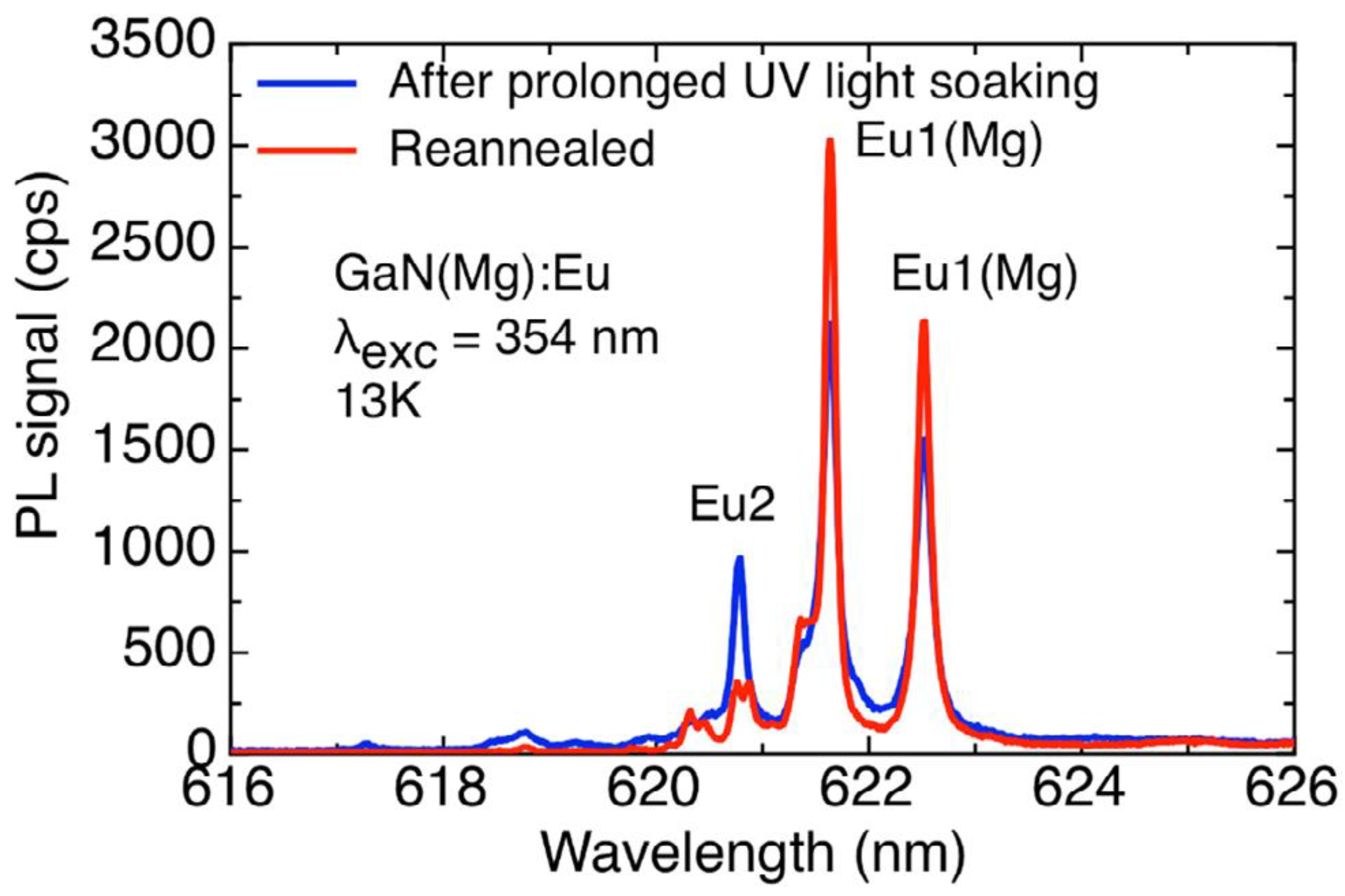
In MOVPE-grown samples, second switching occurs at a leisurely pace, necessitating experimental runs as long as 36,000 s to establish clear trends. However, for a state-of-the-art free-standing GaN(Mg) sample prepared by HVPE at Kyma Technologies, and doped with Eu as described in
Section 2, the blitching process is rapid, and ultimately destructive [
7].
Figure 8 shows the eventual result of such an experiment. Following a first set of runs that resulted in irreversible spectral alterations, the sample was re-annealed at high temperature and pressure, but only partly recovered its original spectral purity. (A sample that has lost the Eu0 dominance of the RT spectrum, shown in
Figure 2, is said to be ‘cooked’.)
4. Discussion
When taken together, the various pieces of experimental evidence summarised in
Section 3 lead us to form a precise microscopic model of the Mg-Eu complexes that form in p-type GaN.
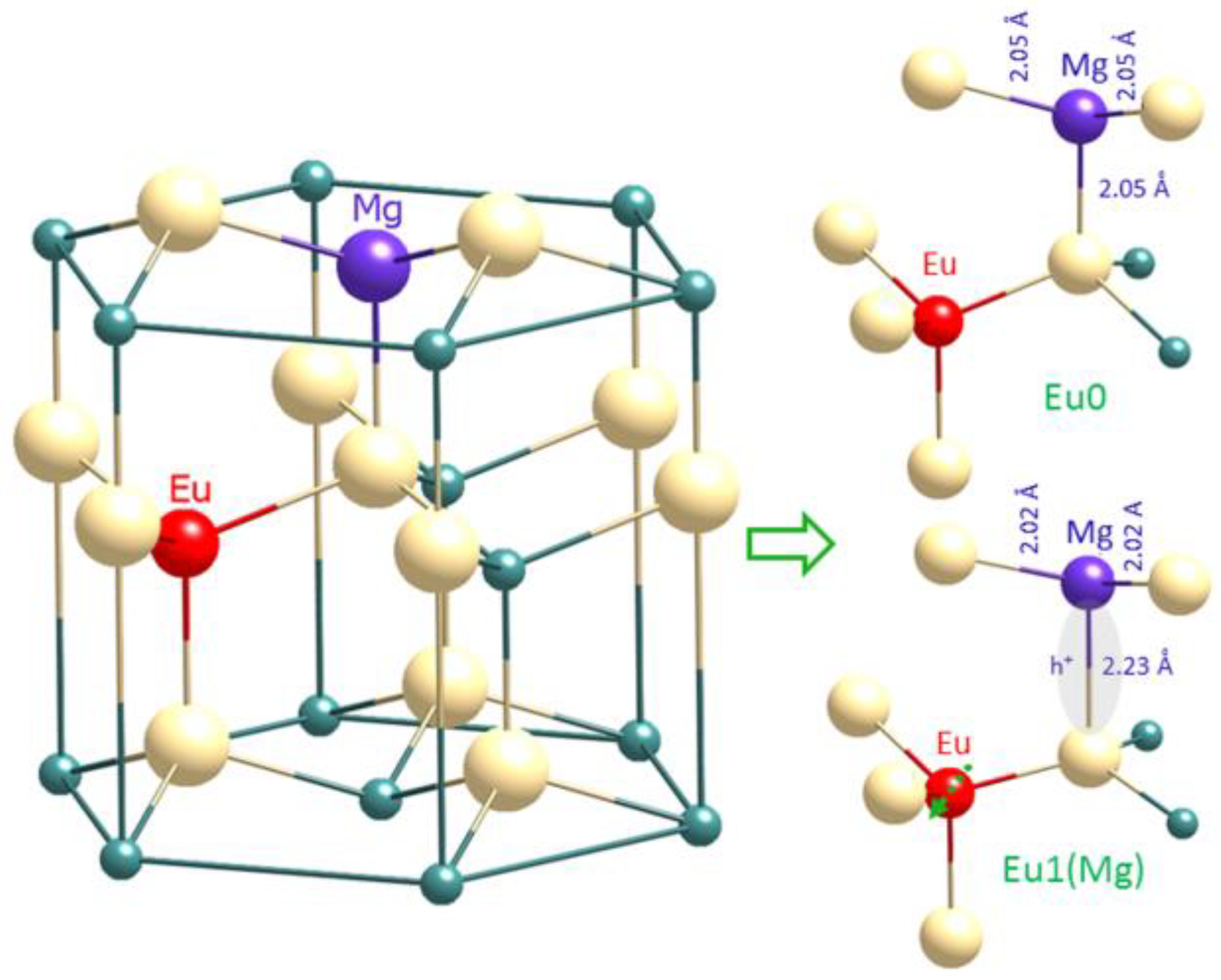
We know from observing maximum Eu0 emission when Mg and Eu concentrations match that the centre is likely to involve a single Eu atom in close association with a single Mg atom. Both Mg2+ and Eu3+ will favour cationic sites in the GaN lattice, so we propose that they both substitute for Ga and share a common nitrogen nearest neighbour.
We next consider the change in symmetry during the switch from Eu0 to Eu1(Mg) on cooling the sample (switchdown). This indicates that a local distortion in the lattice is induced during cooling, a kind of phase change occurring on the nanoscale, and shows the Eu3+ ion to be in a higher symmetry environment in the Eu1(Mg) state.
A further clue regarding the nature of this distortion comes from the activation energy obtained from quantifying the light-induced switching of Eu0 to Eu1(Mg) at low T: this value (27.7 meV) is close to the barrier height predicted to exist between the two Mg acceptor states proposed by Lany and Zunger as the ‘curious case of the shallow Mg
Ga deep state’ [
4]. Hybrid density functional calculations predicted formation of a shallow transient state, effective in the p-type activation, and a deeper ground state; further calculations by Lyons et al. [
5] supported the existence of such a deep state, specifically attributing it to localization of a hole at an axial N atom.
We proposed [
9] that the switching between Eu0 and Eu1(Mg) is the spectroscopic signature of the transition between these two Mg acceptor states, as observed by the nearby Eu ‘spectator’ atom. Localization of a hole on the magnesium’s axial N bond (i.e., the deep ground state) is the stable configuration at low temperature, giving Eu1(Mg); delocalization of the hole at higher temperatures, and the resultant local lattice distortion, results in a change in the crystal field splitting and the switch to Eu0.
Our microscopic model is sketched in
Figure 9.
One effect of the prolonged UV exposure of these materials is the emergence of additional centres whose line splittings reveal successively increased symmetry [
15]. This indicates that a fraction of Mg atoms has
physically migrated to lattice sites away from their Eu partner. This eventually leads to the appearance of the Eu2 centre, identified as the ‘prime’ defect in unimplanted GaN:Eu (i.e., a simple substitutional Eu atom on a cation site); the Mg and Eu must now be sufficiently separated to be entirely disassociated.
Finally, we have made recent observations (to be fully reported at a later date) of the effects of co-implanting the GaN:Mg(Eu) with additional anion species to give triply-doped material. We find that the introduction of excess N atoms enhances Eu0 production and reduces the intensity of vestigial Eu1 lines, supporting Mitchell et al.’s previous identification of this centre as substitutional Eu associated with a nitrogen vacancy [
12]. The introduction of O results in a new centre, with the same symmetry as Eu0, but not exhibiting the switching behaviour. We interpret this as being due to the formation of an Eu-O-Mg defect complex in which the strong Mg-O bond disrupts the ability of a hole to localise in the deep ground state which is central to the Eu0-Eu1(Mg) switching.

 ,
, 


