Comprehensive Review of Hybrid Collagen and Silk Fibroin for Cutaneous Wound Healing


Abstract
:1. Introduction
1.1. Biomaterial
1.2. Cutaneous Wound
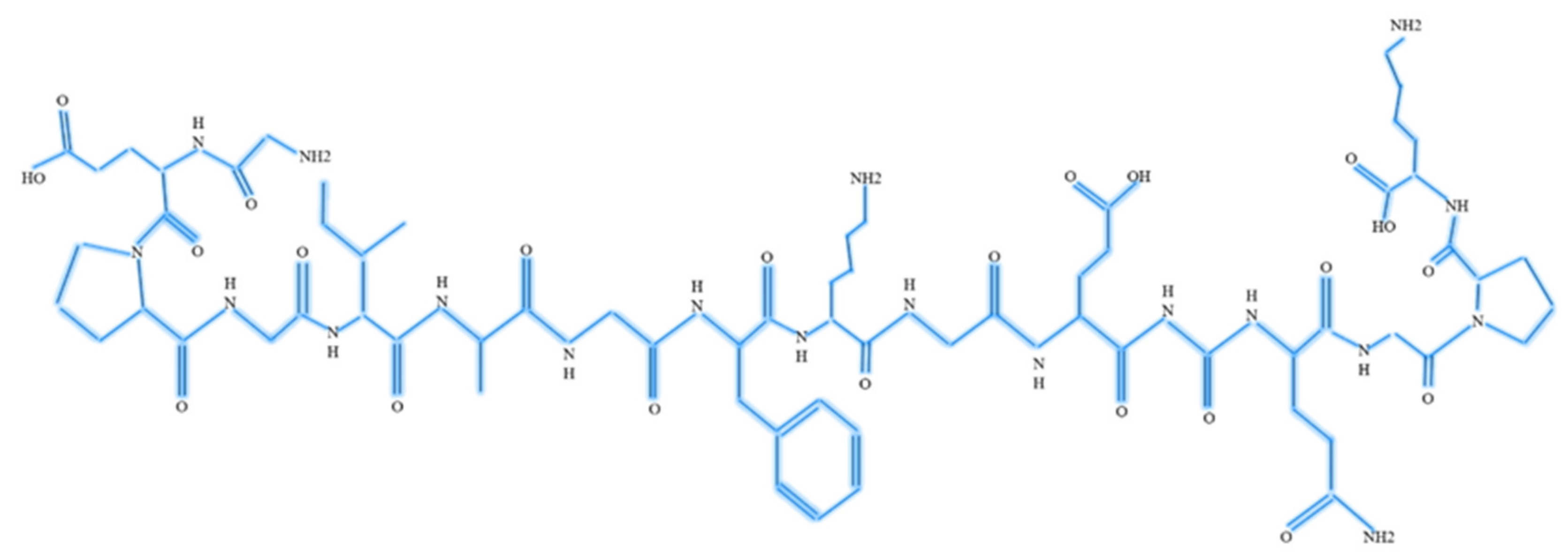
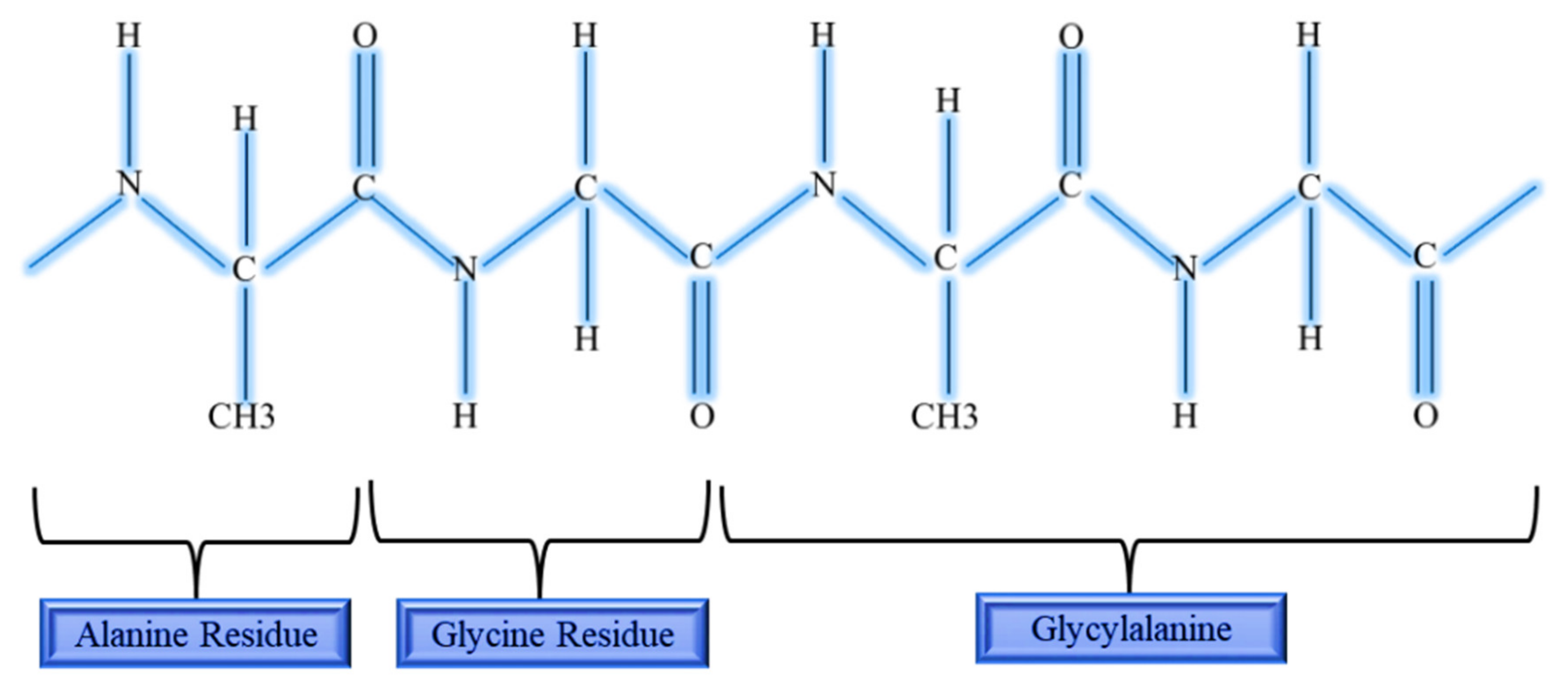
1.3. Collagen
1.4. Silk Fibroin
1.5. Hybrid of Collagen and Silk Fibroin
2. Results
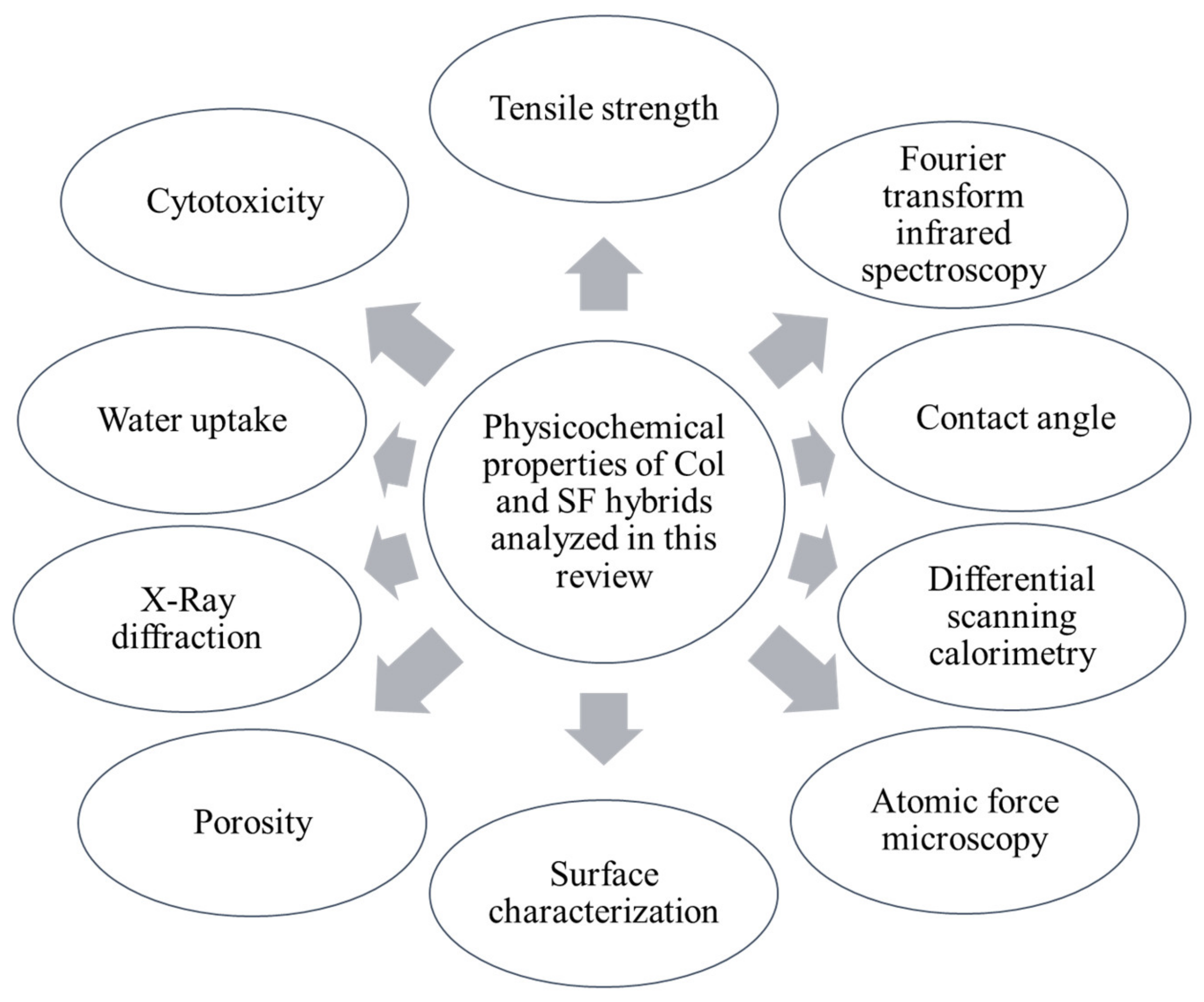
2.1. Characterisation of Hybrid Collagen and Silk Fibroin
2.1.1. Tensile Strength
2.1.2. Fourier Transform Infrared Spectroscopy (FTIR)
2.1.3. Contact Angle
2.1.4. Differential Scanning Calorimetry (DSC)
2.1.5. Atomic Force Microscopy (AFM)
2.1.6. Surface Structure of Hybrid Col and SF
2.1.7. Porosity
2.1.8. X-ray Diffraction Study (XRD)
2.1.9. Water Uptake
2.1.10. Cytotoxicity
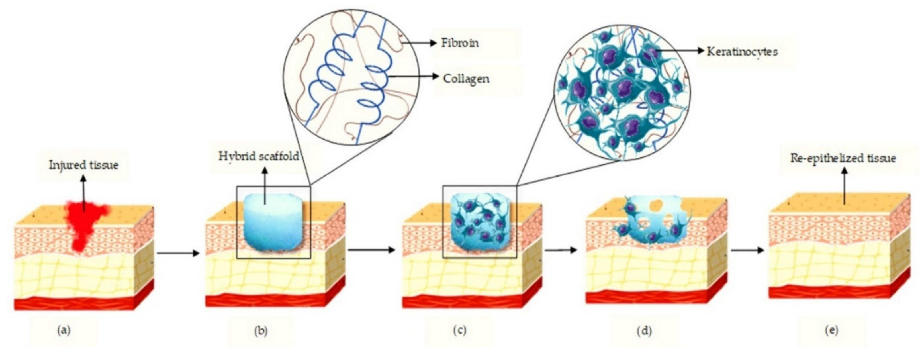
2.2. Role and Physicochemical Influence of Hybrid Col and SF in Wound Healing
3. Discussion
4. Conclusions
Author Contributions
Funding
Acknowledgments
Conflicts of Interest
References
- Busra, M.F.M.; Lokanathan, Y. Recent Development in the Fabrication of Collagen Scaffolds for Tissue Engineering Applications: A Review. Curr. Pharm. Biotechnol. 2019, 20, 992–1003. [Google Scholar] [CrossRef] [PubMed]
- Pavlovic, M. What Are Biomaterials? In Bioengineering; Springer International Publishing: Cham, Switzerland, 2015; pp. 229–244. ISBN 978-3-319-10798-1. [Google Scholar]
- Bhat, S.; Kumar, A. Biomaterials and bioengineering tomorrow’s healthcare. Biomatter 2013, 3, e24717. [Google Scholar] [CrossRef] [PubMed] [Green Version]
- Poologasundarampillai, G.; Nommeots-Nomm, A. Metals, polymers, ceramics, hydrogels. In 3D Printing in Medicine; Geng, H., Ed.; Woodhead Publishing: Tampere, Finland, 2017; pp. 43–71. ISBN 9780081007266. [Google Scholar]
- Zeng, R.; Lin, C.; Lin, Z.; Chen, H.; Lu, W.; Lin, C.; Li, H. Approaches to cutaneous wound healing: Basics and future directions. Cell Tissue Res. 2018, 374, 217–232. [Google Scholar] [CrossRef] [PubMed]
- Wong, S.Y.; Manikam, R.; Muniandy, S. Prevalence and antibiotic susceptibility of bacteria from acute and chronic wounds in Malaysian subjects. J. Infect. Dev. Ctries. 2015, 9, 936–944. [Google Scholar] [CrossRef] [Green Version]
- Fife, C.E.; Carter, M.J. Wound Care Outcomes and Associated Cost Among Patients Treated in US Outpatient Wound Centers: Data From the US Wound Registry. Wounds 2012, 24, 10–17. [Google Scholar] [PubMed]
- Lim, H.W.; Collins, S.A.B.; Resneck, J.S.; Bolognia, J.L.; Hodge, J.A.; Rohrer, T.A.; Van Beek, M.J.; Margolis, D.J.; Sober, A.J.; Weinstock, M.A.; et al. The burden of skin disease in the United States. J. Am. Acad. Dermatol. 2017, 76, 958–972. [Google Scholar] [CrossRef] [Green Version]
- McIntosh, J. What is collagen, and why do people use it? News Today 2017, 2, 1–4. [Google Scholar]
- Kwan, P.; Desmoulière, A.; Tredget, E.E. Molecular and cellular basis of hypertrophic scarring. In Total Burn Care; Elsevier Inc.: Amsterdam, The Netherlands, 2018; p. 465. ISBN 9780323497428. [Google Scholar]
- Ibrahim, M.S.; El-Wassefy, N.A.; Farahat, D.S. Biocompatibility of dental biomaterials. In Biomaterials for Oral and Dental Tissue Engineering; Tayebi, L., Moharamzadeh, K., Eds.; Woodhead Publishing: Boca Raton, FL, USA, 2017; pp. 117–140. ISBN 9780081009673. [Google Scholar]
- Narayanan, N.; Kuang, L.; Del Ponte, M.; Chain, C.; Deng, M. Design and Fabrication of Nanocomposites for Musculoskeletal Tissue Regeneration. In Nanocomposites for Musculoskeletal Tissue Regeneration; Elsevier Inc.: Amsterdam, The Netherlands, 2016; pp. 3–29. ISBN 9781782424758. [Google Scholar]
- Hong, H.; Chaplot, S.; Chalamaiah, M.; Roy, B.C.; Bruce, H.L.; Wu, J. Removing Cross-Linked Telopeptides Enhances the Production of Low-Molecular-Weight Collagen Peptides from Spent Hens. J. Agric. Food Chem. 2017, 65, 7491–7499. [Google Scholar] [CrossRef]
- Cremers, S.; Garnero, P.; Seibel, M.J. Biochemical Markers of Bone Metabolism. In Principles of Bone Biology; Bilezikian, J.P., Raisz, L.G., Martin, T.J., Eds.; Academic Press: New York, NY, USA, 2008; pp. 1857–1881. Volume 2, ISBN 978-0123738868. [Google Scholar]
- Fauzi, M.B.; Lokanathan, Y.; Aminuddin, B.S.; Ruszymah, B.H.I.; Chowdhury, S.R. Ovine tendon collagen: Extraction, characterisation and fabrication of thin films for tissue engineering applications. Mater. Sci. Eng. C 2016, 68, 163–171. [Google Scholar] [CrossRef]
- Ricard-Blum, S. The Collagen Family. Cold Spring Harb. Perspect. Biol. 2011, 3, 1–19. [Google Scholar] [CrossRef] [Green Version]
- Rýglová, Š.; Braun, M.; Suchý, T. Collagen and Its Modifications-Crucial Aspects with Concern to Its Processing and Analysis. Macromol. Mater. Eng. 2017, 302, 1–29. [Google Scholar] [CrossRef]
- Magnus, A. Wound Healing Biomaterials. In Functional Biomaterials; Woodhead Publishing: Boca Raton, FL, USA, 2016; pp. 486–542. Volume 2, ISBN 9780081006061. [Google Scholar]
- Chattopadhyay, S.; Raines, R.T. Collagen-based biomaterials for wound healing. Biopolymers 2014, 101, 821–833. [Google Scholar] [CrossRef] [PubMed] [Green Version]
- Pistone, A.; Sagnella, A.; Chieco, C.; Bertazza, G.; Varchi, G.; Formaggio, F.; Posati, T.; Saracino, E.; Caprini, M.; Bonetti, S.; et al. Silk fibroin film from golden-yellow Bombyx mori is a biocomposite that contains lutein and promotes axonal growth of primary neurons. Biopolymers 2016, 105, 287–299. [Google Scholar] [CrossRef]
- Correlo, V.M.; Oliveira, J.M.; Mano, J.F.; Neves, N.M.; Reis, R.L. Natural Origin Materials for Bone Tissue Engineering—Properties, Processing, and Performance. In Principles of Regenerative Medicine; Atala, A., Lanza, R., Thomson, J.A., Nerem, R.M., Eds.; Elsevier Inc.: Amsterdam, The Netherlands, 2011; pp. 557–586. ISBN 9780123814227. [Google Scholar]
- Martínez-Mora, C.; Mrowiec, A.; García-Vizcaíno, E.M.; Alcaraz, A.; Cenis, J.L.; Nicolás, F.J. Fibroin and Sericin from Bombyx mori Silk Stimulate Cell Migration through Upregulation and Phosphorylation of c-Jun. PLoS One 2012, 7, e42271. [Google Scholar] [CrossRef] [PubMed]
- Tripathy, N.; Perumal, E.; Ahmad, R.; Song, J.E.; Khang, G. Hybrid Composite Biomaterials. In Principles of Regenerative Medicine; Atala, A., Lanza, R.P., Mikos, A.G., Nerem, R.M., Eds.; Elsevier: Seoul, Korea, 2019; pp. 695–714. ISBN 9780128098936. [Google Scholar]
- Neffe, A.T.; Julich-Gruner, K.K.; Lendlein, A. Combinations of biopolymers and synthetic polymers for bone regeneration. In Biomaterials for Bone Regeneration, 1st ed.; Dubruel, P., Van Vlierberghe, S., Eds.; Elsevier: Amsterdam, The Netherlands, 2014; pp. 87–110. ISBN 9780857098108. [Google Scholar]
- Elzoghby, A.O.; Elgohary, M.M.; Kamel, N.M. Implications of Protein and Peptide Based Nanoparticles as Potential Vehicles for Anticancer Drugs. In Advances in Protein Chemistry and Structural Biology; Donev, R., Ed.; Academic Press Inc.: Cambridge, MA, USA, 2015; Volume 98, pp. 169–221. ISBN 978-0-12-802828-5. [Google Scholar]
- Asakura, T.; Suzuki, Y. Silk Fibroin. In Encyclopedia of Polymeric Nanomaterials; Kobayashi, S., Müllen, K., Eds.; Springer: Berlin/Heidelberg, Germany, 2014; pp. 1–7. ISBN 9783642296482. [Google Scholar]
- Saxena, T.; Karumbaiah, L.; Valmikinathan, C.M. Proteins and Poly (Amino Acids). In Natural and Synthetic Biomedical Polymers; Kumbar, S.G., Laurencin, C., Deng, M., Eds.; Elsevier Science: Alpharetta, GA, USA, 2014; pp. 43–65. ISBN 9780123969835. [Google Scholar]
- Łos, M.J.; Panigrahi, S.; Sielatycka, K.; Grillon, C. Successful Biomaterial-Based Artificial Organ-Updates on Artificial Blood Vessels. In Stem Cells and Biomaterials for Regenerative Medicine; Los, M., Hudecki, A., Wieche, E., Eds.; Elsevier: Amsterdam, The Netherlands, 2019; pp. 203–222. ISBN 9780128122785. [Google Scholar]
- Suarato, G.; Bertorelli, R.; Athanassiou, A. Borrowing from nature: Biopolymers and biocomposites as smart wound care materials. Front. Bioeng. Biotechnol. 2018, 6, 137. [Google Scholar] [CrossRef] [PubMed] [Green Version]
- Buitrago, J.O.; Patel, K.D.; El-Fiqi, A.; Lee, J.H.; Kundu, B.; Lee, H.H.; Kim, H.W. Silk fibroin/collagen protein hybrid cell encapsulating hydrogels with tunable gelation and improved physical and biological properties. Acta Biomater. 2018, 69, 218–233. [Google Scholar] [CrossRef] [PubMed]
- Ghezzi, C.E.; Marelli, B.; Donelli, I.; Alessandrino, A.; Freddi, G.; Nazhat, S.N. Multilayered dense collagen silk fibroin hybrid: A platform for mesenchymal stem cell differentiation towards chondrogenic and osteogenic lineages. J. Tissue Eng. Regen. Med. 2015, 11, 2046–2059. [Google Scholar] [CrossRef]
- Cui, B.; Zhang, C.; Gan, B.; Liu, W.; Liang, J.; Fan, Z.; Wen, Y.; Yang, Y.; Peng, X.; Zhou, Y. Collagen-tussah silk fibroin hybrid scaffolds loaded with bone mesenchymal stem cells promote skin wound repair in rats. Mater. Sci. Eng. C 2020, 109, 110611. [Google Scholar] [CrossRef]
- Kim, S.H.; Park, H.S.; Lee, O.J.; Chao, J.R.; Park, H.J.; Lee, J.M.; Ju, H.W.; Moon, B.M.; Park, Y.R.; Song, J.E.; et al. Fabrication of duck’s feet collagen-silk hybrid biomaterial for tissue engineering. Int. J. Biol. Macromol. 2016, 85, 442–450. [Google Scholar] [CrossRef]
- Chowdhury, S.R.; Busra, M.F.M.; Lokanathan, Y.; Ng, M.H.; Law, J.X.; Cletus, U.C.; Idrus, R.B.H. Collagen Type I: A Versatile Biomaterial. In Novel Biomaterials for Regenerative Medicine, Advances in Experimental Medicine and Biology; Chun, H.J., Ed.; Springer Nature Singapore Pte Ltd.: Singapore, 2018; Volume 1077, pp. 389–414. [Google Scholar]
- Rameshbabu, A.P.; Bankoti, K.; Datta, S.; Subramani, E.; Apoorva, A.; Ghosh, P.; Maity, P.P.; Manchikanti, P.; Chaudhury, K.; Dhara, S. Silk Sponges Ornamented with a Placenta-Derived Extracellular Matrix Augment Full-Thickness Cutaneous Wound Healing by Stimulating Neovascularization and Cellular Migration. ACS Appl. Mater. Interfaces 2018, 10, 16977–16991. [Google Scholar] [CrossRef]
- Deen, I.; Rosei, F. Silk fibroin derived polypeptides additives to promote hydroxyapatite nucleation in dense collagen hydrogels. PLoS ONE 2019, 14, e0219429. [Google Scholar] [CrossRef] [PubMed] [Green Version]
- Li, Z.H.; Ji, S.C.; Wang, Y.Z.; Shen, X.C.; Liang, H. Silk fibroin based scaffolds for tissue engineering. Front. Mater. Sci. 2013, 7, 237–247. [Google Scholar] [CrossRef]
- Feng, X.; Xu, P.; Shen, T.; Zhang, Y.; Ye, J.; Gao, C. Influence of pore architectures of silk fibroin/collagen composite scaffolds on the regeneration of osteochondral defects in vivo. J. Mater. Chem. B 2020, 8, 391–405. [Google Scholar] [CrossRef] [PubMed]
- Yeelack, W.; Meesane, J. Preparation and characterization of coated silk fibroin films with mimicked reself assembly type I collagen. In Proceedings of the 6th 2013 Biomedical Engineering International Conference, Amphur Muang, Thailand, 23–25 October 2013; pp. 3–6. [Google Scholar] [CrossRef]
- Zhou, J.; Cao, C.; Ma, X.; Lin, J. Electrospinning of silk fibroin and collagen for vascular tissue engineering. Int. J. Biol. Macromol. 2010, 47, 514–519. [Google Scholar] [CrossRef]
- Sionkowska, A.; Michalska, M.; Walczak, M.; Śmiechowski, K.; Grabska, S. Preparation and characterization of silk fibroin/collagen sponge modified by chemical cross-linking. Mol. Cryst. Liq. Cryst. 2016, 640, 180–190. [Google Scholar] [CrossRef]
- Kittiphattanabawon, P.; Benjakul, S.; Visessanguan, W.; Shahidi, F. Isolation and characterization of collagen from the cartilages of brownbanded bamboo shark (Chiloscyllium punctatum) and blacktip shark (Carcharhinus limbatus). LWT—Food Sci. Technol. 2010, 43, 792–800. [Google Scholar] [CrossRef]
- Hu, K.; Lv, Q.; Cui, F.Z.; Feng, Q.L.; Kong, X.D.; Wang, H.L.; Huang, L.Y.; Li, T. Biocompatible fibroin blended films with recombinant human-like collagen for hepatic tissue engineering. J. Bioact. Compat. Polym. 2006, 21, 23–37. [Google Scholar] [CrossRef]
- Lu, Q.; Feng, Q.; Hu, K.; Cui, F. Preparation of three-dimensional fibroin/collagen scaffolds in various pH conditions. J. Mater. Sci. Mater. Med. 2008, 19, 629–634. [Google Scholar] [CrossRef]
- Lv, Q.; Hu, K.; Feng, Q.L.; Cui, F. Fibroin/collagen hybrid hydrogels with crosslinking method: Preparation, properties, and cytocompatibility. J. Biomed. Mater. Res.—Part A 2007, 84, 198–207. [Google Scholar] [CrossRef]
- Grabska-Zielińska, S.; Sionkowska, A.; Reczyńska, K.; Pamuła, E. Physico-chemical characterization and biological tests of collagen/silk fibroin/chitosan scaffolds cross-linked by dialdehyde starch. Polymers (Basel) 2020, 12, 372. [Google Scholar] [CrossRef] [Green Version]
- Lu, Q.; Hu, K.; Feng, Q.L.; Cui, F. Growth of fibroblast and vascular smooth muscle cells in fibroin/collagen scaffold. Mater. Sci. Eng. C 2009, 29, 2239–2245. [Google Scholar] [CrossRef]
- Carey, S.P.; Kraning-Rush, C.M.; Williams, R.M.; Reinhart-King, C.A. Biophysical control of invasive tumor cell behavior by extracellular matrix microarchitecture. Biomaterials 2012, 33, 4157–4165. [Google Scholar] [CrossRef] [PubMed] [Green Version]
- Ghezzi, C.E.; Marelli, B.; Muja, N.; Hirota, N.; Martin, J.G.; Barralet, J.E.; Alessandrino, A.; Freddi, G.; Nazhat, S.N. Mesenchymal stem cell-seeded multilayered dense collagen-silk fibroin hybrid for tissue engineering applications. Biotechnol. J. 2011, 6, 1198–1207. [Google Scholar] [CrossRef] [PubMed]
- Bellas, E.; Seiberg, M.; Garlick, J.; Kaplan, D.L. In vitro 3D Full-Thickness Skin-Equivalent Tissue Model Using Silk and Collagen Biomaterials. Macromol. Biosci. 2012, 12, 1627–1636. [Google Scholar] [CrossRef] [Green Version]
- Cui, X.A.; Liu, X.; Kong, D.L.; Gu, H.Q. Preparation and characteration of electrospun collagen/silk fibroin complex microfibers. In Proceedings of the World Congress on Medical Physics and Biomedical Engineering, Bejing, China, 26–31 May 2012; Long, M., Ed.; Springer: Berlin/Heidelberg, Germany, 2013; pp. 79–82. [Google Scholar] [CrossRef]
- Sun, K.; Li, H.; Li, R.; Nian, Z.; Li, D.; Xu, C. Silk fibroin/collagen and silk fibroin/chitosan blended three-dimensional scaffolds for tissue engineering. Eur. J. Orthop. Surg. Traumatol. 2014, 25, 243–249. [Google Scholar] [CrossRef]
- Boonrungsiman, S.; Thongtham, N.; Suwantong, O.; Wutikhun, T.; Soykeabkaew, N.; Nimmannit, U. An improvement of silk-based scaffold properties using collagen type I for skin tissue engineering applications. Polym. Bull. 2017, 75, 685–700. [Google Scholar] [CrossRef]
- Ramadass, S.K.; Nazir, L.S.; Thangam, R.; Perumal, R.K.; Manjubala, I.; Madhan, B.; Seetharaman, S. Type I collagen peptides and nitric oxide releasing electrospun silk fibroin scaffold: A multifunctional approach for the treatment of ischemic chronic wounds. Colloids Surf. B Biointerfaces 2019, 175, 636–643. [Google Scholar] [CrossRef]
- Qing, L.; Ren-fu, Q.; Li-hong, C.; Li-hong, H.; En-liang, C.; Hua-hui, H.; Xuan, Z. Acceleration of wound healing by a porous collagen/silk fibroin scaffold carrying zinc oxide nanoparticles. Chin. J. Tissue Eng. Res. 2018, 22, 2161. [Google Scholar] [CrossRef]
- Kim, K.O.; Lee, Y.; Hwang, J.W.; Kim, H.; Kim, S.M.; Chang, S.W.; Lee, H.S.; Choi, Y.S. Wound healing properties of a 3-D scaffold comprising soluble silkworm gland hydrolysate and human collagen. Colloids Surf. B Biointerfaces 2014, 116, 318–326. [Google Scholar] [CrossRef]
- Wu, G.; Ma, X.; Fan, L.; Gao, Y.; Deng, H.; Wang, Y. Accelerating dermal wound healing and mitigating excessive scar formation using LBL modified nanofibrous mats. Mater. Des. 2019, 185, 108265. [Google Scholar] [CrossRef]
- Xue, M.; Jackson, C.J. Extracellular Matrix Reorganization During Wound Healing and Its Impact on Abnormal Scarring. Adv. Wound Care 2015, 4, 119–136. [Google Scholar] [CrossRef] [PubMed] [Green Version]
- Gonzalez, A.C.D.O.; Andrade, Z.D.A.; Costa, T.F.; Medrado, A.R.A.P. Wound healing—A literature review. An. Bras. Dermatol. 2016, 91, 614–620. [Google Scholar] [CrossRef] [Green Version]
- Nikoloudaki, G.; Brooks, S.; Peidl, A.P.; Tinney, D.; Hamilton, D.W. JNK signaling as a key modulator of soft connective tissue physiology, pathology, and healing. Int. J. Mol. Sci. 2020, 21, 1015. [Google Scholar] [CrossRef] [PubMed] [Green Version]
- DiCosmo, F. The Role of Collagen in Wound Healing. Adv. Skin Wound Care 2009, 22, 12–15. [Google Scholar] [CrossRef]
- Sevilla, C.A.; Dalecki, D.; Hocking, D.C. Regional Fibronectin and Collagen Fibril Co-Assembly Directs Cell Proliferation and Microtissue Morphology. PLoS ONE 2013, 8, e77316. [Google Scholar] [CrossRef] [PubMed] [Green Version]
- Guan, G.; Bai, L.; Zuo, B.; Li, M.; Wu, Z.; Li, Y.; Wang, L. Promoted dermis healing from full thickness skin defect by porous silk fibroin scaffolds (PSFSs). Biomed. Mater. Eng. 2010, 20, 295–308. [Google Scholar] [CrossRef]
- Chaudhuri, O.; Gu, L.; Klumpers, D.; Darnell, M.; Bencherif, S.A.; Weaver, J.C.; Huebsch, N.; Lee, H.P.; Lippens, E.; Duda, G.N.; et al. Hydrogels with tunable stress relaxation regulate stem cell fate and activity. Nat. Mater. 2016, 15, 326–334. [Google Scholar] [CrossRef] [Green Version]
- Yannas, I.; Tzeranis, D.; So, P. Surface biology of collagen scaffold explains blocking of wound contraction and regeneration of skin and peripheral nerves. Biomed. Mater. 2015, 11, 014106. [Google Scholar] [CrossRef]
- Junker, J.P.E.; Kamel, R.A.; Caterson, E.J.; Eriksson, E. Clinical Impact upon Wound Healing and Inflammation in Moist, Wet, and Dry Environments. Adv. Wound Care 2013, 2, 348–356. [Google Scholar] [CrossRef] [Green Version]
- Bai, L.; Zhu, L.; Min, S.; Liu, L.; Cai, Y.; Yao, J. Surface modification and properties of Bombyx mori silk fibroin films by antimicrobial peptide. Appl. Surf. Sci. 2008, 254, 2988–2995. [Google Scholar] [CrossRef]
- Zeltz, C.; Gullberg, D. The integrin-collagen connection-a glue for tissue repair? J. Cell Sci. 2016, 129, 653–664. [Google Scholar] [CrossRef] [PubMed] [Green Version]
- Kalaf, E.A.G.; Hixon, K.R.; Kadakia, P.U.; Dunn, A.J.; Sell, S.A. Electrospun biomaterials for dermal regeneration. In Electrospun Materials for Tissue Engineering and Biomedical Applications: Research, Design and Commercialization; Kny, E., Uyar, T., Eds.; Woodhead Publishing: Cambridge, UK, 2017; pp. 179–231. ISBN 9780081022221. [Google Scholar]
- León-López, A.; Morales-Peñaloza, A.; Martínez-Juárez, V.M.; Vargas-Torres, A.; Zeugolis, D.I.; Aguirre-Álvarez, G. Hydrolyzed collagen-sources and applications. Molecules 2019, 24, 4031. [Google Scholar] [CrossRef] [PubMed] [Green Version]
- Wang, M.; Yuan, Q.; Xie, L. Mesenchymal Stem Cell-Based Immunomodulation: Properties and Clinical Application. Stem Cells Int. 2018, 2018, 3057624. [Google Scholar] [CrossRef] [PubMed]
- Hirata, M.; Kobayashi, M.; Matsumoto, C.; Miyaura, C.; Asakura, T.; Inada, M. Cell Shape and Matrix Production of Fibroblasts Cultured on Fibroin-organized Silk Scaffold with Type-II β-turn Structured (Ala-Gly-Ala-Gly-Ser-Gly)n Sequences. J. Health Sci. 2010, 56, 738–744. [Google Scholar] [CrossRef] [Green Version]
- Tanaka, T.; Narazaki, M.; Kishimoto, T. Il-6 in inflammation, Immunity, And disease. Cold Spring Harb. Perspect. Biol. 2014, 6, a016295. [Google Scholar] [CrossRef]
- Luckett-Chastain, L.R.; Gallucci, R.M. Interleukin (IL)-6 modulates transforming growth factor-b expression in skin and dermal fibroblasts from IL-6-deficient mice. Br. J. Dermatol. 2009, 161, 237–248. [Google Scholar] [CrossRef] [Green Version]
- R&D Systems. Cytokines in Wound Healing. Available online: https://www.rndsystems.com/resources/articles/cytokines-wound-healing (accessed on 28 April 2020).
- Abaffy, P.; Tomankova, S.; Naraine, R.; Kubista, M.; Sindelka, R. The role of nitric oxide during embryonic wound healing. BMC Genom. 2019, 20, 815. [Google Scholar] [CrossRef]
- Sangkert, S.; Kamonmattayakul, S.; Lin, C.W.; Meesane, J. A biofunctional-modified silk fibroin scaffold with mimic reconstructed extracellular matrix of decellularized pulp/collagen/fibronectin for bone tissue engineering in alveolar bone resorption. Mater. Lett. 2016, 166, 30–34. [Google Scholar] [CrossRef]


| Author | Aim | Study Design | Follow up | Findings | Conclusion |
|---|---|---|---|---|---|
| Ghezzi and co-workers (2011) [49] | To study the hybridisation of SF and dense Col for cell proliferation | In vitro | 1st, 5th and 7th day | Physicochemical Characterisation -FTIR peaks at 1627 cm−1. -Absence of alteration in structural component. -High toughness. -High tensile strength. Cell–scaffold interaction -Rapid cell growth of mesenchymal stem cell (MSC). -Even distribution of cell. | -The hybrid scaffold supports the viability of human skin cells. -The dermal Col resembles ECM assisting in MSC seeding in the scaffold. |
| Bellas and co-workers (2012) [50] | To develop a 3D human skin equivalent using silk and Col | In vitro | Varies | Physicochemical Characterisation -Not specified Cell–scaffold interaction -Polarised morphology. -Gradual increase of Col-I and Col-IV. -The level of keratin 10 peaks on day 9. -Addition of Transforming growth factor beta (TGF-β) triggers hyper proliferation. | -3D hybrid scaffold supports all type cell proliferation in human skin. |
| Cui and co-workers (2013) [51] | To evaluate the efficacy of Col/SF for biocompatibility of cells | In vitro | 1st, 3rd and 5th day | Physicochemical Characterisation -Scaffold dimeter depends on the SF concentration. -The average tensile strength of the scaffold was 8.7 ± 1.05 MPa when the concentration of SF at 70%. -The amide band I appears as 1646 cm−1, 1647 cm−1, 1647 cm−1, 1652 cm−1,1652 cm−1 for SF concentrations of 0%, 30%, 50%, 70%, and 100%, respectively. -The amide band II appears as 1540 cm−1 for SF concentrations of 0%, 30%, 50%, while 1541 cm−1 for SF concentrations of 70% and 100%, respectively. Cell-scaffold interaction -Proliferation of fibroblasts (L929) was at its peak by day 5. -70% of SF concentration shows greater range of cell proliferation. | -Hybrid scaffold mimics ECM; thus, it supports cell growth and proliferation. |
| Sun and co-workers (2014) [52] | To test the effectiveness of SF incorporated with Col for tissue engineering | In vitro | Varies | Physicochemical Characterisation -The porosity was 94.6 ± 1.1%. -Highly interconnected porous with thick wall. -The water absorption capacity was 1523.7 ± 186.6%. -Young modulus data was 49.7 ± 5.0 KPa. -High compressive characteristic. Cell–scaffold interaction -Rapid proliferation of MSC cells. -Cell infiltration was rapid at the outer surface. -Rate of cell infiltration was at 4 × 10²/HP. -Visibility of cell attachment of at the inner surface. | -Hybrid scaffold suitable for tissue engineering. -Hybrid scaffold supports cell adhesion, growth, and proliferation. |
| Boonrungsiman and co-workers (2017) [53] | To study the effect of hybridisation of silk-based scaffold and Col type I for skin | In vitro | 1st, 3rd and 7th day | Physicochemical Characterisation -Addition of Col Improves porosity and stability. -Unorganised large pores with an increase of SF. -The pore size ranges from 144.09 ± 25.97 μm to 140.67 ± 38.28 μm. -Col concentration of 7.69% and 14.89%. -Intense molecular organisation at 1071 cm−1. -Increase concentration of Col, increase the compressive modulus. -The water-absorption capacity was exceeded up to 1000% within 30 min. -Rapid degradation at day 21. -Scaffold with 0% and 3.61% of Col concentration maintains stability up to 14 days. Cell–scaffold interaction -Fibroblast adhesion was at its peak in the scaffold with 50% of Col concentration. -Transformation of round-shaped fibroblasts into spindle shaped on the first day. -Small pore size enhances cell migration. -Large pore size enhances cell attachment. | -Hybrid scaffold containing 50% of Col concentration promotes a high range of cell adhesion and the proliferation of fibroblasts. |
| Ramadass and co-workers (2019) [54] | To study the hybrid effectiveness of type I Col peptides and nitric oxide releasing electrospun SF scaffold in treating ischemic chronic wounds | In vitro | 1st, 3rd and 5th day | Physicochemical Characterisation -Excellent porous network and void interconnection. -Addition of Col improves hydrophilicity. -No cytotoxic effect. -Presence of antibacterial property. -Nitric oxides reaches a plateau at the 12th h. Cell–scaffold interaction -Excellent adherence of NIH3T3. -Regular morphology of proliferated cell. -Accelerated proliferation of cells. -Extension and spreading of cytoskeleton. | -Hybrid scaffold is proven to be biocompatible and perfect biomaterial for ischemic wound management. |
| Qing and co-workers (2018) [55] | To study the outcome of porous Col/SF scaffold incorporated with zinc oxide nanoparticles in wound healing | In vivo | 1st, 2nd, 4th and 8th week | Physicochemical Characterisation -Optimum size of scaffold was at 500–600 nm. -Residual at the injury site was 3.12 ± 0.02 cm2, 2.75 ± 0.14 cm2, 2.81 ± 0.53 cm2, 2.34 ± 0.12 cm2 for the first, second, fourth and eighth hour. Cell–scaffold interaction -Infiltration of inflammatory cells in the control measures. -Rapid formation of granulation tissue was at the first week. -Positive expression of interleukin. -Increased deposition of mRNA expression at the wound site. -Increased deposition of granulation tissue. -Reduced inflammatory cells at the wound site. -On the 4th week, epidermal tissue exhibits a compact structure. -Rapid reepithelisation at the injury site. | -Hybrid scaffold increases the rate of healing by decreasing the inflammatory response. |
| Cui and co-workers (2020) [32] | To study the hybrid effectiveness of tussah SF and Col loaded with mesenchymal stem cell for wound healing. | In vivo | 1st, 7th, 14th, 21st and 28th day | Physicochemical Characterisation -Porosity ranges from 81% to 84%. -Water absorption capacity was >96%. -WVTR ranges from 52% to 64%. -Scaffold that has been freeze-dried shows positive interconnection and porous morphology. -Degradation occurs at 330 °C and 345 °C. -Scaffold porosity increase proportional to the Col level. -Water vapor transmission rate (WVTR) inversely proportional to Col content. Cell–scaffold interaction -60% of cell successfully adhere to the scaffold. -The rate of cell viability increases with the increase of Col concentration. | -Hybrid scaffold promotes the maturation of blood vessels and accelerates wound healing. |
| Kim and co-workers (2013) [56] | To study the efficacy of human Col and silkworm gland hydrolysate (SSGH) for wound healing | In vitro and in vivo | 3rd, 7th, 10th and 15th day | Physicochemical Characterisation -The porosity ranges from 61% to 81%. -Increased ratio of SSGH decreases the stability of the scaffold. -Greatest protein release was seen at 1:1 and 1:0 ratio of SSGH. Cell–scaffold interaction -Disappearance of debris. -Rapid re-epithelisation. -Rapid expansion of tissue. -Rapid migration of fibroblasts. -Absence of cytotoxicity at SSGH concentration at 0.01 g/mL to 1 g/mL. | -Hybrid scaffold enhance rapid healing from day 10 until day 15. |
| Wu and co-workers (2019) [57] | To study the efficiency of produced nanofibrous mat comprising of (SF)/polycaprolactone (PCL) electrospun with chitosan and Col type I in treating dermal wound and formation of scar | In vitro and In vivo | 3rd, 7th and 14th day | Physico-chemical Characterisation -Increased mechanical strength. -Increased hydrophilic property. -Increased porosity. -Major XRD peak at 21.8°. -High crystallinity structure. -Rough surface -Good binding ability of the nanofibrous mat. Cell–scaffold interaction -Increased cell adhesion in cell counting kit-8 (CCK-8) assay. -Rapid cell attachment, growth, and proliferation. -Increased production of Col. -Reduced in wound-closure timing. -Reduced scar formation. -Decreased wound-healing time. -Reduced in wound exudation. -Reduced inflammation. | -Hybrid scaffold promotes blood capillary distribution. -Complete wound healing achieved at day 14 day. |
© 2020 by the authors. Licensee MDPI, Basel, Switzerland. This article is an open access article distributed under the terms and conditions of the Creative Commons Attribution (CC BY) license (http://creativecommons.org/licenses/by/4.0/).
Share and Cite
Naomi, R.; Ratanavaraporn, J.; Fauzi, M.B. Comprehensive Review of Hybrid Collagen and Silk Fibroin for Cutaneous Wound Healing. Materials 2020, 13, 3097. https://doi.org/10.3390/ma13143097
Naomi R, Ratanavaraporn J, Fauzi MB. Comprehensive Review of Hybrid Collagen and Silk Fibroin for Cutaneous Wound Healing. Materials. 2020; 13(14):3097. https://doi.org/10.3390/ma13143097
Chicago/Turabian StyleNaomi, Ruth, Juthamas Ratanavaraporn, and Mh Busra Fauzi. 2020. "Comprehensive Review of Hybrid Collagen and Silk Fibroin for Cutaneous Wound Healing" Materials 13, no. 14: 3097. https://doi.org/10.3390/ma13143097