Evaluation of Cytotoxicity and Antibacterial Activity of a New Class of Silver Citrate-Based Compounds as Endodontic Irrigants

 , , , and
Abstract
:1. Introduction
2. Materials and Methods
2.1. Cytotoxicity Test
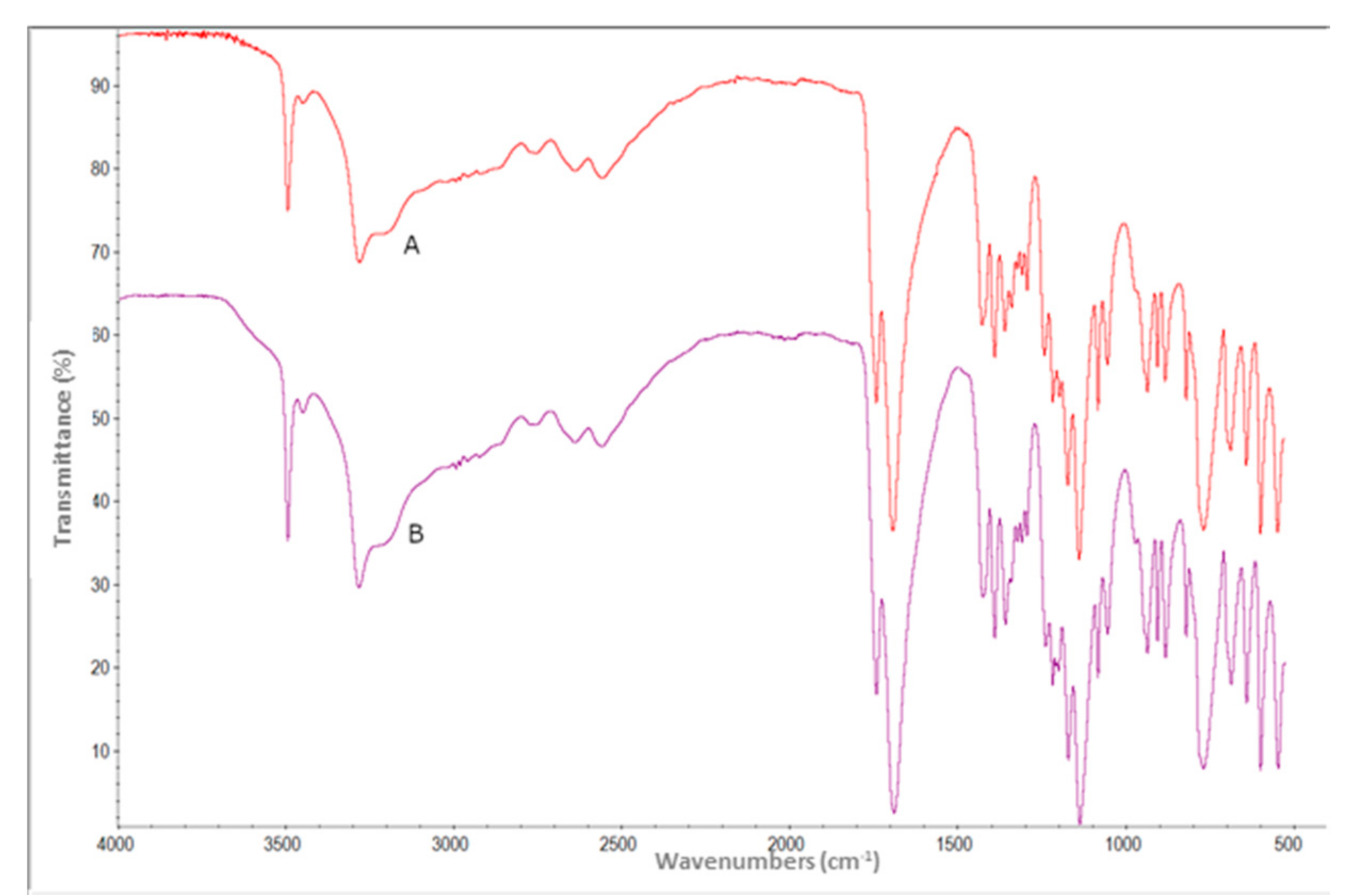
2.2. F.T.-IR Spectroscopy in Attenuated Total Reflectance (A.T.R.)
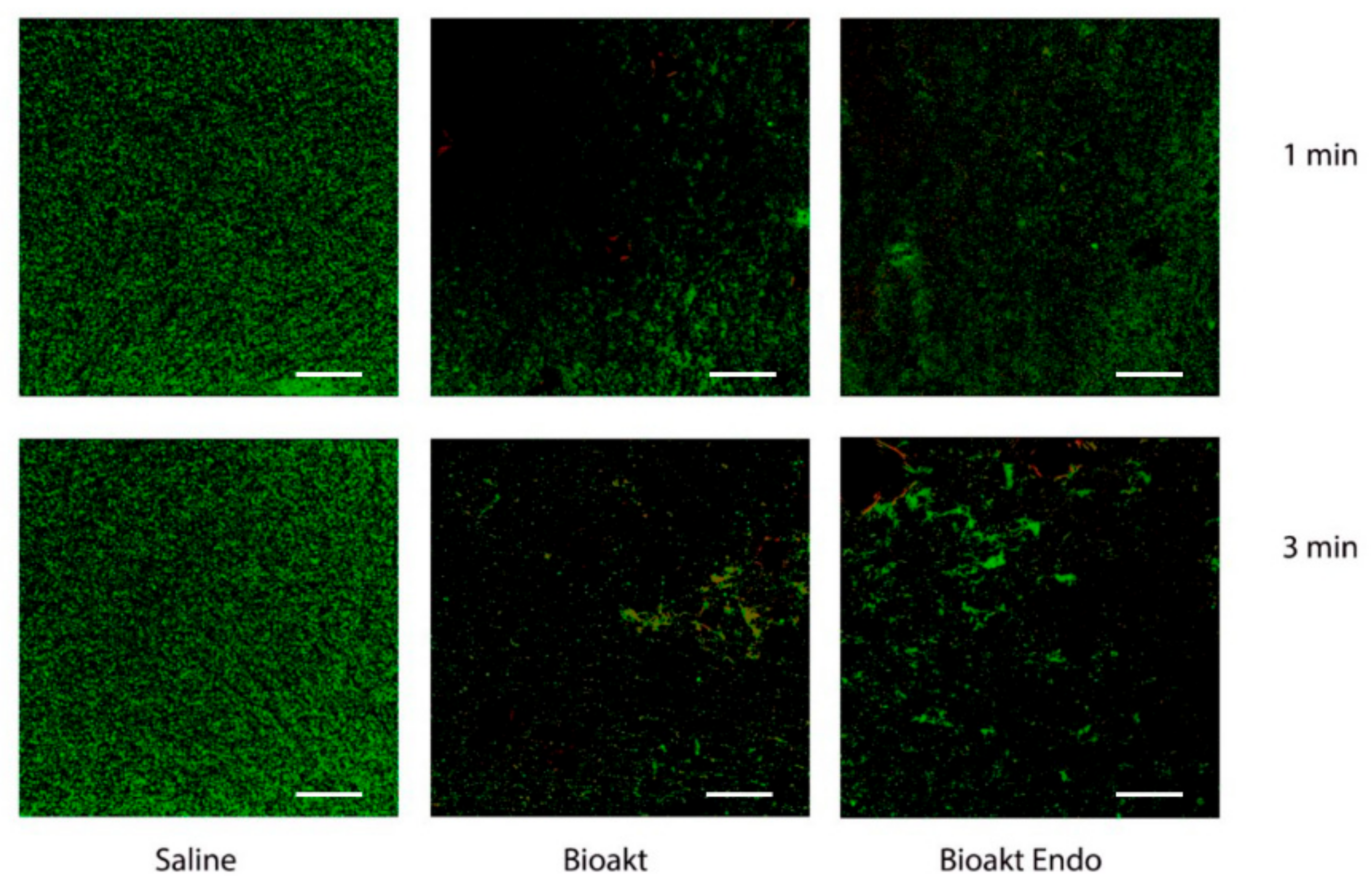
2.3. Assessment of Antimicrobial and Antibiofilm Activity
2.4. Statistical Analysis
3. Results
4. Discussion
5. Conclusions
Supplementary Materials
Author Contributions
Funding
Conflicts of Interest
References
- Zehnder, M. Root Canal Irrigants. J. Endod. 2006, 32, 389–398. [Google Scholar] [CrossRef] [PubMed]
- Peters, O.A.; Laib, A.; Göhring, T.N.; Barbakow, F. Changes in Root Canal Geometry after Preparation Assessed by High-Resolution Computed Tomography. J. Endod. 2001, 27, 1–6. [Google Scholar] [CrossRef] [PubMed]
- Nair, P.; Henry, S.; Cano, V.; Vera, J. Microbial status of apical root canal system of human mandibular first molars with primary apical periodontitis after “one-visit” endodontic treatment. Oral Surg. Oral Med. Oral Pathol. Oral Radiol. Endodontology 2005, 99, 231–252. [Google Scholar] [CrossRef]
- Giardino, L.; Del Fabbro, M.; Cesario, F.; Fernandes, F.S.; De Andrade, F.B. Antimicrobial effectiveness of combinations of oxidant and chelating agents in infected dentine: An ex vivo confocal laser scanning microscopy study. Int. Endod. J. 2017, 51, 448–456. [Google Scholar] [CrossRef] [PubMed]
- Dutner, J.; Mines, P.; Anderson, A. Irrigation Trends among American Association of Endodontists Members: A Web-based Survey. J. Endod. 2012, 38, 37–40. [Google Scholar] [CrossRef] [PubMed]
- Torabinejad, M.; Handysides, R.; Khademi, A.A.; Bakland, L.K. Clinical implications of the smear layer in endodontics: A review. Oral Surg. Oral Med. Oral Pathol. Oral Radiol. Endodontology 2002, 94, 658–666. [Google Scholar] [CrossRef] [PubMed]
- Scelza, M.F.; Teixeira, A.M.; Scelza, P. Decalcifying effect of EDTA-T, 10% citric acid, and 17% EDTA on root canal dentin. Oral Surg. Oral Med. Oral Pathol. Oral Radiol. Endodontology 2003, 95, 234–236. [Google Scholar] [CrossRef] [PubMed]
- Ostby, N. Chelation in root canal therapy: Ethylenediaminetetraacetic acid for cleansing and widening of root canals. Odontol. Tidskr. 1957, 65, 3–11. [Google Scholar]
- McComb, D.; Smith, D.C. A preliminary scanning electron microscopic study of root canals after endodontic procedures. J. Endod. 1975, 1, 238–242. [Google Scholar] [CrossRef]
- Arias-Moliz, M.T.; Ferrer-Luque, C.M.; Espigares-Rodríguez, E.; Liébana-Ureña, J.; Espigares-García, M. Bactericidal activity of phosphoric acid, citric acid, and EDTA solutions against Enterococcus faecalis. Oral Surg. Oral Med. Oral Pathol. Oral Radiol. Endodontology 2008, 106, e84–e89. [Google Scholar] [CrossRef]
- Giardino, L.; Ambu, E.; Becce, C.; Rimondini, L.; Morra, M. Surface Tension Comparison of Four Common Root Canal Irrigants and Two New Irrigants Containing Antibiotic. J. Endod. 2006, 32, 1091–1093. [Google Scholar] [CrossRef]
- Segura, J.J.; Calvo, J.R.; Guerrero, J.M.; Sampedro, C.; Jimenez, A.; Llamas, R. The disodium salt of EDTA inhibits the binding of vasoactive intestinal peptide to macrophage membranes: Endodontic implications. J. Endod. 1996, 22, 337–340. [Google Scholar] [CrossRef]
- Malheiros, C.; Marques, M.M.; Gavini, G. In Vitro Evaluation of the Cytotoxic Effects of Acid Solutions Used as Canal Irrigants. J. Endod. 2005, 31, 746–748. [Google Scholar] [CrossRef] [PubMed]
- Ballal, N.V.; Kundabala, M.; Bhat, S.; Rao, N.; Rao, B.S. A comparative in vitro evaluation of cytotoxic effects of EDTA and maleic acid: Root canal irrigants. Oral Surg. Oral Med. Oral Pathol. Oral Radiol. Endodontology 2009, 108, 633–638. [Google Scholar] [CrossRef] [PubMed]
- Botton, G.; Pires, C.W.; Cadoná, F.C.; Machado, A.K.; Azzolin, V.F.; Cruz, I.B.M.; Sagrillo, M.R.; Praetzel, J.R. Toxicity of irrigating solutions and pharmacological associations used in pulpectomy of primary teeth. Int. Endod. J. 2015, 49, 746–754. [Google Scholar] [CrossRef]
- Wayman, B.E.; Kopp, W.M.; Pinero, G.J.; Lazzari, E. Citric and lactic acids as root canal irrigants in vitro. J. Endod. 1979, 5, 258–265. [Google Scholar] [CrossRef]
- Qian, W.; Shen, Y.; Haapasalo, M. Quantitative Analysis of the Effect of Irrigant Solution Sequences on Dentin Erosion. J. Endod. 2011, 37, 1437–1441. [Google Scholar] [CrossRef]
- Arias-Moliz, M.T.; Ruiz-Linares, M.; Ferrer-Luque, C.M. Irrigating solutions in root canal treatment. Endod. Pract. Today 2019, 13, 131–146. [Google Scholar]
- Amaral, K.F.; Rogero, M.M.; Fock, R.A.; Borelli, P.; Gavini, G. Cytotoxicity analysis of EDTA and citric acid applied on murine resident macrophages culture. Int. Endod. J. 2007, 40, 338–343. [Google Scholar] [CrossRef]
- Chan, C.-P.; Jeng, J.-H.; Hsieh, C.-C.; Lin, C.-L.; Lei, D.; Chang, M.-C. Morphological alterations associated with the cytotoxic and cytostatic effects of citric acid on cultured human dental pulp cells. J. Endod. 1999, 25, 354–358. [Google Scholar] [CrossRef]
- Tomov, G.; Lamprianidis, T.; Zarra, T. Tissue damage after inadvertent citric acid extrusion during root canal treatment: Report of a case. Balk. J. Stom. 2013, 17, 101–106. [Google Scholar]
- Lan, W.C.; Lan, W.H.; Chan, C.P.; Hsieh, C.C.; Chang, M.C.; Jeng, J.-H. The effects of extracellular citric acid acidosis on the viability, cellular adhesion capacity and protein synthesis of cultured human gingival fibroblasts. Aust. Dent. J. 1999, 44, 123–130. [Google Scholar] [CrossRef] [PubMed] [Green Version]
- Tonini, R.; Giovarruscio, M.; Gorni, F.; Ionescu, A.; Brambilla, E.; Mikhailovna, I.M.; Luzi, A.; Pires, P.M.; Sauro, S. In Vitro Evaluation of Antibacterial Properties and Smear Layer Removal/Sealer Penetration of a Novel Silver-Citrate Root Canal Irrigant. Materials 2020, 13, 194. [Google Scholar] [CrossRef] [Green Version]
- Djokić, S. Treatment of various surfaces with silver and its compounds for topical wound dressings, catheter and other biomedical applications. ECS Trans. 2008, 11, 1–12. [Google Scholar] [CrossRef]
- BioAkt Ricerca e tecnologia tutta Italiana—Soluzioni Dentali. Available online: https://www.soluzionidentali.com/Resources/Soluzioni/BioAkt/bioakt.pdf (accessed on 2 April 2020).
- Palza, H. Antimicrobial Polymers with Metal Nanoparticles. Int. J. Mol. Sci. 2015, 16, 2099–2116. [Google Scholar] [CrossRef] [Green Version]
- Manuel, C.; Moore, M.D.; Jaykus, L.-A. Efficacy of a disinfectant containing silver dihydrogen citrate against GI.6 and GII.4 human norovirus. J. Appl. Microbiol. 2016, 122, 78–86. [Google Scholar] [CrossRef]
- Liau, S.Y.; Read, D.C.; Pugh, W.J.; Furr, J.R.; Russell, A.D. Interaction of silver nitrate with readily identifiable groups: Relationship to the antibacterialaction of silver ions. Lett. Appl. Microbiol. 1997, 25, 279–283. [Google Scholar] [CrossRef]
- Peters, O.A. Research that matters - biocompatibility and cytotoxicity screening. Int. Endod. J. 2013, 46, 195–197. [Google Scholar] [CrossRef]
- Bio Akt Endo. Available online: https://www.mc3.store/product-page/bio-akt-endo (accessed on 2 April 2020).
- International Standards Organization. Biological Evaluation of Medical Devices. Part 5: Tests for Cytotoxicity: In Vitro Methods; ISO 10993-Part 5; ISO: Geneva, Switzerland, 2009. [Google Scholar]
- Mosmann, T. Rapid colorimetric assay for cellular growth and survival: Application to proliferation and cytotoxicity assays. J. Immunol. Methods 1983, 65, 55–63. [Google Scholar] [CrossRef]
- Achá, V.; Naveau, H.; Meurens, M. Extractive sampling methods to improve the sensitivity of FTIR spectroscopy in analysis of aqueous liquids its decarbonation. Analusis 1998, 26, 157–163. [Google Scholar] [CrossRef] [Green Version]
- Stuart, C.H.; Schwartz, S.A.; Beeson, T.J.; Owatz, C.B. Enterococcus faecalis: Its Role in Root Canal Treatment Failure and Current Concepts in Retreatment. J. Endod. 2006, 32, 93–98. [Google Scholar] [CrossRef]
- Giardino, L.; Savadori, P.; Generali, L.; Mohammadi, Z.; Del Fabbro, M.; De Vecchi, E.; Bidossi, A. Antimicrobial effectiveness of etidronate powder (Dual Rinse® H.E.D.P.) and two EDTA preparations against Enterococcus faecalis: A preliminary laboratory study. Odontology 2020, 108, 396–405. [Google Scholar] [CrossRef]
- Clinical and Laboratory Standards Institute. Methods for Dilution Antimicrobial Susceptibility Tests for Bacteria That Grow Aerobically; Approved Standard, 9th ed.; C.L.S.I.: Waine, PA, USA, 2012. [Google Scholar]
- MBEC Assay® For High-Throughput Antimicrobial Susceptibility Testing of Biofilms. Available online: https://www.innovotech.ca/wp-content/uploads/2020/01/MBEC-Procedural-Manual-v2_1-3.pdf (accessed on 2 April 2020).
- Zhang, W.; Torabinejad, M.; Li, Y. Evaluation of Cytotoxicity of M.T.A.D. Using the MTT-Tetrazolium Method. J. Endod. 2003, 29, 654–657. [Google Scholar] [CrossRef]
- Masillamoni, C.R.M.; Kettering, J.D.; Torabinejad, M. The biocompatibility of some root canal medicaments and irrigants. Int. Endod. J. 1981, 14, 115–120. [Google Scholar] [CrossRef]
- Greulich, C.; Braun, D.; Peetsch, A.; Diendorf, J.; Siebers, B.; Epple, M.; Köller, M. The toxic effect of silver ions and silver nanoparticles towards bacteria and human cells occurs in the same concentration range. RSC Adv. 2012, 2, 6981–6987. [Google Scholar] [CrossRef]
- Kvitek, L.; Panacek, A.; Prucek, R.; Soukupova, J.; Vanickova, M.; Kolar, M.; Zbořil, R. Antibacterial activity and toxicity of silver – nanosilver versus ionic silver. J. Phys. Conf. Ser. 2011, 304, 012029. [Google Scholar] [CrossRef]
- Piao, M.J.; Kang, K.A.; Lee, I.K.; Kim, H.S.; Kim, S.; Choi, J.Y.; Choi, J.; Hyun, J.W. Silver nanoparticles induce oxidative cell damage in human liver cells through inhibition of reduced glutathione and induction of mitochondria-involved apoptosis. Toxicol. Lett. 2011, 201, 92–100. [Google Scholar] [CrossRef]
- Shim, I.; Choi, K.; Hirano, S. Oxidative stress and cytotoxic effects of silver ion in mouse lung macrophages J774.1 cells. J. Appl. Toxicol. 2016, 37, 471–478. [Google Scholar] [CrossRef]
- Morones-Ramirez, J.R.; Winkler, J.A.; Spina, C.S.; Collins, J.J. Silver Enhances Antibiotic Activity Against Gram-Negative Bacteria. Sci. Transl. Med. 2013, 5, 190ra81. [Google Scholar] [CrossRef] [Green Version]
- Guimarães, L.F.; Fidalgo, T.K.S.; Menezes, G.C.; Primo, L.G.; E Silva-Filho, F.C. Effects of citric acid on cultured human osteoblastic cells. Oral Surg. Oral Med. Oral Pathol. Oral Radiol. Endodontology 2010, 110, 665–669. [Google Scholar] [CrossRef]
- Saghiri, M.A.; Delvarani, A.; Mehrvarzfar, P.; Nikoo, M.; Lotfi, M.; Karamifar, K.; Asgar, K.; Dadvand, S. The impact of pH on cytotoxic effects of three root canal irrigants. Saudi Dent. J. 2011, 23, 149–152. [Google Scholar] [CrossRef] [Green Version]
- Johno, H.; Takahashi, S.; Kitamura, M. Influences of Acidic Conditions on Formazan Assay: A Cautionary Note. Appl. Biochem. Biotechnol. 2010, 162, 1529–1535. [Google Scholar] [CrossRef] [PubMed]
- Zehnder, M.; Schicht, O.; Sener, B.; Schmidlin, P. Reducing Surface Tension in Endodontic Chelator Solutions Has No Effect on Their Ability to Remove Calcium from Instrumented Root Canals. J. Endod. 2005, 31, 590–592. [Google Scholar] [CrossRef]
- Arata, A.B. Disinfectant and Method of Making. International Patent Application No. USOO6197814EB1; GB Patent Application No. 9/169,229; US. 6 March 2001. Available online: https://patentimages.storage.googleapis.com/d0/bc/e0/d0fbd8a4c7ce04/US6197814.pdf (accessed on 2 April 2020).
- Bae, E.-J.; Park, H.-J.; Park, J.-S.; Yoon, J.; Kim, Y.-H.; Choi, K.-H.; Yi, J. Effect of Chemical Stabilizers in Silver Nanoparticle Suspensions on Nanotoxicity. Bull. Korean Chem. Soc. 2011, 32, 613–619. [Google Scholar] [CrossRef] [Green Version]
- Arechabala, B.; Coiffard, C.; Rivalland, P.; Coiffard, L.J.M.; De Roeck-Holtzhauer, Y. Comparison of cytotoxicity of various surfactants tested on normal human fibroblast cultures using the neutral red test, M.T.T. assay and L.D.H. release. J. Appl. Toxicol. 1999, 19, 163–165. [Google Scholar] [CrossRef]
- Hennequin, M.; Pajot, J.; Avignant, D. Effects of different pH values of citric acid solutions on the calcium and phosphorus contents of human root dentin. J. Endod. 1994, 20, 551–554. [Google Scholar] [CrossRef]
- Haznedaroğlu, F. Efficacy of various concentrations of citric acid at different pH values for smear layer removal. Oral Surg. Oral Med. Oral Pathol. Oral Radiol. Endodontology 2003, 96, 340–344. [Google Scholar] [CrossRef]
- Machado-Silveiro, L.F.; González-López, S.; González-Rodríguez, M.P. Decalcification of root canal dentine by citric acid, EDTA and sodium citrate. Int. Endod. J. 2004, 37, 365–369. [Google Scholar] [CrossRef]
- Götze, G.D.R.; Cunha, C.B.C.S.; Primo, L.S.D.S.G.; Maia, L.C. Effect of the sodium hypochlorite and citric acid association on smear layer removal of primary molars. Braz. Oral Res. 2005, 19, 261–266. [Google Scholar] [CrossRef]
- Reis, C.; De-Deus, G.; Leal, F.; Azevedo, E.; Coutinho-Filho, T.; Paciornik, S. Strong effect on dentin after the use of high concentrations of citric acid: An assessment with co-site optical microscopy and E.S.E.M. Dent. Mater. 2008, 24, 1608–1615. [Google Scholar] [CrossRef]
- Demirel, A.; Yüksel, B.N.; Ziya, M.; Gümüş, H.; Doğan, S.; Sari, S. The effect of different irrigation protocols on smear layer removal in root canals of primary teeth: A S.E.M. study. Acta Odontol. Scand. 2019, 77, 380–385. [Google Scholar] [CrossRef]
- Garberoglio, R.; Becce, C. Smear layer removal by root canal irrigants. A comparative scanning electron microscopic study. Oral. Surg. Oral. Med. Oral. Pathol. 1994, 78, 359–367. [Google Scholar] [CrossRef]
- Shokrzadeh, M.; Modanloo, M. An overview of the most common methods for assessing cell viability. J. Res. Med Dent. Sci. 2017, 5, 33. [Google Scholar] [CrossRef]
- Riss, T.; Niles, A.; Moravec, R.; Karassina, N.; Vidugiriene, J. Cytotoxicity Assays: In Vitro Methods to Measure Dead Cells. In Assay Guidance Manual; Eli Lilly & Company and the National Center for Advancing Translational Sciences: Bethesda, MD, USA, 2020; pp. 296–302. [Google Scholar]
- International Standards Organization. Biological Evaluation of Medical Devices. Tests for Irritation and Skin Sensitization ISO 10993-Part 10; ISO: Geneva, Switzerland, 2010. [Google Scholar]
- International Standards Organization. Biological Evaluation of Medical Devices. Part 2: Animal Welfare Requirements. ISO 10993-Part 2; ISO: Geneva, Switzerland, 2006. [Google Scholar]
- Giardino, L.; Bidossi, A.; Del Fabbro, M.; Savadori, P.; Maddalone, M.; Ferrari, L.; Ballal, N.V.; Das, S.; Rao, B.S.S. Antimicrobial activity, toxicity and accumulated hard-tissue debris (A.H.T.D.) removal efficacy of several chelating agents. Int. Endod. J. 2020, 53, 1093–1110. [Google Scholar] [CrossRef]
- Giardino, L.; De Andrade, F.B.; Beltrami, R. Antimicrobial Effect and Surface Tension of Some Chelating Solutions with Added Surfactants. Braz. Dent. J. 2016, 27, 584–588. [Google Scholar] [CrossRef] [PubMed] [Green Version]
- Bates, R.G. Measurement of Effect of Dilution upon pH. Anal. Chem. 1954, 26, 871–874. [Google Scholar] [CrossRef]
- Buchanan, R.L.; Golden, M.H. Interaction of Citric Acid Concentration and pH on the Kinetics of Listeria monocytogenes Inactivation. J. Food Prot. 1994, 57, 567–570. [Google Scholar] [CrossRef]


| Concentration | BioAKT | BioAKT Endo | p-Value |
|---|---|---|---|
| Control | 1.29 ± 0.04 (1.22, 1.35) | 1.25 ± 0.12 (1.06, 1.45) | 0.89 |
| 0.25% | 0.98 ± 0.04 (0.91, 1.05) | 1.05 ± 0.07 (0.95, 1.16) | 0.34 |
| 0.5% | 0.95 ± 0.07 (0.85, 1.06) | 0.95 ± 0.07 (0.83, 1.06) | 1.00 |
| 1.0% | 0.54 ± 0.06 (0.43, 0.64) | 0.53 ± 0.02 (0.49, 0.57) | 1.00 |
| 2.5% | 0.067 ± 0.002 (0.06, 0.08) | 0.070 ± 0.004 (0.06, 0.08) | 0.20 |
| 5.0% | 0.072 ± 0.001 (0.070, 0.074) | 0.077 ± 0.006 (0.068, 0.086 | 0.11 |
| Tukey’s Multiple Comparison Test | Mean Diff, | q | Significance | 95% CI of Diff |
|---|---|---|---|---|
| 0.25% vs. CONTROL | −0.3060 | 13.74 | *** | −0.4061 to −0.2059 |
| 0.5% vs. CONTROL | −0.3352 | 15.06 | *** | −0.4353 to −0.2352 |
| 1% vs. CONTROL | −0.7495 | 33.66 | *** | −0.8496 to −0.6494 |
| 2.5% vs. CONTROL | −1.219 | 54.76 | *** | −1.319 to −1.119 |
| 5% vs. CONTROL | −1.214 | 54.53 | *** | −1.314 to −1.114 |
| 0.25% vs. 0.5% | 0.02924 | 1.313 | ns | −0.07083 to 0.1293 |
| 0.25% vs. 1% | 0.4435 | 19.92 | *** | 0.3434 to 0.5436 |
| 0.25% vs. 2.5% | 0.9131 | 41.01 | *** | 0.8130 to 1.013 |
| 0.25% vs. 5% | 0.9080 | 40.79 | *** | 0.8080 to 1.008 |
| 0.5% vs. 1% | 0.4142 | 18.61 | *** | 0.3142 to 0.5143 |
| 0.5% vs. 2.5% | 0.8838 | 39.70 | *** | 0.7838 to 0.9839 |
| 0.5% vs. 5% | 0.8788 | 39.47 | *** | 0.7787 to 0.9789 |
| 1% vs. 2.5% | 0.4696 | 21.09 | *** | 0.3695 to 0.5697 |
| 1% vs. 5% | 0.4646 | 20.87 | *** | 0.3645 to 0.5646 |
| 2.5% vs. 5% | −0.005032 | 0.2260 | ns | −0.1051 to 0.09504 |
| Tukey’s Multiple Comparison Test | Mean Diff, | q | Significance | 95% CI of Diff |
|---|---|---|---|---|
| 0.25% vs. CONTROL | −0.2003 | 6.165 | ** | −0.3463 to −0.05426 |
| 0.5% vs. CONTROL | −0.3029 | 9.324 | *** | −0.4490 to −0.1569 |
| 1% vs. CONTROL | −0.7252 | 22.32 | *** | −0.8712 to −0.5791 |
| 2.5% vs. CONTROL | −1.183 | 36.40 | *** | −1.329 to −1.037 |
| 5% vs. CONTROL | −1.175 | 36.18 | *** | −1.321 to −1.029 |
| 0.25% vs. 0.5% | 0.1027 | 3.160 | ns | −0.04339 to 0.2487 |
| 0.25% vs. 1% | 0.5249 | 16.16 | *** | 0.3788 to 0.6709 |
| 0.25% vs. 2.5% | 0.9825 | 30.24 | *** | 0.8364 to 1.129 |
| 0.25% vs. 5% | 0.9751 | 30.01 | *** | 0.8290 to 1.121 |
| 0.5% vs. 1% | 0.4222 | 13.00 | *** | 0.2762 to 0.5683 |
| 0.5% vs. 2.5% | 0.8798 | 27.08 | *** | 0.7338 to 1.026 |
| 0.5% vs. 5% | 0.8724 | 26.85 | *** | 0.7264 to 1.018 |
| 1% vs. 2.5% | 0.4576 | 14.08 | *** | 0.3115 to 0.6036 |
| 1% vs. 5% | 0.4502 | 13.86 | *** | 0.3042 to 0.5962 |
| 2.5% vs. 5% | −0.007384 | 0.2273 | ns | −0.1534 to 0.1387 |
| Solutions | Dilution | |||
|---|---|---|---|---|
| MIC | MBC | MBEC 1 min | M.B.E.C. 3 min | |
| BioAKT | 1:16 | 1:8 | 1:8 | 1:16 |
| BioAKT Endo | 1:16 | 1:8 | 1:8 | 1:16 |
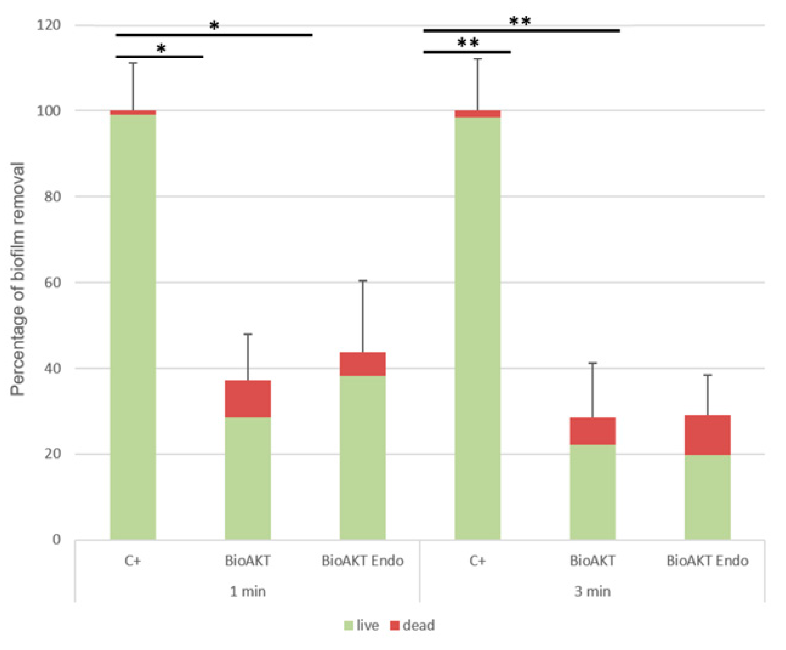
| Solutions | 1 min | 3 min |
|---|---|---|
| Control vs. Bioakt | ns | ns |
| Control vs. BioAKT Endo | ns | * |
| Bioakt vs. BioAKT Endo | ns | ns |
Publisher’s Note: MDPI stays neutral with regard to jurisdictional claims in published maps and institutional affiliations. |
© 2020 by the authors. Licensee MDPI, Basel, Switzerland. This article is an open access article distributed under the terms and conditions of the Creative Commons Attribution (CC BY) license (http://creativecommons.org/licenses/by/4.0/).
Share and Cite
Generali, L.; Bertoldi, C.; Bidossi, A.; Cassinelli, C.; Morra, M.; Del Fabbro, M.; Savadori, P.; Ballal, N.V.; Giardino, L. Evaluation of Cytotoxicity and Antibacterial Activity of a New Class of Silver Citrate-Based Compounds as Endodontic Irrigants. Materials 2020, 13, 5019. https://doi.org/10.3390/ma13215019
Generali L, Bertoldi C, Bidossi A, Cassinelli C, Morra M, Del Fabbro M, Savadori P, Ballal NV, Giardino L. Evaluation of Cytotoxicity and Antibacterial Activity of a New Class of Silver Citrate-Based Compounds as Endodontic Irrigants. Materials. 2020; 13(21):5019. https://doi.org/10.3390/ma13215019
Chicago/Turabian StyleGenerali, Luigi, Carlo Bertoldi, Alessandro Bidossi, Clara Cassinelli, Marco Morra, Massimo Del Fabbro, Paolo Savadori, Nidambur Vasudev Ballal, and Luciano Giardino. 2020. "Evaluation of Cytotoxicity and Antibacterial Activity of a New Class of Silver Citrate-Based Compounds as Endodontic Irrigants" Materials 13, no. 21: 5019. https://doi.org/10.3390/ma13215019