
Effect of Heat Treatment on Structural, Magnetic and Electrical Properties of La2FeMnO6


Abstract
:1. Introduction
2. Materials and Methods
3. Results and Discussion
3.1. Effect of Calcination Temperature on Structural and Magnetic Properties
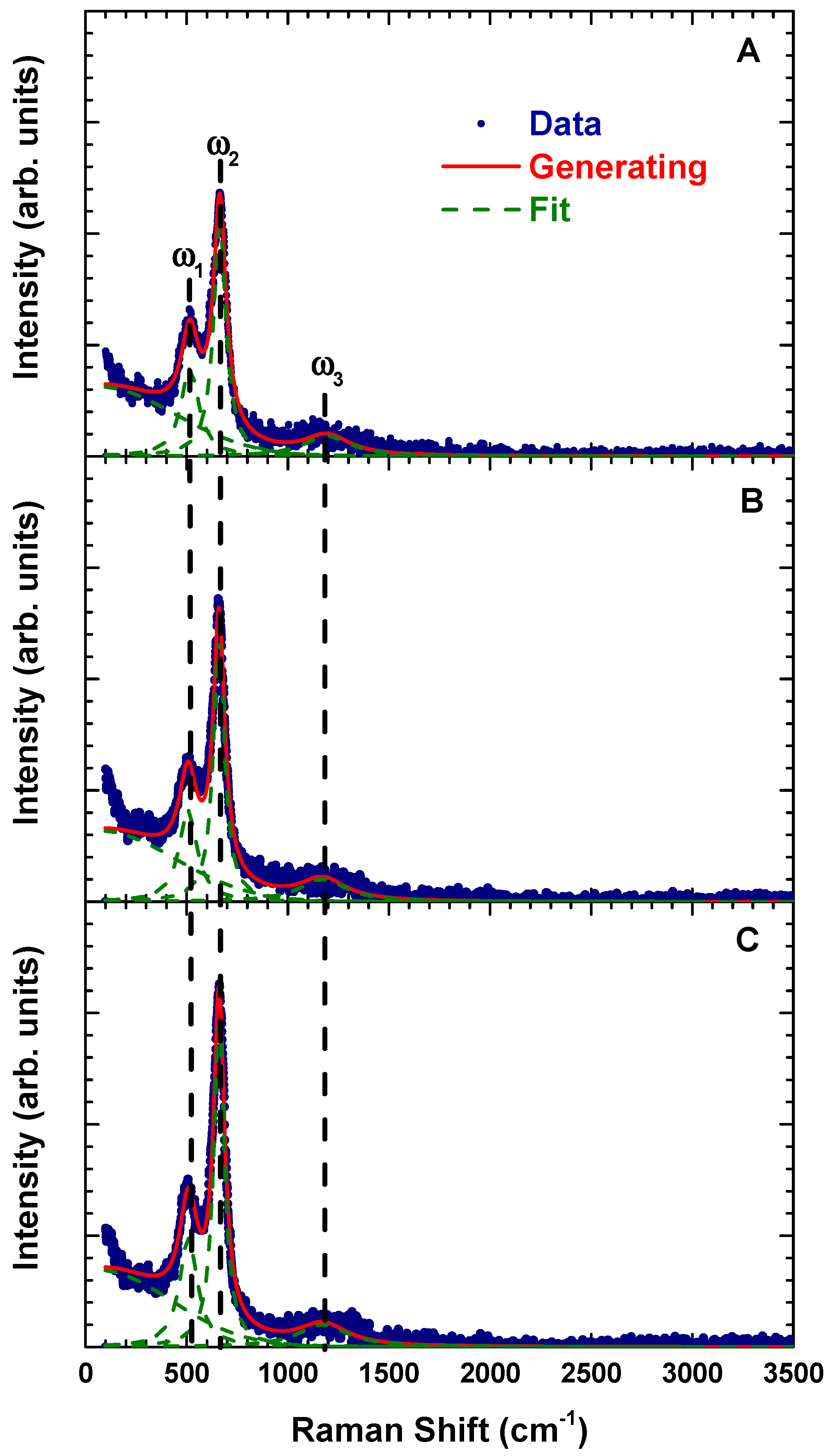
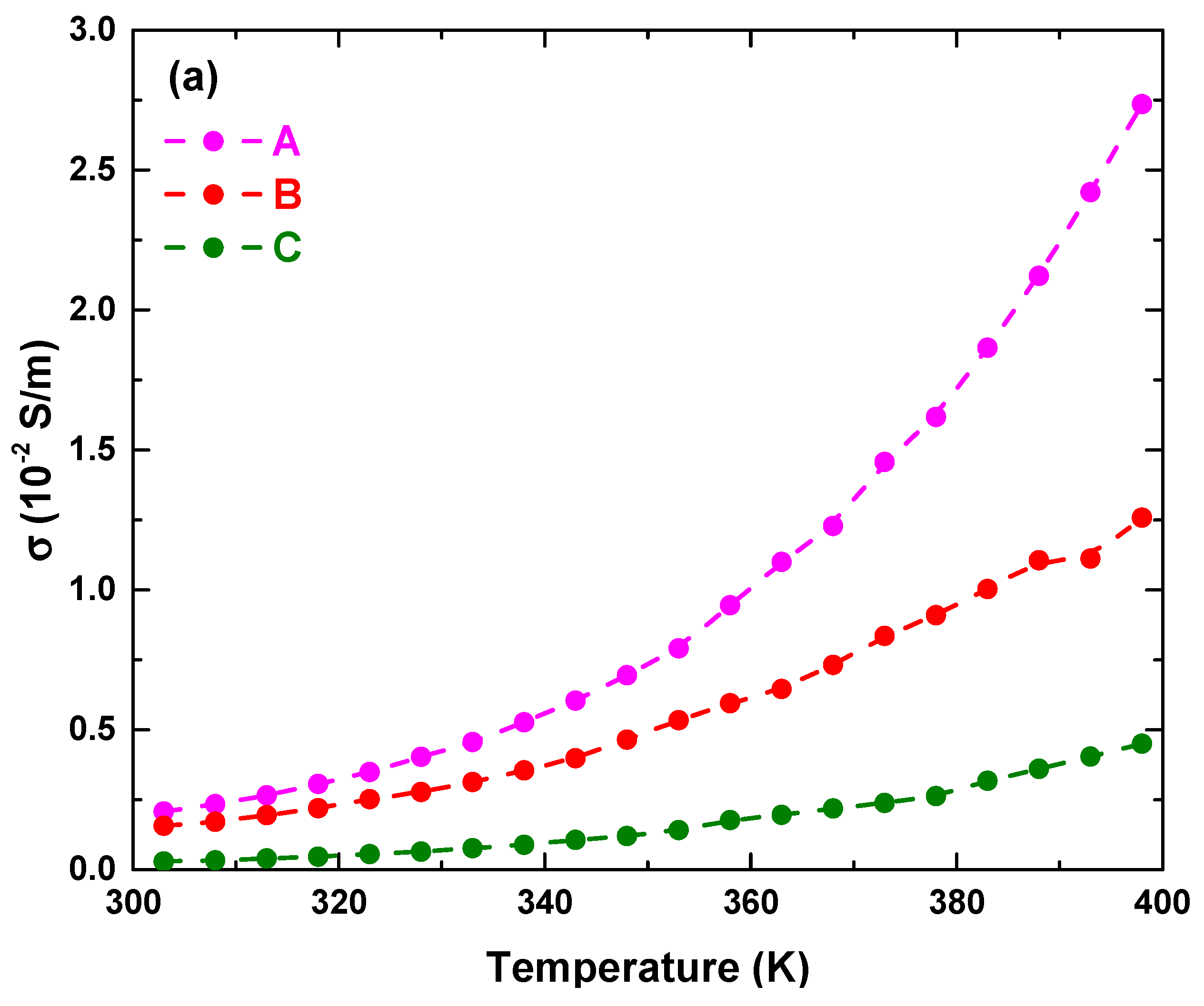
3.2. Effect of Calcination and Sintering Conditions on the Structural and Electrical Properties La2FeMnO6
4. Conclusions
Author Contributions
Funding
Institutional Review Board Statement
Informed Consent Statement
Data Availability Statement
Conflicts of Interest
References
- Qian, Y.; Wu, H.; Kan, E.; Lu, J.; Lu, R.; Liu, Y.; Tan, W.; Xiao, C.; Deng, K. Biaxial strain effect on the electronic and magnetic phase transitions in double perovskite La2FeMnO6: A first-principles study. J. Appl. Phys. 2013, 114, 063713. [Google Scholar] [CrossRef]
- Kobayashi, K.-I.; Kimura, T.; Sawada, H.; Terakura, K.; Tokura, Y. Room-temperature magnetoresistance in an oxide mate-rial with an ordered double—Perovskite structure. Nature 1998, 395, 677–680. [Google Scholar] [CrossRef]
- Palakkal, J.P.; Sankar, C.R.; Paulose, A.P.; Varma, M.R. Hopping conduction and spin glass behavior of La2FeMnO. J. Alloy. Compd. 2018, 743, 403–409. [Google Scholar] [CrossRef]
- Dhilip, M.; Devi, N.A.; Punitha, J.S.; Anbarasu, V.; Kumar, K.S. Conventional synthesis and characterization of cubically ordered La2FeMnO6 double perovskite compound. Vacuum 2019, 167, 16–20. [Google Scholar] [CrossRef]
- Filho, J.D.A.; de Araújo, J.; Morales, M.; Firme, C.; de Oliveira, J. Exchange bias and spin glass in La2FeMnO6 nanoparticles. J. Magn. Magn. Mater. 2019, 471, 177–184. [Google Scholar] [CrossRef]
- Yang, D.; Yang, T.; Mukherjee, P.; Dutton, S.E.; Huo, D.; Carpenter, M.A. Strain coupling and acoustic attenuation associated with glassy magnetic phase transitions in the disordered double perovskite La2FeMnO. Phys. Rev. B 2019, 99, 094314. [Google Scholar] [CrossRef]
- Li, N.; Fan, F.; Sun, F.; Wang, Y.; Zhao, Y.; Liu, F.; Zhang, Q.; Ikuta, D.; Xiao, Y.; Chow, P.; et al. Pressure-enhanced interplay between lattice, spin, and charge in the mixed perovskite La2FeMnO. Phys. Rev. B 2019, 99, 195115. [Google Scholar] [CrossRef] [Green Version]
- Palakkal, J.P.; Neenu Lekshmi, P.; Thomas, S.; Suresh, K.G.; Varma, M.R. Observation of high-temperature magnetic transi-tion and existence of ferromagnetic short-range correlations above transition in double perovskite La2FeMnO. RSC Adv. 2015, 5, 105531–105536. [Google Scholar] [CrossRef]
- Barrozo, P.; Moreno, N.O.; Albino Aguiar, J. Ferromagnetic cluster on La2FeMnO. Adv. Mater. Res. 2014, 975, 122–127. [Google Scholar] [CrossRef]
- Verma, R.; Chauhan, A.; Neha; Batoo, K.M.; Kumar, R.; Hadhi, M.; Raslan, E.H. Effect of calcination temperature on struc-tural and morphological properties of bismuth ferrite nanoparticles. Ceram. Int. 2021, 47, 3680–3691. [Google Scholar] [CrossRef]
- Nasir, M.; Khan, M.; Agbo, S.A.; Bhatt, S.; Kumar, S.; Sen, S. Evidence of cluster-glass and Griffiths-like phases in partially ordered La2FeMnO6 double perovskite. J. Phys. D Appl. Phys. 2020, 53, 375003. [Google Scholar] [CrossRef]
- Ramirez, M.O.; Krishnamurthi, M.; Denev, S.; Kumar, A.; Yang, S.-Y.; Chu, Y.-H.; Saiz, E.; Seidel, J.; Pyatakov, A.P.; Bush, A.; et al. Two-phonon coupling to the antiferromagnetic phase transition in mul-tiferroic BiFeO. Appl. Phys. Lett. 2008, 92, 022511. [Google Scholar] [CrossRef]
- Wiranwetchayan, O.; Promnopas, S.; Phadungdhitidhada, S.; Phuruangrat, A.; Thongtem, T.; Singjai, P.; Thongtem, S. Char-acterization of perovskite LaFeO3 synthesized by microwave plasma method for photocatalytic applications. Ceram. Int. 2019, 45, 4802–4809. [Google Scholar] [CrossRef]
- Cao, E.; Yang, Y.; Cui, T.; Zhang, Y.; Hao, W.; Sun, L.; Peng, H.; Deng, X. Effect of synthesis route on electrical and ethanol sensing characteristics for LaFeO3-δ nanoparticles by citric sol-gel method. Appl. Surf. Sci. 2017, 393, 134–143. [Google Scholar] [CrossRef]
- Gao, F.; Cai, C.; Wang, Y.; Dong, S.; Qiu, X.Y.; Yuan, G.L.; Liu, Z.G.; Liu, J.-M. Preparation of La-doped BiFeO3 thin films with Fe2+ ions on Si substrates. J. Appl. Phys. 2006, 99, 094105. [Google Scholar] [CrossRef] [Green Version]
- Menezes, P.W.; Indra, A.; Gutkin, V.; Driess, M. Bosting electrochemical water oxidation through replacement of Oh Co sites in cobalt oxide spinel with manganese. Chem. Commun. (Camb.) 2017, 53, 8018–8021. [Google Scholar] [CrossRef] [PubMed]
- Xiao, P.; Zhong, L.; Zhu, J.; Hong, J.; Li, J.; Li, H.; Zhu, Y. CO and soot oxidation over macroporous perovskite LaFeO. Catal. Today 2015, 258, 660–667. [Google Scholar] [CrossRef]
- Singh, M.K.; Ryu, S.; Jang, H.M. Polarized Raman scattering of multiferroic BiFeO3 thin films with pseu-do-tetragonal symmetry. Phys. Rev. B 2005, 72, 132101. [Google Scholar] [CrossRef] [Green Version]
- Dhiman, I.; Das, A.; Nigam, A.K.; Gasser, U. Influence of B-site disorderer in La0.5Ca0.5Mn1-xBxO3 (B = Fe, Ru, Al, and Ga) manganites. J. Phys. Condens. Matter 2011, 23, 246006. [Google Scholar] [CrossRef] [PubMed] [Green Version]
- Triyono, D.; Fitria, S.N.; Hanifah, U. Dielectric analysis and electrical conduction mechanism of La1−xBixFeO3 ceramics. RSC Adv. 2020, 10, 18323–18338. [Google Scholar] [CrossRef]


| Parameter | Calcination Temperature (K) | |
|---|---|---|
| 1023 | 1173 | |
| Lattice parameter (Å) | ||
| a | 5.485(1) | 5.507(3) |
| b | 7.778(2) | 7.805(5) |
| c | 5.536(1) | 5.523(2) |
| Volume unit cell (Å3) | 236.2 | 237.4 |
| Average crystallite size (nm) | 213.3 | 325.6 |
| Atomic position (x, y, z) | ||
| La | (0.48, 0.25, 0) | (0.48, 0.25, 0) |
| Fe/Mn | (0, 0, 0) | (0, 0, 0) |
| O1 | (0.28, 0.54, 0.23) | (0.28, 0.54, 0.23) |
| O2 | (0.01, 0.25, 0.05) | (0.01, 0.25, 0.05) |
| Wickoff position | ||
| La | 4c | 4c |
| Fe/Mn | 4a | 4a |
| O1 | 8d | 8d |
| O2 | 4c | 4c |
| Bond distance (Å) | ||
| Fe/Mn–O1 (s) | 1.946 | 1.946 |
| Fe/Mn–O2 (m) | 1.965 | 1.972 |
| Fe/Mn–O1 (l) | 2.019 | 2.022 |
| <Fe/Mn–O> | 1.977 | 1.980 |
| Bond angle (°) | ||
| Fe/Mn–O1–Fe/Mn | 158.652 | 158.612 |
| Fe/Mn–O2–Fe/Mn | 163.486 | 163.577 |
| < Fe/Mn–O–Fe/Mn> | 161.069 | 161.095 |
| Tilt angle (°) | 11.595 | 11.579 |
| Orthorhombic distortion | 0.1392 | 0.1390 |
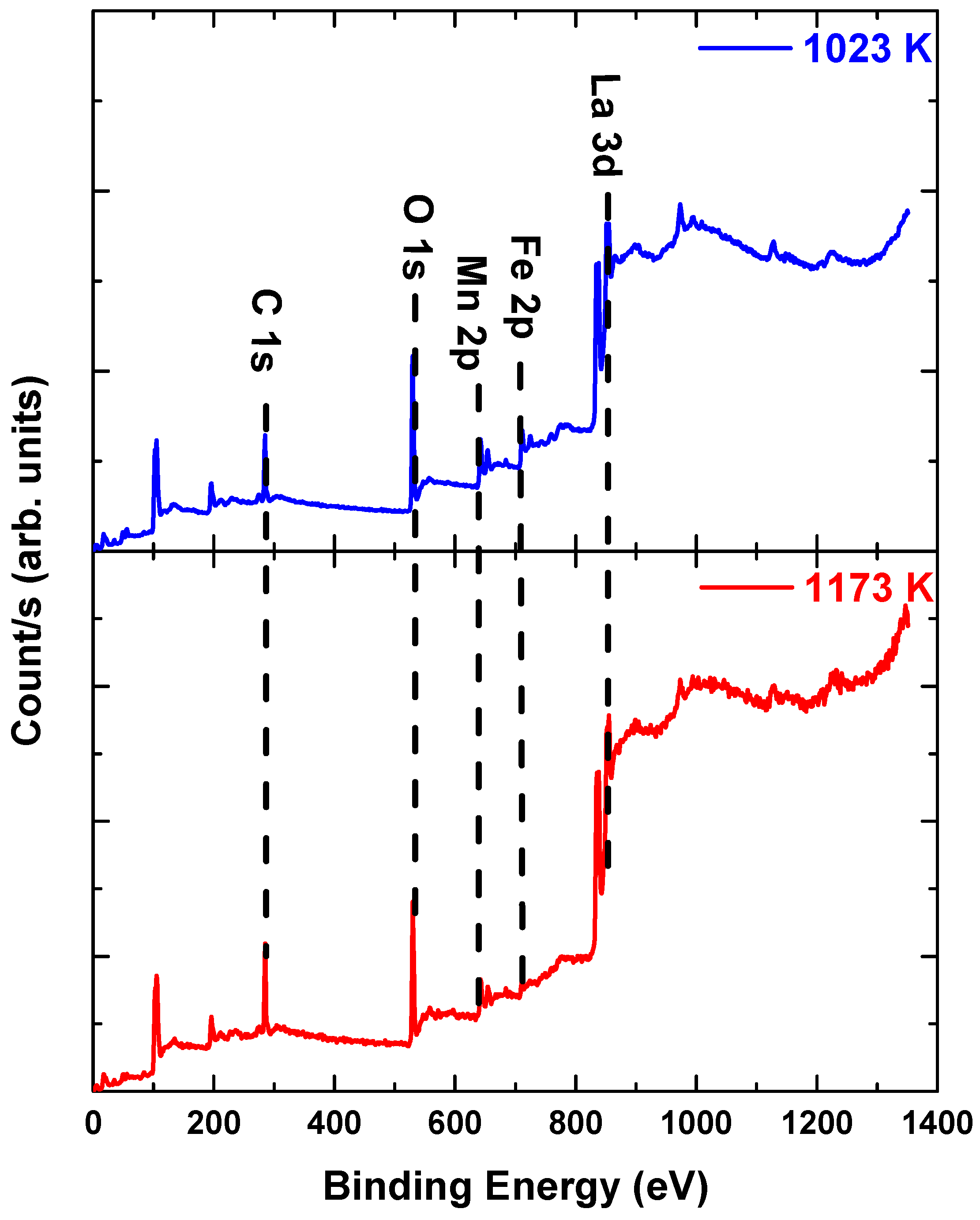
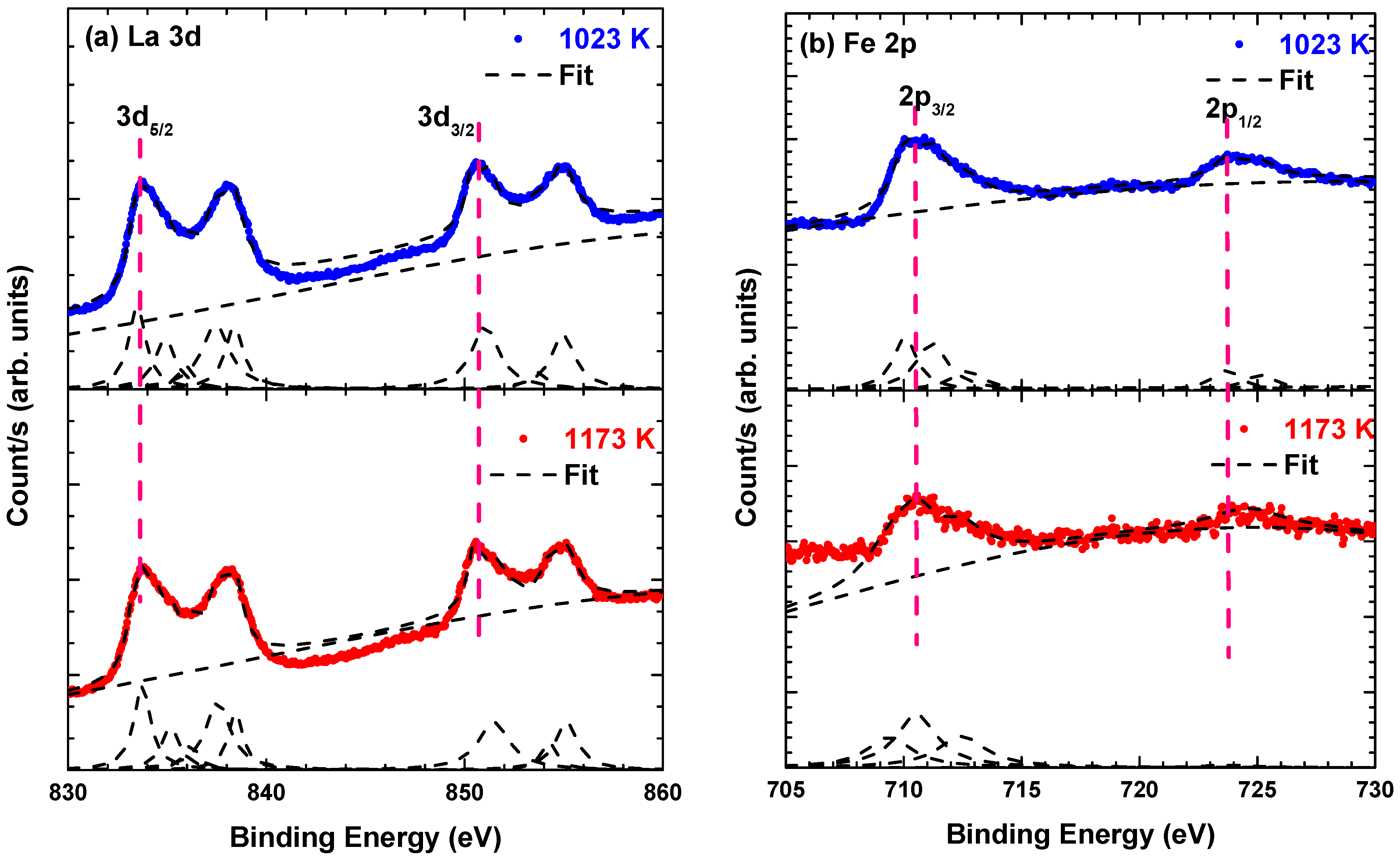
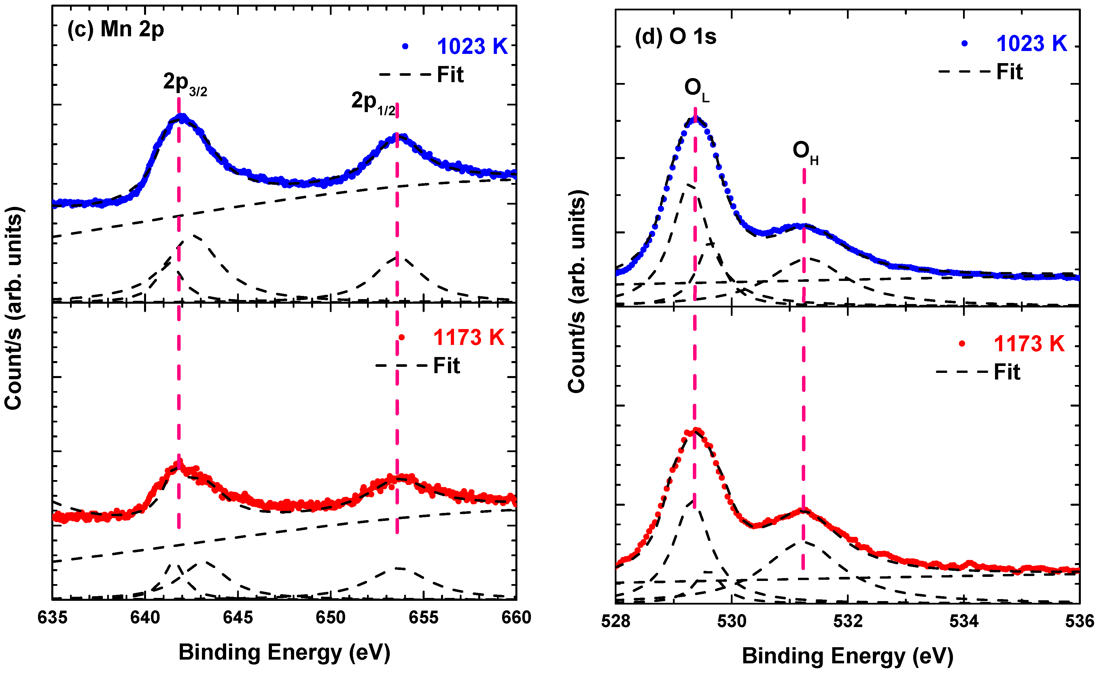
| Calcination Temperature (K) | Binding Energy (eV) | |||||||||||
|---|---|---|---|---|---|---|---|---|---|---|---|---|
| Fe3+ | Fe2+ | Fe4+ | Mn3+ | Mn4+ | OL | OV | OH | |||||
| 2p3/2 | 2p1/2 | 2p3/2 | 2p1/2 | |||||||||
| 1023 | 710.05 | 723.71 | - | 711.19 | 712.68 | 725.13 | 641.31 | 653.65 | 642.57 | 529.3 | 529.8 | 531.3 |
| 1173 | 710.54 | 723.75 | 709.39 | - | 712.76 | 725.19 | 641.25 | 653.56 | 642.14 | 529.6 | 530.0 | 531.7 |
| Caption | Calcination Temperature (K) | |
|---|---|---|
| 1023 | 1173 | |
| Surface atomic composition (%) | ||
| La 3d | 10.2 | 9.76 |
| Fe 2p | 5.18 | 2.30 |
| Mn 2p | 6.27 | 4.34 |
| O 1s | 41.9 | 36.1 |
| C 1s | 36.5 | 47.5 |
| Atomic ratio | ||
| Fe/La | 0.509 | 0.236 |
| Mn/La | 0.617 | 0.445 |
| OH/O | 0.651 | 0.558 |
| Ionic ratio | ||
| Mn3+/(Mn3+ + Mn4+) | 0.475 | 0.609 |
| Fe4+/(Fe2+ + Fe3+ + Fe4+) | 0.589 | 0.418 |
| Calcination Condition (K) | Ms (emu/g) | Mr (emu/g) | Hc (Oe) |
|---|---|---|---|
| 1023 | 0.59 | 0.75 × 10⁻² | 147.7 |
| 1173 | 1.09 | 0.16 | 239.9 |
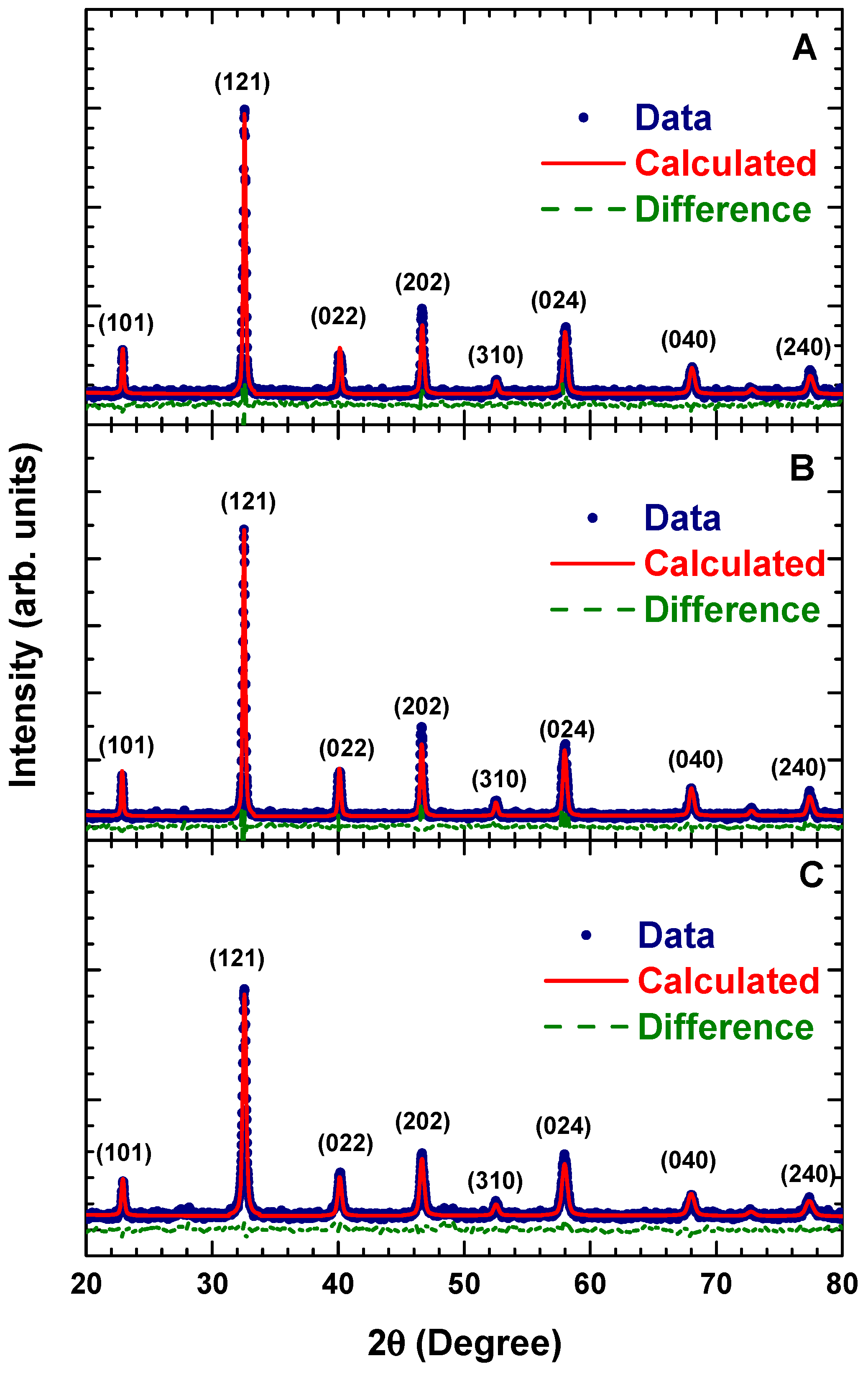
| Parameter | Condition | ||
|---|---|---|---|
| A | B | C | |
| Lattice parameter (Å) | |||
| a | 5.499(3) | 5.503(3) | 5.518(2) |
| b | 7.787(4) | 7.792(4) | 7.811(6) |
| c | 5.533(3) | 5.535(2) | 5.531(3) |
| Volume unit cell (Å3) | 236.9 | 237.3 | 238.4 |
| Average crystallite size (nm) | 664.5 | 736.6 | 393.3 |
| Atomic position (x, y, z) | |||
| La | (0.48, 0.25, 0) | (0.48, 0.25, 0) | (0.48, 0.25, 0) |
| Fe/Mn | (0, 0, 0) | (0, 0, 0) | (0, 0, 0) |
| O1 | (0.28, 0.54, 0.23) | (0.28, 0.54, 0.23) | (0.28, 0.54, 0.23) |
| O2 | (0.01, 0.25, 0.05) | (0.01, 0.25, 0.05) | (0.01, 0.25, 0.05) |
| Wickoff position | |||
| La | 4c | 4c | 4c |
| Fe/Mn | 4a | 4a | 4a |
| O1 | 8d | 8d | 8d |
| O2 | 4c | 4c | 4c |
| Bond distance (Å) | |||
| Fe/Mn—O1 (s) | 1.947 | 1.948 | 1.950 |
| Fe/Mn—O2 (m) | 1.967 | 1.968 | 1.973 |
| Fe/Mn—O1 (l) | 2.022 | 2.023 | 2.026 |
| <Fe/Mn—O> | 1.978 | 1.980 | 1.983 |
| Bond angle (°) | |||
| Fe/Mn—O1—Fe/Mn | 158.650 | 158.648 | 158.626 |
| Fe/Mn—O2—Fe/Mn | 163.512 | 163.516 | 163.565 |
| Tilt angle (°) | 14.396 | 11.587 | 11.579 |
| Orthorhombic distortion | 0.1387 | 0.1386 | 0.1384 |
Publisher’s Note: MDPI stays neutral with regard to jurisdictional claims in published maps and institutional affiliations. |
© 2021 by the authors. Licensee MDPI, Basel, Switzerland. This article is an open access article distributed under the terms and conditions of the Creative Commons Attribution (CC BY) license (https://creativecommons.org/licenses/by/4.0/).
Share and Cite
Triyono, D.; Yunida, Y.; Rafsanjani, R.A. Effect of Heat Treatment on Structural, Magnetic and Electrical Properties of La2FeMnO6. Materials 2021, 14, 7501. https://doi.org/10.3390/ma14247501
Triyono D, Yunida Y, Rafsanjani RA. Effect of Heat Treatment on Structural, Magnetic and Electrical Properties of La2FeMnO6. Materials. 2021; 14(24):7501. https://doi.org/10.3390/ma14247501
Chicago/Turabian StyleTriyono, Djoko, Y Yunida, and Rifqi Almusawi Rafsanjani. 2021. "Effect of Heat Treatment on Structural, Magnetic and Electrical Properties of La2FeMnO6" Materials 14, no. 24: 7501. https://doi.org/10.3390/ma14247501