Abstract
Red–green–blue phosphors excited by ultraviolet (UV) radiation for white light LEDs have received much attention to improve the efficiency, color rendering index (CRI), and chromatic stability. The spectral conversion of a rare-earth ion-doped nonstoichiometric LaO0.65F1.7 host was explored with structural analysis in this report. The nonstoichiometric structure of a LaO0.65F1.7 compound, synthesized by a solid-state reaction using La2O3 and excess NH4F precursors, was analyzed by synchrotron X-ray powder diffraction. The crystallized LaO0.65F1.7 host, which had a tetragonal space group of P4/nmm, contained 9- and 10-coordinated La3+ sites. Optical materials composed of La1−p−qBipTbqO0.65F1.7 (p = 0 and 0.01; q = 0–0.2) were prepared at 1050 °C for 2 h, and the single phase of the obtained phosphors was indexed by X-ray diffraction analysis. The photoluminescence spectra of the energy transfer from Bi3+ to Tb3+ were obtained upon excitation at 286 nm in the nonstoichiometric host lattice. The desired Commission Internationale de l’Eclairage (CIE) values of the phosphors were calculated. The intense green La0.89Bi0.01Tb0.1O0.65F1.7 phosphor with blue and red optical materials was fabricated on a 275 nm UV-LED chip, resulting in white light, and the internal quantum efficiency, CRI, correlated color temperature, and CIE of the pc LED were characterized.
1. Introduction
Phosphor-converted light-emitting diodes (pc LEDs) have been developed as common light sources for various lighting and display industries [1,2,3,4,5]. Yellow garnet phosphors, such as Ce3+-doped Y3Al5O12 compounds, fabricated on blue LED chips are widely used to generate white light sources. LEDs have excellent advantages, such as low cost and easy fabrication; however, they have several weaknesses such as relatively low efficiency, color rendering index (CRI), and chromatic stability [1,2,3,4,5]. Meanwhile, red–green–blue (RGB) pc LEDs excited by ultraviolet (UV) radiation have strengths, such as higher efficiency and a CRI with higher chromatic stability when subjected to different driving currents, compared with blue-excitable pc LEDs [1,2,3,4,5]. Although RGB tri-color phosphors are intricately blended for fabrication on an LED chip, specific mixtures of RGB phosphors can result in tunable color temperatures. When excited by UV radiation in various host lattices, Bi3+ ions can emit radiation in the blue-to-green wavelength regions associated with 6s16p1 to 6s2 transitions [6,7,8,9,10]. The Bi3+ ion, behaving as a donor, can facilitate energy transfer and improve the emission of light from acceptors in the host structures such as Eu3+ or Tb3+ ions [6,7,8,9,10]. The Bi3+ and Eu3+ codoped nonstoichiometric LaO0.65F1.7 phosphor was utilized, as a red emitter, under UV excitation in a previous study [6]. The nonstoichiometric LaO0.65F1.7 host has advantageous optical qualities when compared with stoichiometric LaOF compounds [6,11,12]. Rare-earth ion-doped nonstoichiometric hosts show a broad excitation range with stronger Stark splitting. Furthermore, they have better spectral conversion properties owing to their low nonradiative relaxation caused by the low phonon frequency [6,11,12]. The LaO0.65F1.7 lattice, which is stacked by 9-coordinated LaO2F7 and 10-coordinated LaO3F7 layers along the c-axis with the tetragonal space group P4/nmm, was studied by high-resolution powder neutron diffraction analysis [13]. The single phase of the LaO0.65F1.7 host was synthesized using a high temperature and long reaction period at 1200 °C for 2 d in a nickel-sealed tube [13]. In this study, a LaO0.65F1.7 host was prepared using the flux method at 1050 °C for 2 h in air [6,11]. The nonstoichiometric LaO0.65F1.7 structure was refined using synchrotron powder X-ray diffraction data at room temperature. The luminescence spectra of the La1−p−qBipTbqO0.65F1.7 (p = 0 and 0.01; q = 0–0.2) phosphors were explored by examining the energy-transfer mechanism from Bi3+ to Tb3+. UV-excitable white light was emitted using a green La0.96Bi0.01Tb0.02Eu0.01O0.65F1.7 phosphor with blue and red phosphors on a 275 nm UV-LED chip. The internal quantum efficiency (IQE), CRI, correlated color temperature (CCT), and Commission Internationale de l’Eclairage (CIE) coordinates of the RGB pc LEDs were calculated.
2. Experimental Section
Synchrotron powder X-ray diffraction data of the nonstoichiometric LaO0.65F1.7 host were collected at the PLS-II 6D UNIST-PAL beamline of the Pohang Accelerator Laboratory (PAL). The powdered sample was loaded into a quartz capillary (diameter: 200 μm) and rotated during the data collection to eliminate the preferred orientation effect. Monochromatic X-rays (λ = 0.65303 Å, 18.986 keV) and a charge-coupled device detector (MX225-HS, Rayonix, Evanston, IL, USA) were used in these experiments [14]. The LaO0.65F1.7 structure was refined using the Rietveld refinement program FullProf Suite [15,16]. Phosphors of La1−p−qBipTbqO0.65F1.7 (p = 0 and 0.01; q = 0–0.2) were prepared by heating the appropriate stoichiometric molar amounts of La2O3 (Alfa 99.9%), Bi2O3 (Alfa 99.99%), and Tb4O7 (Alfa 99.9%) with excess NH4F (Alfa 99%) precursor [6,11]. Powdered samples with 1:2 molar ratios of La(Bi,Tb)O3/2 and NH4F were used to prepare the LaO0.65F1.7:Bi3+ and Tb3+ phosphors. The La(Bi,Tb)O3/2 and NH4F precursors were mixed using an agate mortar and pestle and, subsequently, heated at 1050 °C for 2 h in air [6,11]. The La2O3 precursor was used after preheating at 700 °C for 3 h to remove hydroxide from the acquired sample. The phase identification of the La1−p−qBipTbqO0.65F1.7 phosphors was performed using a Shimadzu XRD-6000 powder diffractometer (Cu-Kα radiation). The excitation and emission photoluminescent spectra of the phosphors were measured using a spectrofluorometer (Sinco Fluoromate FS-2, Seoul, Korea). The pc LED was fabricated by packing the green La0.96Bi0.01Tb0.02Eu0.01O0.65F1.7, blue Ba4Ca5Al2Si6O24:Ce,Na, and red LaO0.65F1.7:Bi,Eu phosphors in-between quartz glasses on a 275 nm LED chip (Seoul Semiconductor, Ansan-si, Korea). A spectrometer (USB4000, Ocean Optics, Dunedin, FL, USA) was used to measure the CRI, CCT, CIE coordinates, and IQE of the white-light pc LEDs.
3. Results and Discussion
Rietveld refinement fitting of the synchrotron powder X-ray diffraction data of the nonstoichiometric LaO0.65F1.7 structure at room temperature is shown in Figure 1. Unidentified small impurities at approximately 14° and between 15 and 18°, which are marked, were observed in the experimental XRD data. The refinement and crystal data are presented in Table 1. The nonstoichiometric LaO0.65F1.7 host crystallized in the tetragonal space group P4/nmm, which has a and c cell parameters of a = 4.10058 (6) Å and c = 5.8468 (1) Å. The refined atomic coordinates, equivalent isotropic displacement parameters with Wyckoff positions, and site occupation factors are listed in Table 2.
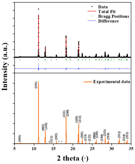
Figure 1.
Synchrotron XRD pattern of a tetragonal LaO0.65F1.7 sample.

Table 1.
Rietveld refinement and crystal data of a LaO0.65F1.7 structure.
Table 2.
Refined atomic coordinates and equivalent isotropic displacement parameters of LaO0.65F1.7.
The formula of the LaO0.65F1.7 structure can be expressed as LaO0.65F(1)0.35F(2)0.86F(3)0.49 based on the calculations of the site occupancies and Wyckoff positions in the unit cell. All possible bonds between the La3+ cations and O2−/F− anions in the host lattice from the crystal data contain La-O4, La-F(1)4, La-F(2)4, and La-F(3)12 as shown in Figure 2 and Table 3.
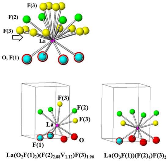
Figure 2.
Polyhedrons of LaO0.65F1.7, 9-coordinated La(O2F(1)2)(F(2)2.88V1.12)F(3)1.96, and 10-coordinated La(O3F(1))(F(2)4)F(3)2 structures.
Table 3.
Selected interatomic distances for LaO0.65F1.7.
In La-F(3)12, there were four long and eight short La-F(3) bond distances of 2.591 (18) and 2.549 (12) Å, respectively. The polyhedron based on the nonstoichiometric LaO0.65F(1)0.35F(2)0.86F(3)0.49 structure refinement can be represented as La(O0.65F(1)0.35)(F(2)0.86V0.14)(F(3)0.49), which contains a vacancy (V) associated with the F(2) anion. In a previous study, the site dependency of 9- and 10-coordinated La3+ sites in Eu3+-doped LaO0.65F1.7 phosphors was reported [6,13]. When Eu3+ ions were substituted in the LaO0.65F1.7 structure, both 9-coordinated no-inversion and 10-coordinated symmetric inversion sites were observed from the Eu3+ transitions of the emission spectra, which showed the 5D0–7F2 electric-dipole and 5D0–7F1 magnetic dipole transitions, respectively [6]. The polyhedrons in the LaO0.65F1.7 structure can be distinguished as both 9-coordinated La(O2F(1)2)(F(2)2.88V1.12)F(3)1.96 and 10-coordinated La(O3F(1))(F(2)4)F(3)2 as shown in Figure 2 [6,13].
The crystallographic phase of the La1−p−qBipTbqO0.65F1.7 (p = 0 and 0.01; q = 0–0.2) phosphors was identified using powder X-ray diffraction (XRD) patterns. The calculated XRD pattern of the tetragonal LaO0.65F1.7 (ICSD 40371) structure is shown in Figure 3a [13]. Figure 3b–d show the XRD patterns of the nonstoichiometric La1−p−qBipTbqO0.65F1.7 phosphors (q = 0.01; q = 0.1; p = 0.01; q = 0.1, respectively). The XRD patterns of the obtained phosphors show a single phase, without any visible impurities, indexed to a tetragonal LaO0.65F1.7 structure. The Tb3+ activator can be located in the 9- and 10-coordinated La3+ sites of the nonstoichiometric LaO0.65F1.7 structure. When small Tb3+ ions (r = 1.04 Å for an eight coordination number (CN); r = 1.095 Å for a nine CN) were substituted for large La3+ ions (r = 1.16 Å for a a 8 CN; r = 1.216 Å for a 9 CN; r = 1.27 Å for a 10 CN) in the LaO0.65F1.7 host lattice, gradual shifts in the positions of the various Bragg reflections to higher angles were observed as shown in Figure 3b,c. The unit cell contraction of the cell parameters in the La0.99Tb0.01O0.65F1.7 and La0.9Tb0.1O0.65F1.7 phosphors occurred from a = 4.0749 (4) and c = 5.8188 (8) Å to a = 4.0689 (3) and c = 5.8057 (7) Å. Meanwhile, when Bi3+ ions (r = 1.17 Å for an eight CN) were substituted for La3+ ions in the La0.9Tb0.1O0.65F1.7 lattice, slight shifts in the positions of the various Bragg reflections to lower angles from the La0.89Bi0.01Tb0.1O0.65F1.7 phosphors (a = 4.0878 (1) and c = 5.8254 (3) Å) were observed as shown in Figure 3c,d, respectively.
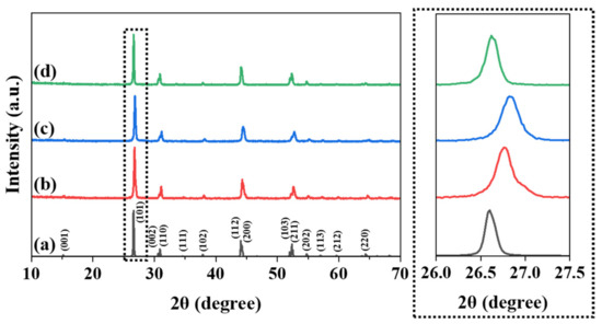
Figure 3.
The calculated XRD patterns of (a) LaO0.65F1.7 (ICSD 40371) and the obtained XRD patterns of La1−p−qBipTbqO0.65F1.7 phosphors (b) q = 0.01; (c) q = 0.1; (d) p = 0.01 and q = 0.1.
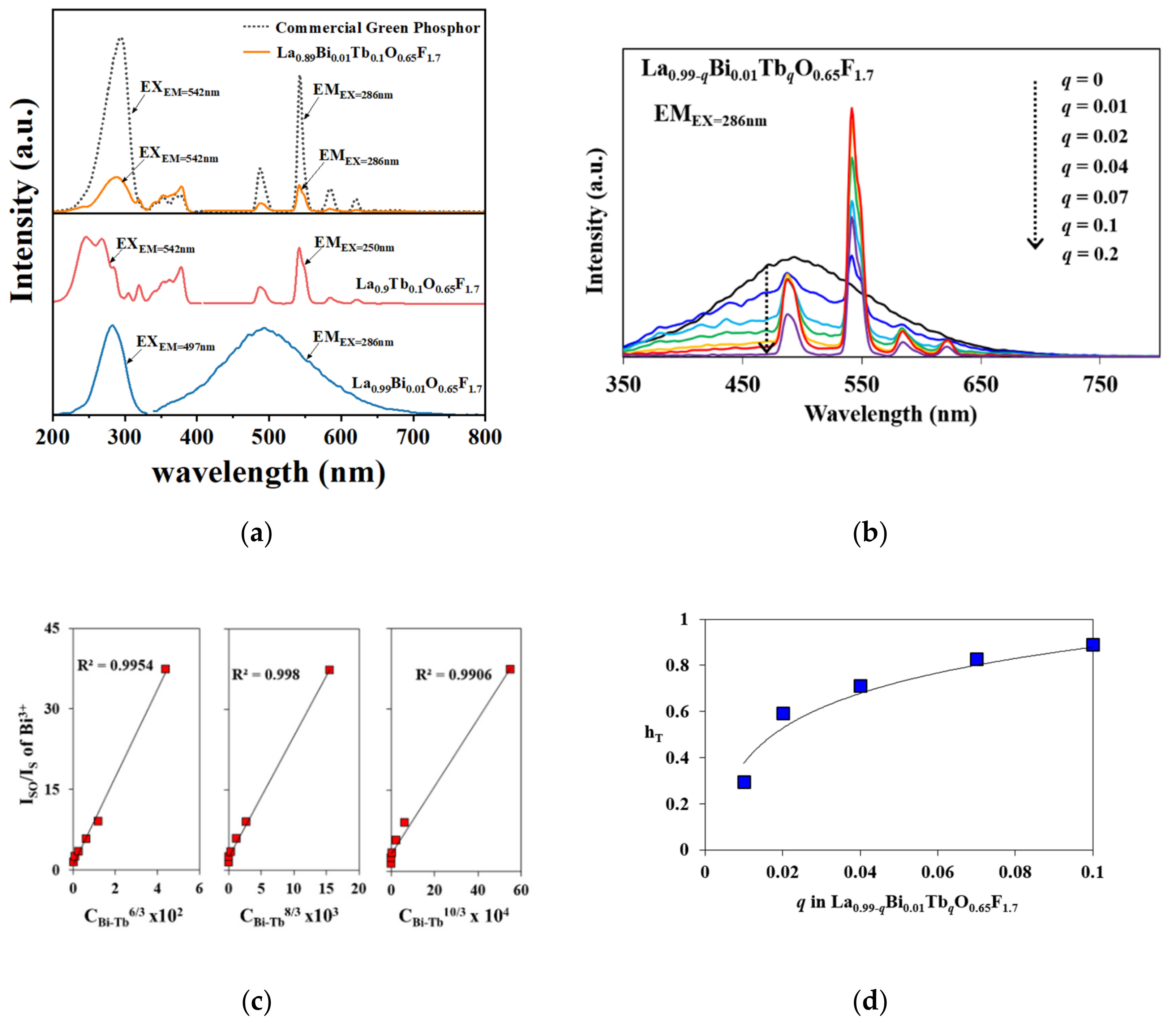
Figure 4a shows the excitation and emission spectra of La0.99Bi0.01O0.65F1.7, La0.9Tb0.1O0.65F1.7, and La0.89Bi0.01Tb0.1O0.65F1.7 phosphors [6]. Previously, the photoluminescence properties of Bi3+-doped LaO0.65F1.7 phosphors were explored. The energy levels of the Bi3+ ions comprise 1S0, 3PJ (J = 0, 1, or 2), and 1P1 states. It is known that the 1S0 → 3P1 and 1P1 transitions arise from spin-orbital coupling, but the 1S0 → 3P0 and 3P2 transitions are forbidden [6,7,8,9,10]. The blue-emitting spectra of the Bi3+-doped LaO0.65F1.7 phosphors, attributed to the 3P1 → 1S0 transitions of the Bi3+ ions, revealed the emission band range from 350 to 650 nm under UV excitation as shown in Figure 4a. The maximum emission intensity was observed in a previous study when the Bi3+ concentration in the host lattice was 1 mol% [6]. The La0.9Tb0.1O0.65F1.7 phosphor was monitored from 400 to 700 nm while under UV excitation. The intense green emission of 5D4–7F5 transition from the Tb3+ ions was observed near 542 nm as shown in Figure 4a. The individual transitions of Bi3+ and Tb3+ ions, with energy transfer from Bi3+ to Tb3+ ions in the La0.99−qBi0.01TbqO0.65F1.7 (q = 0–0.2) phosphors, occurred under an excitation wavelength of 286 nm as shown in Figure 4b. The energy transfer from Bi3+ to Tb3+ operated as a sensitizer and activator, respectively, in the La0.99−qBi0.01TbqO0.65F1.7 (q = 0–0.2) phosphors. The emission of Tb3+ transitions was maximized when the Tb3+ content in the La0.99−qBi0.01TbqO0.65F1.7 (q = 0–0.2) phosphors was q = 0.1, which was compared with commercially available green LaPO4:Ce3+/Tb3+ phosphor in Figure 4a. The green emission of the La0.89Bi0.01Tb0.1O0.65F1.7 phosphor under 286 nm excitation significantly increased by ˃4 times compared to that of the La0.9Tb0.1O0.65F1.7 phosphor because of the energy transfer from Bi3+ to Tb3+ ions as shown in Figure 4b. The energy-transfer efficiency (ηT) was estimated using the following formula:
where IS and ISO are the luminescence intensities of the Bi3+ sensitizer in the presence and absence of a Tb3+ activator, respectively [6,7,8,9,10,17,18,19,20,21]. The energy-transfer mechanism can be represented by linear plots of ISO/IS versus CBi-Tbα/3, where CBi-Tb is the concentration of Bi3+ and Tb3+ ions, with α = 6, 8, or 10, corresponding to dipole–dipole, dipole–quadrupole, and quadrupole–quadrupole interactions, respectively, in accordance with the Dexter theory [17,18,19,20,21,22]. In Figure 4c, when α = 6, 8, and 10, the linear plots showed energy transfers from the Bi3+ to Tb3+ ions in the La0.99−qBi0.01TbqO0.65F1.7 (q = 0–0.2) phosphors with an R2 = 0.9954, 0.998, and 0.9906, respectively. When the value of α was 8, a closer linear plot was determined for the phosphor, and the dipole–quadrupole interaction was involved in the energy-transfer mechanism of the phosphors. The differences on the R-factors between dipole–dipole and dipole–quadrupole mechanisms are quite small when the concentrations of CBi-Tb and CTb were selected (Supplementary Materials, Figure S1). Possibly, there are some arguments that determine the energy-transfer process as the dipole–quadrupole interaction mechanism in the phosphors, because the concentration quenching selectively depends on whether it is a donor–donor or donor–acceptor for C8/3 concentration [23]. The efficiency gradually enhanced from 30 to 89% as the Tb3+ content in the phosphors increased from q = 0.01 to 0.1 as shown in Figure 4d.
ηT = 1 − IS/ISO
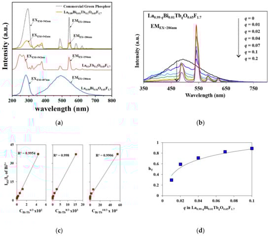
Figure 4.
(a) PL excitation and emission spectra of La0.99Bi0.01O0.65F1.7, La0.9Tb0.1O0.65F1.7, La0.89Bi0.01Tb0.1O0.65F1.7, and commercial green LaPO4:Ce3+/Tb3+ phosphors; (b) the emission spectra of La0.99−qBi0.01EuqO0.65F1.7 (q = 0–0.2) phosphors under 286 nm excitation; (c) the plot of ISO/IS versus CBi-Tbα/3 (α = 6, 8, and 10); (d) energy-transfer efficiency from Bi3+ to Tb3+ in the phosphors.
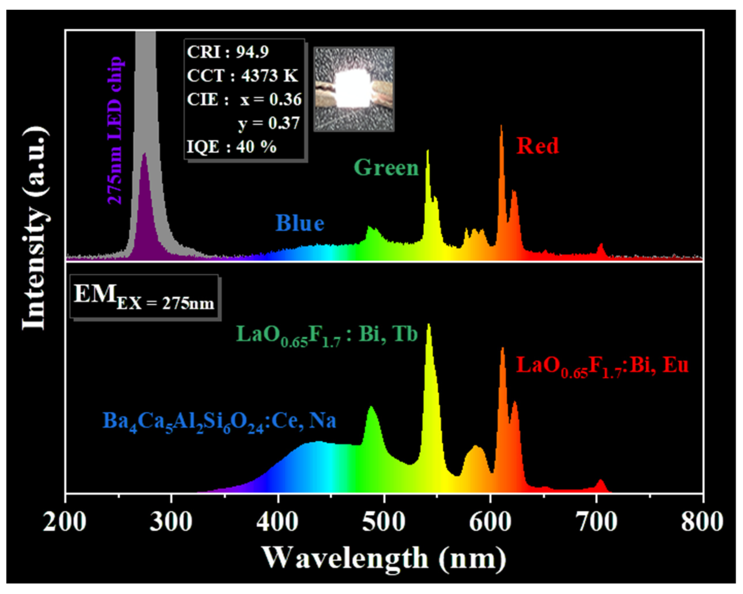
As shown in Figure 5, the chromaticity coordinates, x and y, were in accordance with the desired CIE values from the blue to green wavelength regions for La0.99−qBi0.01TbqO0.65F1.7 (q = 0–0.2) phosphors (EX = 286 nm). When the concentration of Tb3+ ions in the La0.99−qBi0.01TbqO0.65F1.7 (q = 0–0.2) phosphors increased from q = 0 to 0.02 and 0.1, the emission colors exhibited a gradual shift from blue to green emission regions. The CIE values are summarized in the inset of Figure 5, along with the values obtained for the phosphors. The CIE coordinates of the blue and green regions of the phosphor CIE diagram were observed to be x = 0.240 and y = 0.334, x = 0.239 and y = 0.340, x = 0.254 and y = 0.436, and x = 0.267 and y = 0.535 for values of q = 0, 0.01, 0.04, and 0.1, respectively. The emission of the phosphors subject to 312 nm hand-lamp excitation was blue and green. This green-emitting light was adopted for a high color-rendering index to apply to pc UV LEDs. This La0.89Bi0.01Tb0.1O0.65F1.7 phosphor can be prepared as a green-emitting component for fabrication of a 275 nm UV LED chip. The photoluminescence and electroluminescence (EL) spectra resulting from 275 nm UV excitation of the green La0.89Bi0.01Tb0.1O0.65F1.7 phosphor with blue Ba3.8Ce0.1Na0.1Ca5Al2Si6O24 and red La0.94Bi0.01Eu0.05O0.65F1.7 phosphors were monitored as shown in Figure 6, respectively [8,24]. The pc UV LED was prepared at 3.2 V and 20 mA after the phosphors were packaged on a 275 nm LED chip (inset). A CRI (Ra) of 94.9 at CCTs of 4373 K with CIE coordinates of x = 0.36 and y = 0.37 was determined for the 275 nm chip fabricated with the white-light pc UV LED. The three phosphors could generate white light in the 275 nm UV-executable LED chip. The IQE of the phosphors is expressed as ηQE using the following equation [25,26,27,28]:
where LS, ES, and ER are the luminescence integrated emission, excitation spectra of the phosphors, and the integrated excitation spectrum without phosphors in the sphere, respectively. The IQE of the white pc UV LED was approximately 40% under a 275 nm excitation. Color-tunable red, green, and blue phosphors can be diversely fabricated into UV-LED chips, which is an advantage for UV-executable LED applications.
ηQE = ∫LS/(∫ER − ∫ES)
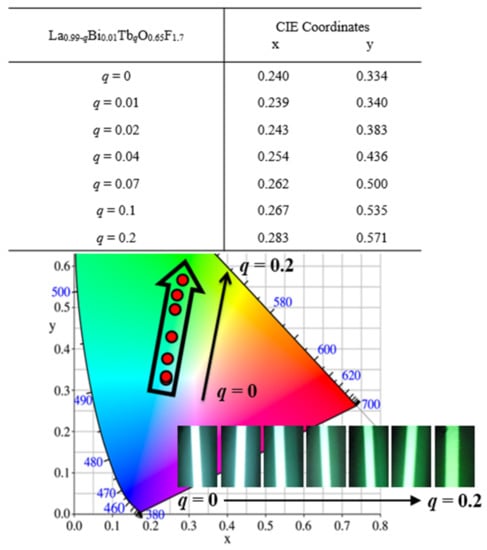
Figure 5.
The chromaticity coordinates with the desired CIE values of La0.99−qBi0.01TbqO0.65F1.7 (q = 0–0.2) phosphors (EX = 286 nm) and photographs of the green emission light of the phosphors under 286 nm radiation.

Figure 6.
The emission spectra of RGB phosphors (La0.94Bi0.01Eu0.05O0.65F1.7, La0.89Bi0.01Tb0.1O0.65F1.7, and Ba3.8Ce0.1Na0.1Ca5Al2Si6O24) under 275 nm UV excitation and the electroluminescence spectra and a photograph of the RGB pc LED under 3.2 V and 20 mA.
4. Conclusions
The nonstoichiometric LaO0.65F1.7 structure was determined as a tetragonal unit cell (P4/nmm) with the cell parameters a = 4.10058 (6) Å and c = 5.8468 (1) Å using synchrotron X-ray powder diffraction. The LaO0.65F1.7 host lattice contained 9- and 10-coordinated La3+ sites in the 2c Wyckoff position. Optical materials composed of La1−p−qBipTbqO0.65F1.7 (p = 0 and 0.01; q = 0–0.2) exhibited photoluminescence spectra characteristic of an energy transfer from Bi3+ to Tb3+ upon excitation with 286 nm involving dipole–quadrupole interactions in the phosphors. The desired Commission Internationale de l’Eclairage values of the blue–green phosphors were calculated. The green La0.96Bi0.01Tb0.02Eu0.01O0.65F1.7 phosphor was fabricated with blue and red phosphors on a 275 nm UV-LED chip, resulting in white light. The IQE was determined to be approximately 40% under a 275 nm excitation with a CRI (Ra) of 94.9 at CCT of 4373 K and CIE coordinates of x = 0.36 and y = 0.37.
Supplementary Materials
The following supporting information can be downloaded at: https://www.mdpi.com/article/10.3390/ma15124222/s1, Figure S1. The plot of ISO/IS versus CTba/3 (a = 6, 8, 10).
Author Contributions
Conceptualization, S.P.; Data curation, S.Y., S.S. and H.H.; Formal analysis, S.Y., S.S., H.H. and S.P.; Funding acquisition, S.P.; Investigation, S.Y., S.S., H.H. and S.P.; Methodology, S.Y.; Project administration, S.P.; Software, S.Y., S.S. and H.H.; Supervision, S.P.; Visualization, S.Y., S.S. and H.H.; Writing—original draft, S.Y.; Writing—review & editing, S.P. All authors have read and agreed to the published version of the manuscript.
Funding
This research was supported by the Basic Science Research Program through the National Research Foundation of Korea (NRF) funded by the Ministry of Education, Science, and Technology (NRF-2018R1D1A3B07048543). This research was supported by the BB21 plus funded by Busan Metropolitan City and the Busan Institute for Talent and Lifelong Education (BIT), Republic of Korea.
Institutional Review Board Statement
Not applicable.
Informed Consent Statement
Not applicable.
Data Availability Statement
The data presented in this study are available on request from the corresponding author.
Conflicts of Interest
The authors declare no conflict of interest.
References
- Ye, S.; Xiao, F.; Parn, Y.X.; Ma, Y.Y.; Zhang, Q.Y. Phosphors in phosphor-converted white light-emitting diodes: Recent advances in materials, techniques and properties. Mater. Sci. Eng. R 2010, 71, 1–34. [Google Scholar] [CrossRef]
- Cao, L.; Li, W.; Devakumar, B.; Ma, N.; Huang, X.; Lee, A.F. Full-Spectrum White Light-Emitting Diodes Enabled by an Efficient Broadband Green-Emitting CaY2ZrScAl3O12:Ce3+ Garnet Phosphor. ACS Appl. Mater. Interfaces 2022, 14, 5643–5652. [Google Scholar] [CrossRef] [PubMed]
- Ahn, Y.N.; Kim, K.D.; Anoop, G.; Kim, G.S.; Yoo, J.S. Design of highly efficient phosphor converted white light-emitting diodes with color rendering indices (R1 − R15) ≥ 95 for artificial lighting. Sci. Rep. 2019, 9, 16848. [Google Scholar] [CrossRef] [PubMed]
- Fang, M.H.; Ni, C.; Zhang, X.; Tsai, Y.T.; Mahlik, S.; Lazarowska, A.; Grinberg, M.; Sheu, H.S.; Lee, J.F.; Cheng, B.M.; et al. Enhance color rendering index via full spectrum employing the important key of cyan phosphor. ACS Appl. Mater. Interfaces 2016, 8, 30677–30682. [Google Scholar] [CrossRef]
- Schubert, E.F.; Kim, J.K. Solid-state light sources getting smart. Science 2005, 308, 1274–1278. [Google Scholar] [CrossRef]
- Yang, S.; Park, S. Bi3+ and Eu3+ Activated Luminescent Behaviors in Non-Stoichiometric LaO0.65F1.7 Structure. Materials 2020, 13, 2326. [Google Scholar] [CrossRef]
- Ning, H.; Tian, L. Enhanced green luminescence in BaZn1.06Al9.94O17:Tb3+ by co-doping with Bi3+ and energy transfer from Bi3+ to Tb3+. Optik 2021, 228, 166218. [Google Scholar] [CrossRef]
- Taikar, D.R. Study of energy transfer from Bi3+ to Tb3+ in Y2O3 phosphor and its application for W-LED. J. Alloys Compd. 2020, 828, 154405. [Google Scholar] [CrossRef]
- Tian, S.; Zhao, L.; Chen, W.; Liu, Z.; Fan, X.; Min, Q.; Yu, H.; Yu, X.; Qiu, J.; Xu, X. Abnormal photo-stimulated luminescence in Ba2Ga2GeO7: Tb3+, Bi3+. J. Lumin. 2018, 202, 414–419. [Google Scholar] [CrossRef]
- Yadav, R.S.; Rai, S.B. Surface analysis and enhanced photoluminescence via Bi3+ doping in a Tb3+ doped Y2O3 nano-phosphor under UV excitation. J. Alloys Compd. 2017, 700, 228–237. [Google Scholar] [CrossRef]
- Noh, W.; Park, S. Synthesis and distinct up-converting behaviors of Er3+, Yb3+ doped LaOF and LaO0.65F1.7 phosphors. Opt. Mater. 2017, 66, 589–594. [Google Scholar] [CrossRef]
- Yang, W.; Park, S. Predominant green emission of Ce3+–Tb3+ activated Y7O6F9 phosphors. RSC Adv. 2016, 6, 12652. [Google Scholar] [CrossRef]
- Laval, J.-P.; Abaouz, A.; Frit, B. High resolution powder neutron diffraction study of the tetragonal anion-excess fluorite-related LaF1.70O0.65 phase. Eur. J. Solid State Inorg. Chem. 1988, 25, 425–434. [Google Scholar] [CrossRef]
- Shin, S.; Yanga, S.; Lee, S.-H.; Shin, T.J.; Park, S. Distinctive occurrences of green-yellow luminescence from orthogermanate-type Ba9Y2(GeO4)6:Ce3+,Na+ phosphors under blue excitation and white-light performance with light-emitting diodes. J. Alloys Compd. 2021, 897, 163213. [Google Scholar] [CrossRef]
- Rodríguez-Carvajal, J.; Roisnel, T. FullProf.98 and WinPLOTR New Windows Applications for Diffraction. Commission on Powder Diffraction. IUCr, Newsletter 20, May–August, 1998.
- Rodríguez-Carvajal, J. Recent developments of the program FullProf”. Commission on Powder Diffraction. IUCr, Newsletter 26, December, 2001.
- Dexter, D.L.; Schulman, J.H. Theory of concentration quenching in inorganic phosphors. J. Chem. Phys. 1954, 22, 1063–1070. [Google Scholar] [CrossRef]
- Li, K.; Fan, J.; Shang, M.; Lian, H.; Lin, J. Sr2Y8(SiO4)6O2:Bi3+/Eu3+: A single-component white-emitting phosphor via energy transfer for UV w-LEDs. J. Mater. Chem. C 2015, 3, 9989–9998. [Google Scholar] [CrossRef]
- Yang, S.; Kim, H.; Park, S. Color-tunable luminescence in Y7(1-m-n-z)Bi7mDy7nEu7zO6F9 (m = 0.001-0.05, n = 0–0.1, z = 0.005, 0.01) phosphors. Opt. Mater. 2018, 77, 154–160. [Google Scholar] [CrossRef]
- Yun, H.; Kim, S.-H.; Park, S. Bi3+, Eu3+-doped Ba9Y2Si6O24 phosphors based on the site-selected substitution. Opt. Mater. 2017, 72, 571–577. [Google Scholar] [CrossRef]
- Zou, Y.; Min, X.; Liu, Z.; Yu, L.; Liu, B. Photoluminescent properties and energy transfer mechanism of Tb3+-Ce3+ doped CaSi2O2N2 oxynitride phosphors. Mater. Res. Bull. 2020, 124, 110769. [Google Scholar] [CrossRef]
- Xia, M.; Zhao, W.; Zhong, J.; Shi, P.; Liao, Z.; Liu, X.; Song, J.; Luo, L.; Ma, L.; Nie, Z. Tunable luminescence of blue-green emitting NaBaBO3:Ce3+,Tb3+ phosphors for near-UV light emitting diodes. J. Lumin. 2020, 220, 116957. [Google Scholar] [CrossRef]
- Sontakke, A.D.; van Bunningen, A.J.; Rabouw, F.T.; Meijers, S.; Meijerink, A. Unraveling the Eu2+ → Mn2+ Energy Transfer Mechanism in w-LED Phosphors. J. Phys. Chem. C 2020, 124, 13902–13911. [Google Scholar] [CrossRef]
- Yang, S.; Park, S. Ca2+-substitution effects in Ba9−xCaxAl2Si6O24:Ce3+,Na+ phosphors. Opt. Mater. 2020, 99, 109548. [Google Scholar] [CrossRef]
- Yang, S.; Park, S. Luminescent performances of Ba9−pCapAl2Si6O24:Eu2+,Mn2+ orthosilicate phosphors along with Ca2+ contents. Opt. Mater. 2021, 114, 11968. [Google Scholar] [CrossRef]
- Yun, H.; Park, S. Blue-white-orange tunable Ba6Ca3YAlSi6O24:Eu2+, Mn2+ phosphors for NUV-pumped LEDs. Opt. Mater. 2018, 86, 600–605. [Google Scholar] [CrossRef]
- Xia, Z.; Liu, R.S.; Huang, K.W.; Drozd, V. Ca2Al3O6F:Eu2+: A green-emitting oxyfluoride phosphor for white light-emitting diodes. J. Mater. Chem. 2012, 22, 15183–15189. [Google Scholar] [CrossRef]
- Liu, C.; Xia, Z.; Lian, Z.; Zhou, J.; Yan, Q. Structure and luminescence properties of green-emitting NaBaScSi2O7:Eu2+ phosphors for near-UV-pumped light emitting diodes. J. Mater. Chem. C 2013, 1, 7139–7147. [Google Scholar] [CrossRef]


Publisher’s Note: MDPI stays neutral with regard to jurisdictional claims in published maps and institutional affiliations. |
© 2022 by the authors. Licensee MDPI, Basel, Switzerland. This article is an open access article distributed under the terms and conditions of the Creative Commons Attribution (CC BY) license (https://creativecommons.org/licenses/by/4.0/).