Article

New Heterotrinuclear CuIILnIIICuII (Ln = Ho, Er) Compounds with the Schiff Base: Syntheses, Structural Characterization, Thermal and Magnetic Properties

Department of General and Coordination Chemistry and Crystallography, Institute of Chemical Sciences, Faculty of Chemistry, Maria Curie-Sklodowska University in Lublin, Maria Curie-Sklodowska Sq. 2, 20-031 Lublin, Poland
*
Author to whom correspondence should be addressed.
Materials 2022, 15(12), 4299; https://doi.org/10.3390/ma15124299
Submission received: 13 April 2022 / Revised: 9 June 2022 / Accepted: 15 June 2022 / Published: 17 June 2022

Abstract

:
New heterotrinuclear complexes with the general formula [Cu2Ln(H2L)(HL)(NO3)2]·MeOH (Ln = Ho (1), Er (2), H4L = N,N′-bis(2,3-dihydroxybenzylidene)-1,3-diaminopropane) were synthesized using compartmental Schiff base ligand in conjugation with auxiliary ligands. The compounds were characterized by elemental analysis, ATR-FTIR spectroscopy, X-ray diffraction, TG, DSC, TG-FTIR and XRD analysis. The N2O4 salen-type ligand coordinates 3d and 4f metal centers via azomethine nitrogen and phenoxo oxygen atoms, respectively, to form heteropolynuclear complexes having CuO2Ln cores. In the crystals 1 and 2, two terminal Cu(II) ions are penta-coordinated with a distorted square-pyramidal geometry and a LnIII ion with trigonal dodecahedral geometry is coordinated by eight oxygen atoms from [CuII(H2L)(NO3)] and [CuII(HL)(NO3)]2− units. Compounds 1 and 2 are stable at room temperature. During heating, they decompose in a similar way. In the first decomposition step, they lose solvent molecules. The exothermic decomposition of ligands is connected with emission large amounts of gaseous products e.g., water, nitric oxides, carbon dioxide, carbon monoxide. The final solid products of decomposition 1 and 2 in air are mixtures of CuO and Ho2O3/Er2O3. The measurements of magnetic susceptibilities and field dependent magnetization indicate the ferromagnetic interaction between CuII and HoIII ions 1.

1. Introduction

In recent years, much progress in the synthesis and investigation of heteronuclear 3d–4f Schiff base coordination compounds has been observed [1,2,3,4,5,6,7,8,9,10,11,12,13,14,15,16,17,18]. Their crystal structures and properties are determined by several factors, e.g., type of the metal ions, metal-to-ligand stoichiometry, the nature and position of the ligands, methods of synthesis (a stepwise reaction or one-pot reaction, Figure 1), type of solvents, kind of coligands, etc.
The salen-type Schiff bases are ligands obtained from salicylaldehyde or its derivatives and different diamines. They usually consist of rigid aromatic rings and flexible aliphatic chains [1,2,3,4,5,6,7,8,9,10,11,12,13,14,15,16,17,18,19]. In the coordination compounds formed by them, 3d and 4f metal ions are captured simultaneously and linked together through two Ophenol atoms. Lanthanide(III) ions behave as hard acids and prefer oxygen to nitrogen donors, whereas 3d metal ions may coordinate to both nitrogens and oxygens. The following salen-type Schiff bases: N,N′-bis(3-methoxysalicylidene)-1,3-diamino-2,2-dimethylpropane [5], N,N′-bis(3-methoxysalicylidene)cyclohexane-1,2-diamine [8], N,N′-bis(3-methoxysalicylidene)-1,3-diamino-2,2-dimethylpropane [11], N,N′-propylenedi(3-methoxysalicylideneimie) [14], 1R,3S)-N′,N″-bis[3-methoxysalicylidene]-1,3-diamino-1,2,2-trimethylcyclopentane [17], that differ by kind of diamines, were used in the synthesis of heteronuclear CuIIHoIII/ErIII complexes. The reported compounds (Figure 2) prepared in different ways and conditions are characterized by interesting structures and properties [5,8,11,14,17].
Among them, there are stepwise synthesized heterobinuclear complexes [Cu(L)(ace)Ln(NO3)3] (where Ln = HoIII, ErIII, ace = acetone, H2L = N,N′-bis(3-methoxysalicylidene)-1,3-diamino-2,2-dimethylpropane) (Figure 2a). In the crystals CuII and HoIII/ErIII, ions are doubly bridged with the phenolate oxygen atoms. The magnetic investigations of the compounds indicated the presence of the ferromagnetic coupling between the HoIII/ErIII and CuII spins [5]. The chiral N,N′-bis(3-methoxysalicylidene)cyclohexane-1,2-diamine (H2L) was applied for the synthesis of the dinuclear complexes [Cu(L)Ln(NO3)3] (where Ln = HoIII, ErIII). The isomorphous complexes are composed of two diphenoxo-bridged CuIILnIII dinuclear clusters (Figure 2b). In the molecular structure, each CuII center adopts a distorted square pyramid geometry. In opposite to them, the two LnIII ions have different coordination environments: one is ten- the other is nine-coordinated. Magnetic investigations of CuIILnIII indicate that CuIIHoIII exhibits field-induced slow magnetic relaxation behaviors [8]. The azido-bridged copper(II)–lanthanide(III) heterotetrametallic complexes [Cu2(valdmpn)2Ln2(N3)6]·2(MeOH)0.5 (where Ln = Ho, Er, H2valdmpn = N,N′-bis(3-methoxysalicylidene)-1,3-diamino-2,2-dimethylpropane) were prepared during a reaction of a metalloligand Cu(valdmpn), a respective lanthanide(III) chloride and sodium azide in methanol. Their structures contain isolated tetranuclear [CuLLn]2 clusters where the HoIII/ErIII centers are bridged by two end-on (EO) azides (Figure 2c). Magnetic investigations of CuII2HoIII2/ErIII2 revealed the SMM behavior for the CuII2HoIII2 complex [11].
The heterometallic coordination polymer [LCuIIErIII(H2O)2(fum)]NO3·3H2O was obtained during reaction of [LCuEr(NO3)3] (L = N,N′-propylenedi(3-methoxysalicylideneiminato) and fumaric acid (H2fum). In the crystals, the {CuEr} nodes (Figure 2d) are connected by fum2− bridges (coordinated only to the lanthanide(III) ions). The magnetic studies show that the values χMT decrease on lowering the temperature [14]. The compound [LCuHo(dca)2(NO3)]n (L = double deprotonated (1R,3S)-N′,N″-bis[3-methoxysalicylidene]-1,3-diamino-1,2,2-trimethylcyclopentane, dca = dicyanamide) is composed of a dinuclear [LCuHo]3+ moiety, one nitrate anion and two bridging dca. Similarly to compounds described above in its crystal structure, the Cu2+ and 4f ions are linked by two μ-phenoxo oxygen atoms of the Schiff base ligand (Figure 2e). The complex is a potential molecule-based multifunctional material indicating optical, ferromagnetic and ferroelectric properties [17]. Literature reported magnetic properties of heteronuclear 3d–4f complexes shows the influence of the lanthanide type on the magnetic exchange coupling interactions between LnIII and paramagnetic 3d metal ions. The mechanism of the 3d–4f interaction are the subject of many studies [20,21,22,23,24,25,26,27]. According to the theoretical model suggested by Kahn et al., the coupling should be antiferromagnetic for the Ln(III) of the first half of the lanthanide row (n < 7) and ferromagnetic for the Ln(III) of the second half of the lanthanide row (n ≥ 7). In determining the magnetic properties of 3d–4f complexes, the orbital angular momentum and spin orbit coupling of unpaired 4f-electron plays a crucial role. In the case of LnIII with 4f1−6 configuration, angular and spin moments are antiparallel in the 2S + 1LJ free-ion ground state (J = L−S). A parallel alignment of the CuII and LnIII spin moment would result in an antiparallel alignment of the angular moment, that is, to an overall antiferromagnetic interaction. For LnIII with configuration 4f7−13 (J = L + S), a parallel alignment of the CuII and LnIII spin moment would lead to an overall ferromagnetic interaction [20]. The investigations of magneto-structural correlation indicate that the exchange interaction in 3d–4f compounds is governed by the value of the dihedral angle between OMO and OLnO planes. For higher value of this angle, the weaker coupling between 3d and 4f metal ions should be anticipated. Superexchange contribution can be awaited for coordination compounds with a planar LnO2M molecular fragment [21,22,23,24,25,26,27].
As a continuation of the investigation on salen-type Schiff base complexes, the aim of this work was to obtain heteronuclear species with N,N′-bis(2,3-dihydroxybenzylidene)-1,3-diaminopropane (the ligand is characterized by the presence in a meta position of a benzene ring –OH substituent instead of –OCH3) and study their properties, as well as investigate the influence of the kind of the additional functional groups in the ligand and kind of lanthanide(III) ions on the structure and feature of the 3d–4f compounds. So far, starting from the N,N′-bis(2,3-dihydroxybenzylidene)-1,3-diaminopropane and respective Cu2+ and Ln3+ salts, we have synthesized heteropolynuclear complexes with different structures and physicochemical properties [28,29,30]. In the case of the first half of the lanthanide row (LaIII, PrIII, NdIII), the inert heterotrinuclear compounds CuIILnIIICuII which differ only in the amount and type of solvent molecules in the outside coordination sphere were obtained. In the crystals of copper(II) and praseodymium(III)/neodymium(III) complexes, the antiferromagnetic coupling of magnetic centers occurred [28]. The hexanuclear cation complex resulted from the simultaneous coordination of two dianionic Schiff bases to CuII and GdIII ions and the forming of trinuclear units [CuII2GdIII] that were connected through bridging nitrate ions. The interaction between neighboring CuII and GdIII ions was ferromagnetic [29]. We also obtained and characterized the heterodinuclear CuIIDyIII compound. Its magnetic measurements showed the weak ferromagnetic interaction between CuII and DyIII ions [30]. It was noticed that in the heterodi-, heterotri- and heterohexanuclear complex crystals reported by us so far, the N,N′-bis(2,3-dihydroxybenzylidene)-1,3-diaminopropane was double deprotonated.
Herein, we report the synthesis and crystal characterization, along with the spectral, thermal and magnetic properties of new heterotrinuclear compounds [Cu2Ln(H2L)(HL)(NO3)2]·MeOH (where Ln = Ho, Er) contained in the crystals dianionic H2L2− and trianionic HL3− Schiff base ligands. The complexes were synthesized in a step-wise manner without the isolation of the mononuclear complex.

2. Materials and Methods

2.1. Materials

The reagent grade chemicals, i.e., 2,3-dihydroxybenzaldehyde (HO)2C6H3CHO, 1,3-diaminopropane NH2(CH2)3NH2, Ho(NO3)3·5H2O, Er(NO3)3·5H2O, Cu(CH3COO)2·H2O were used.

2.2. Synthesis

2.2.1. N,N’-Bis(2,3-dihydroxybenzylidene)-1,3-diaminopropane (H4L)

The ligand H4L was prepared from 1,3-diaminopropane (5 mmol, 0.37 g) and 2,3-dihydroxybenzaldehyde (10 mmol, 1.38 g) in 50 mL of hot methanol according to literature procedures [31]. Yield 80%. Anal. (%) C17H18N2O4. Calcd: C, 64.97; H, 5.73; N, 8.92%. Found: C, 65.20; H, 5.70; N, 9.10%.

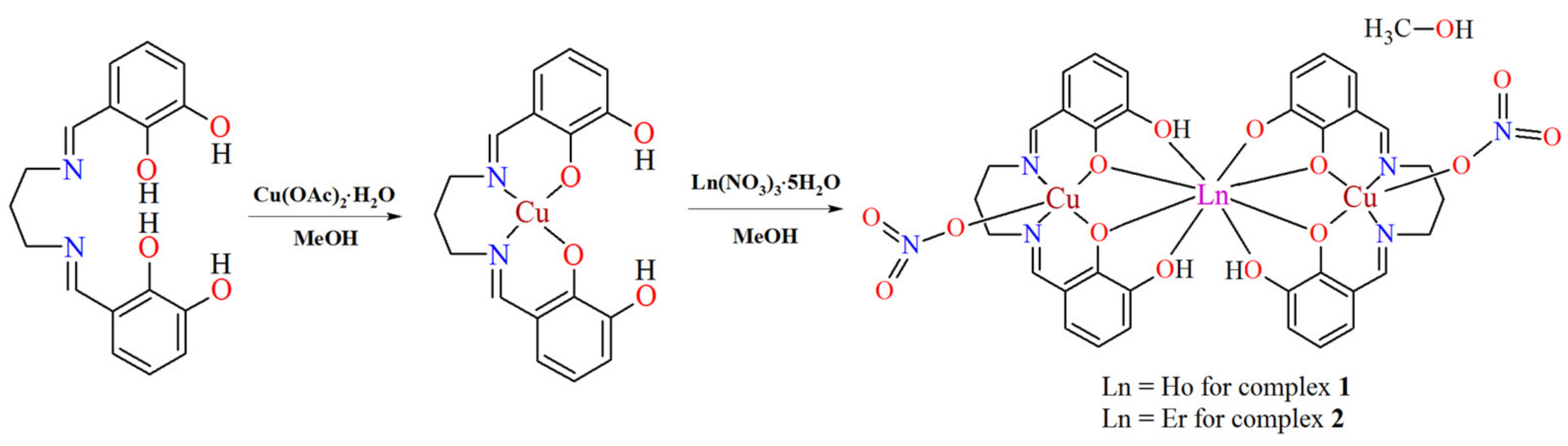
2.2.2. Complexes [Cu2Ln(H2L)(HL)(NO3)2]·MeOH (1, 2)

The complexes were prepared following the same general procedure: a methanol solution (10 mL) of copper acetate monohydrate (0.4 mmol, 0.0799 g) was added to a hot methanol solution (50 mL) of the Schiff base H4L (0.4 mmol, 0.1248 g) to produce a green colored mixture which was magnetically stirred. After 30 min, a methanol solution (5 mL) containing dissolving Ho(NO3)3·5H2O (0.2 mmol, 0.0887 g) or Er(NO3)3·5H2O (0.2 mmol, 0.0882 g) was added and the resulting mixture was stirred for about 30 min. The resulting clear, deep green solutions were left undisturbed in a refrigerator at ~4 °C. The X-ray quality green crystals of the desired compounds were obtained over a period of several days.

Complex [Cu2Ho(H2L)(HL)(NO3)2]·MeOH (1)

Yield 70%, 150 mg. Anal. (%) for C35H35Cu2HoN6O15 (MW: 1071.70). Calcd: C, 39.23; H, 3.29; N, 7.84; Cu, 11.86; Ho, 15.39. Found: C, 39.40; H, 3.10; N, 7.50; Cu, 11.60; Ho, 15.50.

Complex [Cu2Er(H2L)(HL)(NO3)2]·MeOH (2)

Yield 65%, 140 mg. Anal. (%) for C35H35Cu2ErN6O15 (MW: 1074.03). Calcd: C, 39.14; H, 3.28; N, 7.82; Cu, 11.83; Er, 15.57. Found: C, 39.30; H, 3.40; N, 7.60; Cu, 11.50; Er, 15.80.

2.3. Methods

A CHN 2400 Perkin Elmer analyzer was used for determination of C, H and N contents. The metal amounts were determined on an ED XRF spectrophotometer (Canberra-Packard, Schwadorf, Austria). The ATR-FTIR spectra were recorded on a Nicolet 6700 spectrophotometer equipped with the Smart iTR attachment (diamond crystal) over 4000–525 cm−1. Thermal analysis was carried out by the thermogravimetric (TG) and differential scanning calorimetry (DSC) methods using the SETSYS 16/18 analyzer (Setaram, Lyon, France). The samples 7.61 mg (1) and 6.66 mg (2) were heated in open Al2O3 crucibles in air at the range of 20–1000 °C at a heating rate of 10 °C·min−1. TGA Q5000 analyzer (TA Instruments, New Castle, DE, USA) interfaced to the Nicolet 6700 FTIR spectrophotometer (Thermo Scientific, Waltham, MA, USA) were applied for the TG-FTIR analysis. The samples in an open platinum crucible were heated from room temperature to 700 °C (heating rate was 20 °C·min−1). The temperature in the gas cell and transfer line was set to 250 and 240 °C, respectively. XRD analysis of the solid residue was carried out by using PAN Analytical/Empyrean spectrophotometer. The dc magnetic susceptibilities of the compounds were measured on Quantum Design SQUID-VSM magnetometer in a range of 1.8–300 K. The magnetization curves were recorded at 2K in an applied field up to 5 T. Diamagnetic corrections were estimated from Pascal’s constants [32].

X-ray Crystal Structure Determination

Single-crystal data for the complexes were collected on an Oxford Diffraction Xcalibur CCD diffractometer (MoKα radiation, λ = 0.71073Å). The program CrysAlis [33] was used for collecting frames of data, cell refinement and data reduction. A multi-scan absorption correction was applied. Crystal data, data collection and structure refinement details are summarized in Table 1. The structures were solved by direct methods using SHELXS-2018 and refined by the full-matrix least-squares on F2 using the SHELXL-2018 [34] (both programs implemented in WinGX software [35]). All the non-hydrogen atoms were refined with anisotropic displacement parameters. The H-atoms attached to carbon were positioned geometrically and refined applying the riding model [C–H = 0.93–0.99 Å and with Uiso(H) = 1.2 or 1.5 Ueq(C)]. The O-bound H atoms were located on a difference Fourier map and refined freely or with O–H distances restrained to 0.82 Å using DFIX command. The following programs were used to prepare the molecular graphics: ORTEP3 [35] and Mercury [36]. The geometrical calculations were performed using PLATON program [37].

3. Results and Discussion

N,N′-bis(2,3-dihydroxybenzylidene)-1,3-diaminopropane H4L is a multidentate ligand which possess six donor atoms, i.e., two imino nitrogen atoms and four oxygen atoms coming from hydroxyl groups. The ligand can act as a bridge between metal ions through phenoxy groups so as to link the 3d and 4f ions together, therefore, it can be used to synthesize 3d–4f complexes. The inner, smaller N2O2 compartment of the Schiff base may accommodate a borderline acid, e.g., copper(II) ion, whereas the other, bigger O2O2 site selectively binds to hard acids, such as lanthanide(III) ion. Using H4L, the copper(II) acetate and the holmium(III)/erbium(III) nitrate, we obtained the discrete heterotrinuclear complexes of the general formula [Cu2Ln(H2L)(HL)(NO3)2]·MeOH (Ln = Ho 1, Er 2) (Figure 3). The neutral complexes are isostructural, crystalize with one CH3OH solvent molecule and are characterized by the molar ratio between the Schiff base ligand and the 3d and 4f metal ions 2:2:1. The similar values of ionic radious of HoIII and ErIII cations and the same molar ratio of the starting compounds may be the origin of the same crystal structure of 1 and 2.

3.1. Infrared Spectra

The FTIR spectra of 1 and 2 (Table 2, Figure S1) are similar. A broad absorption bands in the 2500–3300 cm−1 region can be attributed to the O–H stretching vibrations of methanol molecule (it interferes with the protonated hydroxyl groups of the N2O4 ligand) that are involved in the strong hydrogen bonds.
A broad absorption bands in the 2500–3300 cm−1 region can be attributed to the O–H stretching vibrations of methanol molecule (it interferes with the protonated hydroxyl groups of the N2O4 ligand) that are involved in the strong hydrogen bonds. This feature is in accordance with the X-ray structures, i.e., methanol molecule acts as a proton acceptor as well as a proton donor. The FTIR spectra of complexes have in common the occurrence of a strong absorption band at 1618 cm−1 1 and 1616 cm−1 2 which is characteristic of the presence of the azomethine group C=N. These bands are shifted towards lower frequencies relative to the free Schiff base 1632 cm−1. This phenomenon is due to the coordination of azomethine nitrogen to the 3d metal ion. The strong bands situated at 1467 cm−1, 1285 cm−1 and 1024 cm−1, respectively, in the spectrum of 1 and 1465 cm−1, 1287 cm−1 and 1024 cm−1 in the spectrum of 2 may be assigned to the monodentate nitrate ligand. The involvement of the phenolic oxygen atoms in the metal-ligand bonding is confirmed by the strong doublet bands observed at 1251 cm−1, 1218 cm−1 1 and 1248 cm−1, 1219 cm−1 2, respectively. The typical absorption band of the νaryl-O vibration is identified in the free ligand spectrum at 1233 cm−1 [4,13,38,39,40,41,42,43,44]. All these spectroscopic features are confirmed by the X-ray structures.

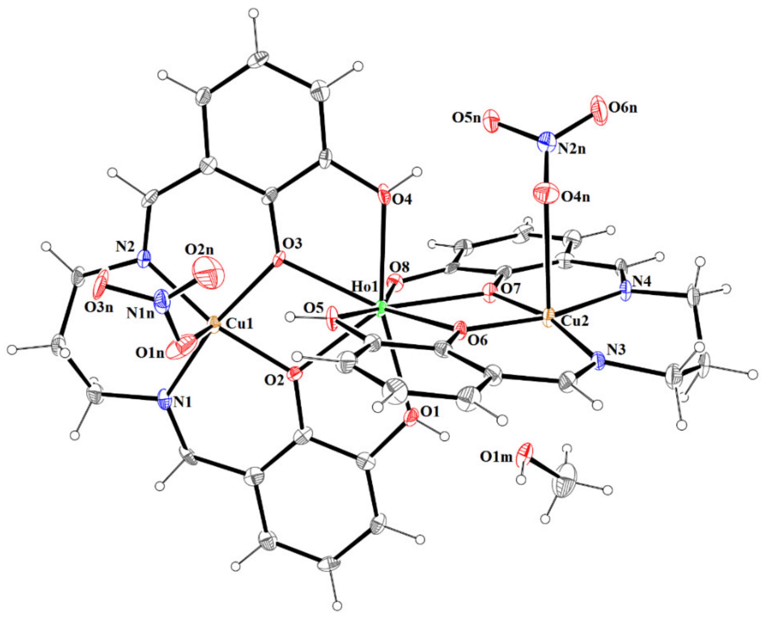
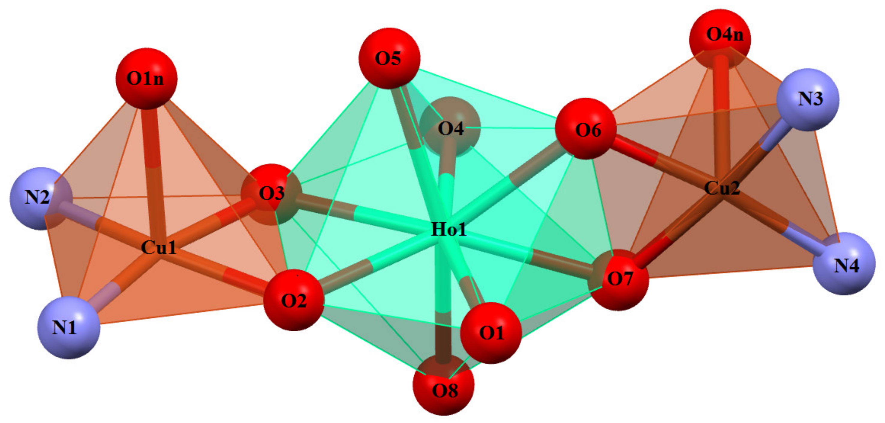
3.2. Crystal and Molecular Structure

The reaction of the Schiff base ligand H4L with copper(II) acetate and lanthanide(III) nitrate result in formation of the trinuclear complexes 1 and 2 which are isomorphous and crystallize in the centrosymmetric monoclinic space group P21/c (Table 1). The asymmetric unit cell of both complexes contains one neutral complex, which consists of two Cu(II) ions, one Ln(III) ion, a dianionic ligand H2L2−, a trianionic ligand HL3−, two nitrite ions and methanol molecule (Figure 4 and Figure S2).
The complex structures are constructed from almost linear trinuclear [Cu2-Ln] units. The values of the Cu-Ln-Cu angle are 167.97(2)° and 168.04(2)°, respectively, for 1 and 2 (Table 3). The distances between the copper(II) and lanthanides(III) ions are within the range 3.4831(7)–3.4977(8) Å.
The HoIII and ErIII ion assume a trigonal dodecahedron [O8] configuration (Figure 5 and Figure S3), while both the partially deprotonated Schiff base ions (H2L2− and HL3−) act in similar chelating coordination modes, i.e., lanthanide(III) ion is coordinated by four oxygen atoms of phenoxide or phenol groups of each ligand. A similar binding type of lanthanide (with partially deprotonated Schiff bases, i.e., one dianionic and one trianionic ligand) was reported for a trinuclear complex of ZnII-TbIII-ZnII ions [45]. The Cu(1) and Cu(2) centers have slightly distorted square pyramidal coordination geometries (Figure 5 and Figure S3) in which the equatorial sites are occupied by two nitrogen and two oxygen atoms of Schiff base ligands. The Cu-O and Cu-N bonds are within normal values (C-O 1.913(4)-1.983(5) Å and C-N 1.960(5)-1.977(5) Å) and comparable to those observed in the related CuII compounds [40,43,46,47,48,49]. The apical positions are occupied by oxygen atoms from monodentate nitrate ions and the bond lengths are found to be significantly longer (distances in the range 2.410(6)-2.649(5) Å) than those of Cu-O in the basal plane.
Moreover, the Cu(2)-O bonds in the axial position are a bit longer from average distances for this kind of connection (2.45 Å) but in literature [40], there are some structures where the Cu-Onitrate bond has similar or higher value. Examples of structures with refcodes and bond lengths are given in Table 4. The planes formed by N2O2 cores around Cu(II) ions of two Schiff bases intersect at an angle of 78,25(1)° for 1 and 78,20(3)° for 2.
The crystal structures of 1 and 2 reveal the presence of intramolecular and intermolecular hydrogen bonds (Table S1). In the crystals 1 and 2, the molecules are linked by O(1)–H(1o)···O(1m) and O(1m)–H(1m)···O(5n)a (symmetry code (a): x−1,y,z) hydrogen bonds forming columns propagating along [100] with C 2 2 10 motifs (Figure 6 and Figure S4). Additional classical hydrogen bonds are supported by weaker non-classical C–H···O contacts, which linked formed columns in 3D supramolecular structure. The partial view of crystal packing for compound 1 and 2 are illustrated in Figures S5 and S6.

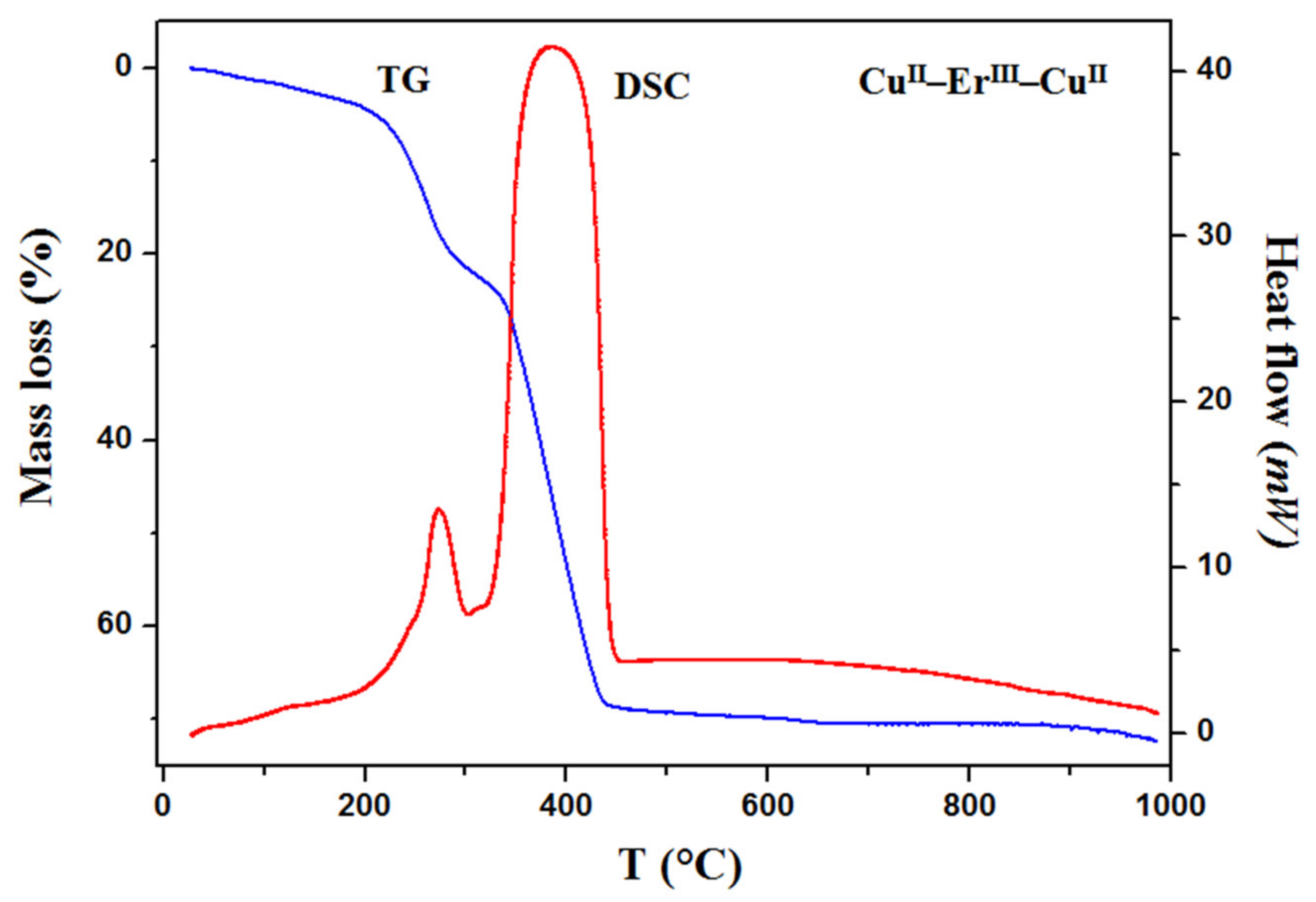
3.3. Thermal Analysis

In order to examine the thermal behavior of the heteronuclear complexes 1 and 2, the thermogravimetric analysis was carried out (Figure 7 and Figure S7). The results of the thermal analysis allow to confirm/evaluate the presence of solvents (e.g., methanol, water) in the structure of compounds and to establish the endothermic and/or exothermic effects connected with different processes such as dehydration, desolvation, melting or decomposition. The TG and DSC curves recorded for both complexes are similar. The mass of samples decreases slowly with the increasing temperature. The first mass loss occurs up to 90 °C and it is assigned to the elimination of one methanol molecule (mass loss: observed 2.60% 1, 2.70% 2, calculated 2.99% 1; 2.98% 2). The small endothermic effect seen on the DSC curves confirms this process. In the case of compound 1, the decomposition process begins immediately after desolvation. The next mass losses recorded at above 200 °C and accompanied with exothermic effects seen on the DSC curves is connected with gradual decomposition of the samples. Additionally, this process is also confirmed by the TG-FTIR analysis (Figure S8).
The recorded TG-FTIR spectra show that carbon dioxide, carbon monoxide and nitric oxide are mainly emitted during this process. The characteristic doublet bands seen at 2240–2400 cm−1 and 670 cm−1, respectively, are assigned to stretching and deformation vibrations of carbon dioxide molecules. The specific bands at 2060–2240 cm−1 are characteristic of carbon monoxide [59]. The solid intermediate products for thermal decomposition could not be identified. The residual mass is about 29.5% 1 and 30.6% 2 (the theoretical values are 31.5% 1 and 32.6% 2). At high temperature, the sublimation of the copper(II) oxide takes place and the differences between values calculated and found can be caused by this process. Mixtures of metal oxides CuO and Ho2O3/Er2O3, experimentally verified by X-ray diffraction powder patterns (Figures S9 and S10) are the final solid products of thermal decomposition of 1 and 2 in air [60].

3.4. Magnetic Properties

Temperature-dependent molar susceptibility measurements of CuII2HoIII 1 and CuII2ErIII 2 were carried out in a magnetic field of 0.1 T at 1.8–300 K. The χMT vs. T curves for 1 and 2 are shown in Figure 8. The magnetic properties of heteronuclear CuIILnIII compounds are governed by three factors: the thermal population of the Stark components of LnIII, the CuII···CuII interactions (including intermolecular interaction) and the CuIILnIII interactions. For CuII2HoIII 1, the χMT value experimentally determined at 300 K is equal to 14.17 cm3Kmol−1, which is slightly smaller than the value 14.82 cm3Kmol−1 calculated for one HoIII (5I8, J = 8, L = 6, S = 2, g = 5/4) and the two CuII (S = 1/2, g = 2) free ions. As the temperature is lowered, χMT keeps a constant value until 150 K, then begins to decrease to 13.91 cm3Kmol−1 at 19 K, next increases to reach a value of 14.70 cm3Kmol−1 at 5.9 K and finally, shows a small decrease to 12.95 cm3Kmol−1 at 1.8 K.
The shape of the χMT vs. T curve is strongly suggestive of the occurrence of two competitive phenomena. The decrease of χMT on lowering of the temperature is most probably governed by the depopulation of the Ho Stark sublevels, or the presence of magnetic anisotropy, or the antiferromagnetic interaction between metal centers, while the increase of the χMT at lower temperatures may be attributed to a ferromagnetic CuIIHoIII coupling. For a Cu2IIErIII 2, the experimental value of χMT product at room temperature is equal to 12.18 cm3Kmol−1 and approximately corresponds to the value 12.23 cm3Kmol−1 calculated for one independent ErIII (4I15/2, S = 3/2, L = 6, J = 15/2, g = 6/5) and two independent CuII ions (S = 1/2, g = 2). As shown in Figure 9, this value decreases by lowering the temperature to 8.05 cm3Kmol−1 at 1.8 K. The reduction of χMT at low temperature should mainly arise from the crystal field splitting of LnIII ions and/or combine the contribution of the overall antiferromagnetic interactions among the metal ions. These results are compatible with the empirical investigations of heterometallic CuII–4f compounds, in which the 4f ions show a spin–orbit coupling [32,61,62,63].
The M vs. H plots (at 2 K) for 1 and 2 are presented in Figure 9. The values of magnetization rise quickly at the low magnetic field whereas at the high magnetic field, the increase of magnetization is slow and linear. The magnetization reaches the values 6.5 μB for 1 and 7.0 μB for 2, respectively, at 5T; these are far from the theoretical saturated values anticipated for one uncoupled lanthanide(III) ion and two copper(II) ions [32,61,62,63].

4. Conclusions

In summary, neutral, heteronuclear CuIILnIIICuII complexes were obtained in a step-wise manner. In the crystal structures of 1 and 2, the smaller CuII ion is exclusively coordinated to the N2O2 compartment of the hexadentate Schiff base ligand, while the O2O2 compartment accommodates a bigger HoIII/ErIII ion. The HoIII/ErIII and CuII ions are double-bridged by two phenoxo oxygen atoms of the N2O4 ligand. The complexes 1 and 2 crystallize as stable at room temperature solvates and their desolvation process is consistent with the loss of methanol molecules. The similar values of ionic radious of HoIII and ErIII cations led to the same coordination number and the same coordination geometry of LnIII ions as well as the similar spectral and thermal properties. The CuII and HoIII centers are ferromagnetically coupled which is in agreement with earlier observations in similar CuIIHoIII compounds. The structural investigations indicate that different (heterodi-, heterotri, -heterohexanuclear) coordination architectures can be received using the same Schiff base as a ligand, but changing the lanthanide(III) ions.

Supplementary Materials

The following supporting information can be downloaded at: https://www.mdpi.com/article/10.3390/ma15124299/s1, Figure S1: FTIR spectra of the free Schiff base H4L and complexes 1 and 2, Figure S2: The molecular structure of 2. Displacement ellipsoids are drawn at the 30% probability level, Figure S3: Coordination polyhedra of Cu(II) and Er(III) cations in the trinuclear complex 2, Figure S4: (a) A partial viewed along the b-axis direction of the crystal packing of 2 with hydrogen bonds shown as dashed lines; (b) A partial viewed along the a-axis direction of the crystal packing of 2 with hydrogen bonds shown as dashed lines, Figure S5: The overall crystal packing of compound 1 showing formation of 3D supramolecular structure, viewed along the a-axis. Hanging contacts were omitted for clarity, Figure S6: The overall crystal packing of compound 2 showing formation of 3D supramolecular structure, viewed along the c-axis. Hanging contacts were omitted for clarity, Figure S7: TG and DCS curves of thermal decomposition of the complex 1 in air, Figure S8: FTIR spectra of gaseous products involved during the complex 2 decomposition, Figure S9: The X-ray powder diffraction patterns of the final products of complex 1 decomposition in air, Figure S10: The X-ray powder diffraction patterns of the final products of complex 2 decomposition in air, Table S1: Hydrogen-bond geometry [Å, °] for compounds 1 and 2.

Author Contributions

Conceptualization, B.C. and D.O.; methodology, B.C., D.O. and A.B.; software, A.B., D.O. and B.C.; formal analysis, D.O., A.B. and B.C.; investigation, B.C., D.O. and A.B.; writing—original draft preparation, B.C., D.O. and A.B.; writing—review and editing, B.C., D.O. and A.B.; visualization, D.O., A.B. and B.C.; supervision, B.C. All authors have read and agreed to the published version of the manuscript.

Funding

This research received no external funding.

Institutional Review Board Statement

Not applicable.

Informed Consent Statement

Not applicable.

Data Availability Statement

The data presented in this study are available on request from the corresponding author. The CIF files have been deposited in the Cambridge Crystallographic Data Center (CCDC). These data can be obtained free of charge from The Cambridge Crystallographic Data Centre via www.ccdc.cam.ac.uk/data_request/cif (or from the CCDC, 12 Union Road, Cambridge CB2 1EZ, UK; Fax: +44-1223-336033; E-mail: [email protected]).

Conflicts of Interest

The authors declare no conflict of interest.

References

  1. Dey, A.; Bag, P.; Kalita, P.; Chandrasekhar, V. Heterometallic CuII–LnIII complexes: Single molecule magnets and magnetic refrigerants. Coord. Chem. Rev. 2021, 432, 213707. [Google Scholar] [CrossRef]
  2. Liua, K.; Shia, W.; Chenga, P. Toward heterometallic single-molecule magnets: Synthetic strategy, structures and properties of 3d–4f discrete complexes. Coord. Chem. Rev. 2015, 289–290, 74–122. [Google Scholar] [CrossRef]
  3. Im, S.Y.; Park, S.J.; Im, H.J.; Lee, S.W. Conversion of Ni–Nd and Ni–Tb compartment compounds into one-dimensional coordination polymers or tetranuclear dimers. Polyhedron 2016, 117, 231–243. [Google Scholar] [CrossRef]
  4. Buta, I.; Shova, S.; Ilies, S.; Manea, F.; Andruh, M.; Costisor, O. Mono- and oligonuclear complexes based on a o-vanillin derived Schiff-base ligand: Synthesis, crystal structures, luminescent and electrochemical properties. J. Mol. Struct. 2022, 1248, 131439. [Google Scholar] [CrossRef]
  5. Ishida, T.; Watanabe, R.; Fujiwara, K.; Okazawa, A.; Kojima, N.; Tanaka, G.; Yoshii, S.; Nojiri, H. Exchange coupling in TbCu and DyCu single-molecule magnets and related lanthanide and vanadium analogs. Dalton Trans. 2012, 41, 13609–13619. [Google Scholar] [CrossRef]
  6. Ghosh, M.; Sepay, S.; Schollmeyer, D.; Sakiyama, H.; Mikuriya, M.; Mal, D.; Gayen, A.; Seikh, M.M.; Saha, S. Spacers directed self-assembly of heterobimetallic copper(II)-lanthanide(III) [Ln = Nd and Gd] moieties: Syntheses, structural diversities and magnetic properties. Polyhedron 2022, 216, 115718. [Google Scholar] [CrossRef]
  7. Cristóvão, B.; Osypiuk, D.; Miroslaw, B.; Bartyzel, A. Heterometallic di- and trinuclear CuIILnIII (LnIII = La, Ce, Pr, Nd) complexes with an alcohol-functionalized compartmental Schiff base ligand: Syntheses, crystal structures, thermal and magnetic studies. Polyhedron 2020, 188, 114703. [Google Scholar] [CrossRef]
  8. Huang, X.-C.; Zhao, X.-H.; Shao, D.; Wang, H.-Y. Syntheses, structures, and magnetic properties of a family of end-on azido-bridged CuII–LnIII complexes. Dalton Trans. 2017, 46, 7232–7241. [Google Scholar] [CrossRef]
  9. Karmakar, M.; Roy, S.; Chattopadhyay, S. Synthesis, structure and properties of homo- and hetero-trinuclear complexes of salicylaldehyde-based di-Schiff bases. Polyhedron 2022, 215, 115652. [Google Scholar] [CrossRef]
  10. Apostol, A.A.; Mihalache, I.; Mocanu, T.; Tutunaru, O.; Pachiu, C.; Gavrila, R.; Maxim, C.; Andruh, M. Luminescent [ZnIILnIII] complexes anchored on graphene: Synthesis and crystal structures of [ZnIIEuIII] and [ZnIITbIII] complexes decorated with pyrene groups. Appl. Organomet. Chem. 2021, 35, e6126. [Google Scholar] [CrossRef]
  11. Wen, H.-R.; Bao, J.; Liu, S.-J.; Liu, C.-M.; Zhang, C.-W.; Tang, Y.-Z. Temperature-controlled polymorphism of chiral CuII–LnIII dinuclear complexes exhibiting slow magnetic relaxation. Dalton Trans. 2015, 44, 11191–11201. [Google Scholar] [CrossRef] [PubMed]
  12. Corredoira-Vázquez, J.; Fondo, M.; Sanmartín-Matalobos, J.; Taboada, P.; García-Deibe, A.M. Filling tricompartmental igands with GdIII and ZnII ions: Some structural and MRI studies. Crystals 2018, 8, 431. [Google Scholar] [CrossRef] [Green Version]
  13. Kori, D.; Dote, Y.; Koikawa, M.; Yamada, Y. Syntheses, crystal structures, and solid-state photoluminescence properties of heterotrinuclear Zn2Ln (Ln: La, Sm, Eu, Tb) complexes derived from 1,4-diaminobutane-based N2O4 compartmental ligand. Polyhedron 2019, 170, 612–621. [Google Scholar] [CrossRef]
  14. Gheorghe, R.; Cucos, P.; Andruh, M.; Costes, J.P.; Donnadieu, B.; Shova, S. Oligonuclear 3d–4f complexes as tectons in designing supramolecular solid-state architectures: Impact of the nature of linkers on the structural diversity. Chem. Eur. J. 2006, 12, 187–203. [Google Scholar] [CrossRef] [PubMed]
  15. Yang, J.-W.; Tian, Y.-M.; Tao, J.; Chen, P.; Li, H.-P.; Zhang, Y.-Q.; Yan, P.-F.; Sun, W.-B. Modulation of the coordination environment around the magnetic easy axis leads to significant magnetic relaxations in a series of 3d–4f Schiff complexes. Inorg. Chem. 2018, 57, 8065–8077. [Google Scholar] [CrossRef] [PubMed]
  16. Hazra, S.; Titiš, J.; Valigura, D.; Boča, R.; Mohanta, S. Bis-phenoxido and bis-acetato bridged heteronuclear {CoIIIDyIII} single molecule magnets with two slow relaxation branches. Dalton Trans. 2016, 45, 7510–7520. [Google Scholar] [CrossRef] [PubMed]
  17. Zhou, Z.; Li, M.-X.; Sui, Y.; Nfor, E.N.; Wang, Z.-X. Two 1D homochiral heterometallic chains: Crystal structures, spectra, ferroelectricity and ferromagnetic properties. RSC Adv. 2020, 10, 7004–7010. [Google Scholar] [CrossRef] [Green Version]
  18. Upadhyay, A.; Das, C.; Langley, S.K.; Murray, K.S.; Srivastava, A.K.; Shanmugam, M. Heteronuclear Ni(II)–Ln(III ) (Ln = La, Pr, Tb, Dy) complexes: Synthesis and single-molecule magnet behavior. Dalton Trans. 2016, 45, 3616–3626. [Google Scholar] [CrossRef]
  19. Gebrezgiabher, M.; Bayeh, Y.; Gebretsadik, T.; Gebreslassie, G.; Elemo, F.; Thomas, M.; Linert, W. Lanthanide-based single-molecule magnets derived from Schiff base ligands of salicylaldehyde derivatives. Inorganics 2020, 8, 66. [Google Scholar] [CrossRef]
  20. Andruh, M.; Ramade, I.; Codjovi, E.; Guillou, O.; Kahn, O.; Trombe, J.C. Crystal structure and magnetic properties of [Ln2Cu4] hexanuclear clusters (where Ln = trivalent lanthanide). Mechanism of the gadolinium(III)-copper(II) magnetic interaction. J. Am. Chem. Soc. 1993, 115, 1822–1829. [Google Scholar] [CrossRef]
  21. Zou, H.-H.; Sheng, L.-B.; Liang, F.-P.; Chena, Z.-L.; Zhang, Y.-Q. Experimental and theoretical investigations of four 3d–4f butterfly single-molecule magnets. Dalton Trans. 2015, 44, 18544–18552. [Google Scholar] [CrossRef] [PubMed]
  22. Rajeshkumar, T.; Jose, R.; Remya, P.R.; Rajaraman, G. Theoretical studies on trinuclear {MnIII2GdIII} and tetranuclear {MnIII2GdIII2} clusters: Magnetic exchange, mechanism of magnetic coupling, magnetocaloric effect, and magneto–structural correlations. Inorg. Chem. 2019, 58, 11927–11940. [Google Scholar] [CrossRef]
  23. Jiang, L.; Liu, Y.; Liu, X.; Tian, J.; Yan, S. Three series of heterometallic NiII–LnIII Schiff base complexes: Synthesis, crystal structures and magnetic characterization. Dalton Trans. 2017, 46, 12558. [Google Scholar] [CrossRef]
  24. Ahmed, N.; Das, C.; Vaidya, S.A.; Srivastava, K.; Langley, S.K.; Murra, K.S.; Shanmugam, M. Probing the magnetic and magnetothermal properties of M(II)–Ln(III) complexes (where M (II)= Ni or Zn; Ln (III)= La or Pr or Gd). Dalton Trans. 2014, 43, 17375–17384. [Google Scholar] [CrossRef] [PubMed]
  25. Singh, S.K.; Beg, M.F.; Rajaraman, G. Role of magnetic exchange interactions in the magnetization relaxation of {3d–4f} single-molecule magnets: A theoretical perspective. Chem. Eur. J. 2016, 22, 672–680. [Google Scholar] [CrossRef] [PubMed]
  26. Maity, S.; Bhunia, P.; Ichihashi, K.; Ishida, T.; Ghosh, A. SMM behaviour of heterometallic dinuclear CuIILnIII (Ln = Tb and Dy) complexes derived from N2O3 donor unsymmetrical ligands. New J. Chem. 2020, 44, 6197–6205. [Google Scholar] [CrossRef]
  27. Fu, X.-X.; Wang, H.-L.; Zou, H.-H.; Quan, B.-H.; Li, B.; Liang, F.-P. Structures and magnetic properties of three heterobimetallic 3d–4f hexanuclear complexes. J. Clust. Sci. 2017, 28, 3229–3239. [Google Scholar] [CrossRef]
  28. Cristóvão, B.; Miroslaw, B. Asymmetry in propeller-like trinuclear diphenoxo-bridged CuII–LnIII–CuII (Ln = La, Pr, Nd) Schiff base complexes—Synthesis, structure and magnetic properties. J. Coord. Chem. 2015, 68, 1602–1615. [Google Scholar] [CrossRef]
  29. Cristóvão, B.; Pełka, R.; Miroslaw, B. A novel hexanuclear CuII4–GdIII2 cluster obtained from heterotrinuclear building blocks. Inorg. Chem. Commun. 2015, 54, 81–84. [Google Scholar] [CrossRef]
  30. Cristóvão, B.; Miroslaw, B. Tautomerism of a compartmental Schiff base ligand and characterization of a new heterometallic CuII–DyIII complex—Synthesis, structure and magnetic properties. Inorg. Chem. Commun. 2015, 52, 64–68. [Google Scholar] [CrossRef]
  31. Zeyrek, C.T.; Elmali, A.; Elerman, Y. Magnetic characterization, synthesis and crystal structure of a heterodinuclear CuIIGdIII Schiff base complex bridged by the two phenolic oxygen atoms. J. Mol. Struct. 2005, 740, 47–52. [Google Scholar] [CrossRef]
  32. Kahn, O. Molecular Magnetism; VCH Publishers Inc.: New York, NY, USA, 1993. [Google Scholar]
  33. Agilent Technologies Ltd. CrysAlis PRO; Agilent Technologies Ltd.: Oxfordshire, UK, 2014. [Google Scholar]
  34. Sheldrick, G.M. Crystal structure refinement with SHELXL. Acta Cryst. C 2015, 71, 3–8. [Google Scholar] [CrossRef] [PubMed]
  35. Farrugia, L.J. WinGX and ORTEP for Windows: An Update. J. Appl. Cryst. 2012, 45, 849–854. [Google Scholar] [CrossRef]
  36. Macrae, C.F.; Sovago, I.; Cottrell, S.J.; Galek, P.T.A.; McCabe, P.; Pidcock, E.; Platings, M.; Shields, G.P.; Stevens, J.S.; Towler, M.; et al. Mercury 4.0: From visualization to analysis, design and prediction. J. Appl. Crystallogr. 2020, 53, 226–235. [Google Scholar] [CrossRef] [Green Version]
  37. Spek, A.L. Single-crystal structure validation with the program PLATON. J. Appl. Cryst. 2003, 36, 7–13. [Google Scholar] [CrossRef] [Green Version]
  38. Cristóvão, B.; Kłak, J.; Miroslaw, B. Synthesis and characterization of CuII–LnIII (Ln = Ho, Tm, Yb or Lu) complexes with N2O4-donor Schiff base ligand. J. Coord. Chem. 2014, 67, 2728–2746. [Google Scholar] [CrossRef]
  39. Singh, B.K.; Prakash, A.; Rajour, H.K.; Bhojak, N.; Adhikari, D. Spectroscopic characterization and biological activity of Zn(II), Cd(II), Sn(II) and Pb(II) complexes with Schiff base derived from pyrrole-2-carboxaldehyde and 2-amino phenol. Spectrochim. Acta A Mol. Biomol. 2010, 76, 376–383. [Google Scholar] [CrossRef] [PubMed]
  40. Cristóvão, B.; Osypiuk, D.; Miroslaw, B.; Bartyzel, A. Syntheses, crystal structures, thermal and magnetic properties of new heterotrinuclear CuII–LnIII–CuII complexes incorporating N2O4-donor Schiff base ligands. Polyhedron 2018, 144, 225–233. [Google Scholar] [CrossRef]
  41. Anitha, C.; Sheela, C.D.; Tharmaraj, P.; Sumathi, S. Spectroscopic studies and biological evaluation of some transition metal complexes of azo Schiff-base ligand derived from (1-phenyl-2,3-dimethyl-4-aminopyrazol-5-one) and 5-((4-chlorophenyl)diazenyl)-2-hydroxybenzaldehyde. Spectrochim. Acta A Mol. Biomol. 2012, 96, 493–500. [Google Scholar] [CrossRef]
  42. Cristóvão, B.; Hnatejko, Z. Lanthanide(III) compounds with the N2O4 Schiff base—Synthesis, spectral, thermal, magnetic and luminescence properties. J. Mol. Struct. 2015, 1088, 50–55. [Google Scholar] [CrossRef]
  43. Osypiuk, D.; Cristóvão, B.; Bartyzel, A. New coordination compounds of CuII with Schiff base ligands—Crystal structure, thermal, and spectral investigations. Crystals 2020, 10, 1004. [Google Scholar] [CrossRef]
  44. Miroslaw, B.; Osypiuk, D.; Cristóvão, B.; Głuchowska, H. Symmetry in recognition of supramolecular synthons–competition between hydrogen bonding and coordination bond in multinuclear CuII–4f complexes with bicompartmental Schiff base ligand. Symmetry 2019, 11, 460. [Google Scholar] [CrossRef] [Green Version]
  45. Olea-Román, D.; Bélanger-Desmarais, N.; Flores-Álamo, M.; Bazán, C.; Thouin, F.; Reber, C.; Castillo-Blum, S.E. Spectroscopic studies of lanthanide complexes of varying nuclearity based on a compartmentalized ligand. Dalton Trans. 2015, 44, 17175–17188. [Google Scholar] [CrossRef] [Green Version]
  46. Datta, A.; Clegg, J.K.; Huang, J.-H.; Pevec, A.; Garribba, E.; Fondo, M. Hydroxo-bridged 1-D coordination polymer of Cu(II) incorporating with salicyladimine precursor: Spectral and temperature dependent magneto structural correlation. Inorg. Chem. Commun. 2012, 24, 216–220. [Google Scholar] [CrossRef]
  47. Bartyzel, A. Synthesis, thermal behaviour and some properties of CuII complexes with N,O donor Schiff bases. J. Therm. Anal. Calorim. 2018, 131, 1221–1236. [Google Scholar] [CrossRef] [Green Version]
  48. Zarei, L.; Asadi, Z.; Dusek, M.; Eigner, V. Homodinuclear Ni(II) and Cu(II) Schiff base complexes derived from O-vanillin with a pyrazole bridge: Preparation, crystal structures, DNA and protein (BSA) binding, DNA cleavage, molecular docking and cytotoxicity study. J. Photochem. Photobiol. A Chem. 2019, 374, 145–160. [Google Scholar] [CrossRef]
  49. Kaczmarek, M.T.; Skrobanska, M.; Zabiszak, M.; Walesa-Chorab, M.; Kubicki, M.; Jastrzab, R. Coordination properties of N,N′-bis(5-methylsalicylidene)-2-hydroxy-1,3-propanediamine with d- and f-electron ions: Crystal structure, stability in solution, spectroscopic and spectroelectrochemical studies. RSC Adv. 2018, 8, 30994–31007. [Google Scholar] [CrossRef] [Green Version]
  50. Bruno, I.J.; Cole, J.C.; Kessler, M.; Luo, J.; Motherwell, W.D.S.; Purkis, L.H.; Smith, B.R.; Taylor, R.; Cooper, R.I.; Harris, S.E.; et al. Retrieval of crystallographically-derived molecular geometry information. J. Chem. Inf. Comput. Sci. 2004, 44, 2133–2144. [Google Scholar] [CrossRef]
  51. Chandrasekhar, V.; Senapati, T.; Dey, A.; Das, S.; Kalisz, M.; Clerac, R. Cyclo- and carbophosphazene-supported ligands for the assembly of heterometallic (Cu2+/Ca2+, Cu2+/Dy3+, Cu2+/Tb3+) complexes: Synthesis, structure, and magnetism. Inorg. Chem. 2012, 51, 2031–2038. [Google Scholar] [CrossRef]
  52. Kashyap, S.; Singh, U.P.; Singh, A.K.; Kumar, P.; Singh, S.P. Synthesis and structural studies of some copper-benzoate complexes. Transit. Met. Chem. 2013, 38, 573–585. [Google Scholar] [CrossRef]
  53. Yang, H.; Meng, Y.-X.; Tian, H.-Q.; Li, D.-C.; Zeng, S.-Y.; Song, Y.; Dou, J.-M. Investigating the effect of lanthanide radius and diamagnetic linkers on the framework of metallacrown complexes. Dalton Trans. 2020, 49, 1955–1962. [Google Scholar] [CrossRef] [PubMed]
  54. Pu, L.-M.; An, X.-X.; Liu, C.; Long, H.-T.; Zhao, L. Insights into crystal structures, supramolecular architectures and antioxidant activities of self-assembled fluorescent hetero-multinuclear [Cu (II)-Ln (III)] (Ln = La, Ce, Pr and Nd) salamo-like complexes. Appl. Organomet. Chem. 2020, 34, e5980. [Google Scholar] [CrossRef]
  55. Mathews, I.I.; Manohar, H. Synthesis, spectral and structural studies on metal complexes of Schiff bases involving vitamin B6 and histamine. J. Chem. Soc. Dalton Trans. 1991, 2289–2294. [Google Scholar] [CrossRef]
  56. Bereau, V.; Dhers, S.; Costes, J.-P.; Duhayon, C.; Sutter, J.-P. Syntheses, structures, and magnetic properties of symmetric and dissymmetric ester-functionalized 3d-4f Schiff base complexes. Eur. J. Inorg. Chem. 2018, 2018, 66–73. [Google Scholar] [CrossRef]
  57. Haribabu, P.; Patil, Y.P.; Hussain Reddy, K.; Nethaji, M. Synthesis, crystal structure, DNA interaction and cleavage activities of mononuclear and trinuclear copper(II) complexes. Transit. Met.Chem. 2014, 39, 167–175. [Google Scholar] [CrossRef]
  58. Singh, Y.P.; Patel, R.N.; Singh, Y.; Choquesillo-Lazarte, D.; Butcher, R.J. Classical hydrogen bonding and stacking of chelate rings in new copper(II) complexes. Dalton Trans. 2017, 46, 2803–2820. [Google Scholar] [CrossRef]
  59. Li, G.; Jones, C.A.; Grassian, V.H.; Larsen, S.C. Selective catalytic reduction of NO2 with urea in nanocrystalline NaY zeolite. J. Catal. 2005, 234, 401–413. [Google Scholar] [CrossRef]
  60. Blake, A.J.; Cherepanov, V.A.; Dunlop, A.A.; Grant, C.M.; Milne, P.E.Y.; Rawson, J.M.; Winpenny, R.E.P. Synthesis, Crystal structures and thermal decomposition studies of a series of copper-lanthanoid complexes of 6-methyl-2-pyridone. J. Chem. Soc. Dalton Trans. 1994, 1, 2719–2727. [Google Scholar] [CrossRef]
  61. Ahmed, N.; Sharma, T.; Spillecke, L.; Koo, C.; Ansari, K.U.; Tripathi, S.; Caneschi, A.; Klingeler, R.; Rajaraman, G.; Shanmugam, M. Probing the origin of ferro-/afntiferromagnetic exchange interactions in Cu(II)–4f complexes. Inorg. Chem. 2022, 61, 5572–5587. [Google Scholar] [CrossRef]
  62. Mahapatra, P.; Ghosh, S.; Koizumi, N.; Kanetomo, T.; Ishida, T.; Drew, M.G.B.; Ghosh, A. Structural variations in (CuL)2Ln complexes of a series of lanthanide ions with a salen-type unsymmetrical Schiff base(H2L): Dy and Tb derivatives as potential single-molecule magnets. Dalton Trans. 2017, 46, 12095. [Google Scholar] [CrossRef]
  63. Ghosh, S.; Gómez García, C.J.; Clemente-Juan, J.M.; Ghosh, A. Key role of size and electronic configuration on the sign and strength of the magnetic coupling in a series of Cu2Ln trimers (Ln = Ce, Gd, Tb, Dy and Er). Magnetochemistry 2016, 2, 2. [Google Scholar] [CrossRef] [Green Version]
Figure 1. Methods synthesis of salen-type Schiff base heteronuclear complex: (a) a stepwise reaction; (b) one-pot reaction (where R = substituent with O-donor atom).
Materials 15 04299 g001
Figure 2. Chemical diagrams of selected heteronuclear complexes CuII-HoIII/ErIII including salen–type Schiff base ligands: (a) [Cu(L)(ace)Ln(NO3)3], (b) [Cu(L)Ln(NO3)3], (c) [Cu2(valdmpn)2Ln2(N3)6]·2(MeOH)0.5, (d) [LCuIIErIII(H2O)2(fum)]NO3·3H2O, (e) [LCuHo(dca)2(NO3)]n (where Ln = Ho, Er).
Materials 15 04299 g002
Figure 3. The scheme of the synthetic route of complexes 1 and 2.
Materials 15 04299 g003
Figure 4. The molecular structure of 1. Displacement ellipsoids are drawn at the 30% probability level.
Materials 15 04299 g004
Figure 5. Coordination polyhedra of Cu(II) and Ho(III) cations in the trinuclear complex 1.
Materials 15 04299 g005
Figure 6. (a) A partial viewed along the b-axis direction of the crystal packing of 1 with hydrogen bonds shown as dashed lines; (b) A partial viewed along the a-axis direction of the crystal packing of 1 with hydrogen bonds shown as dashed lines.
Materials 15 04299 g006
Figure 7. TG and DCS curves of thermal decomposition of the complex 2 in air.
Materials 15 04299 g007
Figure 8. Temperature dependence of experimental χMT and χM−1 versus T for 1 and 2.
Materials 15 04299 g008
Figure 9. Field dependence of the magnetization for complexes 1 and 2 at 2 K.
Materials 15 04299 g009
Table 1. Details of data collection and structure refinement parameters for complexes.
Compound12
CCDC21657832165782
Temperature K120(2)298(2)
Crystal systemmonoclinicmonoclinic
Space groupP21/cP21/c
a (Å)8.5856(3)8.5422(5)
b (Å)30.3312(12)30.3435(15)
c (Å)14.1441(6)14.1234(7)
β (°)101.114(4)101.081(5)
Volume (Å3)3614.2(2)3592.5(3)
Z44
Calculated density (g cm−3)1.9701.986
μ (mm−1)3.4193.573
Absorption correctionmulti-scanmulti-scan
F(000)21282132
Crystal size (mm)0.22 × 0.10 × 0.050.20 × 0.08 × 0.05
θ range (°)2.49 to 26.372.43 to 26.37
Reflections collected/unique 17840/740317336/7613
Rint0.05750.0648
Data/restraints/parameters7403/3/5457613/2/545
GooF on F21.0321.015
Final R indices [I > 2σ(I)]R1 = 0.0461,
wR2 = 0.0734
R1 = 0.0516,
wR2 = 0.0837
R indices (all data)R1 = 0.0761,
wR2 = 0.0833
R1 = 0.0862,
wR2 = 0.0968
Largest diff. peak/hole, e Å−30.961/−0.8922.001/−1.554
Table 2. The selected frequencies (cm−1) of absorption bands in FTIR spectra of Schiff base (H4L), CuII–HoIII–CuII 1 and CuII–ErIII–CuII 2.
H4L12Proposed Assignments
3192 ν(OH) + ν(N–H)
29292924ν(OH) + ν(CHas)
163216181616ν(C=N)
1540, 151715691570ν(C=C)
144614671465ν(C=C) + ν(N–O)complex
139414021404ν(C–H) + ν(CCC)
135513651356δ(O–H)
12851287ω(C–H) (O–H) + ν(N–O)
1233 12511248ν(C–O)
12181219ν(C–O)
118911681167δ(O–H)
112611251119ν(C–C) + tw(C–H)
10881087ν(C–O)methanol
106410691069δ(C–H) + skeletal
10241024ν(N–O)
896 ρ(C–H) + δ(CCC)
865863863γ(O–H)
782781γ(C–H) + ν(N–O)
711741734γ(C–H)
639641δ(C=C) + ring deform.
614615ring deform.
556558ν(M–O)
538 ν(M–N)
Table 3. Selected interatomic distances and bond angles for 1 and 2.
Bond Lengths (Å)
12
Cu(1)–N(1)1.983(5)Cu(1)–N(1)1.977(5)
Cu(1)–N(2)1.978(4)Cu(1)–N(2)1.976(5)
Cu(1)–O(1n)2.417(4)Cu(1)–O(1n)2.409(6)
Cu(1)–O(2)1.946(3)Cu(1)–O(2)1.957(4)
Cu(1)–O(3)1.952(4)Cu(1)–O(3)1.944(4)
Cu(2)–N(3)1.971(4)Cu(2)–N(3)1.959(5)
Cu(2)–N(4)1.971(4)Cu(2)–N(4)1.976(5)
Cu(2)–O(6)1.929(3)Cu(2)–O(6)1.913(4)
Cu(2)–O(7)1.920(4)Cu(2)–O(7)1.932(4)
Cu(2)–O(4n)2.644(4)Cu(2)–O(4n)2.649(5)
Ho(1)–O(1)2.360(4)Er(1)–O(1)2.340(4)
Ho(1)–O(2)2.320(4)Er(1)–O(2)2.303(4)
Ho(1)–O(3)2.316(3)Er(1)–O(3)2.319(4)
Ho(1)–O(4)2.355(3)Er(1)–O(4)2.347(4)
Ho(1)–O(5)2.439(4)Er(1)–O(5)2.221(5)
Ho(1)–O(6)2.343(4)Er(1)–O(6)2.304(4)
Ho(1)–O(7)2.308(4)Er(1)–O(7)2.325(4)
Ho(1)–O(8)2.242(3)Er(1)–O(8)2.409(5)
Ho(1)–Cu(1)3.4977(8)Er(1)–Cu(1)3.4871(7)
Ho(1)–Cu(2)3.4939(8)Er(1)–Cu(2)3.4831(7)
Cu(1)–N(1)1.978(5)
Angles(°)
Cu(1)–O(2)–Ho(1)109.83(14)Cu(1)–O(2)–Er(1)109.65(19)
Cu(1)–O(3)–Ho(1)109.76(15)Cu(1)–O(3)–Er(1)109.48(18)
O(2)–Cu(1)–O(3)76.98(14)O(2)–Cu(1)–O(3)77.08(17)
Cu(2)–O(6)–Ho(1)109.36(15)Cu(2)–O(6)–Er(1)111.0(2)
Cu(2)–O(7)–Ho(1)111.13(15)Cu(2)–O(7)–Er(1)109.47(19)
O(6)–Ho(1)–O(7)62.17(12)O(6)–Er(1)–O(7)62.28(15)
O(6)–Cu(2)–O(7)77.20(15)O(6)–Cu(2)–O(7)77.04(17)
O(2)–Ho(1)–O(3)63.12(12)O(2)–Er(1)–O(3)63.45(14)
φa5.10φc5.23
φb3.40φd3.84
a The dihedral angle between the O(2)–Cu(1)–O(3) plane and the O(2)–Ho(1)–O(3) plane; b The dihedral angle between the O(6)–Cu(2)–O(7) plane and the O(6)–Ho(1)–O(7) plane; c The dihedral angle between the O(2)–Cu(1)–O(3) plane and the O(2)–Er(1)–O(3) plane; d The dihedral angle between the O(6)–Cu(2)–O(7) plane and the O(6)–Er(1)–O(7) plane.
Table 4. The values of a long Cu-Onitrate bond length [50].
RefcodeCu-Onitrate Bond Length [Å]Reference
AZIROS2.626(4)[28]
AZIRIM2.646(4)[28]
CERZAD2.785(4)[40]
CAWYEG2.833(7)[51]
DIFTET2.759(4)[52]
FUBTEE2.714(5)[53]
HUYYOS2.741(3)[54]
KOCMOE2.643(2)[55]
MESBAQ2.718(1)[56]
MESBEU2.734(6)[56]
MIWLEL2.762(4)[57]
SAJTAB2.701(2)[58]
Publisher’s Note: MDPI stays neutral with regard to jurisdictional claims in published maps and institutional affiliations.

