
Efficient Green Synthesis of (Fe3O4) and (NiFe2O4) Nanoparticles Using Star Anise (Illicium verum) Extract and Their Biomedical Activity against Some Cancer Cells


Abstract
:1. Introduction
2. Experimental Section
2.1. Chemicals
2.2. Preparation of the Star Anise Extract
2.3. Synthesis of the Magnetic Nanomaterials
2.4. Instrumentation and Characterization
2.5. In Vitro Cytotoxicity Evaluation
3. Results and Discussion
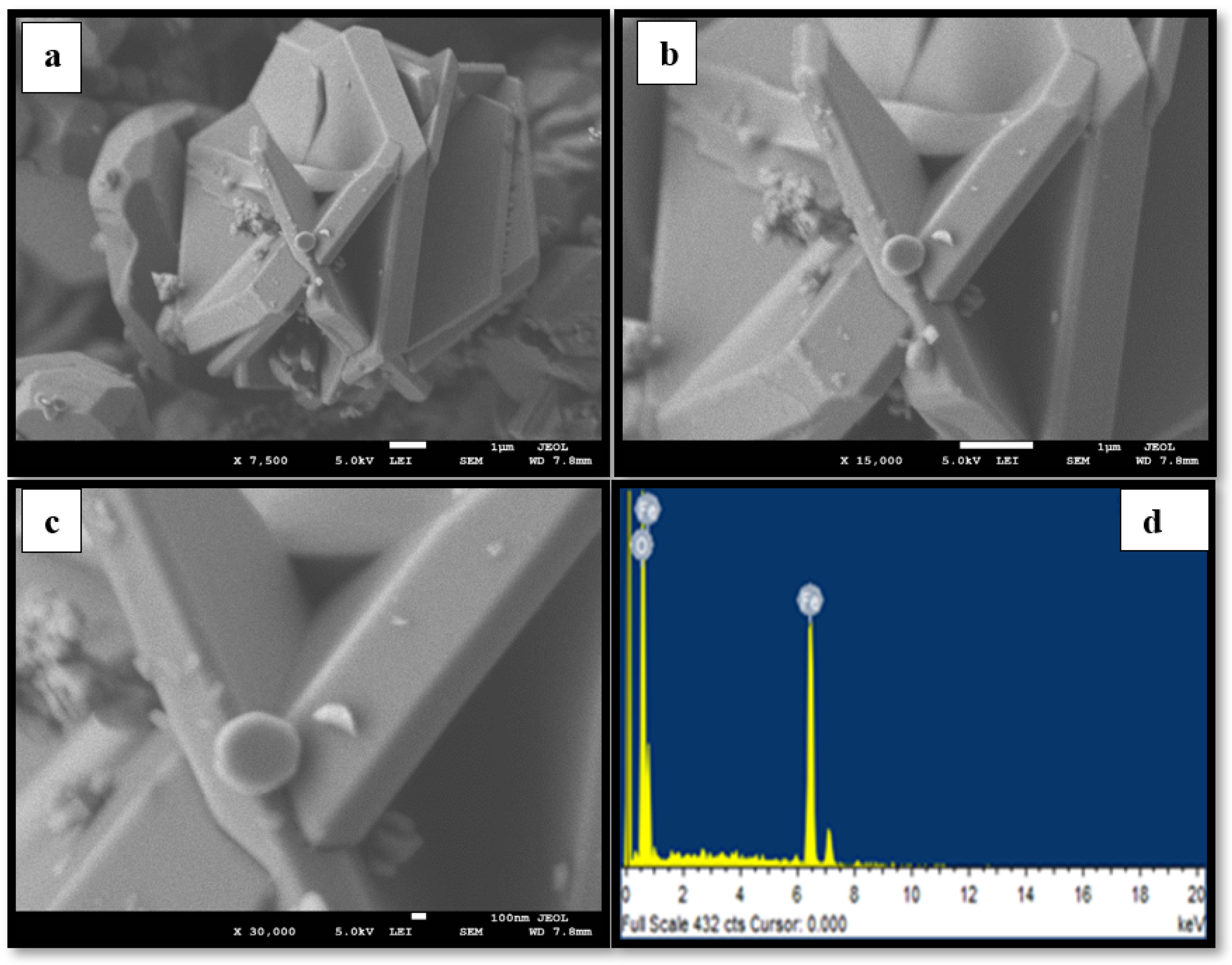
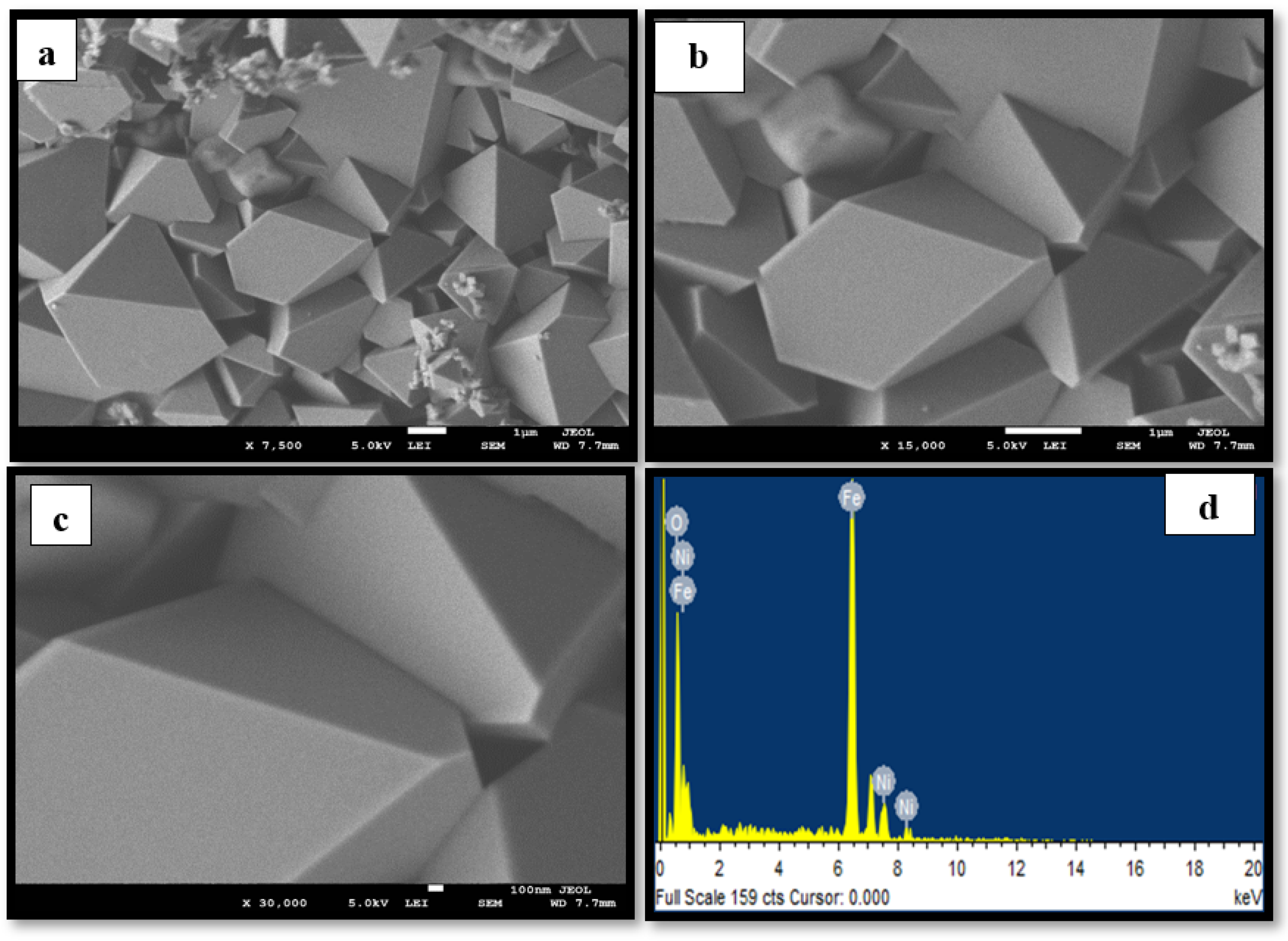
3.1. Morphology
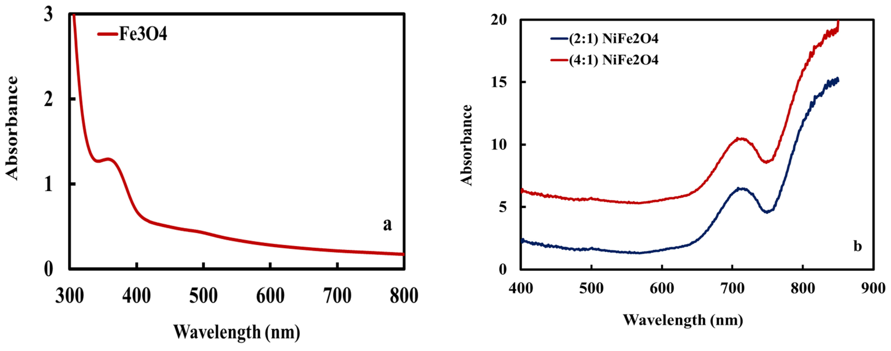
3.2. UV Spectra
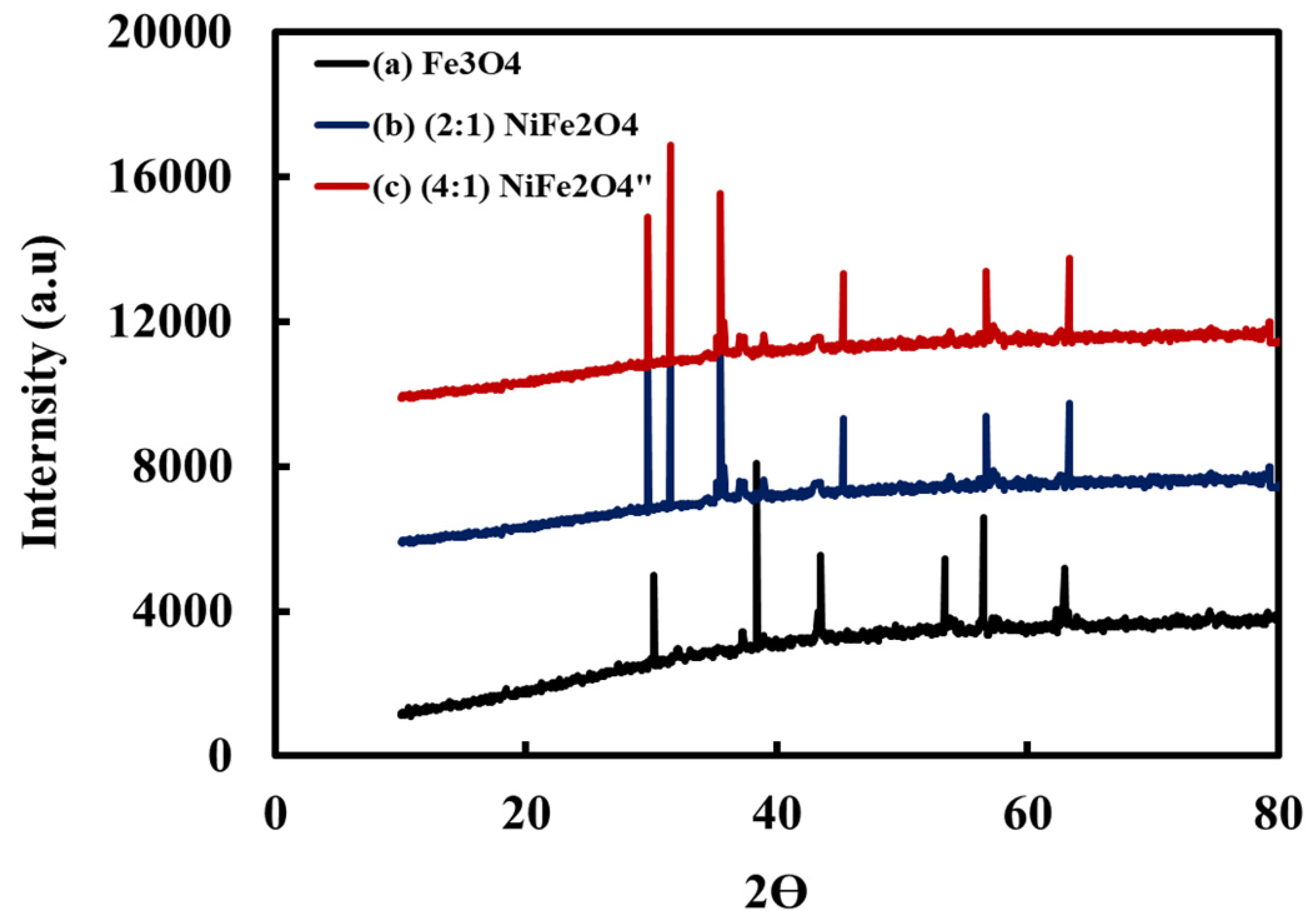
3.3. Crystal Structure
3.4. In Vitro Cytotoxicity
4. Conclusions
Author Contributions
Funding
Institutional Review Board Statement
Informed Consent Statement
Data Availability Statement
Acknowledgments
Conflicts of Interest
References
- Campos, E.; Denise, V.; José, I.; Elizabeth, C.; Rita, C. Synthesis, characterization and applications of iron oxide nanoparticles—A short review. J. Aerosp. Technol. Manag. 2015, 7, 267–276. [Google Scholar] [CrossRef] [Green Version]
- Vedrtnam, A.; Kishor, K.; Sunil, D.; Aman, K. A comprehensive study on structure, properties, synthesis and characterization of ferrites. AIMS Mater. Sci. 2020, 7, 800–835. [Google Scholar] [CrossRef]
- Unsoy, G.; Gunduz, U.; Oprea, O.; Ficai, D.; Sonmez, M.; Radulescu, M.; Alexie, M.; Ficai, A. Magnetite: From synthesis to applications. Curr. Top. Med. Chem. 2015, 15, 1622–1640. [Google Scholar] [CrossRef]
- Rashdan, S.A.; Hazeem, L.J. Synthesis of spinel ferrites nanoparticles and investigating their effect on the growth of microalgae Picochlorum sp. Arab. J. Basic Appl. Sci. 2020, 27, 134–141. [Google Scholar] [CrossRef] [Green Version]
- Klekotka, U.; Dariusz, S.; Simo, S.; Beata, K. Influence of atomic doping on thermal stability of ferrite nanoparticles—Structural and Magnetic Studies. Materials 2021, 14, 100. [Google Scholar] [CrossRef] [PubMed]
- Kharisov, B.; Kharissova, O.; Rasika Dias, H. Iron-Based Nanomaterials in the Catalysis; InTech: Rijeka, Croatia, 2016; pp. 50–55. [Google Scholar]
- Shao, H.; Yoon, T.; Liong, M. Magnetic nanoparticles for biomedical NMR-based diagnostics. Beilstein J. Nanotechnol. 2010, 1, 142–154. [Google Scholar] [CrossRef] [Green Version]
- Tolani, S.C.; Golhar, A.; Rewatkar, K. A review of morphological, structural behaviour and technological applications of ferrites. AIP Conf. Proc. 2019, 2104, 030032. [Google Scholar]
- Avval, Z.; Malekpour, L.; Raeisi, F. Introduction of magnetic and supermagnetic nanoparticles in new approach of targeting drug delivery and cancer therapy application. Drug Metab. Rev. 2020, 52, 157–184. [Google Scholar] [CrossRef]
- Kaur, M.; Kaur, N. Ferrites: Synthesis and applications for environmental remediation, in Ferrites and Ferrates: Chemistry and Applications in Sustainable Energy and Environmental Remediation. ACS Publ. 2016, 113–136. [Google Scholar] [CrossRef]
- Sharma, J.; Pratibha, S.; Gurdip, S.; Hardev, S. Nanoferrites of transition metals and their catalytic activity. In Solid State Phenomena; Trans Tech Publications Ltd.: Bäch, Switzerland, 2016; Volume 241, pp. 126–138. [Google Scholar]
- Lagashetty, A.; Pattar, A.; Ganiger, S.K. Synthesis, characterization and antibacterial study of Ag doped magnesium ferrite nanocomposite. Heliyon 2019, 5, 1760. [Google Scholar] [CrossRef] [Green Version]
- Amiri, M.; Pardakhti, A.; Ahmadi-Zeidabadi, M. Magnetic nickel ferrite nanoparticles: Green synthesis by Urtica and therapeutic effect of frequency magnetic field on creating cytotoxic response in neural cell lines. Colloids Surf. B Biointerfaces 2018, 172, 244–253. [Google Scholar] [CrossRef] [PubMed]
- Majidi, S.; Fatemeh, Z.; Samad, M.; Soleymani, M.; Akbarzadeh, A. Current methods for synthesis of magnetic nanoparticles. Artif. Cells Nanomed. Biotechnol. 2016, 44, 722–734. [Google Scholar] [CrossRef] [PubMed]
- Kaur, R.; Abshar, H.; Nusrat, I.; Samsul, A.; Mahesh, K.; Syed Kalbe, R. Synthesis and surface engineering of magnetic nanoparticles for environmental cleanup and pesticide residue analysis: A review. J. Sep. Sci. 2014, 37, 1805–1825. [Google Scholar] [CrossRef] [PubMed]
- Sharma, R.K.; Dutta, S.; Sharma, S.; Zboril, R.; Varma, R. Fe3O4 (iron oxide)-supported nanocatalysts: Synthesis, characterization and applications in coupling reactions. Green Chem. 2016, 18, 3184–3209. [Google Scholar] [CrossRef]
- Kefeni, K.K.; Msagati, T.A.; Mamba, B.B. Ferrite nanoparticles: Synthesis, characterization and applications in electronic device. Mater. Sci. Eng. B 2017, 215, 37–55. [Google Scholar] [CrossRef]
- Egizbek, K.; Kozlovskiy, A.; Ludzik, K.; Zdorovets, M. Stability and cytotoxicity study of NiFe2O4 nanocomposites synthesized by co-precipitation and subsequent thermal annealing. Ceram. Int. 2020, 46, 16548–16555. [Google Scholar] [CrossRef]
- Dippong, T.; Levei, E.A.; Cadar, O. Recent advances in synthesis and applications of MFe2O4 (M = Co, Cu, Mn, Ni, Zn) nanoparticles. Nanomaterials 2021, 11, 1560. [Google Scholar] [CrossRef]
- Karunakaran, G.; Jagathambal, M.; Van Minh, N. Green synthesis of NiFe2O4 spinel-structured nanoparticles using Hydrangea paniculata flower extract with excellent magnetic property. Miner. Met. Mater. Soc. 2018, 70, 1337–1343. [Google Scholar] [CrossRef]
- Nasrollahzadeh, M.; Mohaddeseh, S.; Mohammad, S. Green Nanotechnology, Interface Science and Technology; Elsevier: Amsterdam, The Netherlands, 2019; pp. 145–198. [Google Scholar]
- Purohit, J.; Chattopadhyay, A.; Singh, N. Green synthesis of microbial nanoparticle: Approaches to application, Microbial Nanobionics; Springer: Berlin/Heidelberg, Germany, 2019; pp. 35–60. [Google Scholar]
- Sorbiun, M.; Shayegan Mehr, E.; Ramazan, A. Biosynthesis of metallic nanoparticles using plant extracts and evaluation of their antibacterial properties. Nanochem. Res. 2018, 3, 1–16. [Google Scholar]
- Jagessar, R. Plant Extracts Based Nanoparticles, a Good Perspective in the Development of Drugs in Nanomedicine. Mod. Approaches Drug Des. 2020, 3, 556. [Google Scholar] [CrossRef]
- Mollarasouli, F.; Zor, E.; Ozcelikay, G.; Ozkan, S. Magnetic nanoparticles in developing electrochemical sensors for pharmaceutical and biomedical applications. Talanta 2021, 226, 122108. [Google Scholar] [CrossRef] [PubMed]
- Wu, K.; Su, D.; Liu, J.; Saha, R.; Wang, J. Magnetic nanoparticles in nanomedicine: A review of recent advances. Nanotechnology 2019, 30, 502003. [Google Scholar] [CrossRef] [PubMed] [Green Version]
- Al-Qasmi, N. Facial eco-friendly synthesis of copper oxide nanoparticles using chia seeds extract and evaluation of its electrochemical activity. Processes 2021, 9, 2027. [Google Scholar] [CrossRef]
- Malik, A.R.; Muhammad, H.; Muhammad, A.; Muhammad, S.; Hafeez, U.; Tuna, N.; Hijaz, A.; Shafiq, A.; Thongchai, B. Lime peel extract induced NiFe2O4 NPs: Synthesis to applications and oxidative stress mechanism for anticancer, antibiotic activity. J. Saudi Chem. Soc. 2022, 26, 101422. [Google Scholar] [CrossRef]
- Amulya, M.; Nagaswarupab, H.; Anil, M.; Ravikumara, C.; Prashanthaa, S.; Kusumaa, K. Sonochemical synthesis of NiFe2O4 nanoparticles: Characterization and their photocatalytic and electrochemical applications. Appl. Surf. Sci. Adv. 2020, 1, 100023. [Google Scholar] [CrossRef]
- Vázquez-Vélez, E.; Martínez, H.; Castillo, F. Degradation of Acid Red 1 Catalyzed by Peroxidase Activity of Iron Oxide Nanoparticles and Detected by SERS. Nanomaterials 2021, 11, 3044. [Google Scholar] [CrossRef]
- Chelike, D.K.; Ananthan, A.; Joydev, A.; Pawan, K.; Koustav, S.; Senthil, A.; Thangavelua, V. Functionalized iron oxide nanoparticles conjugate of multi-anchored Schiff’s base inorganic heterocyclic pendant groups: Cytotoxicity studies. Appl. Surf. Sci. 2020, 501, 143963. [Google Scholar] [CrossRef]
- Thirupathy, C.; Cathrin, S.; Limsa, S.; Sundarama, A.; Hossam, M.; Kaviyarasucd, K. Equilibrium synthesis and magnetic properties of BaFe12O19/NiFe2O4 nanocomposite prepared by co precipitation method. J. King Saud Univ.-Sci. 2020, 32, 1612–1618. [Google Scholar] [CrossRef]
- Yew, Y.; Kamyar, S.; Mikio, M.; Noriyuki, K.; Nurul, B.; Ahmad, K.; Shaza, E.; Kar, X. Green synthesis of magnetite (Fe3O4) nanoparticles using seaweed (Kappaphycus alvarezii) extract. Nanoscale Res. Lett. 2016, 11, 276. [Google Scholar] [CrossRef] [Green Version]
- Khoso, W.A.; Noor, H.; Muhammad, A.; Yousuf, J. Synthesis, characterization and heavy metal removal efficiency of nickel ferrite nanoparticles (NFN’s). Sci. Rep. 2021, 11, 3790. [Google Scholar] [CrossRef]
- Patrizia, L.; Antonio, G.; Rossella, F.; Antonella, A.; Eleonora, M.; Alessio, B.; Luigi, G.; Valli, D.; Nicola, S.; Vito, R. The Evolving Role of Immune Checkpoint Inhibitors in Hepatocellular Carcinoma Treatment. Vaccines 2021, 9, 532. [Google Scholar]


Publisher’s Note: MDPI stays neutral with regard to jurisdictional claims in published maps and institutional affiliations. |
© 2022 by the authors. Licensee MDPI, Basel, Switzerland. This article is an open access article distributed under the terms and conditions of the Creative Commons Attribution (CC BY) license (https://creativecommons.org/licenses/by/4.0/).
Share and Cite
Al-Qasmi, N.; Almughem, F.A.; Jarallah, S.J.; Almaabadi, A. Efficient Green Synthesis of (Fe3O4) and (NiFe2O4) Nanoparticles Using Star Anise (Illicium verum) Extract and Their Biomedical Activity against Some Cancer Cells. Materials 2022, 15, 4832. https://doi.org/10.3390/ma15144832
Al-Qasmi N, Almughem FA, Jarallah SJ, Almaabadi A. Efficient Green Synthesis of (Fe3O4) and (NiFe2O4) Nanoparticles Using Star Anise (Illicium verum) Extract and Their Biomedical Activity against Some Cancer Cells. Materials. 2022; 15(14):4832. https://doi.org/10.3390/ma15144832
Chicago/Turabian StyleAl-Qasmi, Noha, Fahad A. Almughem, Somayah J. Jarallah, and Amani Almaabadi. 2022. "Efficient Green Synthesis of (Fe3O4) and (NiFe2O4) Nanoparticles Using Star Anise (Illicium verum) Extract and Their Biomedical Activity against Some Cancer Cells" Materials 15, no. 14: 4832. https://doi.org/10.3390/ma15144832
APA StyleAl-Qasmi, N., Almughem, F. A., Jarallah, S. J., & Almaabadi, A. (2022). Efficient Green Synthesis of (Fe3O4) and (NiFe2O4) Nanoparticles Using Star Anise (Illicium verum) Extract and Their Biomedical Activity against Some Cancer Cells. Materials, 15(14), 4832. https://doi.org/10.3390/ma15144832