Article

Liquid-Phase-Assisted Catalytic Nitridation of Silicon and In Situ Growth of α-Si3N4

1
The State Key Laboratory of Refractories and Metallurgy, Wuhan University of Science and Technology, Wuhan 430081, China
2
Guangzhou Newlife New Material Co., Ltd., Guangzhou 511356, China
*
Authors to whom correspondence should be addressed.
Materials 2022, 15(17), 6074; https://doi.org/10.3390/ma15176074
Submission received: 19 July 2022 / Revised: 24 August 2022 / Accepted: 30 August 2022 / Published: 1 September 2022

Abstract

:
Si3N4 powders were synthesized with Fe, Co, or Ni as catalysts using Si powder at 1250 °C in a nitrogen atmosphere by liquid-phase-assisted catalytic nitridation synthesis (LPA–CNS). The catalytic effects of the metals on the nitridation of silicon powder were investigated by mixing the powder with 2 wt% by mass of Fe, Co, or Ni in a high-temperature liquid phase in flowing nitrogen. The α-Si3N4 micro-morphology could be effectively changed by adjusting the type of catalyst in the initial reaction mixtures. Fe, Co, and Ni promoted the formation of α-Si3N4 at 1250 °C and controlled the morphology of the α-Si3N4 particles. The hexagonal flakes of α-Si3N4 with a better defined morphology were obtained using Ni as the catalyst, compared to that obtained from the other two catalysts.

1. Introduction

Si3N4-based ceramics have been widely used in steel, aerospace, chemical, and electronic fields due to their strong oxidation resistance, stable chemical properties, and excellent mechanical properties [1,2,3,4,5]. In general, α-Si3N4 powder is preferred to β-Si3N4 powder as a raw material for fabricating compacted bodies because the α-Si3N4 starting powder has a higher reactivity that easily forms elongated Si3N4 grain morphology, which has excellent mechanical properties [4,5]. Due to these appealing properties, Si3N4 has been widely studied and has become the most promising structural material, especially for high temperature applications [6,7]. However, the high cost of fine Si3N4 powders is the prime obstacle to the regular application of Si3N4-based materials. Therefore, the fabrication of α-Si3N4 powders with tailored properties through a cost-effective synthesis method is a research topic of great interest.
At present, the preparation methods of silicon nitride are mainly divided into organic preparation methods and inorganic preparation methods. The organic method is mainly used to synthesize precursors and to prepare silicon nitride through the pyrolysis of the precursors [8,9,10]. Some inorganic preparation methods have been adopted to prepare silicon nitride powder, including the nitridation of Si powder [11], carbothermal reduction nitridation [12,13], and combustion synthesis [14,15]. However, these techniques still suffer various disadvantages, such as a broad particle size distribution, difficulty in size and composition control, powder reunion, high nitridation temperature, and long holding time. High temperature liquid phase synthesis method is an environmentally friendly technology, widely applied in the preparation of powder [16,17,18,19], nano coating [20,21,22,23], and porous materials [24,25,26]. However, there are only a few available reports on the high temperature liquid phase synthesis of silicon nitride powder.
It was well known that transition elements can regulate the kinetics of Si nitridation and accelerate the growth of α-Si3N4 [27,28]. It should be noted that, although it has been reported that metals, metal oxides, metal nitrates, or metal nanoparticles were used as a catalyst for the accelerated nitridation of Si powder [29,30,31,32,33,34], most of the previous studies have focused on the limited choices of catalysts and complex synthesis methods. In addition, the enhancement of α-Si3N4 formation is strictly limited to the presence of molten alloy liquid phases [34,35] but most catalysts do not play a distinctly accelerated role in the information of α-Si3N4 powder. Other research groups have made some attempts on controllable preparation of α-Si3N4 [36,37] including our previous contribution [38,39,40]. However, the influence of different transition metal catalysts on the fabrication of α-Si3N4 powder is still unclear.
In this study, the synthesis of Si3N4 powder with different microstructures from by liquid-phase-assisted catalytic nitridation synthesis (LPA–CNS) in the NaCl–NaF molten salt system. The nitridation processes were carried out simultaneously with the three metal catalysts under identical reaction conditions, such as nitridation temperature, nitridation time and catalyst amount.

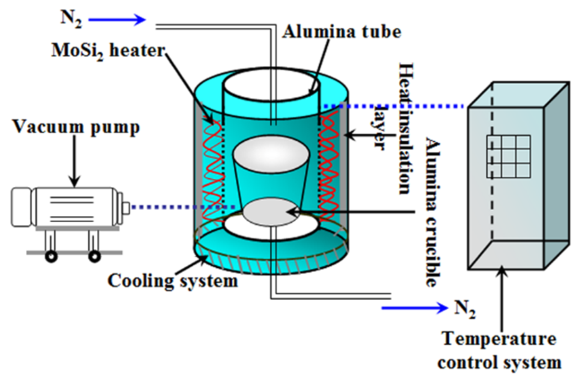
2. Materials and Methods
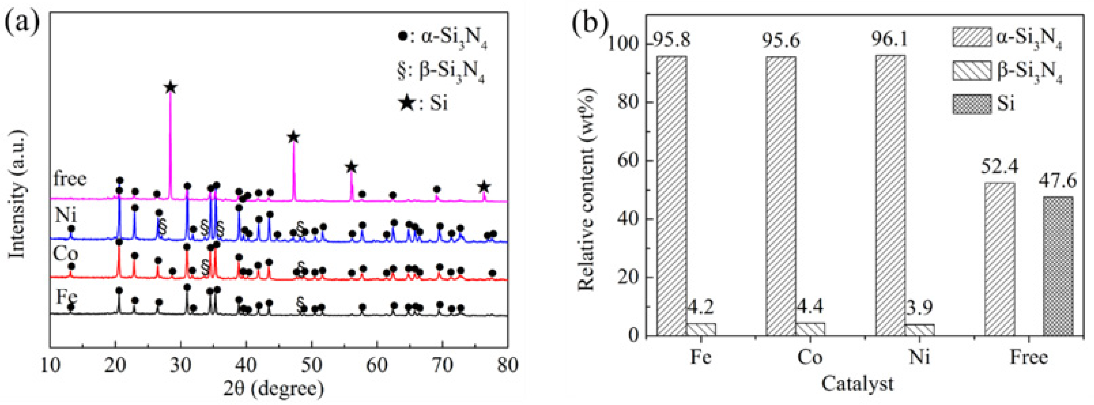
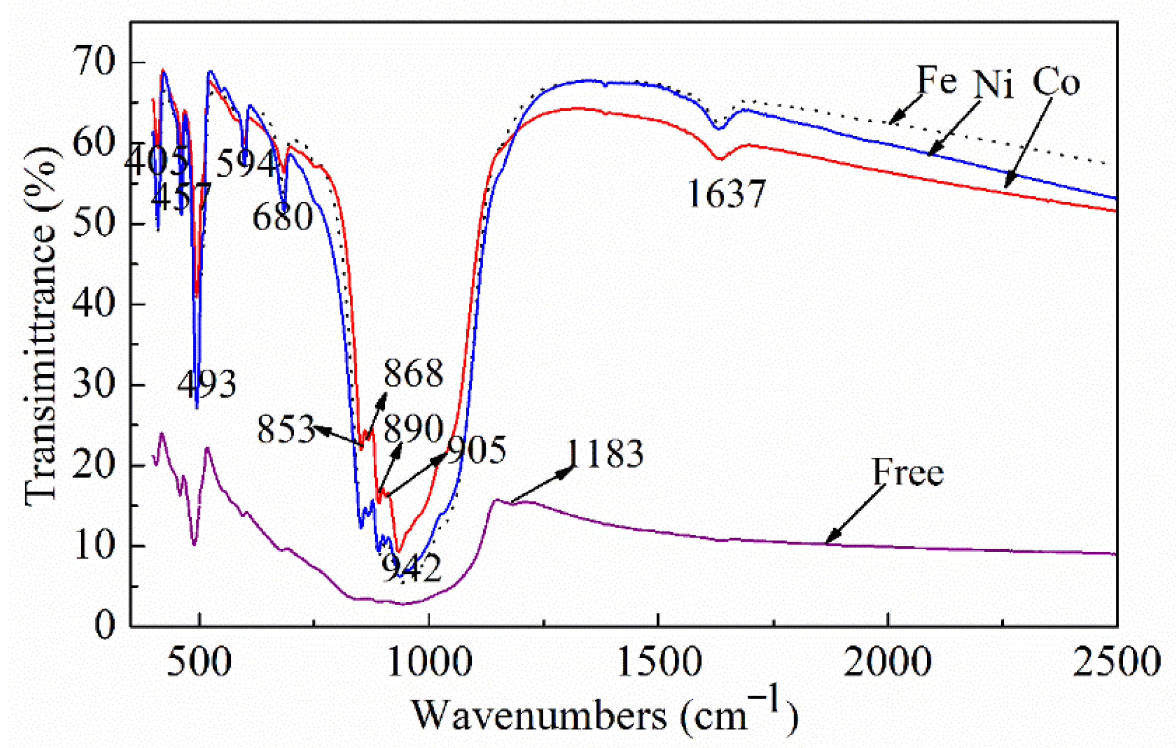
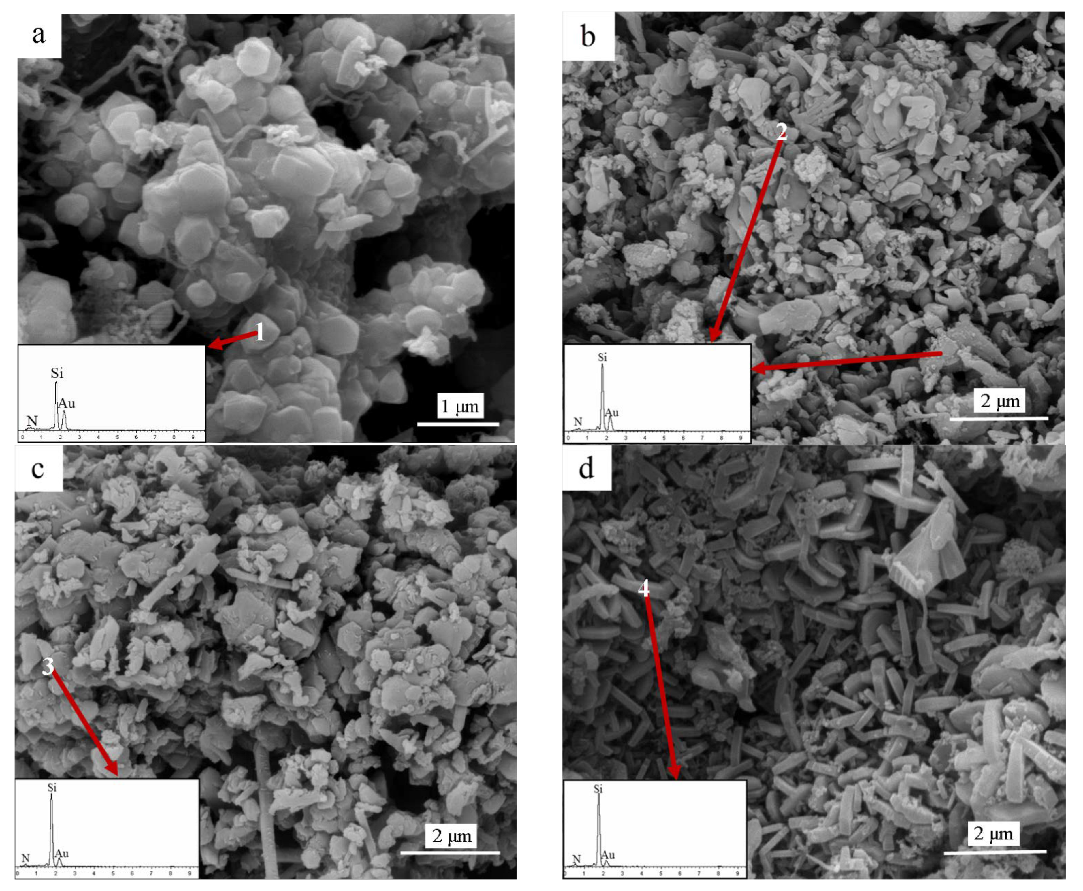
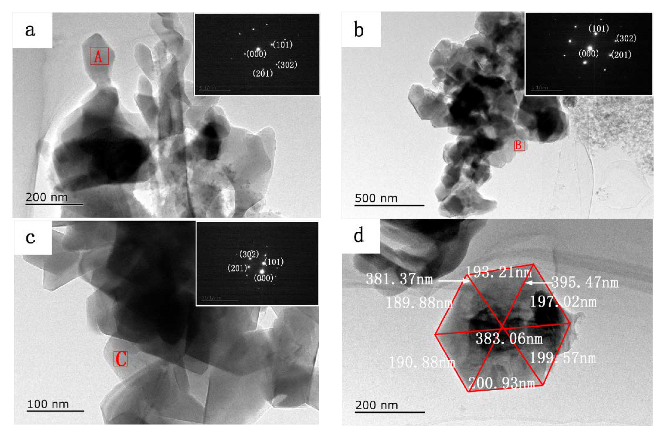
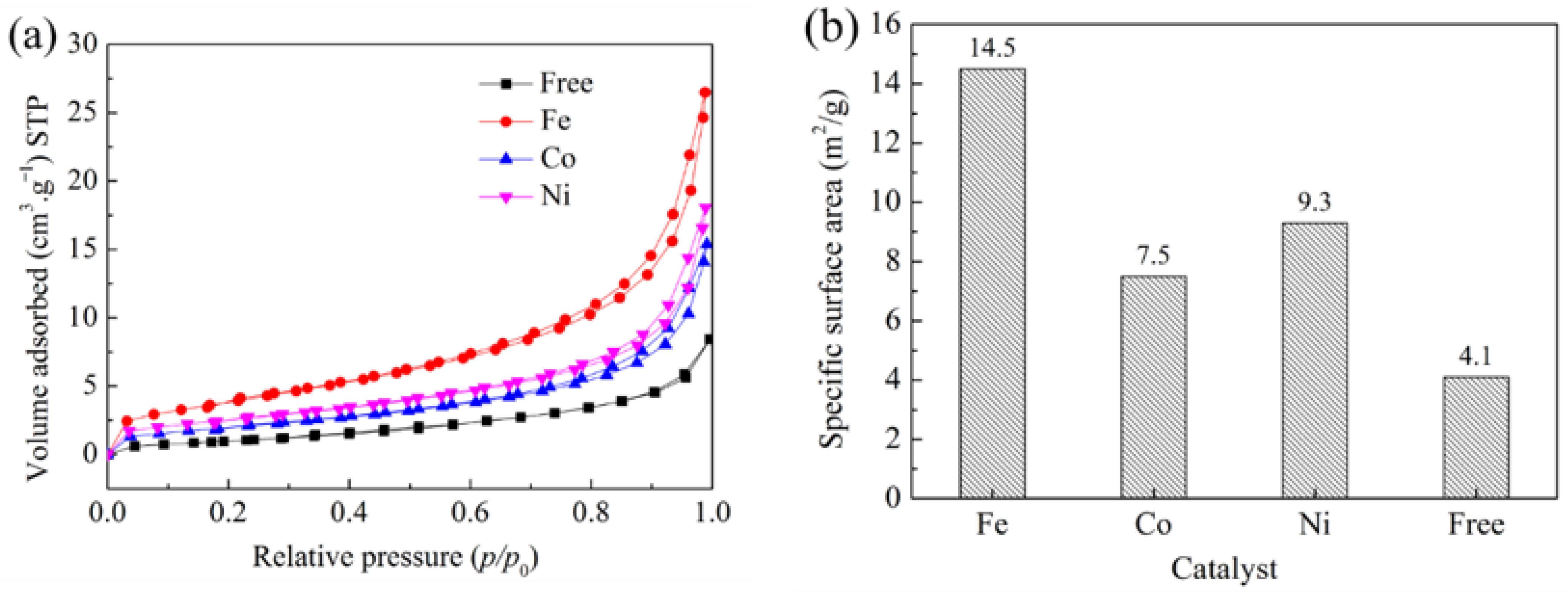
The details of the synthesis process are described as follows: 35 wt% Si powder (99% purity, grain size 9 μm, Beijing Xing Rong Yuan Technology Co. Ltd., Beijing, China); 65 wt% analytical grade salt mixture composed of 95 wt% sodium chloride (NaCl, 99% purity, Sinopharm Chemical Reagent Co., Ltd., Shanghai, China) and 5 wt% sodium fluoride (NaF, 99% purity, Sinopharm Chemical Reagent Co., Ltd., Shanghai, China); and 2 wt% Fe, Co, or Ni powder (99.99% purity, grain size 2 μm, Beijing Xing Rong Yuan Technology Co., Ltd., Beijing, China) were dry-mixed in an alumina mortar for 1 h. Then, five grams of the mixed powder containing Si and Na salts and the catalyst were placed in an alumina crucible. The nitridation of the silicon powder was carried out in a vertical alumina tube furnace, which is shown as a diagram in Figure 1 together with the gas supply system and vacuum system. Since the reaction between N2 and Si is highly exothermic, the samples were initially heated at 1150 °C for 1 h in the alumina-tube furnace. The reaction temperature was then raised to 1250 °C and the reaction was continued for 5 h under a nitrogen atmosphere. After cooling to 25 °C under nitrogen, the as-obtained solidified mass was repeatedly washed with hot distilled water and filtered to remove the residual salts. Finally, the obtained silicon nitride paste, prior to its characterization, was dried in oven at 110 °C for 12 h.
The product was collected, and samples were prepared accordingly for various characterization techniques. The phase composition of the as-prepared samples was identified by X-ray diffraction (XRD, X’Pert Pro, Philips, Eindhoven, The Netherlands) analysis. The spectra from 10° to 80° (2θ) were recorded using Cu Kα radiation (λ = 0.154 nm) at 40 mA and 40 kV, at a scan rate of 2°/min and a step size of 0.03°. ICSD cards No. 035561, 074749 and 043403 were cited, respectively. The infrared (IR) spectra of the samples were recorded on a Fourier transform infrared (FTIR, NICOLET iS50, Thermo Fisher Scientific, Waltham, MA, USA) spectrometer in the range 300–4000 cm−1. The microstructure of the samples was characterized using a field-emission scanning electron microscope (FESEM, Nova nano 400, FEI, Waltham, MA, USA) and a transmission electron microscope (TEM, JEM2100, JEOL, Tokyo, Japan). Samples for SEM analysis were coated with gold. Samples for TEM analysis were dispersed in absolute ethanol by ultrasonication, and a drop of the suspension was placed on a copper grid and air-dried. Selected-area electron diffraction (SAED) along with energy dispersive spectroscopy (EDS) was used to identify the phase composition of the samples. Finally, the property of powder was characterized by Brunauer–Emmett–Teller (BET, Autosorb-1-MP/LP, Quantachrome, Boynton Beach, FL, USA).

3. Results

A series of experiments were performed in which the type of catalyst was varied. There are amounts of unreacted Si and a small amount of α-Si3N4 in the catalyst-free sample, which indicates that only a small amount of Si has been nitrided, as shown in Figure 2a. However, it can be clearly seen that the residual Si peaks (corresponding to (111), (220), (311) and (331)) distinctly decreased or disappeared. Meanwhile, both α-Si3N4 and β-Si3N4 were identified in the samples containing catalyst, indicating that Si powder should be nitrided completely under the effects of Fe, Co, and Ni catalysts. Moreover, Ni catalysts give a higher intensity of α-Si3N4 phase when compared with using Co and Fe, indicating that the amount of α-Si3N4 in the samples containing Ni is more than those in samples containing Co or Fe. The relative contents of crystalline phases in the samples with various catalysts after 5 h nitridation at 1250 °C were calculated by the Rietveld refinement method, and the results are shown in Figure 2b. The addition of transition metal catalyst in Si powder led to a higher content of α-Si3N4 phase than that obtained from the catalyst-free sample. Only 52.4 wt% α-Si3N4 was formed in the catalyst-free samples. On the contrary, the samples containing the Fe, Co, and Ni catalysts demonstrated over 95.6% α-Si3N4 and less than 4.4% β-Si3N4 in the final product. Therefore, adding these transition metals into silicon raw material is a promising choice for the quality improvement of the α-Si3N4 produced by the high temperature liquid phase synthesis method.
The as-prepared α-Si3N4 samples were further characterized by FTIR. The FTIR spectra of the sample in Figure 3 show two absorption bands, one at 800–1100 cm−1 with a peak maximum at ~942 cm−1 and the other at 400–600 cm−1 with a peak maximum at ~493 cm−1, which correspond to the Si-N-Si asymmetric and symmetric stretching modes, respectively [41]. The bands at 942, 890, 853, 680, 594, 493, 457, and 405 cm−1 indicate the distinct absorptions of α-Si3N4 [42]. These bands become sharper in the spectra of the samples with the added catalysts, indicating the formation of a well-defined crystal structure of the α-Si3N4 phase. The peak at 1183 cm−1 corresponds to the stretching vibration of Si–Si bonds owing to unreacted silicon in the catalyst-free sample [43]. The peak at 1637 cm−1 is attributed to the water absorbed during the analysis [44]. The superimposed IR spectrum of α-Si3N4 further confirms the formation of α-Si3N4 as the primary phase in the samples, corroborating with the XRD results. In addition, it confirms the presence of residual Si in the catalyst-free sample, as shown in Figure 3.
The detailed microstructure and elemental composition of the as-prepared Si3N4 samples were examined by FESEM and EDS analysis (insets in Figure 4 and Table 1). SEM images of the samples containing 2 wt% Fe, Co, and Ni are shown in Figure 4. The nitridation product mainly consists of irregularly elongated particles, equiaxed flakes, and short rod-like and hexagonal plate-like grains. As shown in Figure 4a, the catalyst-free sample contains many equiaxial grains with diameters in the micron range, and then numerous grains aggregate to form large particles. Spectra obtained from EDS analysis, given as the inset in Figure 4a, confirm the presence of only N and Si in the granular particles. As seen in Figure 4b, a number of anomalous elongated grains are observed for the samples obtained using the Fe catalyst. Coarse granular agglomerates, composed of α-Si3N4 particles, are randomly distributed in the samples, as shown in Figure 4b. The SEM image in Figure 4c reveals that coarse equiaxial flakes and glossy rod-like grains form agglomerates on the surface of the sample obtained using Co powder as the catalyst, similar to our previous results [38]. On the other hand, the SEM image of the sample obtained using the Ni catalyst (Figure 4d) indicates the formation of well-developed hexagonal plate-like α-Si3N4 grains with the thicknesses of several dozen nanometers, and these grains are randomly distributed in the sample. EDS results (insets in Figure 4b–d and Table 1) show that the anomalous elongated grains, flake-like and rod-like particles, and hexagonal plates consist of Si and N; the elemental amounts correspond to the Si3N4 phase, in agreement with the XRD and IR results.
TEM observations along with SAED analysis were further carried out to identify the crystalline structure of the Si3N4 samples generated at 1250 °C. Figure 5 shows the transmission electron microscopy and SAED images of the samples obtained with Fe, Co, and Ni as catalysts. The TEM image of the sample obtained with Fe as the catalyst (Figure 5a) shows that elongated and granular grains agglomerate to form larger granules. The equiaxial α-Si3N4 flakes aggregate together to form large particles in the product obtained with Co as the catalyst (Figure 5b), as seen in the corresponding SEM image. The TEM image of the sample obtained with Ni as the catalyst (Figure 5c) shows hexagonal Si3N4 plates. The related SAED patterns (insets in Figure 5a–c) confirm that the anomalous elongated grains, flake-like and rod-like particles, and hexagonal plates comprise perfect single-crystalline α-Si3N4, wherein the diffraction spots correspond to the (201) and (101) lattice planes of α-Si3N4 (ICDD card No. 074749), in agreement with the SEM analysis. Figure 5d presents a high-magnification TEM image of the sample, showing a hexagonal plate with edge lengths of about 193 nm, 190 nm, 191 nm, 201 nm, 200 nm, and 197 nm with an α-Si3N4 crystal structure. Combined with the SEM results, an independent α-Si3N4 hexagonal plate with a diameter size of about 395 nm and edge length of about 197 nm can clearly be seen.
Figure 6a shows nitrogen adsorption–desorption isotherms of samples containing different catalysts heated at 1250 °C for 5 h in NaCl–NaF molten salt media. The nitrogen adsorption capacity of samples increased sharply, and the adsorption curve was biased towards the y axis at the low pressure area (P/P0 = 0.0–0.1), indicating that there were stable micropores in the samples. Moreover, the adsorption capacity increased slowly at P/P0 = 0.3–0.8 and increased significantly at P/P0 = 0.9–1.0. It can be confirmed that the adsorption–desorption isotherms of samples are typical type II, according to the IUPAC classification [45]. It is worth noting that the adsorption isotherms of samples, through the addition of catalysts, had a long lagged loop at a high relative pressure area, indicating that there were many mesopores in the samples. On the other hand, the adsorption capacity of sample with Fe catalyst was larger than that of the other two samples. However, the adsorption isotherm of catalyst-free sample has no hysteresis loop. Figure 6b shows the specific surface area of samples without catalyst or by addition of Fe, Co, and Ni powder as catalyst. As shown in Figure 6b, the specific surface area of the sample without catalyst was 4.1 m2/g. In contrast, the specific surface area of samples with Fe, Co, and Ni powder was obviously improved. When Fe powder is used as a catalyst, the specific surface area of the sample reaches 14.5 m2/g.
During Si3N4 synthesis in molten salt system, silicon powder is slightly soluble in the molten salt system under high temperature conditions, and the dissolved silicon exists in the form of atoms or atomic groups. As the temperature increases, the surface tension and viscosity of the molten salt decrease significantly, and the dissolution and diffusion of silicon powder in the molten salt intensifies [22,39]. The introduction of catalysts has a crucial effect on the composition and microstructure of Si3N4 powder in the NaCl–NaF molten salt media. It was worth noting that the crystal shape of α-Si3N4 could be converted to anomalously elongated, granular, or hexagonal plate-like form by the addition of Fe, Co, or Ni. When metal catalysts are present in the samples, the mechanism of Si powder nitridation in the samples is that M-Si (M = Fe, Co and Ni) liquid phase was formed by reactions between metal (Fe, Co or Ni) and Si at low temperature. Moreover, the nucleation and growth of Si3N4 were controlled by different reactions in the molten salt media. Firstly, Si3N4 nucleation was mainly controlled by gas–solid reaction between dissolved Si atom and N2. While the growth of Si3N4 grain was established to be reaction between Si(g)/Si(l) and N/N2 in the system. However, there began to appear liquid phase in the samples containing Fe, Co, and Ni catalysts at different temperature. Therefore, amount of Si(g)/Si(l) in the system varied with catalyst type at nitridation temperature, which also marked Si3N4 crystal preferential growth in different directions. A mechanism for the catalytic of nitridation of Si powder is shown in Figure 7.

4. Conclusions

In summary, to meet the high-quality powder requirements for the preparation of ceramic substrates with low grain boundary size defects, a high α form Si3N4 powder with multiple morphologies was obtained through the nitridation of Si in molten state via the reaction of Si powder mixed with Na salts under N2 atmosphere in the presence of various transition metal catalysts. The nitridation of Si powder with 2 wt% catalysts (Fe, Co, or Ni) was carried out at 1250 °C for 5 h. While only 52.4 wt% α-Si3N4 was present in the catalyst-free sample, there were about 96 wt% α-Si3N4 and 4 wt% β-Si3N4 in the samples obtained using 2 wt% Ni, Fe, and Co, under the same reaction conditions. The addition of Fe, Co, and Ni catalysts not only promoted the formation of α-Si3N4 but also influenced the morphology of α-Si3N4 particles. When using Fe as the catalyst, several anomalously elongated α-Si3N4 grains were obtained, and randomly distributed in the sample. When using Co and Ni as the catalysts, equiaxial flake-like and rod-like α-Si3N4 particles, and hexagonal plate-like α-Si3N4 particles were obtained, respectively. The growth mechanism of α-Si3N4 was confirmed, which was mainly a vapor–solid or vapor–liquid–solid pattern.

Author Contributions

Conceptualization, Z.L. and J.D.; investigation, Z.L. and Z.C.; data curation, writing—original draft preparation, Z.L., Z.C., C.Y., J.D., C.D., X.L. and H.Z.; supervision, funding acquisition, J.D. and X.L.; writing-reviewing and editing, Z.L., Z.C. and J.D. All authors have read and agreed to the published version of the manuscript.

Funding

This research was funded by the Key Research and Development Project of Hubei Province (Grant No. 2020BAA028), the Science Fund for Creative Research Groups of the National Natural Science Foundation of Hubei Province (Grant No.2020CFA038), and the National Natural Science Foundation of China (Grant No. 51972242 and U20A20239).

Institutional Review Board Statement

Not applicable.

Informed Consent Statement

Not applicable.

Data Availability Statement

Data available on request due to privacy restrictions. The data presented in this study are available on request from the corresponding author.

Conflicts of Interest

The authors declare that they have no known competing financial interests or personal relationships that could have appeared to influence the work reported in this paper.

References

  1. Riley, F.L. Silicon nitride and related materials. J. Am. Ceram. Soc. 2000, 83, 245–265. [Google Scholar] [CrossRef]
  2. Krstic, Z.; Krstic, V.D. Silicon nitride: The engineering material of the future. J. Mater. Sci. 2012, 47, 535–552. [Google Scholar] [CrossRef]
  3. Collins, J.F.; Gerby, R.W. New refractory uses for silicon nitride reported. J. Mater. Sci. 1955, 5, 612–615. [Google Scholar] [CrossRef]
  4. Zhu, X.W.; Zhou, Y.; Hirao, K.; Lenčéš, Z. Processing and thermal conductivity of sintered reaction-bonded silicon nitride. I: Effect of Si powder characteristics. J. Am. Ceram. Soc. 2006, 89, 3331–3339. [Google Scholar] [CrossRef]
  5. Xing, H.Y.; Liu, B.Q.; Sun, J.; Zou, B. Mechanical properties of Si3N4 ceramics from an in-situ synthesized α-Si3N4/β-Si3N4 composite powder. Ceram. Int. 2017, 43, 2150–2154. [Google Scholar] [CrossRef]
  6. Bocanegra-Bernal, M.H.; Matovic, B. Mechanical properties of silicon nitride-based ceramics and its use in structural applications at high temperatures. Mater. Sci. Eng. A 2010, 527, 1314–1338. [Google Scholar] [CrossRef]
  7. Santos, C.; Ribeiro, S.; Strecker, K.; Rodrigues, D., Jr.; Silva, C.R.M. Highly dense Si3N4 crucibles used for A1 casting: An investigation of the aluminum ceramic interface at high temperatures. J. Mater. Process. Tech. 2007, 184, 108–114. [Google Scholar] [CrossRef]
  8. Seyferth, D.; Wiseman, G.H.; Prud’homme, C. A liquid silazane precursor to silicon nitride. J. Am. Ceram. Soc. 1983, 66, C-13–C-14. [Google Scholar] [CrossRef]
  9. Burns, G.T.; Chandra, G. Pyrolysis of preceramic polymers in ammonia: Preparation of silicon nitride powders. J. Am. Ceram. Soc. 1989, 72, 333–337. [Google Scholar] [CrossRef]
  10. Isoda, T.; Kaya, H.; Nishii, H.; Funayama, O.; Suzuki, T.; Tashiro, Y. Perhydropolysilazane precursors to silicon nitride ceramics. J. Inorg. Organomet. Polym. 1992, 2, 151–160. [Google Scholar] [CrossRef]
  11. Yao, G.S.; Li, Y.; Jiang, P.; Jin, X.M.; Long, M.L.; Qin, H.X.; Vasant Kumar, R. Formation mechanisms of Si3N4 and Si2N2O in silicon powder nitridation. Solid State Sci. 2017, 66, 50–56. [Google Scholar] [CrossRef]
  12. Chen, K.; Huang, Z.H.; Liu, Y.G.; Fang, M.H.; Huang, J.T.; Xu, Y.G. Synthesis of β-Si3N4 powder from quartz via carbothermal reduction nitridation. Powder Technol. 2013, 235, 728–734. [Google Scholar] [CrossRef]
  13. Ji, H.P.; Huang, Z.H.; Chen, K.; Li, W.J.; Gao, Y.F.; Fang, M.H.; Liu, Y.G.; Wu, X.W. Synthesis of Si3N4 powder with tunable α/β-Si3N4 content from waste silica fume using carbothermal reduction nitridation. Powder Technol. 2014, 252, 51–55. [Google Scholar] [CrossRef]
  14. Wang, Q.S.; Liu, G.H.; Yang, J.H.; Chen, Y.X.; Li, J.T. Preheating-assisted combustion synthesis of β-Si3N4 powders at low N2 pressure. Mater. Res. Bull. 2013, 48, 1321–1323. [Google Scholar] [CrossRef]
  15. Jiang, Y.; Wu, L.E.; Wang, P.L.; Huang, Z.K. Pretreatment and sintering of Si3N4 powder synthesized by the high-temperature self-propagation method. Mater. Res. Bull. 2009, 44, 21–24. [Google Scholar] [CrossRef]
  16. Xue, P.J.; Hu, Y.; Xia, W.R.; Wu, H.; Zhu, X.H. Molten-salt synthesis of BaTiO3 powders and their atomic-scale structural characterization. J. Alloys Comp. 2017, 695, 2870–2877. [Google Scholar] [CrossRef]
  17. Kan, X.Q.; Ding, J.; Zhu, H.X.; Deng, C.J.; Yu, C. Low temperature synthesis of nanoscale titanium nitride via molten-salt mediated magnesiothermic reduction. Powder Technol. 2017, 315, 81–86. [Google Scholar] [CrossRef]
  18. Ding, J.; Guo, D.; Deng, C.J.; Zhu, H.X.; Yu, C. Low-temperature synthesis of nanocrystalline ZrC coatings on flake graphite by molten salts. Appl. Surf. Sci. 2017, 407, 315–321. [Google Scholar] [CrossRef]
  19. Chen, Y.; Deng, C.J.; Wang, X.; Yu, C.; Ding, J.; Zhu, H.X. Evolution of c-ZrN nanopowders in low-carbon MgO–C refractories and their properties. J. Eur. Ceram. Soc. 2021, 41, 963–977. [Google Scholar] [CrossRef]
  20. Ding, J.; Zhu, H.X.; Li, G.Q.; Deng, C.J.; Li, J. Growth of SiC nanowires on wooden template surface using molten salt media. Appl. Surf. Sci. 2014, 320, 620–626. [Google Scholar] [CrossRef]
  21. Zhou, L.; Huang, J.F.; Cao, L.Y.; Ouyang, H.B.; Wu, J.P.; Liu, J.T.; Wang, X.; Hao, W. Synthesis of β-Y2Si2O7 nanowires via a facile molten salt method and their thermal properties. J. Alloys Comp. 2017, 699, 210–215. [Google Scholar] [CrossRef]
  22. Ding, J.; Deng, C.J.; Yuan, W.J.; Zhu, H.X.; Zhang, X.J. Novel synthesis and characterization of silicon carbide nanowires on graphite flakes. Ceram. Int. 2014, 40, 4001–4007. [Google Scholar] [CrossRef]
  23. Li, W.; Wang, X.; Deng, C.J.; Yu, C.; Ding, J.; Zhu, H.X. Molten salt synthesis of Cr3C2-coated flake graphite and its effect on the physical properties of low-carbon MgO–C refractories. Adv. Powder Technol. 2021, 32, 2566–2576. [Google Scholar] [CrossRef]
  24. Ding, J.; Zhu, H.X.; Deng, C.J.; Li, G.Q.; Wang, K.L.; Liu, J.P. Preparation and characterisation of porous biomorphic SiC/C ceramic from molten salt. Ceram. Int. 2015, 41, 11539–11545. [Google Scholar] [CrossRef]
  25. Kan, X.Q.; Ding, J.; Yu, C.; Zhu, H.X.; Deng, C.J.; Li, G.Y. Low-temperature fabrication of porous ZrC/C composite material from molten salts. Ceram. Int. 2017, 43, 6377–6384. [Google Scholar] [CrossRef]
  26. Ding, J.; He, W.Z.; Yu, C.; Liu, L.Z.; Deng, C.J.; Zhu, H.X. Waste biomass derived carbon supported Mo2C/C composite materials for heavy metal adsorption and hydrogen evolution reaction. Mater. Today Commun. 2022, 31, 103646. [Google Scholar] [CrossRef]
  27. Boyer, S.M.; Moulson, A.J. A mechanism for the nitridation of Fe-contaminated silicon. J. Mater. Sci. 1978, 13, 1637–1646. [Google Scholar] [CrossRef]
  28. Pavarajarn, V.; Kimura, S. Catalytic effects of metals on direct nitridation of silicon. J. Am. Ceram. Soc. 2001, 84, 1669–1674. [Google Scholar] [CrossRef]
  29. Gu, Y.J.; Lu, L.L.; Zhang, H.J.; Cao, Y.N.; Li, F.L.; Zhang, S.W. Nitridation of silicon powders catalyzed by cobalt nanoparticles. J. Am. Ceram. Soc. 2015, 98, 1762–1768. [Google Scholar] [CrossRef]
  30. Hyug, H.; Yoshid, K.; Kondo, N.; Kita, H.; Sugai, J.; Okano, H.; Tsuchida, J. Nitridation enhancing effect of ZrO2 on silicon powder. Mater. Lett. 2008, 62, 3475–3477. [Google Scholar] [CrossRef]
  31. Guo, W.M.; Wu, L.X.; Xie, H.; You, Y.; Lin, H.T. Effect of TiO2 additives on nitridation of Si powders. Mater. Lett. 2016, 177, 61–63. [Google Scholar] [CrossRef]
  32. Wang, F.; Qin, X.F.; Yang, L.X.; Meng, Y.F.; Sun, L.X. Synthesis and photoluminescence of Si3N4 nanowires from La/SiO2 composites and Si powders. Ceram. Int. 2015, 41, 1505–1510. [Google Scholar] [CrossRef]
  33. Huang, J.T.; Huang, Z.H.; Yi, S.; Liu, Y.G.; Fang, M.H.; Zhang, S.W. Fe-catalyzed growth of one-dimensional α-Si3N4 nanostructures and their cathodoluminescence properties. Sci. Rep. 2013, 3, 3504. [Google Scholar] [CrossRef] [PubMed]
  34. Pavarajarn, V.; Vongthavorn, T.; Praserthdam, P. Enhancement of direct nitridation of silicon by common metals in silicon nitride processing. Ceram. Int. 2007, 33, 675–680. [Google Scholar] [CrossRef]
  35. Zheng, C.S.; Yan, Q.Z.; Xia, M. Combustion synthesis of SiC/Si3N4-NW composite powders: The influence of catalysts and gases. Ceram. Int. 2012, 38, 4549–4554. [Google Scholar] [CrossRef]
  36. Chen, Y.X.; Yang, J.H.; Jiang, Z.J.; Lin, Z.M.; Li, J.T. Combustion synthesis of α-Si3N4 powder using AC as additive. J. Inorg. Mater. 2012, 27, 169–173. [Google Scholar] [CrossRef]
  37. Cao, Y.G.; Chen, H.; Li, J.T.; Ge, C.C.; Tang, S.Y.; Tang, J.X.; Chen, X. Formation of α-Si3N4 whiskers with addition of NaN3 as catalyst. J. Cryst. Growth 2002, 234, 9–11. [Google Scholar] [CrossRef]
  38. Ding, J.; Zhu, H.X.; Li, G.Q.; Deng, C.J.; Chai, Z.N. Catalyst-assisted synthesis of α-Si3N4 in molten salt. Ceram. Int. 2016, 42, 2892–2898. [Google Scholar] [CrossRef]
  39. Chai, Z.N.; Ding, J.; Deng, C.J.; Zhu, H.X.; Li, G.Q. Ni-catalyzed synthesis of hexagonal plate-like alpha silicon nitride from nitridation of Si powder in molten salt media. Adv. Powder Technol. 2016, 27, 1637–1644. [Google Scholar] [CrossRef]
  40. Chen, Y.; Wang, X.; Deng, C.J.; Yu, C.; Ding, J.; Zhu, H.X. Controllable synthesis of Si@SiC plate@Si3N4 whisker with core-shell structure and their electrochemical performances. Powder Technol. 2021, 377, 324–335. [Google Scholar] [CrossRef]
  41. Alem, A.; Pugh, M.D.; Drew, R.A.L. Reaction bonded silicon nitride foams: The influence of iron disilicide on microstructure and mechanical strength. Ceram. Int. 2015, 41, 4966–4974. [Google Scholar] [CrossRef]
  42. Zhu, H.L.; Han, F.D.; Bi, J.Q.; Bai, Y.J.; Qi, Y.X.; Pang, L.L.; Wang, C.G.; Li, S.J.; Lu, C.W. Facile synthesis of Si3N4 nanocrystals via an organic-inorganic reaction route. J. Am. Ceram. Soc. 2009, 92, 535–538. [Google Scholar] [CrossRef]
  43. Lu, J.G.; Rozgonyi, G.; Rand, J.; Jonczyk, R. Secondary phase inclusions in polycrystalline sheet silicon. J. Cryst. Growth 2004, 269, 599–605. [Google Scholar] [CrossRef]
  44. Batan, A.; Franquet, A.; Vereecken, J.; Reniers, F. Characterization of the silicon nitride thin films deposited by plasma magnetron. Surf. Interface Anal. 2008, 40, 754–757. [Google Scholar] [CrossRef]
  45. Schmit, F.; Bois, L.; Chiriac, R.; Toche, F.; Chassagneux, F.; Descorme, C.; Besson, M.; Khrouz, L. Porous microspheres of manganese-cerium mixed oxides by a polyvinylpyrrolidone assisted solvothermal method. J. Phys. Chem. Solids 2017, 103, 22–32. [Google Scholar] [CrossRef]
Figure 1. Nitridation apparatus for the preparation of α-Si3N4 powder.
Materials 15 06074 g001
Figure 2. XRD patterns of samples and relative content of the crystalline phases in the final products obtained by 5 h of nitridation at 1250 °C using various catalysts. (a) XRD patterns and (b) relative content.
Materials 15 06074 g002
Figure 3. IR spectra of silicon nitride in the samples obtained using various catalysts and the catalyst-free sample.
Materials 15 06074 g003
Figure 4. SEM images and EDS analysis (insets) of final products obtained by nitridation at 1250 °C for 5 h, using various catalysts. (a) catalyst-free, (b) Fe catalyst, (c) Co catalyst, (d) Ni catalyst. The red arrows and the numbers indicate the location of the analysis.
Materials 15 06074 g004
Figure 5. TEM images and SAED patterns (insets, area of A, B, and C) of samples containing obtained using catalysts. (a) 2 wt% Fe, (b) 2 wt% Co, (c) 2 wt% Ni, (d) an individual α-Si3N4 hexagonal plate in the sample obtained using Ni as the catalyst.
Materials 15 06074 g005
Figure 6. Nitrogen adsorption–desorption isotherms and specific surface area of samples containing different catalysts heated at 1250 °C for 5 h in NaCl–NaF molten salt media. (a) nitrogen adsorption–desorption isotherms and (b) specific surface area.
Materials 15 06074 g006
Figure 7. Suggested mechanism for the catalytic nitridation of Si powder.
Materials 15 06074 g007
Table 1. Microarea percent ratio of points 1, 2, 3, and 4 in Figure 4/Atomic%.
ElementNSiAu
140.2550.459.30
248.4044.656.96
337.6355.566.81
451.5541.626.83
Publisher’s Note: MDPI stays neutral with regard to jurisdictional claims in published maps and institutional affiliations.

