
Multiscale Femoral Neck Imaging and Multimodal Trabeculae Quality Characterization in an Osteoporotic Bone Sample

 , , , and
Abstract
:1. Introduction
2. Materials and Methods
2.1. Sample Collection and Preparation
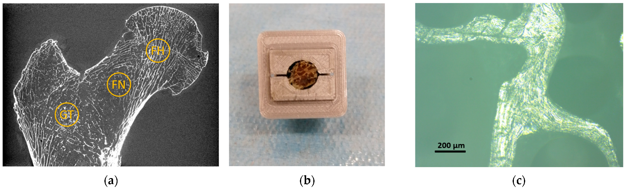
2.2. Sample Extraction and Preparation for Microindentation Test
2.3. X-ray Microtomography Measurements
2.3.1. Image Post-Processing and Analysis
2.3.2. SRµCT (Voxel Size: 0.9 µm)
2.4. Microindentation
2.5. Fourier Transform Infrared Spectroscopy
2.6. Statistical Analysis
3. Results
3.1. Osteocytes Lacunae Characteristics
3.2. Trabeculae Mechanical Properties
3.3. ATR-FTIR Results
4. Discussion
5. Conclusions
Author Contributions
Funding
Institutional Review Board Statement
Informed Consent Statement
Data Availability Statement
Conflicts of Interest
Appendix A. Femoral Neck Macroscale Analysis

| Whole Femoral Neck (51.0 µm) | Femoral Neck Core (4.95 µm) | |||||||
|---|---|---|---|---|---|---|---|---|
| Ct.Th (mm) | Tb.Th (mm) | Tb.Sp (mm) | BVF | Tb.Th (mm) | Tb.Sp (mm) | BVF | Min Tb.Th (mm) | |
| Control | 0.74 ± 0.52 | 0.21 ± 0.10 | 1.03 ± 0.51 | 0.158 | 0.13 ± 0.06 | 1.10 ± 0.29 | 0.079 | 0.057 ± 0.003 |
| OsteopS | 0.73 ± 0.48 | 0.18 ± 0.11 | 1.11 ± 0.59 | 0.137 | 0.12 ± 0.06 | 1.41 ± 0.47 | 0.053 | 0.052 ± 0.005 |
| Diff | −2% | −12% | 8% | −13% | −5% | 28% | −33% | −8% |

Appendix B. Image Processing Effect on OL Characterization

References
- Tresguerres, F.G.F.; Torres, J.; López-Quiles, J.; Hernández, G.; Vega, J.A.; Tresguerres, I.F. The osteocyte: A multifunctional cell within the bone. Ann. Anat.—Anat. Anz. 2020, 227, 151422. [Google Scholar] [CrossRef] [PubMed]
- Hinton, P.V.; Rackard, S.M.; Kennedy, O.D. In Vivo Osteocyte Mechanotransduction: Recent Developments and Future Directions. Curr. Osteoporos. Rep. 2018, 16, 746–753. [Google Scholar] [CrossRef] [PubMed]
- Yavropoulou, M.P.; Yovos, J.G. The molecular basis of bone mechanotransduction. J. Musculoskelet. Neuronal. Interact. 2016, 16, 221–236. [Google Scholar] [PubMed]
- van Oers, R.F.M.; Wang, H.; Bacabac, R.G. Osteocyte Shape and Mechanical Loading. Curr. Osteoporos. Rep. 2015, 13, 61–66. [Google Scholar] [CrossRef] [PubMed] [Green Version]
- Knothe Tate, M.L.; Adamson, J.R.; Tami, A.E.; Bauer, T.W. The osteocyte. Int. J. Biochem. Cell Biol. 2004, 36, 1–8. [Google Scholar] [CrossRef]
- Qiu, S.; Rao, D.S.; Palnitkar, S.; Parfitt, A.M. Differences in osteocyte and lacunar density between Black and White American women. Bone 2006, 38, 130–135. [Google Scholar] [CrossRef]
- Sasaki, M.; Kuroshima, S.; Aoki, Y.; Inaba, N.; Sawase, T. Ultrastructural alterations of osteocyte morphology via loaded implants in rabbit tibiae. J. Biomech. 2015, 48, 4130–4141. [Google Scholar] [CrossRef] [Green Version]
- Katsamenis, O.L.; Chong, H.M.H.; Andriotis, O.G.; Thurner, P.J. Load-bearing in cortical bone microstructure: Selective stiffening and heterogeneous strain distribution at the lamellar level. J. Mech. Behav. Biomed. Mater. 2013, 17, 152–165. [Google Scholar] [CrossRef] [Green Version]
- Ashique, A.M.; Hart, L.S.; Thomas, C.D.L.; Clement, J.G.; Pivonka, P.; Carter, Y.; Mousseau, D.D.; Cooper, D.M.L. Lacunar-canalicular network in femoral cortical bone is reduced in aged women and is predominantly due to a loss of canalicular porosity. Bone Rep. 2017, 7, 9–16. [Google Scholar] [CrossRef] [Green Version]
- Hasegawa, T.; Yamamoto, T.; Hongo, H.; Qiu, Z.; Abe, M.; Kanesaki, T.; Tanaka, K.; Endo, T.; de Freitas, P.H.L.; Li, M.; et al. Three-dimensional ultrastructure of osteocytes assessed by focused ion beam-scanning electron microscopy (FIB-SEM). Histochem. Cell Biol. 2018, 149, 423–432. [Google Scholar] [CrossRef]
- Genthial, R.; Beaurepaire, E.; Schanne-Klein, M.-C.; Peyrin, F.; Farlay, D.; Olivier, C.; Bala, Y.; Boivin, G.; Vial, J.-C.; Débarre, D.; et al. Label-free imaging of bone multiscale porosity and interfaces using third-harmonic generation microscopy. Sci. Rep. 2017, 7, 3419. [Google Scholar] [CrossRef] [PubMed]
- Kamel-ElSayed, S.A.; Tiede-Lewis, L.M.; Lu, Y.; Veno, P.A.; Dallas, S.L. Novel approaches for two and three dimensional multiplexed imaging of osteocytes. Bone 2015, 76, 129–140. [Google Scholar] [CrossRef] [PubMed] [Green Version]
- Yu, B.; Pacureanu, A.; Olivier, C.; Cloetens, P.; Peyrin, F. Assessment of the human bone lacuno-canalicular network at the nanoscale and impact of spatial resolution. Sci. Rep. 2020, 10, 4567. [Google Scholar] [CrossRef] [Green Version]
- Dong, P.; Haupert, S.; Hesse, B.; Langer, M.; Gouttenoire, P.-J.; Bousson, V.; Peyrin, F. 3D osteocyte lacunar morphometric properties and distributions in human femoral cortical bone using synchrotron radiation micro-CT images. Bone 2014, 60, 172–185. [Google Scholar] [CrossRef] [PubMed]
- Carter, Y.; Thomas, C.D.L.; Clement, J.G.; Peele, A.G.; Hannah, K.; Cooper, D.M.L. Variation in osteocyte lacunar morphology and density in the human femur—A synchrotron radiation micro-CT study. Bone 2013, 52, 126–132. [Google Scholar] [CrossRef]
- Suuronen, J.-P.; Hesse, B.; Langer, M.; Bohner, M.; Villanova, J. Evaluation of imaging setups for quantitative phase contrast nanoCT of mineralized biomaterials. J. Synchrotron Rad. 2022, 29, 843–852. [Google Scholar] [CrossRef]
- Van Hove, R.P.; Nolte, P.A.; Vatsa, A.; Semeins, C.M.; Salmon, P.L.; Smit, T.H.; Klein-Nulend, J. Osteocyte morphology in human tibiae of different bone pathologies with different bone mineral density—Is there a role for mechanosensing? Bone 2009, 45, 321–329. [Google Scholar] [CrossRef]
- Bouyer, B.; Leroy, F.; Rudant, J.; Weill, A.; Coste, J. Burden of fractures in France: Incidence and severity by age, gender, and site in 2016. Int. Orthop. 2020, 44, 947–955. [Google Scholar] [CrossRef] [Green Version]
- Soldati, E.; Rossi, F.; Vicente, J.; Guenoun, D.; Pithioux, M.; Iotti, S.; Malucelli, E.; Bendahan, D. Survey of MRI Usefulness for the Clinical Assessment of Bone Microstructure. IJMS 2021, 22, 2509. [Google Scholar] [CrossRef]
- Chang, G.; Boone, S.; Martel, D.; Rajapakse, C.S.; Hallyburton, R.S.; Valko, M.; Honig, S.; Regatte, R.R. MRI assessment of bone structure and microarchitecture: Bone Structure and Microarchitecture. J. Magn. Reson. Imaging 2017, 46, 323–337. [Google Scholar] [CrossRef]
- Soldati, E.; Pithioux, M.; Guenoun, D.; Bendahan, D.; Vicente, J. Assessment of Bone Microarchitecture in Fresh Cadaveric Human Femurs: What Could Be the Clinical Relevance of Ultra-High Field MRI. Diagnostics 2022, 12, 439. [Google Scholar] [CrossRef] [PubMed]
- Alford, A.I.; Kozloff, K.M.; Hankenson, K.D. Extracellular matrix networks in bone remodeling. Int. J. Biochem. Cell Biol. 2015, 65, 20–31. [Google Scholar] [CrossRef] [PubMed]
- Wu, D.; Isaksson, P.; Ferguson, S.J.; Persson, C. Young’s modulus of trabecular bone at the tissue level: A review. Acta Biomater. 2018, 78, 1–12. [Google Scholar] [CrossRef] [PubMed]
- Boskey, A.L. Matrix Proteins and Mineralization: An Overview. Connect. Tissue Res. 1996, 35, 357–363. [Google Scholar] [CrossRef]
- Gong, B.; Mandair, G.S.; Wehrli, F.W.; Morris, M.D. Novel Assessment Tools for Osteoporosis Diagnosis and Treatment. Curr. Osteoporos. Rep. 2014, 12, 357–365. [Google Scholar] [CrossRef]
- Pérez-Sáez, M.J.; Herrera, S.; Prieto-Alhambra, D.; Nogués, X.; Vera, M.; Redondo-Pachón, D.; Mir, M.; Güerri, R.; Crespo, M.; Díez-Pérez, A.; et al. Bone Density, Microarchitecture, and Tissue Quality Long-term After Kidney Transplant. Transplantation 2017, 101, 1290–1294. [Google Scholar] [CrossRef]
- Soldado-Folgado, J.; Lerma-Chippirraz, E.; Arrieta-Aldea, I.; Bujosa, D.; García-Giralt, N.; Pineda-Moncusi, M.; Trenchs-Rodríguez, M.; Villar-García, J.; González-Mena, A.; Díez-Pérez, A.; et al. Bone density, microarchitecture and tissue quality after 1 year of treatment with dolutegravir/abacavir/lamivudine. J. Antimicrob. Chemother. 2020, 75, 2998–3003. [Google Scholar] [CrossRef]
- Ovejero Crespo, D. Microindentation: A New Technique for Bone Quality Assessment. Adv. Ther. 2020, 37, 47–54. [Google Scholar] [CrossRef]
- Diez-Perez, A.; Güerri, R.; Nogues, X.; Cáceres, E.; Peña, M.J.; Mellibovsky, L.; Randall, C.; Bridges, D.; Weaver, J.C.; Proctor, A.; et al. Microindentation for in vivo measurement of bone tissue mechanical properties in humans. J. Bone Miner. Res. 2010, 25, 1877–1885. [Google Scholar] [CrossRef] [Green Version]
- Greenwood, C.; Clement, J.; Dicken, A.; Evans, J.P.O.; Lyburn, I.; Martin, R.M.; Rogers, K.; Stone, N.; Zioupos, P. Towards new material biomarkers for fracture risk. Bone 2016, 93, 55–63. [Google Scholar] [CrossRef]
- Keen, C.E.; Whittier, D.E.; Firminger, C.R.; Edwards, W.B.; Boyd, S.K. Validation of Bone Density and Microarchitecture Measurements of the Load-Bearing Femur in the Human Knee Obtained Using In Vivo HR-pQCT Protocol. J. Clin. Densitom. 2021, 24, 651–657. [Google Scholar] [CrossRef] [PubMed]
- Von Euw, S.; Wang, Y.; Laurent, G.; Drouet, C.; Babonneau, F.; Nassif, N.; Azaïs, T. Bone mineral: New insights into its chemical composition. Sci. Rep. 2019, 9, 8456. [Google Scholar] [CrossRef] [PubMed] [Green Version]
- Paschalis, E.P.; Gamsjaeger, S.; Klaushofer, K. Vibrational spectroscopic techniques to assess bone quality. Osteoporos. Int. 2017, 28, 2275–2291. [Google Scholar] [CrossRef] [PubMed]
- Garcia, I.; Chiodo, V.; Ma, Y.; Boskey, A. Evidence of altered matrix composition in iliac crest biopsies from patients with idiopathic juvenile osteoporosis. Connect. Tissue Res. 2016, 57, 28–37. [Google Scholar] [CrossRef] [Green Version]
- Boskey, A.L.; Donnelly, E.; Boskey, E.; Spevak, L.; Ma, Y.; Zhang, W.; Lappe, J.; Recker, R.R. Examining the Relationships Between Bone Tissue Composition, Compositional Heterogeneity, and Fragility Fracture: A Matched Case-Controlled FTIRI Study: Association of Ftiri Variables and Fragility Fractures. J. Bone Miner. Res. 2016, 31, 1070–1081. [Google Scholar] [CrossRef] [Green Version]
- Jin, Y.; Zhang, T.; Lui, Y.F.; Sze, K.Y.; Lu, W.W. The measured mechanical properties of osteoporotic trabecular bone decline with the increment of deformation volume during micro-indentation. J. Mech. Behav. Biomed. Mater. 2020, 103, 103546. [Google Scholar] [CrossRef] [PubMed]
- Jadzic, J.; Milovanovic, P.; Cvetkovic, D.; Ivovic, M.; Tomanovic, N.; Bracanovic, M.; Zivkovic, V.; Nikolic, S.; Djuric, M.; Djonic, D. Mechano-structural alteration in proximal femora of individuals with alcoholic liver disease: Implications for increased bone fragility. Bone 2021, 150, 116020. [Google Scholar] [CrossRef]
- O’Leary, T.J.; Rice, H.M.; Greeves, J.P. Biomechanical Basis of Predicting and Preventing Lower Limb Stress Fractures During Arduous Training. Curr. Osteoporos. Rep. 2021, 19, 308–317. [Google Scholar] [CrossRef]
- McCreadie, B.R.; Hollister, S.J.; Schaffler, M.B.; Goldstein, S.A. Osteocyte lacuna size and shape in women with and without osteoporotic fracture. J. Biomech. 2004, 37, 563–572. [Google Scholar] [CrossRef]
- Kollmannsberger, P.; Kerschnitzki, M.; Repp, F.; Wagermaier, W.; Weinkamer, R.; Fratzl, P. The small world of osteocytes: Connectomics of the lacuno-canalicular network in bone. New J. Phys. 2017, 19, 073019. [Google Scholar] [CrossRef]
- Rolvien, T.; Milovanovic, P.; Schmidt, F.N.; Kroge, S.; Wölfel, E.M.; Krause, M.; Wulff, B.; Püschel, K.; Ritchie, R.O.; Amling, M.; et al. Long-Term Immobilization in Elderly Females Causes a Specific Pattern of Cortical Bone and Osteocyte Deterioration Different From Postmenopausal Osteoporosis. J. Bone Miner. Res. 2020, 35, 1343–1351. [Google Scholar] [CrossRef] [PubMed] [Green Version]
- Renault, J.-B.; Carmona, M.; Tzioupis, C.; Ollivier, M.; Argenson, J.-N.; Parratte, S.; Chabrand, P. Tibial subchondral trabecular bone micromechanical and microarchitectural properties are affected by alignment and osteoarthritis stage. Sci. Rep. 2020, 10, 3975. [Google Scholar] [CrossRef] [PubMed] [Green Version]
- Gustafson, M.B.; Martin, R.B.; Gibson, V.; Storms, D.H.; Stover, S.M.; Gibeling, J.; Griffin, L. Calcium buffering is required to maintain bone stiffness in saline solution. J. Biomech. 1996, 29, 1191–1194. [Google Scholar] [CrossRef] [Green Version]
- RX Solutions SAS, 3D X-ray Tomography Systems. Available online: http://www.3d-skenovani.cz/wp-content/uploads/2021/05/2021_RX_SOLUTIONS_CT_SERIE.pdf (accessed on 29 September 2022).
- Soldati, E.; Vicente, J.; Guenoun, D.; Bendahan, D.; Pithioux, M. Validation and Optimization of Proximal Femurs Microstructure Analysis Using High Field and Ultra-High Field MRI. Diagnostics 2021, 11, 1603. [Google Scholar] [CrossRef]
- Brun, F.; Pacilè, S.; Accardo, A.; Kourousias, G.; Dreossi, D.; Mancini, L.; Tromba, G.; Pugliese, R. Enhanced and Flexible Software Tools for X-ray Computed Tomography at the Italian Synchrotron Radiation Facility Elettra. Fundam. Inform. 2015, 141, 233–243. [Google Scholar] [CrossRef]
- van Aarle, W.; Palenstijn, W.J.; De Beenhouwer, J.; Altantzis, T.; Bals, S.; Batenburg, K.J.; Sijbers, J. The ASTRA Toolbox: A platform for advanced algorithm development in electron tomography. Ultramicroscopy 2015, 157, 35–47. [Google Scholar] [CrossRef] [Green Version]
- Baker, D.R.; Mancini, L.; Polacci, M.; Higgins, M.D.; Gualda, G.A.R.; Hill, R.J.; Rivers, M.L. An introduction to the application of X-ray microtomography to the three-dimensional study of igneous rocks. Lithos 2012, 148, 262–276. [Google Scholar] [CrossRef]
- Paganin, D.; Mayo, S.C.; Gureyev, T.E.; Miller, P.R.; Wilkins, S.W. Simultaneous phase and amplitude extraction from a single defocused image of a homogeneous object. J. Microsc. 2002, 206, 33–40. [Google Scholar] [CrossRef]
- Brun, E.; Ferrero, C.; Vicente, J. Fast Granulometry Operator for the 3D Identification of Cell Structures. Fundam. Inform. 2017, 155, 363–372. [Google Scholar] [CrossRef]
- Brun, E.; Vicente, J.; Topin, F.; Occelli, R. iMorph: A 3D morphological tool to fully analyze all kind of cellular materials. Cell. Met. Struct. Funct. Appl. 2008, 6. [Google Scholar]
- Benouali, A.-H.; Froyen, L.; Dillard, T.; Forest, S.; N’guyen, F. Investigation on the influence of cell shape anisotropy on the mechanical performance of closed cell aluminium foams using micro-computed tomography. J. Mater. Sci. 2005, 40, 5801–5811. [Google Scholar] [CrossRef]
- Özarslan, E.; Vemuri, B.C.; Mareci, T.H. Generalized scalar measures for diffusion MRI using trace, variance, and entropy: Generalized Scalar Measures for Diffusion MRI. Magn. Reson. Med. 2005, 53, 866–876. [Google Scholar] [CrossRef] [PubMed]
- Oliver, W.C.; Pharr, G.M. An improved technique for determining hardness and elastic modulus using load and displacement sensing indentation experiments. J. Mater. Res. 1992, 7, 1564–1583. [Google Scholar] [CrossRef]
- Zysset, P.K. Indentation of bone tissue: A short review. Osteoporos. Int. 2009, 20, 1049–1055. [Google Scholar] [CrossRef] [PubMed]
- Ren, F.; Ding, Y.; Leng, Y. Infrared spectroscopic characterization of carbonated apatite: A combined experimental and computational study: Ir Spectroscopic Characterization of CAp. J. Biomed. Mater. Res. 2014, 102, 496–505. [Google Scholar] [CrossRef]
- Rehman, I.; Bonfield, W. Characterization of hydroxyapatite and carbonated apatite by photo acoustic FTIR spectroscopy. J. Mater. Sci. Mater. Med. 1997, 8, 1–4. [Google Scholar] [CrossRef]
- Nesseri, E.; Boyatzis, S.C.; Boukos, N.; Panagiaris, G. Optimizing the biomimetic synthesis of hydroxyapatite for the consolidation of bone using diammonium phosphate, simulated body fluid, and gelatin. SN Appl. Sci. 2020, 2, 1892. [Google Scholar] [CrossRef]
- Elliott, J.C. Synthetic and Biological Carbonate Apatites and Other Calcium Orthophosphates. In Studies in Inorganic Chemistry; Elsevier: Amsterdam, The Netherlands, 1994; Volume 18, ISBN 978-1-4832-9031-7. [Google Scholar]
- Paschalis, E.P. Fourier Transform Infrared Imaging of Bone. In Bone Research Protocols; Idris, A.I., Ed.; Methods in Molecular Biology; Springer: New York, NY, USA, 2019; Volume 1914, pp. 641–649. Available online: http://link.springer.com/10.1007/978-1-4939-8997-3_34 (accessed on 29 October 2022)ISBN 978-1-4939-8996-6.
- Schmidt, F.N.; Zimmermann, E.A.; Campbell, G.M.; Sroga, G.E.; Püschel, K.; Amling, M.; Tang, S.Y.; Vashishth, D.; Busse, B. Assessment of collagen quality associated with non-enzymatic cross-links in human bone using Fourier-transform infrared imaging. Bone 2017, 97, 243–251. [Google Scholar] [CrossRef] [Green Version]
- Zhai, M.; Lu, Y.; Fu, J.; Zhu, Y.; Zhao, Y.; Shang, L.; Yin, J. Fourier transform infrared spectroscopy research on subchondral bone in osteoarthritis. Spectrochim. Acta Part A Mol. Biomol. Spectrosc. 2019, 218, 243–247. [Google Scholar] [CrossRef]
- Ren, F.Z.; Leng, Y. Carbonated Apatite, Type-A or Type-B? KEM 2011, 493–494, 293–297. [Google Scholar] [CrossRef]
- Madupalli, H.; Pavan, B.; Tecklenburg, M.M.J. Carbonate substitution in the mineral component of bone: Discriminating the structural changes, simultaneously imposed by carbonate in A and B sites of apatite. J. Solid State Chem. 2017, 255, 27–35. [Google Scholar] [CrossRef] [PubMed]
- Ortali, C.; Julien, I.; Vandenhende, M.; Drouet, C.; Champion, E. Consolidation of bone-like apatite bioceramics by spark plasma sintering of amorphous carbonated calcium phosphate at very low temperature. J. Eur. Ceram. Soc. 2018, 38, 2098–2109. [Google Scholar] [CrossRef] [Green Version]
- Pedrosa, M.; Ferreira, M.T.; Batista de Carvalho, L.A.E.; Marques, M.P.M.; Curate, F. The association of osteochemometrics and bone mineral density in humans. Am. J. Phys. Anthropol. 2021, 176, 434–444. [Google Scholar] [CrossRef] [PubMed]
- Fleet, M.E.; Liu, X.; King, P.L. Accommodation of the carbonate ion in apatite: An FTIR and X-ray structure study of crystals synthesized at 2–4 GPa. Am. Mineral. 2004, 89, 1422–1432. [Google Scholar] [CrossRef]
- Stepien, K.R.; Yoder, C.H. Europium-Doped Carbonated Apatites. Minerals 2022, 12, 503. [Google Scholar] [CrossRef]
- Figueiredo, M.; Fernando, A.; Martins, G.; Freitas, J.; Judas, F.; Figueiredo, H. Effect of the calcination temperature on the composition and microstructure of hydroxyapatite derived from human and animal bone. Ceram. Int. 2010, 36, 2383–2393. [Google Scholar] [CrossRef]
- Rey, C.; Collins, B.; Goehl, T.; Dickson, I.R.; Glimcher, M.J. The carbonate environment in bone mineral: A resolution-enhanced fourier transform infrared spectroscopy study. Calcif Tissue Int. 1989, 45, 157–164. [Google Scholar] [CrossRef]
- Sotiropoulou, S.; Sciutto, G.; Tenorio, A.L.; Mazurek, J.; Bonaduce, I.; Prati, S.; Mazzeo, R.; Schilling, M.; Colombini, M.P. Advanced analytical investigation on degradation markers in wall paintings. Microchem. J. 2018, 139, 278–294. [Google Scholar] [CrossRef] [Green Version]
- Magni, P.A.; Lawn, J.; Guareschi, E.E. A practical review of adipocere: Key findings, case studies and operational considerations from crime scene to autopsy. J. Forensic Leg. Med. 2021, 78, 102109. [Google Scholar] [CrossRef]
- Ammarullah, M.I.; Santoso, G.; Sugiharto, S.; Supriyono, T.; Kurdi, O.; Tauviqirrahman, M.; Winarni, T.I.; Jamari, J. Tresca stress study of CoCrMo-on-CoCrMo bearings based on body mass index using 2D computational model. Tribol. J. 2022, 33, 31–38. [Google Scholar]
- Zysset, P.K.; Edward Guo, X.; Edward Hoffler, C.; Moore, K.E.; Goldstein, S.A. Elastic modulus and hardness of cortical and trabecular bone lamellae measured by nanoindentation in the human femur. J. Biomech. 1999, 32, 1005–1012. [Google Scholar] [CrossRef]
- Hengsberger, S.; Kulik, A.; Zysset, P. Nanoindentation discriminates the elastic properties of individual human bone lamellae under dry and physiological conditions. Bone 2002, 30, 178–184. [Google Scholar] [CrossRef]
- Hoffler, C.E.; Moore, K.E.; Kozloff, K.; Zysset, P.K.; Brown, M.B.; Goldstein, S.A. Heterogeneity of bone lamellar-level elastic moduli. Bone 2000, 26, 603–609. [Google Scholar] [CrossRef]
- Kokot, G.; Makuch, A.; Skalski, K.; Bańczerowski, J. Mechanical properties of cancellous tissue in compression test and nanoindentation. BME 2018, 29, 415–426. [Google Scholar] [CrossRef]
- Pawlikowski, M.; Skalski, K.; Bańczerowski, J.; Makuch, A.; Jankowski, K. Stress–strain characteristic of human trabecular bone based on depth sensing indentation measurements. Biocybern. Biomed. Eng. 2017, 37, 272–280. [Google Scholar] [CrossRef]
- Fratzl-Zelman, N.; Roschger, P.; Gourrier, A.; Weber, M.; Misof, B.M.; Loveridge, N.; Reeve, J.; Klaushofer, K.; Fratzl, P. Combination of Nanoindentation and Quantitative Backscattered Electron Imaging Revealed Altered Bone Material Properties Associated with Femoral Neck Fragility. Calcif. Tissue Int. 2009, 85, 335–343. [Google Scholar] [CrossRef] [Green Version]
- Dall’Ara, E.; Karl, C.; Mazza, G.; Franzoso, G.; Vena, P.; Pretterklieber, M.; Pahr, D.; Zysset, P. Tissue properties of the human vertebral body sub-structures evaluated by means of microindentation. J. Mech. Behav. Biomed. Mater. 2013, 25, 23–32. [Google Scholar] [CrossRef]
- Milovanovic, P.; Potocnik, J.; Djonic, D.; Nikolic, S.; Zivkovic, V.; Djuric, M.; Rakocevic, Z. Age-related deterioration in trabecular bone mechanical properties at material level: Nanoindentation study of the femoral neck in women by using AFM. Exp. Gerontol. 2012, 47, 154–159. [Google Scholar] [CrossRef]
- Laval-Jeantet, A.-M.; Bergot, C.; Carroll, R.; Garcia-Schaefer, F. Cortical bone senescence and mineral bone density of the humerus. Calcif. Tissue Int. 1983, 35, 268–272. [Google Scholar] [CrossRef]
- Tassani, S.; Öhman, C.; Baruffaldi, F.; Baleani, M.; Viceconti, M. Volume to density relation in adult human bone tissue. J. Biomech. 2011, 44, 103–108. [Google Scholar] [CrossRef]
- Burghardt, A.J.; Link, T.M.; Majumdar, S. High-resolution Computed Tomography for Clinical Imaging of Bone Microarchitecture. Clin. Orthop. Relat. Res. 2011, 469, 2179–2193. [Google Scholar] [CrossRef] [PubMed] [Green Version]
- Soldati, E.; Escoffier, L.; Gabriel, S.; Ogier, A.C.; Chagnaud, C.; Mattei, J.P.; Cammilleri, S.; Bendahan, D.; Guis, S. Assessment of in vivo bone microarchitecture changes in an anti-TNFα treated psoriatic arthritic patient. PLoS ONE 2021, 16, e0251788. [Google Scholar] [CrossRef] [PubMed]
- Johansson, M.V.; Testa, F.; Perrier, P.; Vicente, J.; Bonnet, J.P.; Moulin, P.; Graur, I. Determination of an effective pore dimension for microporous media. Int. J. Heat Mass Transf. 2019, 142, 118412. [Google Scholar] [CrossRef] [Green Version]
- Tjong, W.; Kazakia, G.J.; Burghardt, A.J.; Majumdar, S. The effect of voxel size on high-resolution peripheral computed tomography measurements of trabecular and cortical bone microstructure: HR-pQCT voxel size effects on bone microstructural measurements. Med. Phys. 2012, 39, 1893–1903. [Google Scholar] [CrossRef] [PubMed] [Green Version]
- Wang, B.; Overgaard, S.; Chemnitz, J.; Ding, M. Cancellous and Cortical Bone Microarchitectures of Femoral Neck in Rheumatoid Arthritis and Osteoarthritis Compared with Donor Controls. Calcif. Tissue Int. 2016, 98, 456–464. [Google Scholar] [CrossRef]
- Li, Z.-C.; Dai, L.-Y.; Jiang, L.-S.; Qiu, S. Difference in subchondral cancellous bone between postmenopausal women with hip osteoarthritis and osteoporotic fracture: Implication for fatigue microdamage, bone microarchitecture, and biomechanical properties. Arthritis Rheum. 2012, 64, 3955–3962. [Google Scholar] [CrossRef] [PubMed] [Green Version]
- Du, J.; Brooke-Wavell, K.; Paggiosi, M.A.; Hartley, C.; Walsh, J.S.; Silberschmidt, V.V.; Li, S. Characterising variability and regional correlations of microstructure and mechanical competence of human tibial trabecular bone: An in-vivo HR-pQCT study. Bone 2019, 121, 139–148. [Google Scholar] [CrossRef] [Green Version]
- Souzanchi, M.F.; Palacio-Mancheno, P.; Borisov, Y.A.; Cardoso, L.; Cowin, S.C. Microarchitecture and bone quality in the human calcaneus: Local variations of fabric anisotropy. J. Bone Miner. Res. 2012, 27, 2562–2572. [Google Scholar] [CrossRef] [Green Version]
- Jun, B.-J.; Vasanji, A.; Ricchetti, E.T.; Rodriguez, E.; Subhas, N.; Li, Z.-M.; Iannotti, J.P. Quantification of regional variations in glenoid trabecular bone architecture and mineralization using clinical computed tomography images: Glenoid Trabecular Bone Architectural Variations. J. Orthop. Res. 2017. Available online: https://onlinelibrary.wiley.com/doi/10.1002/jor.23620 (accessed on 9 July 2021). [CrossRef] [Green Version]
- Issever, A.S.; Vieth, V.; Lotter, A.; Meier, N.; Laib, A.; Newitt, D.; Majumdar, S.; Link, T.M. Local Differences in the Trabecular Bone Structure of the Proximal Femur Depicted with High-Spatial-Resolution MR Imaging and Multisection CT. Acad. Radiol. 2002, 9, 1395–1406. [Google Scholar] [CrossRef]
- van Oostwaard, M. Osteoporosis and the Nature of Fragility Fracture: An Overview. In Fragility Fracture Nursing; Hertz, K., Santy-Tomlinson, J., Eds.; Perspectives in Nursing Management and Care for Older Adults; Springer International Publishing: Cham, Switzerland, 2018; pp. 1–13. ISBN 978-3-319-76680-5. Available online: http://link.springer.com/10.1007/978-3-319-76681-2_1 (accessed on 8 January 2022).
- Bovik, A.C.; Huang, T.S.; Munson, D.C. The Effect of Median Filtering on Edge Estimation and Detection. IEEE Trans. Pattern Anal. Mach. Intell. 1987, PAMI-9, 181–194. [Google Scholar] [CrossRef] [PubMed]


| Gender | Age (y) | Leg Pos | Height (m) | DXA (g/cm2) | |||
|---|---|---|---|---|---|---|---|
| Total | Neck | Troch. 2 | |||||
| Control | Female | 95 | Right | 1.63 | 0.939 | 0.898 | 0.883 |
| OsteopS 1 | Female | 96 | Right | 1.65 | 0.480 | 0.423 | 0.419 |
| Total Values | OsteopS | Control | Diff | |
| Nb of Analyzed Regions | 5 | 5 | - | |
| Bone Volume (mm3) | 0.28 | 0.45 | - | |
| OL Number | 4030 | 6649 | - | |
| OL Volume (mm3) | 0.0016 | 0.0023 | - | |
| OL Density (104 mm−3) | 1.44 | 1.49 | −3% | |
| Bone Porosity (%) | 0.59 | 0.52 | +13% | |
| Mean Values | OsteopS (mean ± SD) | Control (mean ± SD) | Diff | |
| OL Volume (µm3) | 358.08 ± 165.00 | 287.10 ± 160.00 | 25% * | |
| OL Surface (µm2) | 225.53 ± 13.75 | 195.00 ± 10.11 | 16% * | |
| Lacunar Density (104 mm−3) | 1.60± 0.33 | 1.56 ± 0.16 | 3% | |
| OL Region of Action (104 µm−3) | 5.7 ± 2.7 | 6.0 ± 4.0 | −5% | |
| OL Principal Axes (µm) | a (length) | 12.13 ± 0.46 | 11.18 ± 0.57 | 8% |
| b (width) | 6.68 ± 0.32 | 6.19 ± 0.13 | 8% * | |
| c (depth) | 4.40 ± 0.13 | 4.09 ± 0.12 | 8% * | |
| OL Shape (Ad) | a/b | 1.90 ± 0.11 | 1.89 ± 0.09 | 1% |
| b/c | 1.54 ± 0.06 | 1.53 ± 0.06 | 1% | |
| a/c | 2.85 ± 0.07 | 2.82 ± 0.17 | 1% | |
| a/(b + c) | 1.13 ± 0.05 | 1.12 ± 0.05 | 1% | |
| OL Sphericity (Ad) | 0.80 ± 0.01 | 0.79 ± 0.02 | 0% | |
| OL Fractal Anisotropy (Ad) | 0.47 ± 0.02 | 0.46 ±0.02 | 1% | |
Publisher’s Note: MDPI stays neutral with regard to jurisdictional claims in published maps and institutional affiliations. |
© 2022 by the authors. Licensee MDPI, Basel, Switzerland. This article is an open access article distributed under the terms and conditions of the Creative Commons Attribution (CC BY) license (https://creativecommons.org/licenses/by/4.0/).
Share and Cite
Soldati, E.; Roseren, F.; Guenoun, D.; Mancini, L.; Catelli, E.; Prati, S.; Sciutto, G.; Vicente, J.; Iotti, S.; Bendahan, D.; et al. Multiscale Femoral Neck Imaging and Multimodal Trabeculae Quality Characterization in an Osteoporotic Bone Sample. Materials 2022, 15, 8048. https://doi.org/10.3390/ma15228048
Soldati E, Roseren F, Guenoun D, Mancini L, Catelli E, Prati S, Sciutto G, Vicente J, Iotti S, Bendahan D, et al. Multiscale Femoral Neck Imaging and Multimodal Trabeculae Quality Characterization in an Osteoporotic Bone Sample. Materials. 2022; 15(22):8048. https://doi.org/10.3390/ma15228048
Chicago/Turabian StyleSoldati, Enrico, Flavy Roseren, Daphne Guenoun, Lucia Mancini, Emilio Catelli, Silvia Prati, Giorgia Sciutto, Jerome Vicente, Stefano Iotti, David Bendahan, and et al. 2022. "Multiscale Femoral Neck Imaging and Multimodal Trabeculae Quality Characterization in an Osteoporotic Bone Sample" Materials 15, no. 22: 8048. https://doi.org/10.3390/ma15228048