Review

Lanthanide-Doped Upconversion Luminescent Nanoparticles—Evolving Role in Bioimaging, Biosensing, and Drug Delivery

1
SVKM’s Dr. Bhanuben Nanavati College of Pharmacy, Mumbai 400 056, India
2
Department of Pharmaceutics, SVKM’s Dr. Bhanuben Nanavati College of Pharmacy, Mumbai 400 056, India
3
Department of Pharmaceutical Chemistry, SVKM’s Dr. Bhanuben Nanavati College of Pharmacy, Mumbai 400 056, India
4
The Novel Drug & Vaccine Delivery Systems Facility, Department of Chemistry and Biochemistry, Laurentian University, Sudbury, ON P3E2C6, Canada
*
Authors to whom correspondence should be addressed.
Materials 2022, 15(7), 2374; https://doi.org/10.3390/ma15072374
Submission received: 9 February 2022 / Revised: 19 March 2022 / Accepted: 21 March 2022 / Published: 23 March 2022
(This article belongs to the Special Issue Advanced Biomaterials Design and Sensing Application)

Abstract

:
Upconverting luminescent nanoparticles (UCNPs) are “new generation fluorophores” with an evolving landscape of applications in diverse industries, especially life sciences and healthcare. The anti-Stokes emission accompanied by long luminescence lifetimes, multiple absorptions, emission bands, and good photostability, enables background-free and multiplexed detection in deep tissues for enhanced imaging contrast. Their properties such as high color purity, high resistance to photobleaching, less photodamage to biological samples, attractive physical and chemical stability, and low toxicity are affected by the chemical composition; nanoparticle crystal structure, size, shape and the route; reagents; and procedure used in their synthesis. A wide range of hosts and lanthanide ion (Ln3+) types have been used to control the luminescent properties of nanosystems. By modification of these properties, the performance of UCNPs can be designed for anticipated end-use applications such as photodynamic therapy (PDT), high-resolution displays, bioimaging, biosensors, and drug delivery. The application landscape of inorganic nanomaterials in biological environments can be expanded by bridging the gap between nanoparticles and biomolecules via surface modifications and appropriate functionalization. This review highlights the synthesis, surface modification, and biomedical applications of UCNPs, such as bioimaging and drug delivery, and presents the scope and future perspective on Ln-doped UCNPs in biomedical applications.

Graphical Abstract

1. Introduction

Nanotechnology has ushered in a paradigm shift following recent breakthroughs and recognition as one of the most crucial areas of upcoming technology [1]. In layman’s terms, it is a method of manipulating at the atomic/molecular level with materials so small that nothing can be constructed any smaller for its utilization in the design, characterization, synthesis, and application of materials, structures, and devices. It has applications in a variety of systems, including physical, chemical, and biological systems with diameters ranging from atoms to submicrons [2]. Nanoparticles (NPs) have always existed in nature, mostly in the form of dust and smoke with a diameter ranging from 1 to 100 nanometers [3]. They have a wide range of applications in life sciences from basic biophysical studies to clinical therapies. NPs have a high specific surface area that confers high binding capability and distinctive optical features [4]. Usually, when light emission occurs, the wavelength of emitted light is longer than that of the excitation light, implying that the emitted photon energy is lower than the absorbed one. However, this phenomenon can be reversed in some cases, i.e., the emission wavelength is shorter than that of the incident light. This distinct nonlinear anti-Stokes phenomenon occurs via a process called as upconversion. Furthermore, when a nanoparticle absorbs energy, a portion of that energy can be converted into a form of electromagnetic radiation with energies greater than thermal radiation, resulting in luminescence. Upconverting nanoparticles with luminescent properties, also known as upconverting luminescent nanoparticles (UCLNPs), have piqued scientific interest as they have a unique nonlinear optical feature wherein two or more photons are sequentially absorbed and ultraviolet/visible/near-infrared light is emitted at a wavelength shorter than the excitation wavelength [5,6]. Figure 1 depicts the synthetic procedures of UCNPs along with their properties and potential application in biomedical fields.
The upconversion luminescence (UCL) mechanism takes place due to interaction between the low-energy incident photons and the long-lived intermediate state of the luminescent entity. Excited-state absorption (ESA), photon avalanche (PA), and energy transfer upconversion (ETU) are the three primary mechanisms of UCL. The material is excited to a higher energy level via energy transfer, excited-state absorption, and triplet-triplet annihilation, and subsequently emits a high-energy photon [7]. Upconversion efficiency can be achieved by co-doping sensitizer ions along with activator ions having a closely matched intermediate-excited state [8,9,10]. The doping process involves a rational design that offers optimal interactions of a network of sensitizer and activator ions. (Figure 2). The inert shell does not contain the sensitizer or activator ion; hence, the shell layer removes the energy transfer route from the activator (or sensitizer) to the surface-quenching centers and reduces the possibility of nonradiative transition translating to increased upconversion efficiency. The upconversion efficiency of the NPs depends on the distance between the dopants and this makes the doping concentration, a deciding factor in maximizing the energy transfer process and consequently the luminescence performance of the NP [11,12].
The properties of UCNPs can be modulated using suitable host–dopants combinations, core–shell nanostructures, and energy exchange of nanostructures with “alien species” (such as organic dyes, quantum dots) [13]. These attractive features make UCNPs an excellent biomaterial for multimodal tumor imaging, drug delivery, cell labeling, sensing, PDT, and photothermal therapy (PTT) [14,15,16].

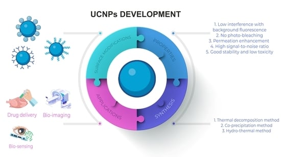
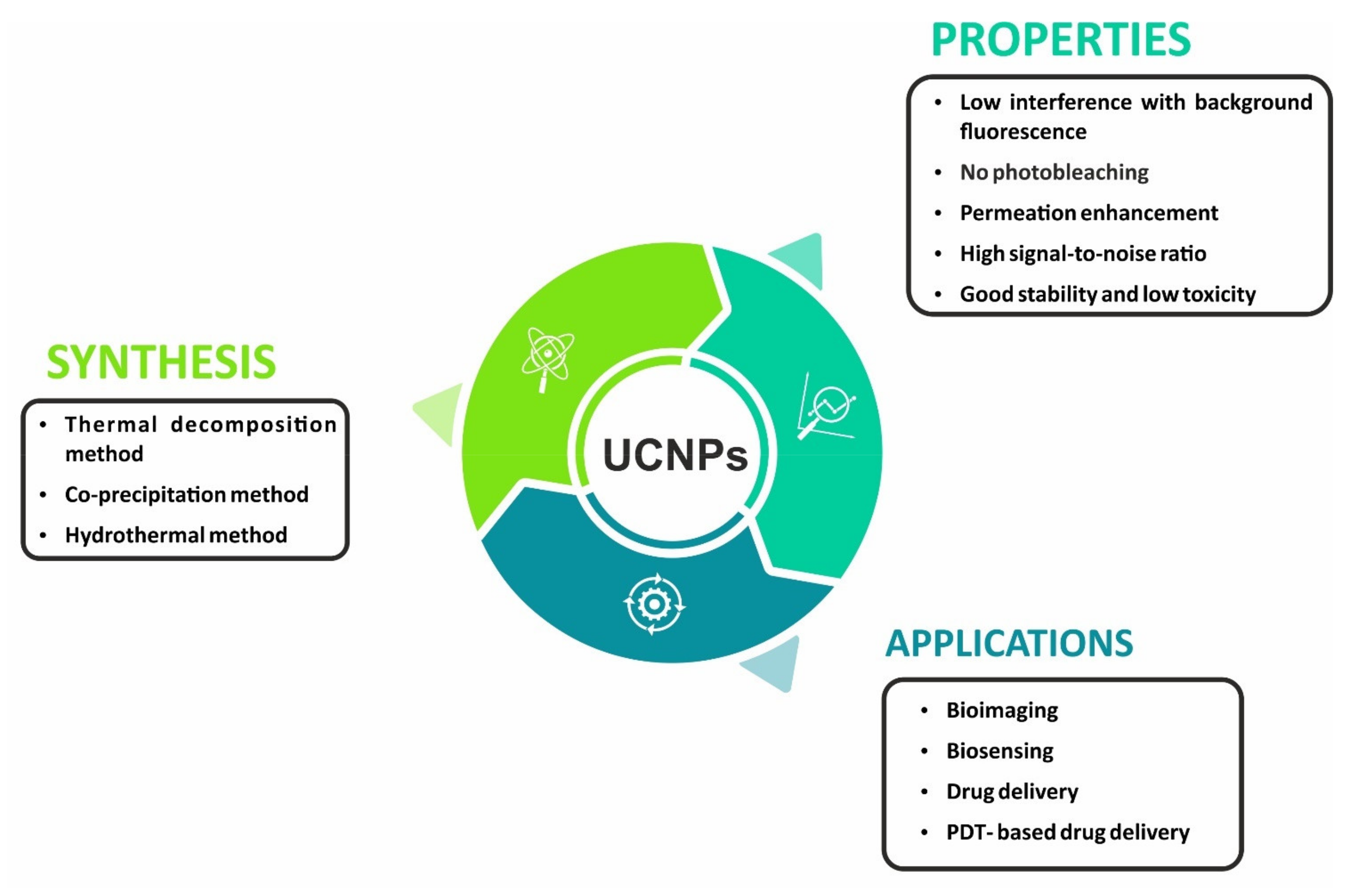
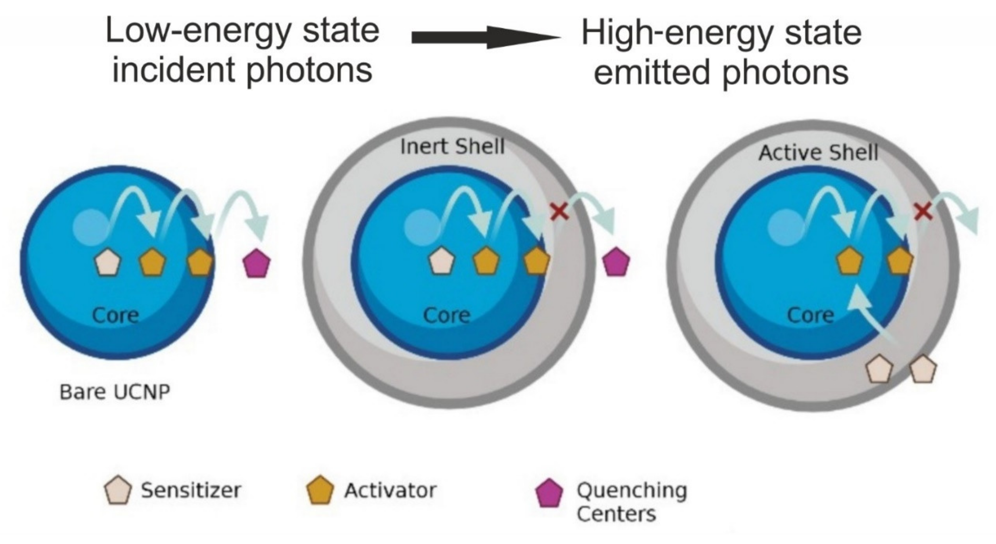
2. Properties and Composition of UCNPs

UCNPs possess several advantages including (i) absence of autofluorescence, resulting in enhanced signal-to-noise ratio and higher detection sensitivity; (ii) deeper biological tissue near-infrared (NIR) light penetration, causing less photodamage; and (iii) low-power NIR-laser-based excitation. The UCNP signal can be quantified because there is a direct correlation between the number of particles and the signal strength. Furthermore, there is extensive scope for multiplexing in UCNPs because the emission spectrum of the signal is very narrow and one can have any number of colors in one image. The additional features of UCNPs include narrow emission peaks, multiplexing, better chemical and physical stability, low toxicity, and no photobleaching.
UCNPs consist of inorganic host molecules with a rare earth (RE)-based lanthanide/actinide dopant incorporated in the host’s lattice. Photon upconversion is reported for a variety of dopants embedded in suitable host molecules, for example, solids doped with transition metal ions such as Ti2+, Ni2+, Mo3+, Re4+, or Os4+. Lanthanide-doped (Ln3+) materials have the highest upconversion efficiencies at room temperature. Luminescence is highly dependent on the transition of electrons in the 4f subshell. Lanthanides are metal ions having their 4f energy level filled and the valence electrons are shielded from external interactions. While all lanthanides (from lanthanum to lutetium) undergo upconversion, only erbium (III), holmium (III), and thulium (III) can absorb and advance to the specified levels of the visible and UV ranges because their inner shell electrons are shielded by the 5s 5p subshells, resulting in a large number of metastable energy states, making them amenable to upconversion (Figure 3). Even though a single lanthanide ion can induce the upconversion (UC) effect, co-doping is normally preferred to enhance the UC efficiency, since most lanthanide ions have low absorption cross-sections, resulting in weak emission [17]. The absorption can be increased by raising the dopant concentration of lanthanide ions per single nanocrystal. However, high doping concentrations result in a phenomenon called concentration quenching, which limits the quantity of dopant used. A high amount can lead to radiation-free deactivation and cross-relaxation processes, hence must be kept below 2 mol % to avoid loss of excitation energy. It is necessary to co-dope sensitizer ions alongside activator ions with a closely matched intermediate-excited state to achieve high upconversion efficiency. These highly absorbing sensitizer ions provide efficient nonradiative energy transfer to activator ions and result in improved absorption [18,19]; Yb3+ ions are the most often utilized sensitizers for Er3+, Tm3+, or Ho3+ doped UCNPs [20]. A few examples of nanoparticles with upconversion luminescence include NaYF4:Yb:Er [21,22], Y2O3:Yb:Er [23], Gd2O2S:Eu3+ [24,25], NaPrF4:Yb:Tm [26], and Cs3Lu2Br9:Er3+ [27]. Of these, Ln-doped UCNPs, NaYF4 co-doped with Yb3+/Er3+ or Yb3+/Tm3+ nanoparticles demonstrated the highest UC efficiency with usage in cellular and in vivo animal imaging [28].

3. Synthesis of UCNPs

Achieving high luminescent efficiency of UCNPs is a major goal; therefore, synthesizing UCNPs using appropriate techniques is essential to obtain UCNPs with well-defined size, shape, content, and phase. UCNPs are reported to be generally synthesized by three methods: thermal decomposition, co-precipitation, and hydrothermal synthesis.

3.1. Thermal Decomposition Method

Thermal decomposition method is based on the traditional solvent thermal method, and includes addition of trifluoroacetic acid RE salt and RE halide to the reaction system, which decomposes at high temperatures. This method is used to produce phase-pure single-crystalline UCNPs of uniform size within short time duration. Different sizes and shapes of NaYF4: Yb3+/Tm3+ or YB3+/Er3+ UCNPs can be generated by adjusting reaction time, reaction temperature, and reagent concentration. When a trifluoroacetate, such as Na (CF3COO), is dissolved in high-boiling organic solvents such as dimethylformamide (DMF) and dimethyl sulfoxide (DMSO) with the help of surfactants having polar capping groups and long hydrocarbon chains, such as oleic acid (OA), and omeylamine (OM), NaF is formed [29]. A rare-earth-doped metal trifluoroacetate is added to NaF, resulting in formation of α-NaYF4: Yb3+/Tm3+ or α-NaYF4: Yb3+/Er3+ UCNPs [30]. The thermal decomposition method comprises four steps that are depicted in Figure 4 [31]. It is essential to maintain appropriate reaction conditions (high temperature and pressure, capping ligand, heating and cooling rates, reaction duration, solvent and reagent concentrations) as UCNPs are sensitive to oxygen impurities and are anhydrous, requiring long reaction time and higher reaction temperatures for synthesis to yield high-quality monodispersed nanoparticles of desired nanocrystal morphology and size. OA and OM (capping ligands) bind to the surface of NPs via outward hydrophobic alkyl chains, making it hydrophobic. This method releases toxic fluorinated and oxyfluorinated carbon species into the environment. The synthetic particles are usually oil-soluble and have a high toxicity. Surface modification strategies are used to improve the water solubility, biocompatibility, and reduce the toxicity of UCNPs; their functionalization also expands the scope of their application. Thermal decomposition is the most common method used for synthesis high-quality UCNPs, in spite of some limitations pertaining to its industrial application. In most cases, the synthesis process requires the use of high temperatures, expensive and air-sensitive precursors and solvents, and is accompanied by the generation of toxic byproducts.

3.2. Co-Precipitation Method

The co-precipitation method is a simple cost-effective operational process, requires mild temperature conditions, produces fewer harmful byproducts, and provides a better solution from an industrial translational perspective, offering more environmentally friendly reagents containing inorganic RE salts. This approach is used to make ultrasmall Ln-UCNPs (2–10 nm) with improved crystallinity and luminescence efficiency [32]. Firstly, the formation of tiny amorphous Ln-UCNPs is controlled by a coordinating ligand at room temperature. Polyvinylpyrrolidone (PVP) may be used as the surface-coordinating ligand. The temperature is then increased to induce particle growth, which leads to the formation of nanocrystals via the Ostwald ripening mechanism [33]. Chen et al. constructed a dye-sensitized core–shell NaGdF4: Yb, Er@NaGdF4:Yb, Nd UCNPs to detect H2S for its application in cell and living body research via the co-precipitation method [34]. Liu et al. proposed a two-step reaction methodology for the solvothermal co-precipitation method to synthesize ultrasmall core–shell UCNPs, which resulted in a strong luminescence as a prototype for preparing UCNPs with high efficiency [35]. Lei et al. synthesized NaBiF4:Yb3+/Er3+ under room temperature via a simple hydrothermal method combined with a succedent calcining process. The substitution of yttrium with bismuth species considerably reduced the reaction conditions while maintaining upconversion luminescence, making them a potential candidate for lighting and solar cell applications [36].
Yi et al. synthesized α-NaYF4: Yb, Er UCNPs via a homogeneous nucleation process by injecting a RE-EDTA complex into NaF solution with vigorous stirring. The resulting UC fluorescent intensity of the UCNP was too low for biological labeling. Hence, an annealing treatment was given to enhance the UC fluorescent intensity [37]. Annealing of capping reagents results in carbonization and the hydrophilicity of the NPs is decreased. Hence, surface modification is required to improve the hydrophilicity of these NPs and to allow further increase in the size of NPs. This limits the utility of the co-precipitation method for the synthesis of RE-doped NaYF4 UCNPs for biological applications.

3.3. Hydrothermal Method

The hydrothermal method is usually carried out in a special closed reactor under high temperature and high pressure with water or organic solvents, and produces fewer harmful byproducts. In this method, a chemical reaction occurs when positive ions and negative ions are exposed to temperatures and pressures above their critical points in polar liquids, resulting in the formation of UCNPs. Zhang et al. used the hydrothermal method for the synthesis of Yb3+ and Er3+ co-doped NaYF4 upconversion luminescent materials and Ag nanoparticles coated with SiO2, which enhances the luminous intensity and has low cytotoxicity because of the SiO2 coating [38]. Nampi et al. synthesized single-crystalline, stable, aqueous Yb3+/Er3+doped BaYF5 UCNPs with polyethyleneimine (PEI) via hydrothermal route. With appropriate surface modifications, these particles could be adopted for biosensing of disease markers and bioimaging applications [39]. Heer et al. described a solution-based method for increasing the solubility of lanthanide precursors in supercritical polar solvents that favors the nanoparticles’ development rate. Surfactants are also added in the formulation process as growth control agents for nanocrystals [40]. The nucleation and growth stages are separated by the creation of an Ln3+ surfactant complex. The particle size distribution and morphology may be fine-tuned by adjusting the surfactant/Ln3+ ratio [29]. After the nanoparticles’ development stage is completed, the surfactant serves to cap ligands, limiting further aggregation [41]. Yi et al. synthesized UCNPs using the hydrothermal method and the results show that Ni2+ doping increases the UCL intensity, resulting in a change in morphology (hexagons to nanorods) with increasing size [42]. However, the disadvantage of this method is that the resulting UCNPs usually have a wider particle size distribution, and byproduct remnants can occasionally attach to the surface of UCNPs, making removal difficult. It should be highlighted that the hydrothermal preparation method’s future industrialization is still hindered by the long reaction time, which can range from 12 to 24 h or even longer. Apart from that, high pressures, large solvent volumes, and poor reproducibility are all major concerns that must be addressed.
In addition to these synthetic routes, ionothermal, microwave-assisted heating, sol-gel, microemulsion method, and combustion are reported for the synthesis of Ln-doped UCNPs. Microwave-assisted heating, for example, is a one-step process that takes much less time and energy than conventional methods and is regarded as a green method for synthesizing nanocrystals [43]. The combustion and microemulsion processes are utilized less commonly due to various intrinsic limitations such as difficulty to control the size and agglomeration, and poor light performance. Table 1 lists the methods used in the synthesis of UCNPs, along with their advantages and disadvantages.

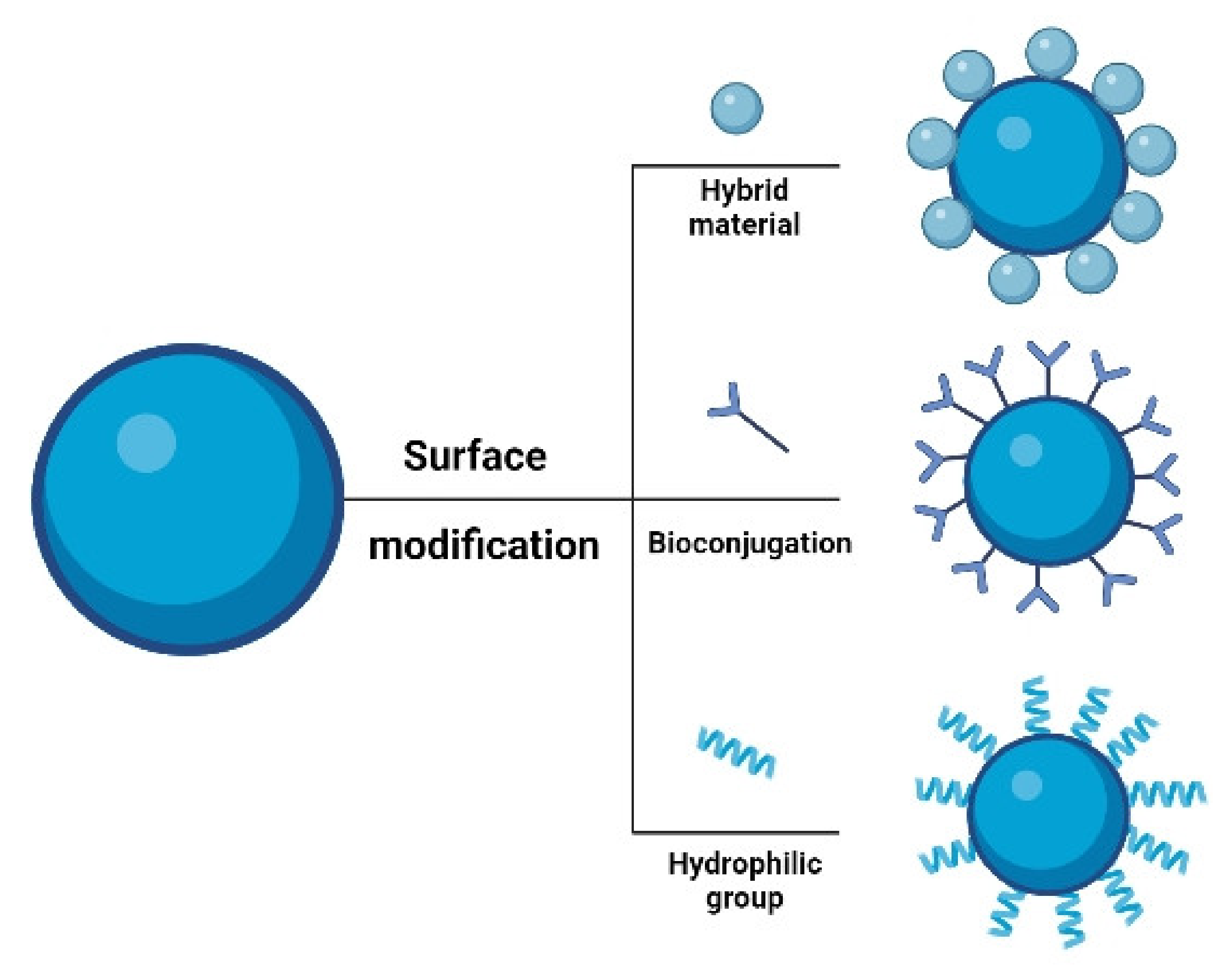
4. Surface Modification of UCNPs

Using a combination of energy migration and core–shell structural engineering to improve properties for a wide range of activators would widen the range of applications for lanthanide-doped nanoparticles [53,54,55]. Surface modification is required for specific nanomaterials to fulfil various biological activities, in biosciences such as disease therapy (particularly cancer) [56,57], detection [58], and immunoassay [59]. Understanding surface functionalization of Ln-doped UCNPs is critical for improving UC efficiency and aqueous solubility [60]. UCNPs are typically hydrophobic; hence, creating water-soluble Ln-doped UCNPs is critical for biological applications. Hydrophilic ligands coat the surface of UCNPs and can be dispersed in nonpolar organic solvents such as hexane and heptane. They lack functional groups for conjugation with biomolecules, such as carboxyl or amino groups. On the other hand, UCNPs capped with hydrophobic surfactants are not biocompatible and cannot be used immediately as they do not disperse in water. The surface characteristics of UCNPs determine their biocompatibility in in vitro and in vivo [61]. As a result, surface modification with an inorganic shell layer and an organic capping ligand is favored to overcome these difficulties. The surface of the NP can be coated by selection of an appropriate polymer or surfactant such as polyethyleneglycol (PEG), polyethylenimine (PEI), polyvinylalcohol (PVA), carboxydextran, or oleic acid (OA). Ligand removal, ligand oxidation, layer-by-layer deposition, acid treatment, and ligand exchange are some of the methods used to alter the surface properties of UCNPs, conferring high hydrophilic attributes [62,63]. Several surface modifications and depicted in Figure 5. Kostiv et al. synthesized uniformly sized NaYF4:Yb3+/Tm3+@NaYF4-PEG-Alk nanoparticles and bioconjugated it with a click reaction of pAbF-azide or SA azide for its bioanalytical applications, such as immunoassays [64]. Wang et al. [65] used a robust ligand exchange technique to develop a novel method for converting hydrophobic inorganic UCNPs to hydrophilic UCNPs. They were synthesized using the hydrothermal method and converted into carboxyl-modified UCNPs by replacing the original capping OA ligands on the surface of nanocrystals with PAA in a diethylene glycol (DEG) solvent at a high reaction temperature, rendering water-soluble nanocrystals.

5. Applications of UCNPs

UCNPs are reported to be used across a diverse spectrum of biological applications such as bioimaging, therapeutics, drug delivery, and biosensing. Discussion of these applications is elaborated in this section.

5.1. UCNPs in Bioimaging

Life science and nanomedicine have advanced at an incredible rate to improve life quality, which has encouraged significant research in the fields of bioimaging and technology. Bioimaging is a noninvasive technique for visualizing biological processes, allowing observation of subcellular structures, cells, tissues, and even complete multicellular creatures. UCNPs have versatility in generating nanoplatforms that have imaging and therapeutic modalities [66,67]. In addition, they have potential biomedical applications in early-stage diagnosis and monitoring therapy intervention, and have emerged as a novel carrier for small animal imaging, including tumor-targeted imaging, lymphatic imaging, and vascular imaging [68]. As UCNPs use a two-photon absorption mechanism, their energy emission and upconversion efficiency are higher when compared to other traditional technologies involving organic dyes and quantum dots. Hence, they have demonstrated promising results in optical-imaging-guided drug delivery and have become exclusive candidates in the field of bioimaging (in vivo and in vitro imaging of animal tissues) due to their unique photophysical properties such as lack of autofluorescence and deep-tissue-reaching resulting from luminescence after NIR excitation, resistance to photobleaching, and blinking [69]. In vitro cellular imaging involves targeting Ln-doped UCNPs to a subcellular component such as a membrane protein. The enhanced cellular uptake efficiency of positively charged UCNPs results in brighter in vitro cellular imaging. UCNPs can be employed to improve image contrast and sensitivity of in vivo imaging [70,71]. However, more research and testing are needed to fully comprehend the effect of nanoparticle size on optical properties to help optimize them for in vitro luminescence imaging [72,73,74]. Various imaging modalities used in pre-clinical studies and imaging science are depicted in Figure 6. Of these imaging modalities, PET/MRI shows the greatest clinical potential because MRI uses protons present in the soft tissue in the human body contrast paired with PET sensitivity. MRI produces better in vivo images together with good deep-tissue contrast and spatial resolution because tissue penetration is limited to a few mm [75].
Tian et al. synthesized a novel Nd3+-sensitized Er@Y@Nd@Gd core@multishell UCNP with carboxy-terminated silica and UEA-I. This UCNP@SiO2-UEA-I has high SW480 tumor-targeting potential. The ultrasmall SW480 tumor in the Balb/c nude mouse is observed using in vivo UCL imaging with UCNP@ SiO2-UEA-I. The findings suggest that the red UCL-emitted UCNP@SiO2-COOH bioconjugates with a minimized heating effect have much potential for sensitive deep-tissue biomedical imaging; synthesized UCNP@SiO2-UEA-I can serve as an efficient optical probe for early diagnosis of SW480 tumors [76]. A multifunctional nanocluster bomb (UCGM nanoparticles), CeOx, graphite-C3N4 (g-C3N4) NPs, and metformin (Met) were developed to alleviate hypoxia by oxidizing H2O2 to O2. In vivo UCL was used to monitor the distribution of UCGM NPs after they were injected into HepG2 tumor-bearing mice. Meanwhile, g-C3N4 NPs were released from UCGM NPs and, due to their tiny size, they penetrate tumor tissue deeply. Before the IV injection of UCGM NPs, there was no in vivo reinforcement in CT in the tumor, but after the injection, there was a significant CT signal in the tumor. UCGM NPs can serve as multifunctional theranostic agent for use in PTT/PDT-based therapy guided by UCL/CT/MRI trimodel imaging in vivo because of their great capacity to combat tumor hypoxia [77].
NaYF4 is conventionally the most notable system that has been employed in cellular and in vivo imaging. Hence, small-animal imaging and deep-tissue imaging are performed using NaYF4 UCNPs doped with Er3+ or Tm3+ that emit 800 nm NIR light, and have a better contrast that can be further improved by separating the long-lived luminescence from scattered light by tissue components using time-gated measurements [78]. In addition, there is significant use of UCNPs for super-resolution imaging based on stimulated emission depletion microscopy (STED), as reported by the Kolesov et al. [79]. The development of UCNP probes has promoted the translation of UCNPs application in cellular imaging. It is believed that UCNPs comprising both opportunities and limitations will attract great concern as probes for super-resolution microscopy [80]. Table 2 summarizes recent studies on the development of UCNPs for bioimaging, together with their composition/modifier and the synthesis route used to create UCNPs.

5.2. UCNPs in Biosensing

Many optically based biosensing methods have great potential for monitoring biomedical substances at clinically relevant levels; however, many of these methods encounter the problem of serum or whole blood autofluorescence and upconversion materials help resolve this issue. UCNPs exhibit minimal autofluorescence and deep tissue penetration, allowing them to be used in biological and environmental monitoring, detection, and sensing. Once UCNPs are synthesized, they can be easily functionalized and utilized as sensing nanoprobes to detect circulating cancer biomarkers. The capabilities of Ln-doped UCNPs in various biological sensing/detection depend on resonance energy transfer (RET) [88]. Fluorescence resonance energy transfer (FRET) is a nonradioactive process that describes the transfer of energy from a donor fluorophore to an acceptor fluorophore via a nonradiative dipole−dipole coupling [89,90] (Figure 7). These FRET systems were created by combining UCNP’s as an energy donor and organic dyes or QDs that act as an energy acceptor [91]. FRET typically offers greater freedom for upconverted emission wavelengths than the one which is formed solely by the Ln3+ ions. The FRET systems, which were developed using upconversion nanoparticles and gold nanoparticles for the detection system, have significant implications for biosensing and are frequently used in UCNP-based homogeneous tests. Zhen et al. proposed the use of UCNPs with confined emitters and bared surfaces as the luminophore and Ca2+ as a proof-of-concept target to develop a LRET-based nanoprobe. By simply altering the Ca2+ receptor into different recognition units, such as peptides, aptamers, and small-molecule ligands, this technique can be adapted to build numerous UC nanoprobes [92]. Their theory proposed the sandwich structure upconversion nanoparticles (SWUCNPs) with a core–inner-shell–outer-shell architecture, wherein the emitting ions (Ln3+) are precisely placed in the inner shell near the particle surface, close to external energy acceptors.
Several research groups have reported the usage of Ln doped-UCNPs in FRET-based detection [93]. Nd3+-UCNPs sensor is an excellent emitter that has low autofluorescence and a high penetration depth to biological samples. The oleic ligands from the core@shell UCNPs were readily removed by acid treatment, resulting in water-dispersible Nd3+-UCNPs. An ultrasensitive and selective approach for detecting miRNA with surface functionalized Au NPs-thiolated single-stranded DNA was proposed based on the chiroplasmonic and upconversion luminescence features of Au-UCNP pyramids as intracellular nanoprobes. The results indicate that an ultrasensitive and efficient chiral nanostructure-based detection approach can be used to identify biological systems [94]. Another intriguing aspect of UCNPs is their luminescence intensity, which is substantially temperature dependent. The nanoparticle-based thermometer provides a variety of alternatives for measuring the two-dimensional distribution of temperature and is important for understanding subcellular processes [95]. Lin et al. created a model of a multilayer nanocomposite structure NaYbF4:2%Er@NaYF4@MSN@Au@SiO2@Ag2S to depict the temperature distribution among nanoparticles. They concluded that these temperature-sensitive luminous probes, which are nanoscale in size, can be useful tools for high-resolution thermal sensing in micro areas [96]. Wolfbeis et al. studied temperature sensing with UCNPs of various sizes and RE dopants and found that the core–shell structured hexagonal (NaYF4:Yb20%Er2%)/NaYF4 UCNPs are better suited for temperature sensing because they can resolve temperature differences of less than 0.5 °C in the physiological range between 20 and 45 °C [97]. Additionally, Li et al. developed a new type of fluorescence temperature fiber optic sensor that uses NaYF4:Er3+, Yb3+ nanocrystals as the sensing unit. Results suggest that such UCNPs are highly stable and reliable, and prove the rationality of fluorescence fiber optic sensor’s design and its feasibility [98]. As a result, UCNPs are a great alternative for the design of temperature sensors, which account for the majority of the sensor market worldwide. It will promote the development of temperature sensors in industrial detection and other areas. Table 3 presents a summary of recent research on the development of UCNPs as attractive nanocarriers in biosensing applications.

5.3. UCNPs in Drug Delivery

The ability of nanocarriers to encapsulate poorly soluble drugs and minimize drug side effects is a significant advantage over traditional drug delivery systems. Nanocarriers are amenable for functionalization with imaging moieties and targeting ligands, further incrementing their functionality. UCNP-based drug delivery systems are reported to enhance the efficacy of a wide range of drug payloads and improve the solubility, stability, biodistribution, and pharmacokinetics of drugs [107,108]. Ln-doped UCNPs facilitate cell endocytosis and have good therapeutic effect due to their tiny particle size [109]. These nanoparticles subsequently release the drug and deliver them into tumor cells within a specified time limit. They improve the efficacy of the controlled drug release while reducing cell death, adverse effects, and tissue damage. UCNP-based nanocomposites are used as drug delivery systems and drug monitoring devices to address the needs of disease diagnostics and therapeutics.
Photoinduced reactions such as photocleavage and photoisomerization are used in drug delivery systems. UCNPs, by virtue of their ability to emit UV light on excitation with NIR light, serve as attractive materials for photoinduced drug delivery. Photocleavage and photoisomerization of light-sensitive molecules can be induced by Yb3+ and Er3+ co-doped UCNPs and they can emit UV light under 980 nm NIR excitation, which is primarily used as probes for in vitro and in vivo bioimaging. UCNPs can enhance the efficiency of drug delivery when used to induce photocleavable reactions in presence of NIR light. Combination of the high penetration depth of NIR light and low energy requirement of UCNPs makes them compatible for use with economical CW lasers instead of high-energy pulsed lasers [110].
Wang et al. [111] prepared oleic-acid-capped β-NaGdF4: Yb3+, Er 3+ @β-NaGdF4 UCNP UCNPs coated with NIR light-absorbing polymer polydopamine (PDA) using a water-in-oil microemulsion method, to obtain monodisperse, stable, noncytotoxic core–shell-structured nanospheres UCNP@PDA core–shell nanocomposites. These hydrophobic NPs were subsequently modified with amino-terminated polyethylene glycol (mPEG-NH2) to improve the stability of UCNP@PDA in physiological conditions. The PDA shell exhibited a strong photothermal effect and provided an active surface for loading doxorubicin (DOX) via π–π stacking and hydrogen-bonding interactions. Owing to the high UCL emission, T1 relaxivity value, and CT contrast enhancement of UCNP cores, trimodal imaging (UCL/MRI/CT) of a mouse-bearing colorectal (SW620) tumor was achieved by PEGylated UCNP@PDA with a 5 nm thickness PDA shell (UCNP@PDA 5 -PEG). DOX-loaded UCNP@PDA 5 -PEG (UCNP@PDA 5 -PEG-DOX) demonstrated excellent synergistic efficacy in in vitro cell culture and in vivo animal experiments. Their results suggested that drug-loaded UCNP@PDA core–shell nanocomposite can be used as an efficient nanoplatform for biomedical applications, including chemo photothermal therapy and multimodality imaging. The UCNP@PDA 5 -PEG-DOX, in combination with 808 nm NIR-laser irradiation, exhibited a synergistic interaction between PTT and the enhanced chemotherapy, resulting in complete eradication of tumor without regrowth. The leakage study, hemolysis assay, histology analysis, and blood biochemistry assay revealed that these nanocomposites had negligible organ toxicity. These results indicate that UCNP@PDA 5 -PEG can be applied as efficient multimodality contrast agents for UCL, MR, and CT imaging. The advantages of this strategy include its simplicity as the dopamine monomers can be directly polymerized on the UCNP surface via water-in-oil microemulsion technique, and the thicknesses of PDA shell can be controlled by variation in the number of dopamine monomers used in the reaction mixture; its amenability to functionalization by reaction with thiol and amino-terminated molecules via Michael addition or Schiff base reaction; its utility as an efficient nanoprobe for UCL/MRI/CT multimodality imaging and the application of the PDA shell as a drug carrier with high photothermal conversion agent resulting in chemophotothermal synergistic killing of tumors.
Liu et al. [112] developed a unique nanolongan delivery system that utilized a combination of ferroptosis−apoptosis (co-deliver an iron element and a chemotherapeutic drug) for improved anticancer efficacy of DOX. This comprised one core (UCNP) in one gel particle (Fe3+ cross-linked oxidized starch) with several on-demand conversions. The charge conversion of nanolongan surface sourced from 2,3-dimethyl maleic anhydride (DMMA) decoration conferred long circulation for utilizing the EPR effect and enabled more efficient uptake by tumor cells accompanied with subsequent lysosome escape. The core UCNP with the light conversion from NIR to UV circumvented the impediment of limited penetration depth and enabled the reduction of Fe3+ to Fe2+ (Figure 8).
The nanolongan gel network developed could be deconstructed owing to this valence conversion, leading to the rapid release of Fe2+ and DOX. Cytotoxicity studies of the formulations developed (DGU: Fe; DGU: Fe + L; Dox; DGU: Fe/Dox and DGU: Fe/Dox + L) using the CCK8 assay on 4T1 and MCF7 cell lines indicated a dose-dependent response with DGU: Fe/Dox+ L formulation being the most cytotoxic, emphasizing the merits of co-operation of ferroptosis and apoptosis in the nanolongan formulations. Biodistribution studies in the 4T1-xenografted mouse model demonstrated that DMMA decoration resulted in prolongation of circulation time and increased tumor cell uptake via tumor-targeted delivery. In vivo efficacy studies in 4T1-xenograft mice indicated that DGU: Fe/Dox + L treatment showed enhanced therapeutic effect and complete tumor elimination at a higher dose, resulting in a 100% survival rate at 55 days along with a significant antimetastasis effect. Safety studies indicated that the DGU: Fe/Dox + L group showed lower toxicity than DOX due to targeted delivery to tumor. Their work demonstrated superior anticancer efficacy for this combination and can be extended to other anticancer agents for improved therapeutic efficacy. Table 4 presents a summary of recent research on the development of UCNPs as attractive nanocarriers in drug delivery.

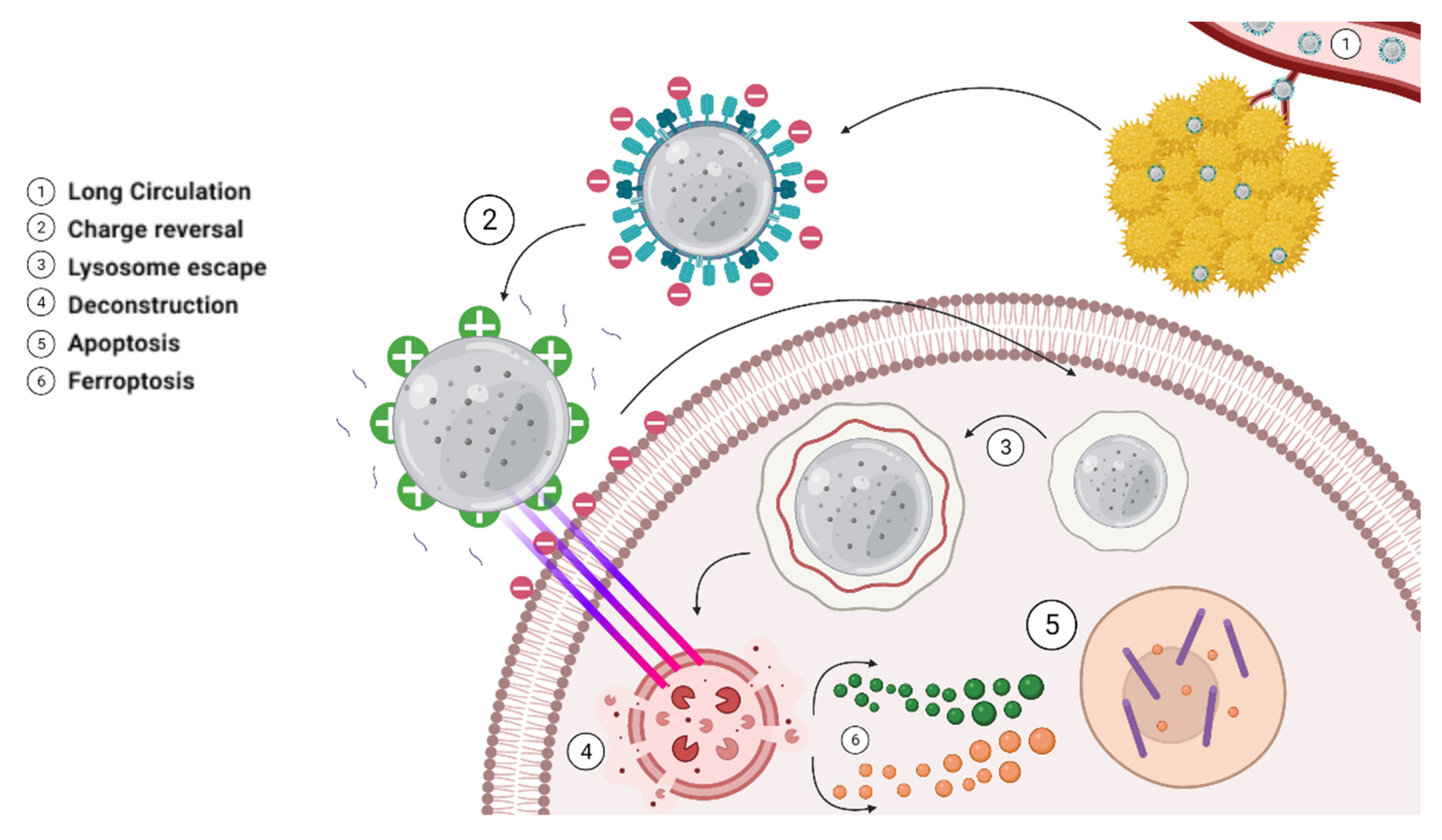
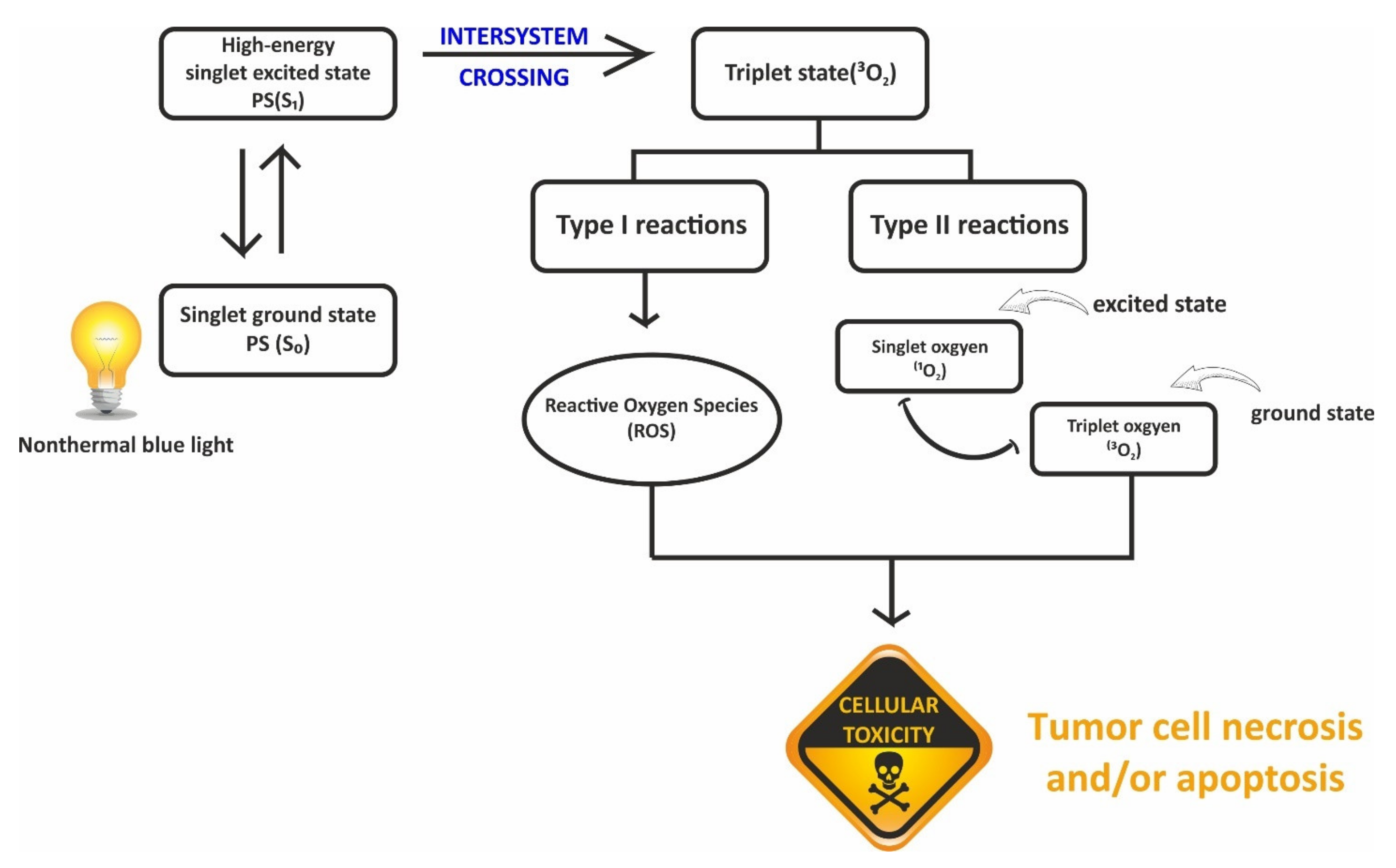
5.4. UCNPs in Photodynamic Therapy (PDT)-Based Drug Delivery

PDT employs photodynamic effects to diagnose and treat several diseases including prostate cancer [121,122] and offers the advantage of low invasiveness and toxicity [123]. It involves three components, which include photosensitizer (PS) molecules, light source, and oxygen within the tissue at the disease site [124]. The principle of PDT showing the activation of the photosensitizer due to the excitation with light and the energy transfer to molecular oxygen is depicted in Figure 9. A light source is required for functioning in addition to the photosensitizers. Any light source with the appropriate wavelength and intensity can be utilized. Reactive oxygen species (ROS) created during PDT can eliminate tumors in a variety of ways, including directly triggering tumor cell necrosis and/or apoptosis, ultimately leading to cellular toxicity [125].
One of the most significant benefits of PDT is to treat the lesion area selectively under light irradiation while leaving normal tissues unaffected [126]. The engagement of UCNPs in PDT is clinically significant because it offers a novel way to treat deep-tissue tumors. It is based on the fact that UCNPs can efficiently convert deeply penetrating NIR light to visible wavelengths, which can activate photosensitizers, resulting in the production of cytotoxic 1O2. The PDT effect of the UCNP-based delivery system is evaluated by combining oleic acid-coated UCNPs with angiopep-2/cholesterol-conjugated poly (ethylene glycol) and hydrophobic photosensitizers [127]. The results showed that ANG-IMNPs could deliver dual photosensitizers to brain astrocytoma tumors selectively, resulting in successful PDT/PTT conjugation and a significantly enhanced median survival. The therapeutic efficacy of ANG-IMNPs demonstrated in this study implies that they can overcome the blood–brain barrier (BBB) and develop a glioblastoma multiforme (GBM) treatment. PDT research is undertaken to expand the platform of photosensitizers. It has the advantage of being able to provide the drug directly to the patient because cancer cells, due to their increased metabolism and absorption, only congregate in large numbers in tumors, causing no harm to healthy cells.

6. Pharmacokinetics (PK) of UCNPs

The absorption, distribution, metabolism, elimination, and toxicity (ADMET) together describe a drug’s overall disposition via pharmacokinetics, or what the body does to a drug, which plays a major role in drug development. The pharmacokinetic profile of UCNPs is an area with very few studies reported in comparison to the development of UCNPs for diverse biomedical applications. This is vital for assessing the feasibility of their translation to the clinic for therapeutic and diagnostic applications. Few toxicity studies of UCNPs developed as drug carriers and contrast agents are reported in mice with a vast number of UCNPs in vitro toxicity studies reported on different cell lines [128]. Most of the in vivo toxicity studies utilized the intravenous route of administration, although one study by Ortgies et al. reported toxicity studies of UCNPs developed for multiplexed imaging and drug delivery via the oral route of administration, which is the most common route for drug delivery.
Sun et al. [128] developed 50 nm-sized silica-coated NaYF4: Yb, Er NPs (NaYF4:Yb, Er@SiO2) and evaluated their bioavailability, biodistribution, and toxicity in mice via the oral and intravenous routes by using TEM and ICP–MS. Their results demonstrated that the biodistribution was a function of the route of administration, the UCNPs post-oral administration were absorbed in the intestine via Peyer’s patches as confirmed by TEM studies and the UCNPs via intravenous route were observed to be trapped in the hepatocytes. NaYF4: Yb, Er@SiO2 nanoparticles were found to accumulate in the bone, stomach, and intestine on oral administration, and in the liver and spleen on intravenous dosing. They did not report any significant toxicity of NaYF4: Yb, Er@SiO2 post 14 days oral dosing at 100 mg/kg.
Li et al. [129] investigated the long-term in vivo distribution and toxicity of polyacrylic acid-coated NaYF4 upconversion nanophosphors (PAA-UCNPs) as NIR- to-NIR luminescence probes. The results of in vitro cytotoxicity studies showed that PAA-UCNPs had no significant effects on the proliferation of KB cells with retention of high cellular viability of more than 80% after 48 h incubation with PAA-UCNPs (480 mg/mL). Biodistribution studies illustrated that a high amount of PAA-UCNPs was found in the liver and spleen with a slow renal clearance. The results of in vivo toxicity studies in mice demonstrated that the mice survived for 115 days without any evident (observational, histological, hematological, and biochemical) toxic effects post intravenous dosing of 15 mg/kg of PAA-UCNPs. Their studies provide encouraging evidence in terms of safety and biodistribution for translating the use of PAA-UCNPs for long-term in vivo imaging to the clinic.

7. Outlook and Perspectives

Upconversion nanoparticles represent a unique class of lanthanide-doped nanomaterials capable of converting near-infrared excitation into visible and ultraviolet emission. Their unique optical properties have expanded the landscape of their applications, ranging from drug delivery to bioimaging and biosensing. In this review, we presented current strategies used in the efficient synthesis of UCNPs and recent developments in their utility in biomedical applications. The intrinsic advantages of UCNPs, such as no background autofluorescence, minimal background tissue damage, and long penetration depth in biological tissues, are anticipated to keep UCNPs at the forefront of biological imaging and therapeutics. Their unique optical properties make them amenable to appropriate functionalization and they can be designed for effective delivery and release of drug payloads in response to a specific stimulus. Several NIR-responsive drug delivery systems based on UCNPs are reported in the literature, which appear to be promising additions in detection, bioimaging, drug delivery, and PDT, especially in cancer therapeutics. Some radiology departments have started using these modalities as the next-generation technology. There is scope for improving the efficiency of upconversion to promote wider utilization under in vivo settings and the development of surface treatment techniques for targeted drug delivery.
Furthermore, the toxicity of these nanoparticles has lately been thoroughly addressed and the results of these investigations indicate that UCNPs show reduced toxicity in both in vitro and in vivo studies. Although various UCNP-based theranostic nanoplatforms have showcased immense potential, very few have entered into the clinical stage. Limited information on the long-term in vivo biological effects is one of the major barriers to the clinical translation of UCNPs. Given the complexity of many UCNP carrier systems, the question of the stability of these structures in the human biological environment for extended periods remains a biosafety concern. As a result, more research into the long-term stability of UCNPs in biological systems is required. Furthermore, once these UCNP-based nanoparticles are ingested, their function within the body and interaction with the nervous and immune systems is an important aspect that needs more explorative studies.
Another area that needs to be improved is the efficient integration of commercially available imaging equipment with upconversion nanocrystals. These nanocrystals, in contrast to fluorescent dyes and quantum dots, require excitation in the infrared regime, which means that standard commercial equipment cannot be used for quantitative measurements. Advances in fundamental nanophenomena in upconversion necessitate complex instrumentation, and a collaborative effort across a wide range of disciplines will have a significant impact in these areas. The detrimental effect of concentration quenching in luminescent materials restricts access to a high level of luminescence intensity, thereby limiting their future applications. The limitation imposed by the concentration quenching threshold becomes a real issue for nanoscale luminescent materials.
UCNPs have attractive applications in biomedical and healthcare sectors including drug delivery, diagnostics, and theranostics. These applications depend on biocompatible products without or with negligible hazard to human cells, tissues, and organs. Translation of UCNPs is associated with challenges of reproducible synthesis, effective control over the NP size and shape, preparation of a biocompatible surface architecture with biorecognition elements for labeling target sites, light activation, theranostics, and drug delivery. Safety concerns and socioeconomic uncertainties are some of the important barriers to translational process in nanomedicine. Several deficiencies need to be addressed such as how they fit into clinical pathways, data consistency, and imaging techniques, but the potential is undeniable. With the present scenario of extensive research in nanotechnology development, it is conceivable that UCNPs will soon be placed among the mainstream nanoprobes that will be commonly utilized in both laboratories and clinics.

Author Contributions

Conceptualization, T.K. and P.J.; writing—original draft preparation, P.J.; writing—review and editing, T.K., M.M. and A.O.; supervision, T.K. and A.O.; formal analysis, M.M. and A.O. All authors have read and agreed to the published version of the manuscript.

Funding

This research received no external funding.

Acknowledgments

The authors would like to acknowledge the help rendered by Saachi Mehta, SVKM’s Dr. Bhanuben Nanavati College of Pharmacy, for drawing the figures in this manuscript.

Conflicts of Interest

There are no conflicts to declare.

References

  1. Bayda, S.; Adeel, M.; Tuccinardi, T.; Cordani, M.; Rizzolio, F. The history of nanoscience and nanotechnology: From chemical-physical applications to nanomedicine. Molecules 2020, 25, 112. [Google Scholar] [CrossRef] [PubMed] [Green Version]
  2. Nasrollahzadeh, M.; Sajadi, S.M.; Sajjadi, M.; Issaabadi, Z. An Introduction to Nanotechnology, 1st ed.; Elsevier Ltd.: Amsterdam, The Netherlands, 2019; Volume 28, ISBN 9780128135860. [Google Scholar]
  3. Talapin, D.V.; Shevchenko, E.V. Introduction: Nanoparticle chemistry. Chem. Rev. 2016, 116, 10343–10345. [Google Scholar] [CrossRef] [PubMed] [Green Version]
  4. Guerrini, L.; Alvarez-Puebla, R.A.; Pazos-Perez, N. Surface modifications of nanoparticles for stability in biological fluids. Materials 2018, 11, 1154. [Google Scholar] [CrossRef] [PubMed] [Green Version]
  5. Tao, K.; Sun, K. Upconversion nanoparticles. In Photonanotechnology for Therapeutics and Imaging; Elsevier: Amsterdam, The Netherlands, 2020; pp. 147–176. [Google Scholar]
  6. Haase, M.; Schäfer, H. Upconverting nanoparticles. Angew. Chem.-Int. Ed. 2011, 50, 5808–5829. [Google Scholar] [CrossRef]
  7. Das, A.; Bae, K.; Park, W. Enhancement of upconversion luminescence using photonic nanostructures. Nanophotonics 2020, 9, 1359–1371. [Google Scholar] [CrossRef]
  8. Chen, X.; Peng, D.; Ju, Q.; Wang, F. Photon upconversion in core-shell nanoparticles. Chem. Soc. Rev. 2015, 44, 1318–1330. [Google Scholar] [CrossRef] [PubMed]
  9. Tu, L.; Liu, X.; Wu, F.; Zhang, H. Excitation energy migration dynamics in upconversion nanomaterials. Chem. Soc. Rev. 2015, 44, 1331–1345. [Google Scholar] [CrossRef] [Green Version]
  10. Wen, S.; Zhou, J.; Zheng, K.; Bednarkiewicz, A.; Liu, X.; Jin, D. Advances in highly doped upconversion nanoparticles. Nat. Commun. 2018, 9, 2415. [Google Scholar] [CrossRef]
  11. Wei, W.; Chen, G.; Baev, A.; He, G.S.; Shao, W.; Damasco, J.; Prasad, P.N. Alleviating Luminescence Concentration Quenching in Upconversion Nanoparticles through Organic Dye Sensitization. J. Am. Chem. Soc. 2016, 138, 15130–15133. [Google Scholar] [CrossRef]
  12. Viger, M.L.; Live, L.S.; Therrien, O.D.; Boudreau, D. Reduction of self-quenching in fluorescent silica-coated silver Nnanoparticles. Plasmonics 2008, 3, 33–40. [Google Scholar] [CrossRef]
  13. Li, X.; Zhang, F.; Zhao, D. Lab on upconversion nanoparticles: Optical properties and applications engineering via designed nanostructure. Chem. Soc. Rev. 2015, 44, 1346–1378. [Google Scholar] [CrossRef] [PubMed]
  14. Gao, D.; Guo, X.; Zhang, X.; Chen, S.; Wang, Y.; Chen, T.; Huang, G.; Gao, Y.; Tian, Z.; Yang, Z. Multifunctional Phototheranostic Nanomedicine for Cancer Imaging and Treatment; Elsevier Ltd.: Amsterdam, The Netherlands, 2020; Volume 5, ISBN 0086298266328. [Google Scholar]
  15. Liu, B.; Li, C.; Xing, B.; Yang, P.; Lin, J. Multifunctional UCNPs@PDA-ICG nanocomposites for upconversion imaging and combined photothermal/photodynamic therapy with enhanced antitumor efficacy. J. Mater. Chem. B 2016, 4, 4884–4894. [Google Scholar] [CrossRef] [PubMed]
  16. Zhou, B.; Shi, B.; Jin, D.; Liu, X. Controlling upconversion nanocrystals for emerging applications. Nat. Nanotechnol. 2015, 10, 924–936. [Google Scholar] [CrossRef] [PubMed]
  17. Cheng, X.; Ge, H.; Wei, Y.; Zhang, K.; Su, W.; Zhou, J.; Yin, L.; Zhan, Q.; Jing, S.; Huang, L. Design for Brighter Photon Upconversion Emissions via Energy Level Overlap of Lanthanide Ions. ACS Nano 2018, 12, 10992–10999. [Google Scholar] [CrossRef] [PubMed]
  18. Wang, Z.; Meijerink, A. Concentration Quenching in Upconversion Nanocrystals. J. Phys. Chem. C 2018, 122, 26298–26306. [Google Scholar] [CrossRef] [Green Version]
  19. Eliseeva, S.V.; Bünzli, J.C.G. Lanthanide luminescence for functional materials and bio-sciences. Chem. Soc. Rev. 2010, 39, 189–227. [Google Scholar] [CrossRef]
  20. Vu, D.T.; Tsai, Y.C.; Le, Q.M.; Kuo, S.W.; Lai, N.D.; Benisty, H.; Lin, J.Y.; Kan, H.C.; Hsu, C.C. A synergy approach to enhance upconversion luminescence emission of rare-earth nanophosphors with million-fold enhancement factor. Crystals 2021, 11, 1187. [Google Scholar] [CrossRef]
  21. Kavand, A.; Serra, C.A.; Blanck, C.; Lenertz, M.; Anton, N.; Vandamme, T.F.; Mély, Y.; Przybilla, F.; Chan-Seng, D. Controlled Synthesis of NaYF4:Yb, Er Upconversion Nanocrystals as Potential Probe for Bioimaging: A Focus on Heat Treatment. ACS Appl. Nano Mater. 2021, 4, 5319–5329. [Google Scholar] [CrossRef]
  22. Nie, Z.; Ke, X.; Li, D.; Zhao, Y.; Zhu, L.; Qiao, R.; Zhang, X.L. NaYF4:Yb,Er,Nd@NaYF4:Nd Upconversion Nanocrystals Capped with Mn:TiO2 for 808 nm NIR-Triggered Photocatalytic Applications. J. Phys. Chem. C 2019, 123, 22959–22970. [Google Scholar] [CrossRef]
  23. Unal, F.; Kaya, F.; Kazmanli, K. Synthesis, characterization and radioluminescence properties of erbium-doped yttria phosphors. Int. J. Miner. Metall. Mater. 2021, 28, 1983–1990. [Google Scholar] [CrossRef]
  24. Xie, J.; Liu, B.; Qong, Q.; Xu, Z.; Jin, Z.; Ma, W. Eu3+/Eu2+/Tb3+ co-activated single-phase Gd2O2S: A high-performance white light emitting phosphor for light emitting diode. Mater. Express 2021, 11, 54–62. [Google Scholar] [CrossRef]
  25. Lin, H.; Luo, Q.; Tong, W.Y.; Jiang, C.; Huang, R.; Peng, H.; Zhang, L.C.; Travas-Sejdic, J.; Duan, C.G. Facile preparation of rare-earth semiconductor nanocrystals and tuning of their dimensionalities. RSC Adv. 2015, 5, 86885–86890. [Google Scholar] [CrossRef]
  26. Zhang, J.; Zhao, S.; Xu, Z.; Zhang, L.; Zuo, P.; Wu, Q. Near-infrared light-driven photocatalytic NaYF4:Yb,Tm@ZnO core/shell nanomaterials and their performance. RSC Adv. 2019, 9, 3688–3692. [Google Scholar] [CrossRef] [Green Version]
  27. Hehlen, M.P.; Frei, G.; Güdel, H.U. Dynamics of infrared-to-visible up-conversion in Cs3Lu2Br9: 1%Er3+. Phys. Rev. B Condens. Matter. 1994, 50, 16264–16273. [Google Scholar] [CrossRef] [PubMed]
  28. Boyer, J.C.; Vetrone, F.; Cuccia, L.A.; Capobianco, J.A. Synthesis of colloidal upconverting NaYF4 nanocrystals doped with Er3+, Yb3+ and Tm3+, Yb3+ via thermal decomposition of lanthanide trifluoroacetate precursors. J. Am. Chem. Soc. 2006, 128, 7444–7445. [Google Scholar] [CrossRef] [PubMed]
  29. Mai, H.X.; Zhang, Y.W.; Sun, L.D.; Yan, C.H. Size- and phase-controlled synthesis of monodisperse NaYF4:Yb,Er nanocrystals from a unique delayed nucleation pathway monitored with upconversion spectroscopy. J. Phys. Chem. C 2007, 111, 13730–13739. [Google Scholar] [CrossRef]
  30. Zhang, Y.W.; Sun, X.; Si, R.; You, L.P.; Yan, C.H. Single-crystalline and monodisperse LaF3 triangular nanoplates from a single-source precursor. J. Am. Chem. Soc. 2005, 127, 3260–3261. [Google Scholar] [CrossRef]
  31. Wang, M.; Abbineni, G.; Clevenger, A.; Mao, C.; Xu, S. Upconversion nanoparticles: Synthesis, surface modification and biological applications. Nanomed. Nanotechnol. Biol. Med. 2011, 7, 710–729. [Google Scholar] [CrossRef] [Green Version]
  32. Ibarra-Ruiz, A.M.; Rodríguez Burbano, D.C.; Capobianco, J.A. Photoluminescent nanoplatforms in biomedical applications. Adv. Phys. X 2016, 1, 194–225. [Google Scholar] [CrossRef] [Green Version]
  33. Wang, F.; Deng, R.; Liu, X. Preparation of core-shell NaGdF4 nanoparticles doped with luminescent lanthanide ions to be used as upconversion-based probes. Nat. Protoc. 2014, 9, 1634–1644. [Google Scholar] [CrossRef]
  34. Chen, H.; Wang, W.; Ji, C.; Wang, L. Dye-sensitized core–shell NaGdF4:Yb,Er@NaGdF4:Yb,Nd upconversion nanoprobe for determination of H2S. Spectrochim. Acta-Part A Mol. Biomol. Spectrosc. 2021, 248, 119281. [Google Scholar] [CrossRef] [PubMed]
  35. Liu, D.; Yan, J.; Wang, K.; Wang, Y.; Luo, G. Continuous synthesis of ultrasmall core-shell upconversion nanoparticles via a flow chemistry method. Nano Res. 2022, 15, 1199–1204. [Google Scholar] [CrossRef]
  36. Wang, Y.; Zhu, Y.; Xu, X.; Huang, J.; Lu, Z.; Qiu, D. One-step synthesis and upconversion luminescence properties of hierarchical In2O3:Yb3+,Er3+ nanorod flowers. RSC Adv. 2017, 7, 54500–54505. [Google Scholar] [CrossRef] [Green Version]
  37. Wei, Y.; Lu, F.; Zhang, X.; Chen, D. Synthesis and characterization of efficient near-infrared upconversion Yb and Tm codoped NaYF4 nanocrystal reporter. J. Alloys Compd. 2007, 427, 333–340. [Google Scholar] [CrossRef]
  38. Zhang, W.; Lu, Y.; Zang, Y.; Han, J.; Xiong, Q.; Xiong, J. SiO2 coated up-conversion nanomaterial doped with ag nanoparticles for micro-CT imaging. Nanomaterials 2021, 11, 3395. [Google Scholar] [CrossRef]
  39. Nampi, P.P.; Vakurov, A.; Viswambharan, H.; Schneider, J.E.; Brydson, R.; Millner, P.A.; Saha, S.; Jose, G. Barium yttrium fluoride based upconversion nanoparticles as dual mode image contrast agents. Mater. Sci. Eng. C 2021, 124, 111937. [Google Scholar] [CrossRef]
  40. Sun, Y.; Chen, Y.; Tian, L.; Yu, Y.; Kong, X.; Zhao, J.; Zhang, H. Controlled synthesis and morphology dependent upconversion luminescence of NaYF4:Yb, Er nanocrystals. Nanotechnology 2007, 18, 275609. [Google Scholar] [CrossRef]
  41. Li, C.; Yang, J.; Quan, Z.; Yang, P.; Kong, D.; Lin, J. Different microstructures of β-NaYF4 fabricated by hydrothermal process: Effects of pH values and fluoride sources. Chem. Mater. 2007, 19, 4933–4942. [Google Scholar] [CrossRef]
  42. Yi, M.; Liu, Y.; Gao, H.; Huang, Z.; Liang, J.; Mao, Y. Upconversion effective enhancement of NaYF4:Yb3+/Er3+ nanoparticles by Ni2+ doping. J. Mater. Sci. 2018, 53, 1395–1403. [Google Scholar] [CrossRef]
  43. Reddy, K.L.; Prabhakar, N.; Rosenholm, J.M.; Krishnan, V. Core-shell structures of upconversion nanocrystals coated with silica for near infrared light enabled optical imaging of cancer cells. Micromachines 2018, 9, 400. [Google Scholar] [CrossRef] [Green Version]
  44. MacKenzie, L.E.; Alvarez-Ruiz, D.; Pal, R. Low-temperature open-air synthesis of PVP-coated NaYF 4 :Yb,Er,Mn upconversion nanoparticles with strong red emission. R. Soc. Open Sci. 2022, 9, 211508. [Google Scholar] [CrossRef] [PubMed]
  45. Zheng, L.; Huang, X.; Zhong, J.; Wang, Z.; Cheng, X. Upconversion luminescence and temperature sensing properties of NaGd(WO4)2:Yb3+/Er3+@SiO2 core-shell nanoparticles. RSC Adv. 2021, 11, 3981–3989. [Google Scholar] [CrossRef]
  46. Liu, G.; Sun, Z.; Jia, M.; Fu, Z.; Zhang, A.; Li, P. One pot synthesis and optimized luminescent intensity of Gd2(WO4)3: Yb3+/Ho3+@SiO2 nanoparticles for biological application. J. Lumin. 2019, 206, 1–5. [Google Scholar] [CrossRef]
  47. Mohan, M.; Poddar, R. Polymerically engineered upconversion nanoparticles (UCNPs) as a contrast agent for functionally modified optical coherence tomography (OCT). Mater. Sci. Eng. C 2021, 121, 111841. [Google Scholar] [CrossRef]
  48. Rafique, R.; Baek, S.H.; Phan, L.M.T.; Chang, S.J.; Gul, A.R.; Park, T.J. A facile hydrothermal synthesis of highly luminescent NaYF4:Yb3+/Er3+ upconversion nanoparticles and their biomonitoring capability. Mater. Sci. Eng. C 2019, 99, 1067–1074. [Google Scholar] [CrossRef]
  49. Du, P.; Zhang, P.; Kang, S.H.; Yu, J.S. Hydrothermal synthesis and application of Ho3+-activated NaYbF4 bifunctional upconverting nanoparticles for in vitro cell imaging and latent fingerprint detection. Sens. Actuators B Chem. 2017, 252, 584–591. [Google Scholar] [CrossRef]
  50. Lakshmanan, A.; Akasov, R.A.; Sholina, N.V.; Demina, P.A.; Generalova, A.N.; Gangadharan, A.; Sardar, D.K.; Lankamsetty, K.B.; Khochenkov, D.A.; Khaydukov, E.V.; et al. Nanocurcumin-loaded UCNPs for cancer theranostics: Physicochemical properties, in vitro toxicity, and in vivo imaging studies. Nanomaterials 2021, 11, 2234. [Google Scholar] [CrossRef]
  51. Guo, X.; Wu, W.; Li, Y.; Zhang, J.; Wang, L.; Ågren, H. Recent research progress for upconversion assisted dye-sensitized solar cells. Chin. Chem. Lett. 2021, 32, 1834–1846. [Google Scholar] [CrossRef]
  52. Liang, X.; Fan, J.; Zhao, Y.; Jin, R. Core-Shell Structured NaYF4:Yb,Er Nanoparticles with Excellent Upconversion Luminescent for Targeted Drug Delivery. J. Clust. Sci. 2021, 32, 1683–1691. [Google Scholar] [CrossRef]
  53. Liu, S.; Huang, J.; Yan, L.; Song, N.; Zhang, P.; He, J.; Zhou, B. Multiphoton ultraviolet upconversion through selectively controllable energy transfer in confined sensitizing sublattices towards improved solar photocatalysis. J. Mater. Chem. A 2021, 9, 4007–4017. [Google Scholar] [CrossRef]
  54. Li, H.; Wang, X.; Ohulchanskyy, T.Y.; Chen, G. Lanthanide-Doped Near-Infrared Nanoparticles for Biophotonics. Adv. Mater. 2021, 33, 2000678. [Google Scholar] [CrossRef] [PubMed]
  55. Gao, C.; Zheng, P.; Liu, Q.; Han, S.; Li, D.; Luo, S.; Temple, H.; Xing, C.; Wang, J.; Wei, Y.; et al. Recent advances of upconversion nanomaterials in the biological field. Nanomaterials 2021, 11, 2474. [Google Scholar] [CrossRef] [PubMed]
  56. Zha, S.; Chau, H.F.; Chau, W.Y.; Chan, L.S.; Lin, J.; Lo, K.W.; Cho, W.C.; Yip, Y.L.; Tsao, S.W.; Farrell, P.J.; et al. Dual-Targeting Peptide-Guided Approach for Precision Delivery and Cancer Monitoring by Using a Safe Upconversion Nanoplatform. Adv. Sci. 2021, 8, 2002919. [Google Scholar] [CrossRef] [PubMed]
  57. Nehra, M.; Uthappa, U.T.; Kumar, V.; Kumar, R.; Dixit, C.; Dilbaghi, N.; Mishra, Y.K.; Kumar, S.; Kaushik, A. Nanobiotechnology-assisted therapies to manage brain cancer in a personalized manner. J. Control. Release 2021, 338, 224–243. [Google Scholar] [CrossRef] [PubMed]
  58. Zhao, X.; Wang, Y.; Li, J.; Huo, B.; Huang, H.; Bai, J.; Peng, Y.; Li, S.; Han, D.; Ren, S.; et al. A fluorescence aptasensor for the sensitive detection of T-2 toxin based on FRET by adjusting the surface electric potentials of UCNPs and MIL-101. Anal. Chim. Acta 2021, 1160, 338450. [Google Scholar] [CrossRef] [PubMed]
  59. Raiko, K.; Lyytikäinen, A.; Ekman, M.; Nokelainen, A.; Lahtinen, S.; Soukka, T. Supersensitive photon upconversion based immunoassay for detection of cardiac troponin I in human plasma. Clin. Chim. Acta 2021, 523, 380–385. [Google Scholar] [CrossRef] [PubMed]
  60. Ghosh, P.; De La Rosa, E.; Oliva, J.; Solis, D.; Kar, A.; Patra, A. Influence of surface coating on the upconversion emission properties of LaPO4:Yb/Tm core-shell nanorods. J. Appl. Phys. 2009, 105, 113532. [Google Scholar] [CrossRef]
  61. Singh, R.; Dumlupinar, G.; Andersson-Engels, S.; Melgar, S. Emerging applications of upconverting nanoparticles in intestinal infection and colorectal cancer. Int. J. Nanomed. 2019, 14, 1027–1038. [Google Scholar] [CrossRef] [Green Version]
  62. Li, Z.; Liang, T.; Wang, Q.; Liu, Z. Strategies for Constructing Upconversion Luminescence Nanoprobes to Improve Signal Contrast. Small 2020, 16, 1905084. [Google Scholar] [CrossRef]
  63. Sedlmeier, A.; Gorris, H.H. Surface modification, and characterization of photon-upconverting nanoparticles for bioanalytical applications. Chem. Soc. Rev. 2015, 44, 1526–1560. [Google Scholar] [CrossRef] [Green Version]
  64. Kostiv, U.; Farka, Z.; Mickert, M.J.; Gorris, H.H.; Velychkivska, N.; Pop-Georgievski, O.; Pastucha, M.; Odstrčilíková, E.; Skládal, P.; Horák, D. Versatile Bioconjugation Strategies of PEG-Modified Upconversion Nanoparticles for Bioanalytical Applications. Biomacromolecules 2020, 21, 4502–4513. [Google Scholar] [CrossRef] [PubMed]
  65. Guo, T.; Deng, Q.; Fang, G.; Gu, D.; Yang, Y.; Wang, S. Upconversion fluorescence metal-organic frameworks thermo-sensitive imprinted polymer for enrichment and sensing protein. Biosens. Bioelectron. 2016, 79, 341–346. [Google Scholar] [CrossRef]
  66. Liu, H.; Li, J.; Hu, P.; Sun, S.; Shi, L.; Sun, L. Facile synthesis of Er3+/Tm3+ co-doped magnetic/luminescent nanosystems for possible bioimaging and therapy applications. J. Rare Earths 2022, 40, 11–19. [Google Scholar] [CrossRef]
  67. Liu, G.; Jiang, F.; Chen, Y.; Yu, C.; Ding, B.; Shao, S.; Jia, M.; Ma, P.; Fu, Z.; Lin, J. Superior Temperature Sensing of Small-Sized Upconversion Nanocrystals for Simultaneous Bioimaging and Enhanced Synergetic Therapy; Elsevier Inc.: Amsterdam, The Netherlands, 2020; Volume 24, ISBN 8643185262614. [Google Scholar]
  68. Zhou, J.; Liu, Z.; Li, F. Upconversion nanophosphors for small-animal imaging. Chem. Soc. Rev. 2012, 41, 1323–1349. [Google Scholar] [CrossRef] [PubMed]
  69. González-Béjar, M.; Francés-Soriano, L.; Pérez-Prieto, J. Upconversion nanoparticles for bioimaging and regenerative medicine. Front. Bioeng. Biotechnol. 2016, 4, 47. [Google Scholar] [CrossRef] [Green Version]
  70. Bartosik, P.B.; Fitzgerald, J.E.; El Khatib, M.; Yaseen, M.A.; Vinogradov, S.A.; Niedre, M. Prospects for the use of upconverting nanoparticles as a contrast agent for enumeration of circulating cells in vivo. Int. J. Nanomed. 2020, 15, 1709–1719. [Google Scholar] [CrossRef] [Green Version]
  71. Li, D.; Wen, S.; Kong, M.; Liu, Y.; Hu, W.; Shi, B.; Shi, X.; Jin, D. Highly Doped Upconversion Nanoparticles for in Vivo Applications under Mild Excitation Power. Anal. Chem. 2020, 92, 10913–10919. [Google Scholar] [CrossRef]
  72. Mohan, M.; Maurya, S.K.; Kumar, K.; Poddar, R. In Vitro Imaging of Animal Tissue with Upconversion Nanoparticles (UCNPs) as a Molecular Probing Agent Using Swept Source Optical Coherence Tomography (SSOCT). J. Med. Biol. Eng. 2020, 40, 251–263. [Google Scholar] [CrossRef]
  73. Gerelkhuu, Z.; Huy, B.T.; Sharipov, M.; Jung, D.; Phan, T.L.; Conte, E.D.; Lee, Y.I. One-step synthesis of NaLu80−xGdxF4:Yb183+/Er23+(Tm3+) upconversion nanoparticles for in vitro cell imaging. Mater. Sci. Eng. C 2018, 86, 56–61. [Google Scholar] [CrossRef]
  74. Hoshyar, N.; Gray, S.; Han, H.; Bao, G. The effect of nanoparticle size on in vivo pharmacokinetics and cellular interaction. Nanomedicine 2016, 11, 673–692. [Google Scholar] [CrossRef] [Green Version]
  75. Lu, W.; Hong, H.; Cai, W. Radio-nanomaterials for biomedical applications: State of the art. Eur. J. Nanomed. 2016, 8, 151–170. [Google Scholar] [CrossRef] [PubMed]
  76. Tian, R.; Zhao, S.; Liu, G.; Chen, H.; Ma, L.; You, H.; Liu, C.; Wang, Z. Construction of lanthanide-doped upconversion nanoparticle-Uelx Europaeus Agglutinin-I bioconjugates with brightness red emission for ultrasensitive in vivo imaging of colorectal tumor. Biomaterials 2019, 212, 64–72. [Google Scholar] [CrossRef] [PubMed]
  77. Jiang, W.; Zhang, C.; Ahmed, A.; Zhao, Y.; Deng, Y.; Ding, Y.; Cai, J.; Hu, Y. H2O2-Sensitive Upconversion Nanocluster Bomb for Tri-Mode Imaging-Guided Photodynamic Therapy in Deep Tumor Tissue. Adv. Healthc. Mater. 2019, 8, 1900972. [Google Scholar] [CrossRef]
  78. Zheng, X.; Zhu, X.; Lu, Y.; Zhao, J.; Feng, W.; Jia, G.; Wang, F.; Li, F.; Jin, D. High-Contrast Visualization of Upconversion Luminescence in Mice Using Time-Gating Approach. Anal. Chem. 2016, 88, 3449–3454. [Google Scholar] [CrossRef] [PubMed] [Green Version]
  79. Kolesov, R.; Reuter, R.; Xia, K.; Stöhr, R.; Zappe, A.; Wrachtrup, J. Super-resolution upconversion microscopy of praseodymium-doped yttrium aluminum garnet nanoparticles. Phys. Rev. B-Condens. Matter Mater. Phys. 2011, 84, 153413. [Google Scholar] [CrossRef]
  80. Dong, H.; Sun, L.D.; Yan, C.H. Lanthanide-Doped Upconversion Nanoparticles for Super-Resolution Microscopy. Front. Chem. 2021, 8, 1231. [Google Scholar] [CrossRef]
  81. Xing, H.; Bu, W.; Zhang, S.; Zheng, X.; Li, M.; Chen, F.; He, Q.; Zhou, L.; Peng, W.; Hua, Y.; et al. Multifunctional nanoprobes for upconversion fluorescence, MR and CT trimodal imaging. Biomaterials 2012, 33, 1079–1089. [Google Scholar] [CrossRef]
  82. Zhao, N.; Wu, B.; Hu, X.; Xing, D. NIR-triggered high-efficient photodynamic and chemo-cascade therapy using caspase-3 responsive functionalized upconversion nanoparticles. Biomaterials 2017, 141, 40–49. [Google Scholar] [CrossRef]
  83. Ren, F.; Ding, L.; Liu, H.; Huang, Q.; Zhang, H.; Zhang, L.; Zeng, J.; Sun, Q.; Li, Z.; Gao, M. Ultra-small nanocluster mediated synthesis of Nd3+-doped core-shell nanocrystals with emission in the second near-infrared window for multimodal imaging of tumor vasculature. Biomaterials 2018, 175, 30–43. [Google Scholar] [CrossRef]
  84. Mimun, L.C.; Ajithkumar, G.; Rightsell, C.; Langloss, B.W.; Therien, M.J.; Sardar, D.K. Synthesis and characterization of Na(Gd0.5Lu0.5)F4: Nd3+, a core-shell free multifunctional contrast agent. J. Alloys Compd. 2017, 695, 280–285. [Google Scholar] [CrossRef] [Green Version]
  85. Li, Z.; Zhang, Y. An efficient and user-friendly method for the synthesis of hexagonal-phase NaYF4:Yb, Er/Tm nanocrystals with controllable shape and upconversion fluorescence. Nanotechnology 2008, 19, 345606. [Google Scholar] [CrossRef] [PubMed]
  86. Xing, H.; Bu, W.; Ren, Q.; Zheng, X.; Li, M.; Zhang, S.; Qu, H.; Wang, Z.; Hua, Y.; Zhao, K.; et al. A NaYbF4: Tm3+ nanoprobe for CT and NIR-to-NIR fluorescent bimodal imaging. Biomaterials 2012, 33, 5384–5393. [Google Scholar] [CrossRef] [PubMed]
  87. Liu, S.; Li, W.; Gai, S.; Yang, G.; Zhong, C.; Dai, Y.; He, F.; Yang, P.; Suh, Y.D. A smart tumor microenvironment responsive nanoplatform based on upconversion nanoparticles for efficient multimodal imaging guided therapy. Biomater. Sci. 2019, 7, 951–962. [Google Scholar] [CrossRef] [PubMed]
  88. De Guereñu, A.L.; Bastian, P.; Wessig, P.; John, L.; Kumke, M.U. Energy transfer between TM-doped upconverting nanoparticles and a small organic dye with large stokes shift. Biosensors 2019, 9, 9. [Google Scholar] [CrossRef] [Green Version]
  89. Su, Q.; Feng, W.; Yang, D.; Li, F. Resonance energy transfer in upconversion nanoplatforms for selective biodetection. Acc. Chem. Res. 2017, 50, 32–40. [Google Scholar] [CrossRef]
  90. Xu, S.; Dong, B.; Zhou, D.; Yin, Z.; Cui, S.; Xu, W.; Chen, B.; Song, H. Paper-based upconversion fluorescence resonance energy transfer biosensor for sensitive detection of multiple cancer biomarkers. Sci. Rep. 2016, 6, 23406. [Google Scholar] [CrossRef] [Green Version]
  91. Jo, E.J.; Mun, H.; Kim, M.G. Homogeneous Immunosensor Based on Luminescence Resonance Energy Transfer for Glycated Hemoglobin Detection Using Upconversion Nanoparticles. Anal. Chem. 2016, 88, 2742–2746. [Google Scholar] [CrossRef]
  92. Li, Z.; Lv, S.; Wang, Y.; Chen, S.; Liu, Z. Construction of LRET-based nanoprobe using upconversion nanoparticles with confined emitters and bared surface as a luminophore. J. Am. Chem. Soc. 2015, 137, 3421–3427. [Google Scholar] [CrossRef]
  93. Mo, J.; Shen, L.; Xu, Q.; Zeng, J.; Sha, J.; Hu, T.; Bi, K.; Chen, Y. An Nd3+-Sensitized Upconversion Fluorescent Sensor for Epirubicin Detection. Nanomaterials 2019, 9, 1700. [Google Scholar] [CrossRef] [Green Version]
  94. Li, S.; Xu, L.; Ma, W.; Wu, X.; Sun, M.; Kuang, H.; Wang, L.; Kotov, N.A.; Xu, C. Dual-Mode Ultrasensitive Quantification of MicroRNA in Living Cells by Chiroplasmonic Nanopyramids Self-Assembled from Gold and Upconversion Nanoparticles. J. Am. Chem. Soc. 2016, 138, 306–312. [Google Scholar] [CrossRef]
  95. Gong, Z.; Wu, T.; Chen, X.; Guo, J.; Zhang, Y.; Li, Y. Upconversion Nanoparticle Decorated Spider Silks as Single-Cell Thermometers. Nano Lett. 2021, 21, 1469–1476. [Google Scholar] [CrossRef]
  96. Lin, X.; Kong, M.; Wu, N.; Gu, Y.; Qiu, X.; Chen, X.; Li, Z.; Feng, W.; Li, F. Measurement of Temperature Distribution at the Nanoscale with Luminescent Probes Based on Lanthanide Nanoparticles and Quantum Dots. ACS Appl. Mater. Interfaces 2020, 12, 52393–52401. [Google Scholar] [CrossRef] [PubMed]
  97. Sedlmeier, A.; Achatz, D.E.; Fischer, L.H.; Gorris, H.H.; Wolfbeis, O.S. Photon upconverting nanoparticles for luminescent sensing of temperature. Nanoscale 2012, 4, 7090–7096. [Google Scholar] [CrossRef] [PubMed] [Green Version]
  98. Li, X.; Qian, C.; Shen, R.; Xiao, H.; Zhao, W.; Ye, S. Fluorescence fiber optic temperature sensor based on fused upconversion luminescent nanoparticles. Opt. Express 2018, 26, 30753. [Google Scholar] [CrossRef] [PubMed]
  99. Gu, T.; Li, Z.; Ren, Z.; Li, X.; Han, G. Rare-earth-doped upconversion nanocrystals embedded mesoporous silica nanoparticles for multiple microRNA detection. Chem. Eng. J. 2019, 374, 863–869. [Google Scholar] [CrossRef]
  100. Cheng, Z.H.; Liu, X.; Zhang, S.Q.; Yang, T.; Chen, M.L.; Wang, J.H. Placeholder Strategy with Upconversion Nanoparticles-Eriochrome Black T Conjugate for a Colorimetric Assay of an Anthrax Biomarker. Anal. Chem. 2019, 91, 12094–12099. [Google Scholar] [CrossRef]
  101. Wei, R.; Wei, Z.; Sun, L.; Zhang, J.Z.; Liu, J.; Ge, X.; Shi, L. Nile Red Derivative-Modified Nanostructure for Upconversion Luminescence Sensing and Intracellular Detection of Fe3+ and MR Imaging. ACS Appl. Mater. Interfaces 2016, 8, 400–410. [Google Scholar] [CrossRef]
  102. Ren, H.; Long, Z.; Shen, X.; Zhang, Y.; Sun, J.; Ouyang, J.; Na, N. Sandwich DNA Hybridization Fluorescence Resonance Energy-Transfer Strategy for miR-122 Detection by Core-Shell Upconversion Nanoparticles. ACS Appl. Mater. Interfaces 2018, 10, 25621–25628. [Google Scholar] [CrossRef]
  103. Qiu, Z.; Shu, J.; Tang, D. Near-Infrared-to-Ultraviolet Light-Mediated Photoelectrochemical Aptasensing Platform for Cancer Biomarker Based on Core-Shell NaYF4:Yb,Tm@TiO2 Upconversion Microrods. Anal. Chem. 2018, 90, 1021–1028. [Google Scholar] [CrossRef]
  104. Ma, L.; Liu, F.; Lei, Z.; Wang, Z. A novel upconversion@polydopamine core@shell nanoparticle based aptameric biosensor for biosensing and imaging of cytochrome c inside living cells. Biosens. Bioelectron. 2017, 87, 638–645. [Google Scholar] [CrossRef]
  105. Zhang, X.; Wang, Y.; Deng, H.; Xiong, X.; Zhang, H.; Liang, T.; Li, C. An aptamer biosensor for CA125 quantification in human serum based on upconversion luminescence resonance energy transfer. Microchem. J. 2021, 161, 105761. [Google Scholar] [CrossRef]
  106. Hu, S.; Xu, H.; Zhou, B.; Xu, S.; Shen, B.; Dong, B.; Yin, Z.; Xu, S.; Sun, L.; Lv, J.; et al. Double Stopband Bilayer Photonic Crystal Based Upconversion Fluorescence PSA Sensor. Sens. Actuators B Chem. 2021, 326, 128816. [Google Scholar] [CrossRef]
  107. Navya, P.N.; Kaphle, A.; Srinivas, S.P.; Bhargava, S.K.; Rotello, V.M.; Daima, H.K. Current trends, and challenges in cancer management and therapy using designer nanomaterials. Nano Converg. 2019, 6, 23. [Google Scholar] [CrossRef] [PubMed] [Green Version]
  108. Patra, J.K.; Das, G.; Fraceto, L.F.; Campos, E.V.R.; Rodriguez-Torres, M.D.P.; Acosta-Torres, L.S.; Diaz-Torres, L.A.; Grillo, R.; Swamy, M.K.; Sharma, S.; et al. Nano based drug delivery systems: Recent developments and future prospects 10 Technology 1007 Nanotechnology 03 Chemical Sciences 0306 Physical Chemistry (incl. Structural) 03 Chemical Sciences 0303 Macromolecular and Materials Chemistry 11 Medical and He. J. Nanobiotechnol. 2018, 16, 71. [Google Scholar] [CrossRef] [PubMed] [Green Version]
  109. Han, S.; Samanta, A.; Xie, X.; Huang, L.; Peng, J.; Park, S.J.; Teh, D.B.L.; Choi, Y.; Chang, Y.T.; All, A.H.; et al. Gold and Hairpin DNA Functionalization of Upconversion Nanocrystals for Imaging and In Vivo Drug Delivery. Adv. Mater. 2017, 29, 1700244. [Google Scholar] [CrossRef]
  110. Gargas, D.J.; Chan, E.M.; Ostrowski, A.D.; Aloni, S.; Altoe, M.V.P.; Barnard, E.S.; Sanii, B.; Urban, J.J.; Milliron, D.J.; Cohen, B.E.; et al. Engineering bright sub-10-nm upconverting nanocrystals for single-molecule imaging. Nat. Nanotechnol. 2014, 9, 300–305. [Google Scholar] [CrossRef]
  111. Liu, F.; He, X.; Lei, Z.; Liu, L.; Zhang, J.; You, H.; Zhang, H.; Wang, Z. Facile preparation of doxorubicin-loaded upconversion@polydopamine nanoplatforms for simultaneous in vivo multimodality imaging and chemophotothermal synergistic therapy. Adv. Healthc. Mater. 2015, 4, 559–568. [Google Scholar] [CrossRef]
  112. Bao, W.; Liu, X.; Lv, Y.; Lu, G.H.; Li, F.; Zhang, F.; Liu, B.; Li, D.; Wei, W.; Li, Y. Nanolongan with Multiple On-Demand Conversions for Ferroptosis-Apoptosis Combined Anticancer Therapy. ACS Nano 2019, 13, 260–273. [Google Scholar] [CrossRef]
  113. Song, X.; Yan, T.; Tian, F.; Li, F.; Ren, L.; Li, Q.; Zhang, S. Aptamer Functionalized Upconversion Nanotheranostic Agent With Nuclear Targeting as the Highly Localized Drug-Delivery System of Doxorubicin. Front. Bioeng. Biotechnol. 2021, 9, 38. [Google Scholar] [CrossRef]
  114. Xiang, J.; Tong, X.; Shi, F.; Yan, Q.; Yu, B.; Zhao, Y. Near-infrared light-triggered drug release from UV-responsive diblock copolymer-coated upconversion nanoparticles with high monodispersity. J. Mater. Chem. B 2018, 6, 3531–3540. [Google Scholar] [CrossRef]
  115. Liu, Y.; Zhang, C.; Liu, H.; Li, Y.; Xu, Z.; Li, L.; Whittaker, A. Controllable synthesis of up-conversion nanoparticles UCNPs@MIL-PEG for pH-responsive drug delivery and potential up-conversion luminescence/magnetic resonance dual-mode imaging. J. Alloys Compd. 2018, 749, 939–947. [Google Scholar] [CrossRef]
  116. Zhang, R.; Yao, R.; Ding, B.; Shen, Y.; Shui, S.; Wang, L.; Li, Y.; Yang, X.; Tao, W. Fabrication of upconverting hybrid nanoparticles for near-infrared light triggered drug release. Adv. Mater. Sci. Eng. 2014, 2014, 169210. [Google Scholar] [CrossRef] [Green Version]
  117. Lin, M.; Gao, Y.; Diefenbach, T.J.; Shen, J.K.; Hornicek, F.J.; Park, Y.I.; Xu, F.; Lu, T.J.; Amiji, M.; Duan, Z. Facial Layer-by-Layer Engineering of Upconversion Nanoparticles for Gene Delivery: Near-Infrared-Initiated Fluorescence Resonance Energy Transfer Tracking and Overcoming Drug Resistance in Ovarian Cancer. ACS Appl. Mater. Interfaces 2017, 9, 7941–7949. [Google Scholar] [CrossRef]
  118. Wang, X.; Yang, Y.; Liu, C.; Guo, H.; Chen, Z.; Xia, J.; Liao, Y.; Tang, C.Y.; Law, W.C. Photo- and pH-responsive drug delivery nanocomposite based on o-nitrobenzyl functionalized upconversion nanoparticles. Polymer 2021, 229, 123961. [Google Scholar] [CrossRef]
  119. Liu, J.N.; Bu, W.; Pan, L.M.; Zhang, S.; Chen, F.; Zhou, L.; Zhao, K.L.; Peng, W.; Shi, J. Simultaneous nuclear imaging and intranuclear drug delivery by nuclear-targeted multifunctional upconversion nanoprobes. Biomaterials 2012, 33, 7282–7290. [Google Scholar] [CrossRef]
  120. Chen, G.; Jaskula-Sztul, R.; Esquibel, C.R.; Lou, I.; Zheng, Q.; Dammalapati, A.; Harrison, A.; Eliceiri, K.W.; Tang, W.; Chen, H.; et al. Neuroendocrine Tumor-Targeted Upconversion Nanoparticle-Based Micelles for Simultaneous NIR-Controlled Combination Chemotherapy and Photodynamic Therapy, and Fluorescence Imaging. Adv. Funct. Mater. 2017, 27, 1604671. [Google Scholar] [CrossRef]
  121. Osuchowski, M.; Osuchowski, F.; Latos, W.; Kawczyk-Krupka, A. The use of upconversion nanoparticles in prostate cancer photodynamic therapy. Life 2021, 11, 360. [Google Scholar] [CrossRef]
  122. Gheewala, T.; Skwor, T.; Munirathinam, G. Photosensitizers in prostate cancer therapy. Oncotarget 2017, 8, 30524–30538. [Google Scholar] [CrossRef] [Green Version]
  123. Wang, C.; Cheng, L.; Liu, Y.; Wang, X.; Ma, X.; Deng, Z.; Li, Y.; Liu, Z. Imaging-guided pH-sensitive photodynamic therapy using charge reversible upconversion nanoparticles under near-infrared light. Adv. Funct. Mater. 2013, 23, 3077–3086. [Google Scholar] [CrossRef]
  124. Wang, C.; Cheng, L.; Liu, Z. Upconversion nanoparticles for photodynamic therapy and other cancer therapeutics. Theranostics 2013, 3, 317–330. [Google Scholar] [CrossRef] [Green Version]
  125. Xie, A.; Li, H.; Hao, Y.; Zhang, Y. Tuning the Toxicity of Reactive Oxygen Species into Advanced Tumor Therapy. Nanoscale Res. Lett. 2021, 16, 142. [Google Scholar] [CrossRef]
  126. Jin, F.; Liu, D.; Xu, X.; Ji, J.; Du, Y. Nanomaterials-based photodynamic therapy with combined treatment improves antitumor efficacy through boosting immunogenic cell death. Int. J. Nanomed. 2021, 16, 4693–4712. [Google Scholar] [CrossRef] [PubMed]
  127. Tsai, Y.C.; Vijayaraghavan, P.; Chiang, W.H.; Chen, H.H.; Liu, T.I.; Shen, M.Y.; Omoto, A.; Kamimura, M.; Soga, K.; Chiu, H.C. Targeted delivery of functionalized upconversion nanoparticles for externally triggered photothermal/photodynamic therapies of brain glioblastoma. Theranostics 2018, 8, 1435–1448. [Google Scholar] [CrossRef] [PubMed]
  128. Zhou, M.; Ge, X.; Ke, D.M.; Tang, H.; Zhang, J.Z.; Calvaresi, M.; Gao, B.; Sun, L.; Su, Q.; Wang, H. The bioavailability, biodistribution, and toxic effects of silica-coated upconversion nanoparticles in vivo. Front. Chem. 2019, 7, 218. [Google Scholar] [CrossRef] [PubMed]
  129. Xiong, L.; Yang, T.; Yang, Y.; Xu, C.; Li, F. Long-term in vivo biodistribution imaging and toxicity of polyacrylic acid-coated upconversion nanophosphors. Biomaterials 2010, 31, 7078–7085. [Google Scholar] [CrossRef] [PubMed]
Figure 1. Synthesis, properties, and applications of UCNPs.
Materials 15 02374 g001
Figure 2. Structure of core–shell upconversion nanoparticle.
Materials 15 02374 g002
Figure 3. Components of UCNPs and mechanism of energy transfer in UCNPs.
Materials 15 02374 g003
Figure 4. Synthesis of α-NaYF4: Yb3+/Tm3+ or α-NaYF4: Yb3+/Er3+ UCNPs via thermal decomposition method.
Materials 15 02374 g004
Figure 5. Surface modification of UCNPs for modulating its properties.
Materials 15 02374 g005
Figure 6. Development of UCNPs in imaging modalities.
Materials 15 02374 g006
Figure 7. Mechanism of FRET resulting in fluorescence.
Materials 15 02374 g007
Figure 8. UCNP and DOX were loaded into gel nanoparticles and modified with PEI and DMMA to construct a nanolongan schematic with multiple transformations and corresponding anticancer mechanisms.
Materials 15 02374 g008
Figure 9. Type I and Type II reactions in PDT (photodynamic therapy).
Materials 15 02374 g009
Table 1. Methods for synthesis of UCNPs.
MethodsConditionsAdvantagesDisadvantagesExamplesReferences
Thermal decompositionHigher temperature with an anhydrous anaerobic environmentHigh uniformity and monodisperse crystals, high luminous efficiencyExpensive, toxic byproducts are formedNaYF4, NaYbF4, LiYF4[44,45]
Co-precipitation methodSoluble salt solution, precipitant, coordinating ligandCost-effective with a simple operation process, ultrasmall UCNPs can be formed, usually requiring post treatmentLack of particle size controlNaGdF4, LaF3,
BaY5
[46,47]
Hydrothermal methodHigher temperature and pressure conditionsSimple and inexpensive, good control of morphology and the size of crystals can control the shape and size of the productNanocrystal growth process cannot be observedNaYF4, NaYbF4, YVO4, BaYF2[48,49]
Microemulsion methodAppropriate surfactant to stabilize a micelle and/or to control the growth of nanocrystalsSimple operation process, narrow size, high stabilityIn most cases, calcination or annealing is usually requiredLaF3, NaYF4[50]
Combustion methodExplosive reaction by heating, the reaction temperature is generally 500–3000 °CFaster reaction time and less utilization of energy; controllable product quantityPoor product purity and luminescenceBa5(PO4)3OH: Er3+/Yb3+
Na3Y(PO4)2: Er3+/Yb3+
[51]
Sol-gel processing methodHigh luminescence intensity due to high crystallinity at high annealing temperatureInexpensive precursors; small product size and simple proceduresBroad particle size and unsuitable for bioapplicationGdVO4[52]
Table 2. Composition and synthesis of UCNPs for bioimaging application.
Composition/ModifierResultsRoute of SynthesisReferences
β-NaY/GdF4: Yb, Er, Tm (UCNP)Targets the lymphatic node, used for MR and CT imagingThermal decomposition[81]
NaYF4:Yb3+, Er3+
/DEVD peptide
In vitro and in vivo fluorescence results demonstrated the potency of tumor cell killing and significant suppression of tumor growth without any detectable side effectsHydrothermal method[82]
NaYF4:5%Nd@NaGdF4/DSPE-PEG2000Strongest photoluminescence among the resultant NCs for NIR-II fluorescence imaging, and possess strong paramagnetism and X-ray attenuation for MRI and CT imagingLiquid–solid-solution[83]
NaLuF4:Gd3+/Yb3+/Tm3+/Oleic acidUsed for fluorescence imaging/MRISolvothermal method[84]
NaYbF4:Tm3+/PEGCT and strong NIR-fluorescent imaging that demonstrates both high in vitro and in vivo performances in the dual-bioimaging; very low cytotoxicityUser-friendly method [85][86]
NaYF4: Yb, Er@NaYF4: Yb, Nd UCNPs /Folate–chitosanEffective UCL/CT imaging and combined chemotherapy and photothermal therapy-[87]
Table 3. UCNPs used in biosensing.
MechanismBiomarkerProbesLimit of
Detection (LOD)
ApplicationsReference
FluorescenceCaF2:RE3+@MSN+ Fe3O4Oligonucleotide100 nMMultiple breast cancer-related miRNA biomarkers.[99]
FluorescenceDipicolinic acid (DPA)UCNPs−TPP/EBT0.9 μMAnalysis of DPA in human serum.[100]
Luminescence resonance energy transferFe3+NaYF4:Yb,Er,Tm@NaGdF4/ Nile red derivative (NRD) fluorescent106.2 nMDevelopment of mPEG-UCNPs-NRD nanostructure used for detecting the intracellular Fe3+.[101]
Fluorescence resonance energy transferMicrorna-122NaGdF4@NaGdF4: Yb,Er@DNA10−13 MSandwich-hybridization observed between miR-122 and the designed DNAs.[102]
Photoelectrochemical (PEC) aptasensingCarcinoembryonic antigenNaYF4:Yb, Tm@TiO2 upconversion microrods3.6 pg/mLNIR light-mediated PEC aptasensing system exhibiting a PEC response towards target CEA and its detection.[103]
FluorescenceCyt c aptamerNaYF4:Yb,Er@
NaGdF4@PDA@AP
20 nMIntracellular Cyt c evaluation using UCNP@PDA@AP.[104]
Luminescence resonance energy transferCarbohydrate antigen125 (CA125)Polyacrylic acid (PAA)-coated UCNPs9.0 × 10−3 U/mL−1CA125 quantification in human serum, construction of point-of-care testing (POCT) devices.[105]
FluorescenceProstate-specific antigen (PSA)Anti-PSA antibodies0.01 ng/mLBiochip sensor for early diagnosis of cancer markers.[106]
Table 4. UCNPs in drug delivery.
Material
Composition
Payload Drug in UCNPsUCL Excitation (nm)Therapeutic
Efficacy/Drug Loading Efficiency
Release ProfileResultsReferences
UCNPs@PDL PDL-poly-D-lysineDOX980 nm--Nanotheranostic agent developed to achieve highly localized therapy with great therapeutic efficacy against malignant tumors[113]
NaYF4:Yb3+, Tm3+DOX980 nm-Increase in DOX release by activation of NIR lightDevelopment of NIR light-triggered drug release of encapsulated DOX molecules by using UCNP/polymer nanomaterials in diblock copolymer self-assembly[114]
UCNPs@MIL-PEGDOX980 nmTherapeutic efficacy-60%Less than 20% at pH = 7.4
UCNPs@MIL-100–60% after 30 h at pH 7.4 and 80% after 50 h; UCNPs@MIL-PEG reaches less than 20% at pH 7.4
Application of multifunctional UCNPs@MIL-PEG nanoparticles for UCL/MR dual-mode imaging and pH-responsive anticancer drug delivery[115]
NaYF4: Tm3+, Yb3+Nile Red980 nm--Synthesized hybrid nanoparticles release the Nile red in response to a NIR-triggered drug release stimulus[116]
NaYF4: Yb,Er/PAA/PEI nanoparticlesMDR1-siRNA980 nmDrug loading rate: 34.1%50% MDR1-siRNA released from UCNP/PAA/PEI/MDR1-siRNA complexUCNP nano complex—effective in gene silencing in paclitaxel-resistant ovarian cancer cells and resensitizes them to paclitaxel treatment[117]
UCNPs@SiO2@PNBAM/MAADOX980 nmDrug loading rate: 7.23 wt%Release rate constants and the correlation coefficients 4.15 × 10⁻⁶ and 0.98 (pH 7.4 and visible light), 2.64 × 10⁻⁵ and 0.99 (NIR light), 3.26 × 10⁻⁵ and 0.97 (pH 4.5 and visible light), 2.59 × 10⁻⁴ and 0.99 (pH 4.5 and NIR light), respectivelyNIR irradiation and acidic conditions are beneficial to drug release; this controlled release feature makes the nanocomposite a promising carrier of drugs[118]
NaYF4:Er/Yb@NaGdF4 ePEGDOX980 nm--Nuclear-targeted UCNPs-based theranostic systems combined with MR/optical imaging for cell nuclei and direct nuclear drug delivery functionalities to deliver drugs into the cell nuclei more efficiently[119]
NaYF4:Yb/Tm/Erhydrophobic AB3980 nmLoading efficiency: 16.7 wt%Released without the 980 nm laser (<14 wt%) after 16 h. With a 10 min irradiation of 980 nm laser—nearly 75 wt% of drugs released after 16 hA superior chemotherapy efficacy, whereas in vivo studies demonstrated that AB3-loaded UCNP-based micelles capable of targeted combination chemotherapy and PDT—provides a better antitumor efficacy compared to chemotherapy or PDT alone, without any apparent systemic toxicity[120]
Publisher’s Note: MDPI stays neutral with regard to jurisdictional claims in published maps and institutional affiliations.

