Article

Mechanical Characteristics of the Flebogrif System—The New System of Mechano-Chemical Endovenous Ablation

1
Chair and Department of Vascular Surgery and Angiology, Medical University of Lublin, 20-081 Lublin, Poland
2
Department of Mechanical Engineering and Automation, Faculty of Production Engineering, University of Life Sciences in Lublin, 20-612 Lublin, Poland
3
Department of Vascular Surgery, General and Transplant Surgery, Wroclaw Medical University, 50-556 Wrocław, Poland
*
Authors to whom correspondence should be addressed.
Materials 2022, 15(7), 2599; https://doi.org/10.3390/ma15072599
Submission received: 3 March 2022 / Revised: 23 March 2022 / Accepted: 30 March 2022 / Published: 1 April 2022

Abstract

:
Non-thermal endovenous ablations, due to the lowest probability of complications, are the new method of treating chronic venous insufficiency—one of the most common diseases globally. The Flebogrif system (Balton Sp. z o.o., Warsaw, Poland) is a new mechano-chemical ablation system causing the mechanical damage of endothelium that allows for better sclerosant penetration into its wall. The purpose of the article is to provide mechanical characteristics in the form of force–displacement dependence for a single cutting element, and a bundle of cutting elements of Flebogrif as a whole for different levels of protrusion of the bundle of cutting elements. A TA.HD plus (Stable Micro Systems, Godalming, UK) analyzer equipped with special handles, was used for characteristics testing. The head movement speed used was 5 mm·s−1. The Flebogrif system was tested for three cutting element protrusion levels: L = Lmax, L = 0.9·Lmax, and L = 0.8·Lmax. Before testing, geometric measurement of the spacing of the cutting elements for three proposed protrusions was performed. It was established that decreasing the working length of the cutting elements will increase their rigidity, and, as a result, increase the force exerted on the internal surface of the vein wall. The obtained characteristics will allow for specifying contact force variability ranges and the corresponding diameter ranges of operated veins.

1. Introduction

Chronic venous insufficiency (CVI) is one of the most common diseases globally. It is estimated to occur in 40–60% women and 25–45% men [1]. It is characterized by complex and diverse lesion pathology and type, from telangiectasia and venulectasias, through varicose veins, to thrombotic complications and trophic ulcerations. In most cases, venous hypertension is caused by the insufficiency and reflux of one or more great saphenous veins (GSV), resulting in varicose vein development. Due to the development of endovenous techniques for the last 30 years, stripping has been almost completely replaced with percutaneous procedures. Thermal ablations (endovenous laser ablation—EVLA and radiofrequency ablation—RFA) are strongly recommended by international societies in the treatment of patients with GVS insufficiency and are preferred to surgical treatment and sclerotherapy [2,3]. Due to possible side effects of thermal energy on the adjacent tissues, thermal ablations require careful tumescent anesthesia. Although complications occur rarely, the introduction of non-thermal techniques, including mechano-chemical ablations, have provided new perspectives for treating patients with CVI [4]. Mechano-chemical ablations are less effective than thermal ablations, but they do not cause such complications as burns or nerve damage [5]. MOCA (mechano-chemical ablation) is a recently introduced technique that combines chemical damage through sclerosant foam injection with an endothelial spasm performed by a rotating wire or radial cutting hooks, commercially patented as Clarivein (Merit Medical, Utah, South Jordan, UT, USA) and Flebogrif (Balton, Warsaw, Poland), respectively. The endothelial and medial mechanical damage is purported to enhance the penetration of the sclerosant in the vessel wall and the subsequent vasoconstriction, as proven in ex vivo and animal models [6,7]. Thus, it is reasonable to assume that recanalization rates would be lower than with mechanical or chemical ablation treatments alone. Clarivein has been compared to other techniques such as EVLA and RFA with satisfying results, proving a high safety profile and low recanalization rates [8,9]. Results from a randomized controlled trial published in December 2019 confirm a shorter operative time, lower rates of postoperative phlebitis, and a significantly shorter time to return to work following treatment [10]. The Flebogrif system is a new mechano-chemical ablation method. It consists of five retractable elements with sharp tips for the mechanical damage of endothelium, allowing for the stronger contraction of a vessel and better penetration of a sclerosant into its wall. The spacing of cutting elements of up to 28 mm in diameter, after removing them from the sheath, allows for performing an effective procedure inside veins with a diameter of more than 15 mm, which distinguishes Flebogrif from other thermal and non-thermal ablation techniques. Due to its simple design and low procedure-related costs, it is an interesting option, even more so when considering the good results obtained during a three-year follow-up [11]. Different treatment efficacy depending on the vein diameter prompted the authors to perform tests to establish the maximum system efficacy range and to search for solutions to potentially improve it. Unlike Clarivein, no study has yet been conducted that observes the histological effects of Flebogrif cutting hooks on the endothelium and whether it indeed increases sclerosant penetration in the vessel wall, nor has a comparison been made on its benefit over other comparable techniques.
The purpose of this article was to provide mechanical characteristics in the form of force–displacement dependence for a single cutting element and a bundle of cutting elements of Flebogrif as a whole for different levels of protrusion of the bundle of cutting elements. The results obtained were used to specify the contact force on the vein wall in its diameter function and the protrusion level of the bunch of the cutting elements.

2. Materials and Methods

Four Flebogrif systems (Balton Sp. z o.o., Warsaw, Poland) were tested. Flebogrif is a system for the mechano-chemical ablation of superficial veins in the form of a 90 cm catheter with an internal canal with a polycarbonate tube inside, for which there is a steel clamping ring with five flexible cutting elements at the end. Cutting elements are pre-formed springs that dilate after being removed from the sheath, creating a pattern shown in Figure 1a.
The cutting function is performed by the tips of the flexible elements bundled into a knife shape (Figure 1b), which are located on the vertices of a regular pentagon after expansion. After the arms of the working portion are released, the system is withdrawn in a uniform motion from the positioning site to the vein puncture site while administering 1 mL of 3% polidocanol foam per 5 cm of the treated vein. Scarification depth depends on the pre-shaping and elasticity of the cutting elements, the extent of their protrusion from the sheath, vein diameter, size and shape of the cutting element tips, as well as their angle relative to the inner vein surface. If a cutting element exerts too much pressure on the vein wall, it may cause perforation, while too little pressure may result in a lack of procedure efficacy or premature vessel recanalization. The procedural technique using Flebogrif is described in detail by Ciostek and Zubilewicz [12,13]. Studies by Rybak et al. on an animal model have confirmed the superior efficacy of the system with mechanical ablation supplemented by the effects of a sclerosant [14].
To determine the compression force characteristics of the cutting elements, two measurement methods were used, whose concepts are shown in Figure 2. The ends of sections of silk cord were fixed to the cutting element tips and then threaded through a stationary ring. The other ends of the cords were secured in the jaws of a universal testing machine head. The first measurement consisted of the measurement of the total tension of all cords (Figure 2a), and the second consisted of the measurement of the tension of a single cord (Figure 2b). If friction is not considered, the measured force values are the total force acting on all cutting elements (first measurement) and the force acting on a single cutting element (second measurement). However, it should be noted that there is some ambiguity as to the length of the flexible elements, as these lean loosely against the edge of a polycarbonate tube located inside the catheter.
To carry out the measurement item shown in Figure 2, an instrument was used to fix the Flebogrif tip, which is shown together with the Flebogrif in Figure 3.
The structure of the instrument enabled a permanent fixation of the tip while retaining the ability to slide out the central portion of the catheter equipped with steel cutting elements. A plastic insert with a hole was placed in the vertical portion of the instrument that consisted of an aluminum tube. The Flebogrif tip was located inside the insert with the cutting elements protruding. Their tips were bonded using cyanoacrylate glue to sections of silk cord with a diameter of 0.18 mm and a tensile strength of 50 N. The other ends of the tensioning cords were bundled, threaded through a stationary ceramic grommet, and secured in the jaws of a universal testing machine head. Since the length of the system’s cutting elements was changed during testing, the vertical position of the grommet was also adjusted to maintain the horizontal position of the tensioning cords. The grommet position could be changed by vertically sliding the grommet grip, which was fixed to the instrument using a push screw.
A TA.HD plus analyzer (Stable Micro Systems, Godalming, UK) was utilized for strength testing (Figure 4). During the experiment, the lower jaw of the universal testing machine remained stationary, while the upper jaw moved at a constant speed of 5 mm⋅s−1. Testing was performed for three cutting element protrusion lengths: L = Lmax, L = 0.9⋅Lmax, and L = 0.8⋅Lmax. The protrusion lengths were set at the other end of the catheter by adjusting the shaft protrusion level using the appropriate distance plates. The measurement results were the cord tensile strength characteristics as a function of vertical displacement x of the universal testing machine head.
Strength testing was preceded by measurements of Flebogrif’s geometry. Mean lengths of the diagonals p and sides a of a pentagon (distances between the blades) were determined for the three shaft protrusion levels, L = Lmax = 18.4 mm, L = 0.9⋅Lmax = 16.6 mm, and L = 0.8⋅Lmax = 14.7 mm. Based on the measurement results, the diameter df of the circumcircle of a regular pentagon was calculated. The values of the pentagon’s diagonals p were substituted in the following relationship:
d f = p 50 + 10 5 ( 5 1 ) / 10 ,
If the measurements of the pentagon’s sides a are used, the diameter of the circumcircle can be calculated using the following formula:
d f = a 50 + 10 5 / 5 ,
The results of the measurements and calculations are provided in Table 1.

3. Results and Discussion

Figure 5 shows the typical characteristics of the relationship of force as a function of displacement F(x) for the measurement of total force (acting on all cutting elements) for the different levels of protrusion.
The relationships shown are linear with regard to their values and are characterized by a high correlation coefficient. For the protrusion of L = Lmax, the mean correlation coefficient is R2 = 0.995, while for the protrusions of L = 0.9⋅Lmax and L = 0.8⋅Lmax the mean R2 values were 0.971 and 0.978, respectively. Mean slope values of the straight lines for the protrusion levels used are 0.067, 0.127, and 0.232, respectively. Small rapid fluctuations in the force value are visible throughout the head displacement range characteristics, with an amplitude that does not exceed 0.02 N. These fluctuations are caused by friction between the cord and the ring, and result entirely from the measurement method adopted. These fluctuations do not affect the shape of the characteristics with regard to the mean force values.
A comparison of the force–displacement characteristics obtained for the different lengths of protrusion of the cutting element bundle is shown in Figure 6. The regression lines used to approximate the force distributions show a tendency towards an increase in the slope. This seems wholly justified, as shortening an elastic beam increases its rigidity.
Examples of force–displacement characteristics for a single cutting element for different levels of protrusion are presented in Figure 7.
As expected, force–displacement characteristics for single cutting elements are also similar to straight lines. Mean regression coefficient values for subsequent protrusions, starting from the highest, were 0.937, 0.916, and 0.910, respectively. Mean slope values of the straight lines were 0.016, 0.027, and 0.039.
The maximum force values obtained for a single cutting element are approximately five-fold lower than the values obtained for all cutting elements (from 4.25 for the maximum protrusion to 5.95 for the protrusion of L = 0.8⋅Lmax). The comparison of force–displacement characteristics obtained for the examples of single cutting elements and the applied protrusions is presented in Figure 8. Similarly, as in the case of the bundle protrusion, the regression lines used to approximate the force distribution for a single cutting element show an upward tendency for decreasing protrusion values.
Figure 9 shows aggregated charts with characteristics of a single cutting element force acting on the vein wall as a function of the vein diameter for individual Flebogrif protrusions obtained with the use of the measurement results for the total force acting on all cutting elements (Figure 9a), and the forces acting on a single cutting element (Figure 9b). As for the use of measurement results obtained for the total force acting on all cutting elements, the force of a single cutting element acting on a vein wall was calculated using the following formula
F f = F / n
where F—total force acting on all cutting elements, n—number of cutting elements in a bundle.
In the case of using the results of measurements of the force of a single cutting element, the force acting on the vein wall Ff = F. In order to calculate the displacement x, for which a value of the force acting on a vein wall was read, the following relationship was used
x = ( d f d v ) / 2
where df—the diameter of a circumcircle of the Flebogrif cutting elements, dv—vein diameter.
On the basis of the charts presented, it is possible to determine what protrusions should be used to achieve the contact force value falling in the pre-assumed value range. Using the chart in Figure 9a, it may be stated that, e.g., the assumed range of the contact force acting on the vein Ff ∈ 〈0.08–0.14〉 N will be achieved for a complete protrusion of the Flebogrif Lmax and vein diameters between 13.8 and 22.7 mm, the protrusion of 0.9⋅Lmax and vein diameters between 16.5 and 21.1 mm, and the protrusion of 0.8⋅Lmax and vein diameters between 12.5 and 15.2 mm.
The introduction of minimally invasive endovenous ablation techniques has revolutionized the GVS insufficiency treatment. Various endovenous techniques have been evaluated in many prospective and randomized studies [8,15,16,17]. Most of them were used for the treatment of the great saphenous vein insufficiency, and only a few for the small saphenous vein (SSV). The results obtained for endothermal techniques proved to be very satisfactory also during the long-term follow-up, with the occlusion rate range being 88–95%. This was shown in the current guidelines, which recommend these methods as the treatment of choice for the GSV insufficiency and, with some limitations, the SSV insufficiency. An alternative solution for thermal ablations was the introduction in Europe in 2010 of the Clarivein system for non-thermal, mechano-chemical ablation that combined mechanical endothelium damage caused by a rotating angled catheter tip with a simultaneous infusion of a liquid sclerosant. Due to the lack of a thermal effect, this method in particular is dedicated to the small saphenous vein treatment [18]. Previous studies show the high efficacy of the method, reaching up to 87–91% during a 3-year follow-up [16,19]. The mechanical injury of the vein wall appears to be the initial and pivotal event that initiates damage of the endothelium and enables penetration of the sclerosant into deeper layers of the venous wall. Mechano-chemical obliteration should be more effective in comparison with standard chemical ablation, since the former should result in a more severe and diffuse inflammatory process that leads to fibrosis of the vein. Ciostek et al. performed a pilot study of the Flebogrif catheter in clinical settings in the years 2011–2013. He included 40 patients presenting with class C2–C6 of chronic venous disease and with incompetence of the great or small saphenous veins demonstrated by a duplex Doppler. Thirty-nine patients completed the study. The efficacy of the procedure—defined as occlusion of the treated saphenous vein—assessed at follow-up visits was: after one month 97.4% (38/39), after 3 months 94.9% (37/39), after 6 months 89.7% (35/39), and after 12 months 89.7% (35/39) [12].
In two other studies, the Flebogrif system presents similar to the Clarivein 3-year efficacy of 92% and clinical success demonstrated by improvement of VCSS (Venous Clinical Severity Score) [11,20]. Only one study compared Flebogrif with EVLA in a randomized controlled trial, with occlusion rates of 96% vs. 98%, respectively [10]. The development of deep vein thrombosis after Flebogrif occurred in 0.3% of the patients within 12 months of follow-up. The data on ClariVein reported deep vein thrombosis in 0.0% to 1.0% of patients [21].
Due to the number of recanalizations increasing with the increase in the treated vein diameter, the maximum transverse diameter was considered to be 15 mm [22]. Naturally the treatment of wider veins is also possible; however, it usually requires the use of higher energy, very carefully performed tumescence, and is also associated with a higher risk of recurrence.
The performed experiment showed that the maximum effect was achieved for all tested ranges of the cutting element spacing with vein diameters of 12.5–22.7 mm.
Due to the wide cutting element spacing, and the comparable and strong mechanical effect on the vein wall shown in the study, the Flebogrif system can be effectively used in a much broader vein diameter range when compared to alternative endovenous treatment methods [23].

4. Conclusions

  • The method established and used to measure the acting force both for the cutting element bundle and for a single cutting element allowed for an effective determination of their mechanical characteristics.
  • The tests allowed for the determination of the value range for the Flebogrif cutting element contact force acting on the vein surface, depending on the protrusion of the system from the sheath introducer.
  • Decreasing the working length of the Flebogrif cutting elements results in an increase in their rigidity, which is associated with the increase in contact forces acting on the internal surface of the vein. As a result, this leads to the determination of the force variability ranges and corresponding diameter ranges of operated veins.
  • On the basis of Flebogrif testing, it may be stated that the values of contact force acting on a vein falling in the range of Ff ∈ 〈0.08–0.14〉 N can be achieved for a complete catheter protrusion of Lmax and vein diameters between 13.8 and 22.7 mm, the protrusion of 0.9⋅Lmax and vein diameters between 16.5 and 21.1 mm, and the protrusion of 0.8⋅Lmax and vein diameters between 12.5 and 15.2 mm.

Author Contributions

Conceptualization, P.T., M.B. and K.G.; methodology, M.B., P.K. and Z.S.; software, Z.S., D.J. and M.A.; validation, P.T., K.G. and T.Z.; formal analysis, M.B., P.K. and Z.S.; investigation, P.K., M.B. and Z.S.; resources, P.T., D.J. and M.A.; writing—original draft preparation, M.B., P.T., P.K., D.J. and M.A.; writing—review and editing, M.B., P.T., P.K.; visualization, M.B.; supervision, K.G., D.J. and M.A.; project administration, K.G., T.Z. and P.T.; funding acquisition, T.Z., K.G. and P.T. All authors have read and agreed to the published version of the manuscript.

Funding

This research received no external funding.

Institutional Review Board Statement

Not applicable.

Informed Consent Statement

Not applicable.

Data Availability Statement

Data are contained within the article.

Conflicts of Interest

The authors declare no conflict of interest.

References

  1. Eklof, B.G.; Perrin, M.; Delis, K.T.; Rutherford, R.B.; Gloviczki, P. Updated terminology of chronic venous disorders: The VEIN-TERM transatlantic interdisciplinary consensus document. J. Vasc. Surg. 2009, 49, 498–501. [Google Scholar] [CrossRef] [PubMed] [Green Version]
  2. Gloviczki, P.; Comerota, A.J.; Lohr, M.; Dalsing, M.C.; Eklöf, B.G.; Gillespie, D.L.; Gloviczki, M.L.; Lohr, J.M.; McLafferty, R.B.; Meissner, M.H.; et al. The care of patients with varicose veins and associated chronic venous diseases: Clinical practice guidelines of the Society for Vascular Surgery and the American Venous Forum. J. Vasc. Surg. 2011, 53, 2S–47S. [Google Scholar] [CrossRef] [PubMed] [Green Version]
  3. Wittens, C.; Davies, A.H.; Bwkgaard, N.; Broholm, R.; Cavezzi, A.; Chastanet, S.; De Wolf, M.; Eggen, C.; Giannoukas, A.; Gohel, M.; et al. Editor’s Choice—Management of Chronic Venous Disease: Clinical Practice Guidelines of the European Society for Vascular Surgery (ESVS). Eur. J. Vasc. Endovasc. Surg. 2015, 49, 678–737. [Google Scholar] [CrossRef] [PubMed] [Green Version]
  4. Elias, S.; Raines, J.K. Mechanochemical tumescentless endovenous ablation: Final results of the initial clinical trial. Phlebology 2012, 27, 67–72. [Google Scholar] [CrossRef] [PubMed]
  5. Boersma, D.; van Eekeren, R.R.J.P.; Kelder, H.J.C.; Werson, D.A.B.; Holewijn, S.; Schreve, M.A.; Reijnen, M.M.P.J.; de Vries, J.P.P.M. Mechanochemical endovenous ablation versus radiofrequency ablation in the treatment of primary small saphenous vein insufficiency (MESSI trial): Study protocol for a randomized controlled trial. Trials 2014, 15, 421. [Google Scholar] [CrossRef] [Green Version]
  6. Whiteley, M.S.; Dos Santos, S.J.; Lee, C.T.; Li, J.M. Mechanochemical ablation causes endothelial and medial damage to the vein wall resulting in deeper penetration of sclerosant compared with sclerotherapy alone in extrafascial great saphenous vein using an ex vivo model. J. Vasc. Surg. Ven. Lymphat. Disord. 2017, 5, 370–377. [Google Scholar] [CrossRef]
  7. Boersma, D.; van Haelst, S.T.W.; van Eekeren, R.R.J.P.; Vink, A.; Reijnen, M.M.J.P.; de Vries, J.P.P.M.; de Borst, G.J. Macroscopic and Histologic Analysis of Vessel Wall Reaction After Mechanochemical Endovenous Ablation Using the ClariVein OC Device in an Animal Model. Eur. J. Vasc. Endovasc. Surg. 2017, 53, 290–298. [Google Scholar] [CrossRef] [Green Version]
  8. Leung, C.C.M.; Carradice, D.; Wallace, T.; Chetter, I.C. Endovenous laser ablation versus mechanochemical ablation with ClariVein® in the management of superficial venous insufficiency (LAMA trial): Study protocol for a randomised controlled trial. Trials 2016, 17, 421. [Google Scholar] [CrossRef] [Green Version]
  9. Lane, T.; Bootun, R.; Dharmarajah, B.; Lim, C.S.; Najem, M.; Renton, S.; Sritharan, K.; Davies, A.H. A multi-centre randomised controlled trial comparing radiofrequency and mechanical occlusion chemically assisted ablation of varicose veins—Final results of the Venefit versus Clarivein for varicose veins trial. Phlebology 2017, 32, 89–98. [Google Scholar] [CrossRef] [Green Version]
  10. Tawfik, A.M.; Sorour, W.A.; El-Laboudy, M.E. Laser ablation versus mechanochemical ablation in the treatment of primary varicose veins: A randomized clinical trial. J. Vasc. Surg. Ven. Lymphat. Disord. 2020, 8, 211–215. [Google Scholar] [CrossRef]
  11. Iłżecki, M.; Terlecki, P.; Przywara, S.; Zubilewicz, T. Single-centre experience with mechanochemical ablation of insufficient veins with the Flebogrif® catheter in a 36-month follow-up. Phlebol. Rev. 2021, 29, 32–37. [Google Scholar] [CrossRef]
  12. Ciostek, P.; Kowalski, M.; Woźniak, W.; Miłek, T.; Myrcha, P.; Migda, B. Phlebogriffe—A new device for mechanochemical ablation of incompetent saphenous veins: A pilot study. Phlebol. Rev. 2015, 23, 72–77. [Google Scholar] [CrossRef] [Green Version]
  13. Zubilewicz, T.; Terlecki, P.; Terlecki, K.; Przywara, S.; Rybak, J.; Iłżecki, M. Application of endovenous mechanochemical ablation (MOCA) with FlebogrifTM to treat varicose veins of the lower extremities: A single center experience over 3 months of observation. Acta Angiol. 2017, 22, 137–142. [Google Scholar] [CrossRef]
  14. Rybak, Z.; Janeczek, M.; Dobrzynski, M.; Wujczyk, M.; Czerski, A.; Kuropka, P.; Noszczyk-Nowak, A.; Szymonowicz, M.; Sender-Janeczek, A.; Wiglusz, K.; et al. Study of Flebogrif®—A New Tool for Mechanical Sclerotherapy—Effectiveness Assessment Based on Animal Model. Nanomaterials 2021, 11, 544. [Google Scholar] [CrossRef]
  15. Bishawi, M.; Bernstein, R.; Boter, M.; Draughn, D.; Gould, C.F.; Hamilton, C.; Koziarski, J. Mechanochemical ablation in patients with chronic venous disease: A prospective multicenter report. Phlebology 2014, 29, 397–400. [Google Scholar] [CrossRef]
  16. Holewijn, S.; van Eekeren, R.R.J.P.; Vahl, A.; de Vries, J.P.P.M.; Reijnen, M.M.P.J.; Werson, D.; Cuijpers-Patist, B.; Boersma, D.; Bosma, J.; Kolkert, J.L.P.; et al. Two-year results of a multicenter randomized controlled trial comparing Mechanochemical endovenous Ablation to RADiOfrequeNcy Ablation in the treatment of primary great saphenous vein incompetence (MARADONA trial). J. Vasc. Surg. Ven. Lymphat. Disord. 2019, 7, 364–374. [Google Scholar] [CrossRef]
  17. Mohamed, A.H.; Leung, C.; Wallace, T.; Smith, G.; Carradice, D.; Chetter, I. A Randomized Controlled Trial of Endovenous Laser Ablation Versus Mechanochemical Ablation with ClariVein in the Management of Superficial Venous Incompetence (LAMA Trial). Ann. Surg. 2021, 273, e188–e195. [Google Scholar] [CrossRef]
  18. Baccellieri, D.; Apruzzi, L.; Ardita, V.; Favia, N.; Saracino, C.; Carta, N.; Melissano, G.; Chiesa, R. Early results of mechanochemical ablation for small saphenous vein incompetency using 2% polidocanol. J. Vasc. Surg. Ven. Lymphat. Disord. 2021, 9, 683–690. [Google Scholar] [CrossRef]
  19. Shaidakov, E.V.; Grigoryan, A.G.; Ilyukhin, E.A.; Bulatov, V.L.; Rosukhovskiy, D.A. Radiofrequency ablation or stripping of large-diameter incompetent great saphenous varicose veins with C2 or C3 disease. J. Vasc. Surg. Ven. Lymphat. Disord. 2016, 4, 45–50. [Google Scholar] [CrossRef]
  20. Alozai, T.; Huizing, E.; Schreve, M.; Mooij, M.C.; van Vlijmen, C.J.; Wisselink, W.; Ünlü, Ç. A systematic review and meta-analysis of mechanochemical endovenous ablation using Flebogrif for varicose veins. J. Vasc. Surg. Ven. Lymphat. Disord. 2022, 10, 248–257. [Google Scholar] [CrossRef]
  21. Vos, C.G.; Ünlü, Ç.; Bosma, J.; van Vlijmen, C.J.; de Nie, A.J.; Schreve, M.A. A systematic review and meta-analysis of two novel techniques of nonthermal endovenous ablation of the great saphenous vein. J. Vasc. Surg. Ven. Lymphat. Disord. 2017, 5, 880–896. [Google Scholar] [CrossRef]
  22. Kemaloğlu, C. Saphenous vein diameter is a single risk factor for early recanalization after endothermal ablation of incompetent great saphenous vein. Vascular 2019, 27, 537–541. [Google Scholar] [CrossRef]
  23. Soliman-Hamad, M.A. Mechano-Chemical Endo-Venous Ablation of Varicose Veins with Flebogrif Occlusion Catheter. Med. J. Cairo Univ. 2019, 87, 3749–3754. [Google Scholar] [CrossRef] [Green Version]
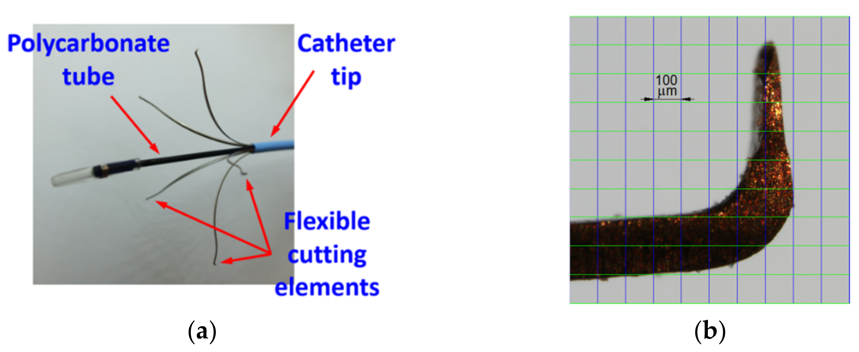
Figure 1. Flebogrif view: (a) Tip with five cutting elements; (b) Zoom-in of the cutting element.
Materials 15 02599 g001
Figure 2. Schematics of the measurement of forces exerted by the Flebogrif cutting elements: (a) Measurement of the total force of all cutting elements; (b) Measurement of the force of a single cutting element.
Materials 15 02599 g002
Figure 3. Instrument used to fix the Flebogrif in the measurement position: (a) Fixation of the testing instrument in the universal testing machine jaws; (b) Structure of the instrument used to fix the Flebogrif tip.
Materials 15 02599 g003
Figure 4. View of the measurement stand.
Materials 15 02599 g004
Figure 5. Sample F(x) characteristics for all cutting elements for the following protrusions: (a) L = Lmax, (b) L = 0.9⋅Lmax, (c) L = 0.8⋅Lmax.
Materials 15 02599 g005
Figure 6. The aggregated F(x) chart for all cutting elements.
Materials 15 02599 g006
Figure 7. Examples of F(x) characteristics for a single cutting element with protrusion of: (a) L = Lmax, (b) L = 0.9⋅Lmax, (c) L = 0.8⋅Lmax.
Materials 15 02599 g007
Figure 8. Aggregated F(x) chart for single cutting elements.
Materials 15 02599 g008
Figure 9. The aggregated Ff (dv) dependence charts created with the use of: (a) the results of force measurements for all cutting elements; (b) the results of force measurements for a single cutting element.
Materials 15 02599 g009
Table 1. Results of measurements and calculations of Flebogrif geometrical value.
Materials 15 02599 i001
Protrusion Levels
L
Side
a (mm)
Diagonal
p (mm)
Calculated Diameter
df (mm)
L = Lmax20.633.234.5
21.532.5
19.831.8
20.532.9
19.632.5
L = 0.9⋅Lmax16.325.227.3
17.225.1
16.524.8
17.125.2
16.424.6
L = 0.8⋅Lmax11.017.718.6
11.317.8
10.817.6
11.217.7
10.117.7
Publisher’s Note: MDPI stays neutral with regard to jurisdictional claims in published maps and institutional affiliations.