Article

In-Situ Growth of ZnO Whiskers on Ti2ZnC MAX Phases

1
Jiangsu Key Laboratory of Advanced Metallic Materials, School of Materials Science and Engineering, Southeast University, Nanjing 211189, China
2
Liyang Zichen New Material Technology Co., Ltd., Changzhou 213000, China
*
Authors to whom correspondence should be addressed.
Materials 2023, 16(10), 3610; https://doi.org/10.3390/ma16103610
Submission received: 7 April 2023 / Accepted: 6 May 2023 / Published: 9 May 2023

Abstract

:
ZnO whiskers have many applications, such as in medical and photocatalysis fields. In this study, an unconventional preparation approach is reported, realizing the in-situ growth of ZnO whiskers on Ti2ZnC. The weak bonding between the layer of Ti6C-octahedron and the Zn-atom layers leads to the easy extraction of Zn atoms from Ti2ZnC lattice points, resulting in the formation of ZnO whiskers on the Ti2ZnC surface. This is the first time that ZnO whiskers have been found to grow in-situ on Ti2ZnC substrate. Further, this phenomenon is amplified when the size of the Ti2ZnC grains is mechanically reduced by ball-milling, which bodes a promising route to prepare ZnO in-situ on a large scale. Additionally, this finding can also help us better understand the stability of Ti2ZnC and the whiskering mechanism of MAX phases.

1. Introduction

ZnO is a functional semiconductor with a wide band gap (3.37 eV) and a considerable exciton binding energy (60 meV) at room temperature [1,2], which can be used in sensors [3,4] and photocatalysis [5,6,7,8,9]. In addition, because of its nontoxicity and biocompatibility, ZnO can also be used in medical fields [10]. In 2006, Wang et al. [11] successfully used the tip of a conductive atomic force microscope probe to bend ZnO whiskers, perfectly converting this part of mechanical energy into electrical energy, and invented nanogenerators, which opened a door for the study of ZnO materials. ZnO whiskers have also been widely studied as an efficient photocatalyst for the degradation of organic pollutants in water treatment [5,6] by which organic pollutants are decomposed into small molecules and less-harmful products such as CO2 and H2O [12]. The preparation of ZnO is the foundation for all of its applications and may affect its application effect. Therefore, the preparation methods for ZnO are important and always draw attention from both academic and industrial circles.
The primary methods for preparing ZnO whiskers include vapor deposition [13,14,15] and the hydrothermal method [16,17,18]. Physical vapor deposition (PVD) consists of vaporizing the raw material of ZnO powder and then forming a ZnO nanomaterial from the gaseous state without a catalyst. Since ZnO has a high melting point (1975 °C), the temperature required for the synthesis of ZnO is very high [13]; as for chemical vapor deposition (CVD), in addition to the above physical changes, chemical reactions such as oxidation and compounding also occur. The formation and morphology of ZnO are affected by many factors such as temperature, gas pressure, gas flow, and catalyst [18]. Compared with vapor deposition, the hydrothermal method has the advantages of low temperature and simple equipment, and it is suitable for large-scale preparation. However, the hydrothermal process usually requires a seeded substrate for highly dense arrayed ZnO whiskers. The interface in the presence of seeds has defects, leading to low adhesion [19]. In addition, these methods may need to transfer the as-synthesized ZnO for some specific application purposes. Sometimes, it is significant to immobilize ZnO on substrates, for example when it is used as photocatalyst, to realize a sustainable operation as well as good photocatalyst recovery [20,21,22], because the recovery of photocatalysts after water treatment is complicated and costly [12,23]. Therefore, the in-situ preparation of ZnO is the requirement of the technology development.
Recently, an interesting phenomenon of whisker growth on MAX phases has drawn increasing attention [24,25,26,27,28]. The MAX phases are layered and hexagonal early transition-metal carbides and nitrides, with a general formula of Mn+1AXn (where n = 1, 2, 3; M: early transition metal; A: A-group element; X: C or N) [29,30,31,32]. They combine the excellent properties of metals and ceramics, such as good electrical conductivity, high temperature oxidation resistance, and corrosion resistance [33,34]. However, due to the weak bonding between the M6X-octahedron layer and the A-atom layer, A-site atoms can be easily released from the lattice points of the MAX phase during mechanical exfoliation [24,25,27]. So far, there have been some reports [26,27,28,35,36] confirming that A-site metal whiskers (such as Ga, Sn, and In whiskers) can grow on the corresponding MAX phases (Cr2GaC, Ti2SnC, and Ti2InC, respectively). Zn-containing MAX phases (e.g., Ti2ZnC, V2ZnC, and Ti3ZnC2) were successfully synthesized by Huang Qing et al. [37]. If Zn atoms can be extracted from the Ti2ZnC precursor and be made to oxidize in-situ, the in-situ preparation of ZnO whiskers on the MAX phase substrates may be realized. Considering that MAX phases are corrosion-resistant and conductive [29,33], they would be the ideal substrates for the specific applications of ZnO whiskers, and realizing ZnO whiskers grown in-situ on MAX phases would be beneficial for a host of applications that require the immobilization of ZnO whiskers.
Herein, Ti2ZnC was used to grow ZnO whiskers in-situ. The results bode a new method for the preparation of ZnO whiskers, which does not require very high temperature and gas flow like traditional vapor deposition and is expected to be promising in applications that require the immobilization of ZnO whiskers. In addition, the size of Ti2ZnC particles is reduced after ball-milling and more Zn atoms diffuse out of the Ti2ZnC, resulting in more ZnO whiskers being grown on the surface of the sample. The findings not only help us better understand the stability of Ti2ZnC but also help us further understand the whiskering mechanism of MAX phases, which would be utilized for ZnO whisker preparation.

2. Materials and Methods

Commercial powders (TiC: 99.9%, 300 mesh, Nanjing crystal chemical Co., Nanjing, China; Ti: 99.99%, ≥300 mesh, Aladdin, Shanghai, China; Al: 99.7%, 300 mesh, Zhongnuo new materials Co., Beijing, China) were used as raw materials to synthesize Ti2AlC following previous work [38,39]. First, the powders of TiC, Ti, and Al were mixed according to a molar ratio of 0.95:1.05:1.05. Afterwards, the mixture was placed in an alumina crucible and then sintered at 1400 °C for 1 h under the protection of Ar gas to obtain the precursor Ti2AlC. The as-synthesized Ti2AlC was mixed with ZnCl2 by a molar ratio of 1:1.5 using an agate mortar under the protection of nitrogen in a glovebox. The mixed Ti2AlC/ZnCl2 was treated at 550 °C for 5 h in an Ar atmosphere. Then, the reactant was washed with deionized water and dried at 40 °C, to exclude the interference of the elemental Zn in the as-synthesized Ti2ZnC, and an acid pickling treatment was carried out, soaking the as-synthesized Ti2ZnC in a 1 mol/L HCL solution for 5 h with continuous stirring. Finally, the pickled Ti2ZnC was collected by suction filtration and then dried in an oven under 40 °C, and thus the target MAX phase Ti2ZnC was obtained. The obtained Ti2ZnC powder was divided into two groups. One group of Ti2ZnC powder was directly cold-pressed into disc samples under 800 MPa and then heated at 400 °C for 48 h, and the other group was ball-milled and then pressed into disc specimens.
To reduce the grain size of the Ti2ZnC, it was ball-milled in stainless steel jars with steel milling balls, the ratio of the ball to the Ti2ZnC was 10:1, the rotating speed was 700 rpm, and the milling time was 18 h. After the ball-milling, the as-milled Ti2ZnC powder was cold-pressed into discs under 800 MPa and then heated at 400 °C for 48 h.
The phase composition of Ti2ZnC samples under different conditions was characterized by using an X-ray diffractometer (XRD, Haoyuan, DX–2700BH, Cu-Kα radiation) operating at 40 kV and 30 mA, wherein the 2-theta scanning range was 5° to 65°. The morphologies of samples were characterized by scanning electron microscopy (SEM, FEI Sirion 200, Hillsboro, OR, USA), wherein the applied electron beam acceleration voltage was 12–15 kV and the transmission electron microscope (TEM, Thermo Talos F200X, Waltham, MA, USA) equipped with an energy spectrum probe (EDS, Oxford X-Max 50, Abingdon, UK) was also used to further characterize ZnO whiskers.

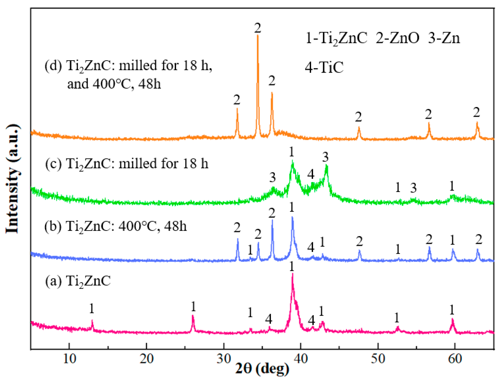
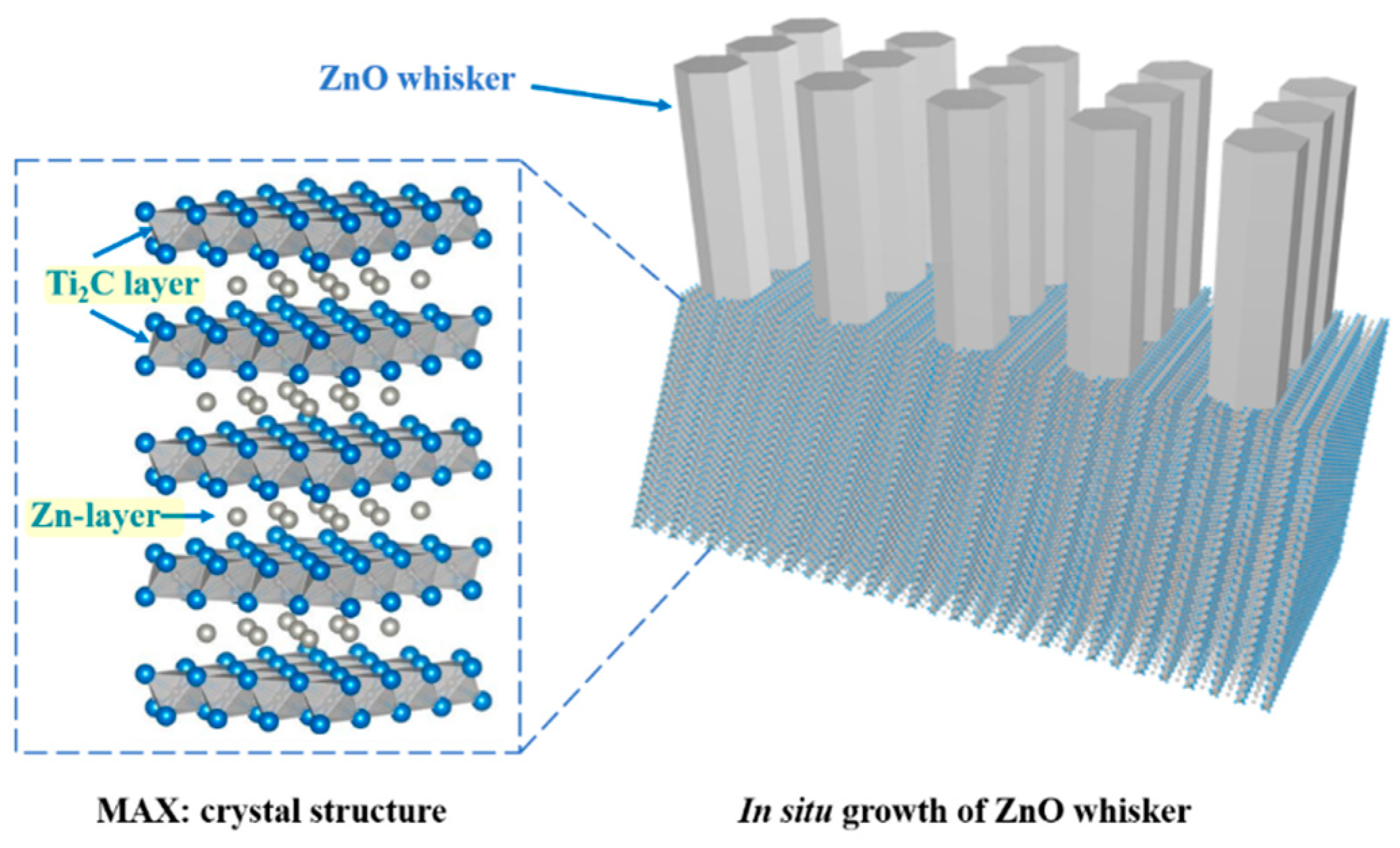
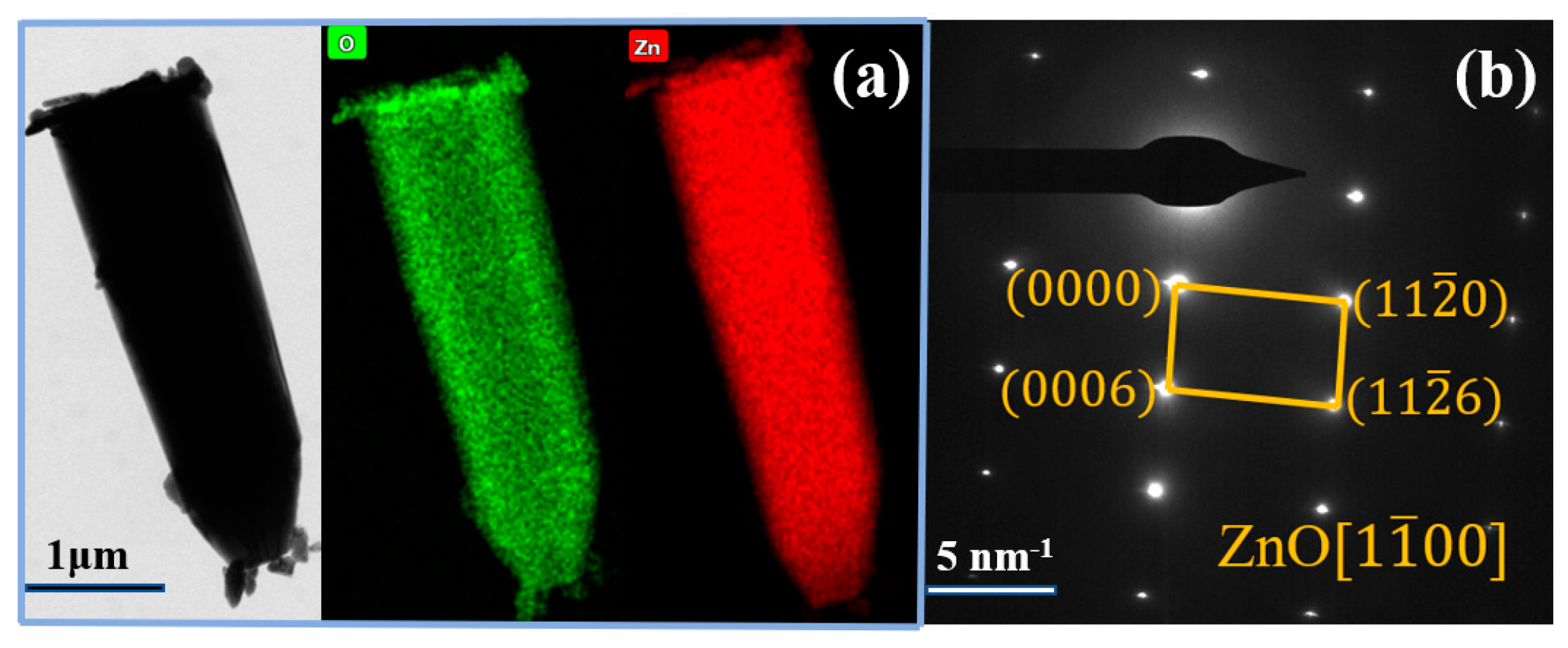
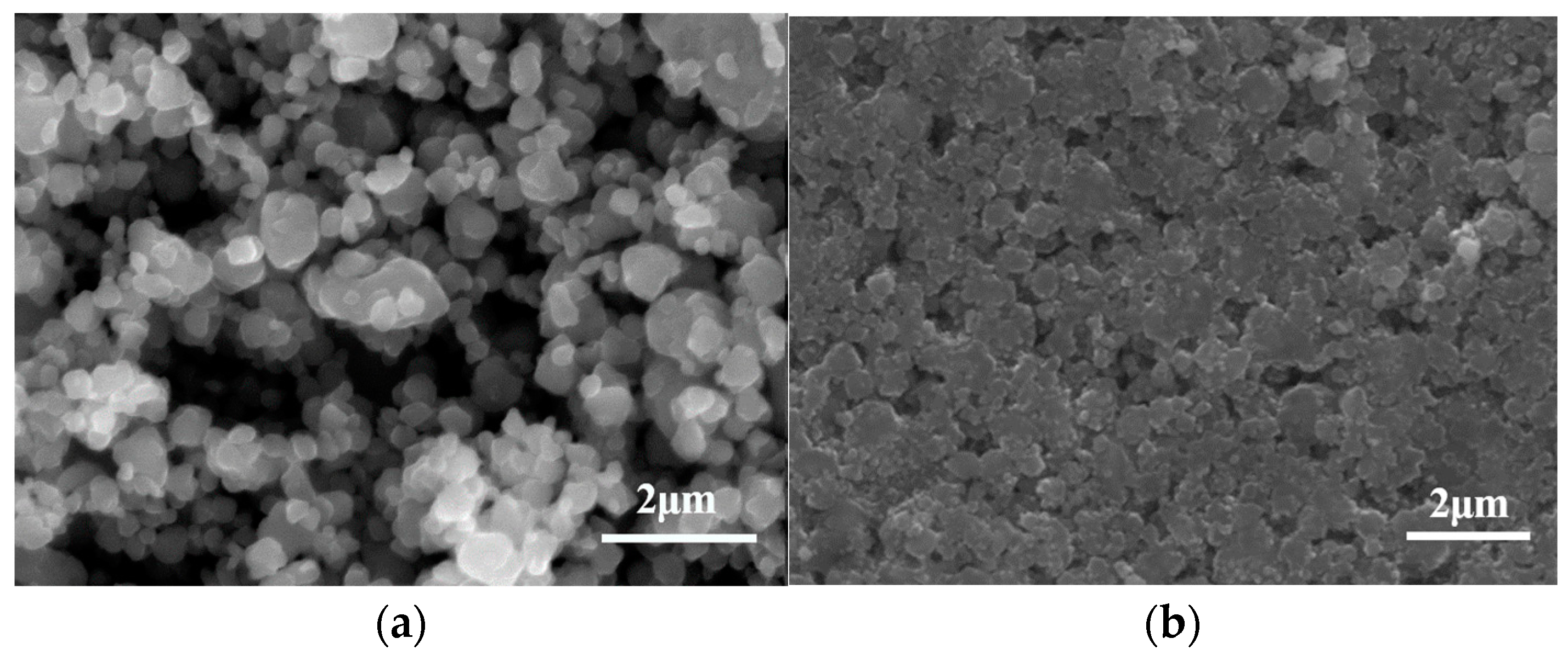
3. Results and Discussion

Figure 1a shows the XRD pattern of the as-synthesized Ti2ZnC, which is consistent with the work of Huang et al. [37]. Figure 2a shows the SEM characterization of the as-synthesized Ti2ZnC. After being heated at 400 °C for 48 h, the Ti2ZnC sample was characterized again, and new XRD peaks indexed to ZnO appeared, as shown in Figure 1b. The SEM image of the sample after heating, shown in Figure 2b, shows that some Zn atoms diffuse out from the lattice points of Ti2ZnC and then are oxidized by oxygen in air, thus forming ZnO whiskers that are closely connected with the Ti2ZnC matrix. The ZnO whiskers grown on the Ti2ZnC correspond to the new XRD peaks of ZnO. Zhang et al. [40] reported that when Ti2SnC was heated to 400 °C, some Sn atoms would precipitate from the lattice points of Ti2SnC, and Ti2SnC was partially decomposed into Ti2C and Sn. Because the diffusion process mainly depends on temperature, the precipitation of Sn atoms is also strongly affected by temperature. The precipitation of Sn atoms is almost invisible under 200 °C. When the temperature rises to 400 °C, the precipitation of Sn atoms can be clearly observed. When the temperature rises to 600 °C, this phenomenon becomes more obvious. In this work, the soaking temperature was set to 400 °C to make Zn atoms diffuse out of the Ti2ZnC as much as possible. It is reasonable to ascribe this whisker growth process to the Zn atom precipitation from the lattice points of Ti2ZnC. Like other MAX phases, Ti2ZnC has a layered structure, as illustrated in Figure 3. The bonds between the Zn layer and Ti2C layer are relatively weak, which indicates that Ti-Zn bonds are easy to break under elevated temperature and some Zn atoms can diffuse out from the lattice points of Ti2ZnC. Then, the Zn atoms on the surface are oxidized by oxygen in the air, forming ZnO whiskers.
Since the Zn atoms in the Ti2ZnC lattice are the Zn source for the growth of ZnO whiskers, how will the Ti2ZnC change with the increase in temperature during the heating process? The Ti2ZnC sample was characterized by in-situ XRD. The whole process was carried out from room temperature to 400 °C, the heating rate was 10 °C/min, and the sample was characterized at 100 °C, 200 °C, 300 °C and 400 °C, respectively. It can be seen from Figure 4 that the XRD results at four different heating temperatures all contain the diffraction peak of Ti2C (6.6°). The generation of this diffraction peak is due to the etching of the A-site atoms and the increase in the interlayer distance of the MAX phase. However, the existence of diffraction peaks corresponding to (002), (004) and (106) of Ti2ZnC at four temperatures means that only some Zn atoms diffuse out from the lattice points of Ti2ZnC, and Ti2ZnC and Ti2C exist in the sample at the same time. The partially enlarged figure on the right shows that with the increase in temperature, the diffraction peaks corresponding to (002), (004) and (103) of Ti2ZnC shift slightly to the left. Furthermore, with the increase in temperature, the intensity of the diffraction peaks of TiC and ZnO gradually increase, indicating that some Ti2ZnC decompose into TiC and Zn and Zn reacts with O2 in the air at high temperature, thus forming ZnO. This can be confirmed by the phenomenon that the intensity of the diffraction peaks corresponding to (100), (002), and (101) of ZnO increases with increasing temperature.
Figure 5 shows a ZnO whisker observed under TEM. The corresponding element mapping (Figure 5b,c) shows that O and Zn elements are uniformly distributed throughout the whisker. The selected area electron diffraction (SAED) pattern of the whisker is shown in Figure 5b, which can be assigned to the ( 11 2 ¯ 0 ), ( 0006 ), and ( 11 2 ¯ 6 ) planes of ZnO along the [ 1 1 ¯ 00 ] axis, indicating that it is a single-crystal ZnO whisker.
Since the Zn atoms feeding the ZnO whisker growth are from the Ti2ZnC substrate, if we can make more Zn atoms come out of the substrate, more ZnO whiskers are expectable. Mechanically decomposing the MAX phase by ball-milling is an effective way to obtain small-sized carbides and A-site metal whiskers, and Tian et al. [25] reported that when Ti2SnC was ball-milled for 7 h, after cold-pressing at 1000 MPa and storing at room temperature (RT) for 30 days, a large number of Sn whiskers grew on the surface of the substrate. The study shows that with the increase in ball-milling time, the damage degree of ball-milling to Ti2SnC is intensified, and more active Sn atoms release from the lattice points of Ti2SnC, which serve as the source of Sn whisker growth. The same phenomenon also occurs in Ti2InC. Therefore, ball-milling was employed to mechanically decompose Ti2ZnC. The typical morphology of Ti2ZnC particles after ball-milling is shown in Figure 6a. Compared with Figure 2a, the size of Ti2ZnC particles is greatly reduced by ball-milling. Figure 1c shows the XRD of the ball-milled Ti2ZnC. Comparing the XRD data of Ti2ZnC before (Figure 1a) and after ball-milling (Figure 1c), it is found that the XRD peaks of the samples are broadened after ball-milling, indicating that the Ti2ZnC has been refined to a great extent. This result is consistent with the particle refinement observed in Figure 6a. In addition, the diffraction peaks of Zn can also be seen, indicating that the mechanochemical decomposition of Ti2ZnC has occurred and more Zn atoms are released from the lattice of the Ti2ZnC after mechanical exfoliation.
Then, the ball-milled mixture was cold-pressed into discs under 800 MPa (Figure 6b) and heated at 400 °C for 48 h in the air. In Figure 7a, it can be seen that a large number of whiskers grow on the surface of the sample, and the EDS analysis result (Figure 7b) suggests that the whisker is composed of Zn and O elements, with Ti impurity accounting for 0.83 at.%. In addition, Figure 1d shows that the ZnO diffraction peaks are very obvious, indicating that these whiskers are ZnO whiskers. Because a large number of ZnO whiskers grown on the surface of the sample cover the Ti2ZnC matrix and Ti2ZnC has poor crystallinity after ball-milling while ZnO has high crystallinity, Figure 1d shows very strong ZnO diffraction peaks, while the diffraction peaks of Ti2ZnC are almost invisible. There are two reasons for the growth of a large number of ZnO whiskers. On the one hand, Ti2ZnC was mechanically decomposed and many more Zn atoms came out of the lattice of the Ti2ZnC after ball-milling, and these Zn atoms combine with O atoms to form whiskers. On the other hand, according to Figure 2b, some Zn atoms released from the lattice points of Ti2ZnC under 400 °C, and these Zn atoms contributed to the growth of ZnO whiskers. From Figure 7, it is found that the ZnO whiskers were longer and thinner than those observed in Figure 2b. This is because the reduced size of Ti2ZnC particles after ball-milling provides more sites for whisker growth.
The ZnO whiskers grown on the surface of the sample were collected to characterize their optical properties. The UV-Vis spectra for the grown ZnO whiskers are shown in Figure 8a, which shows that the grown ZnO whiskers possess very strong absorption for UV light. The band gap energy is calculated by Tauc’s plot, to obtain (αhν)2 vs hν graphs and extrapolate the linear segment of the curves. Figure 8b shows the Tauc’s plot of the grown ZnO whiskers, and the band gap of the grown ZnO whiskers is calculated to be 3.10 eV.
The Raman spectra of the grown ZnO whiskers are displayed in Figure 9a. The whiskers exhibit a characteristic wurtzite ZnO Raman peak at 437 cm−1. In addition, the low-intensity peaks observed at 331 cm−1 and 573 cm−1 can be attributed to the presence of different types of defects such as oxygen vacancies and interstitials [41]. The photoluminescence spectroscopy (PL) spectra of the grown ZnO whiskers are shown in Figure 9b with λex = 325 nm. The spectra are characterized by a strong emission peak centered around 376 nm, which is attributed to the UV near-band-edge emission of ZnO. In addition, there is a wide green emission peak around 520 nm, which can be attributed to the presence of defects [42]. The intensity of the green emission peak is lower than that of the UV emission peak, indicating that the ZnO whiskers grown on the surface of the sample have low defect concentrations and good crystallinity.
The ZnO whisker growth suggests the low-temperature instability of the Ti2ZnC, similar to Ti2SnC [40]. Specifically, when the grain size is reduced, more Zn atoms can diffuse out of the Ti2ZnC, and then oxidized, resulting in more ZnO whiskers. This consolidates the growth mechanism proposed for the formation of massive Sn whiskers found in Ti2SnC [24]. The findings here, on the one hand, may be harnessed to engineer a new method for the in-situ preparation of ZnO whiskers based on the Zn-containing MAX phase, and on the other hand, would help us to better understand the stability of Ti2ZnC and the whiskering mechanism of MAX phases.

4. Conclusions

In this study, the in-situ growth of ZnO whiskers on Ti2ZnC was realized by heating Ti2ZnC at 400 °C for 48 h. Ball-milling (700 rpm, 18 h) can reduce the size of Ti2ZnC particles and therefore extract more Zn atoms from the lattice points of Ti2ZnC, resulting in the growth of a large number of ZnO whiskers on the surface of Ti2ZnC. The ZnO whiskers grown on the surface of the sample (ball-milled and aged at 400 °C for 48 h) are thinner than those grown on the directly heated Ti2ZnC substrate (aging at 400 °C for 48 h), which is because the reduced size of Ti2ZnC particles after ball-milling provides more sites for whisker growth. This phenomenon could be harnessed to develop a new method for the in-situ preparation of ZnO whiskers. The findings here will also assist us in comprehensively understanding the stability of Ti2ZnC and promote the study of the whiskering mechanism of MAX phases. Additionally, the in-situ growth of ZnO on Ti2ZnC is expected to be promising for applications that require the immobilization of ZnO whiskers.

Author Contributions

Conceptualization, Y.R. and P.Z.; methodology, Y.R. and P.Z.; software, Y.R. and P.Z.; validation, Y.R., H.X. and P.Z.; investigation, Y.R.; resources, F.W., Z.T. and Q.Z.; data curation, Y.Z. and Z.T.; writing—original draft preparation, Y.R. and P.Z.; writing—review and editing, Y.R., Z.T., P.Z. and Z.S.; supervision, P.Z. and Z.S. All authors have read and agreed to the published version of the manuscript.

Funding

This research was funded by the National Natural Science Foundation of China (52171033 and 51731004), and the Natural Science Foundation of Jiangsu Province (BK20201283).

Institutional Review Board Statement

Not applicable.

Informed Consent Statement

Not applicable.

Data Availability Statement

Not applicable.

Acknowledgments

The authors gratefully acknowledge the financial support from the National Natural Science Foundation of China (52171033 and 51731004), and the Natural Science Foundation of Jiangsu Province (BK20201283).

Conflicts of Interest

The authors declare no conflict of interest.

References

  1. Wlazlo, M.; Haras, M.; Kolodziej, G.; Szawcow, O.; Ostapko, J.; Andrysiewicz, W.; Kharytonau, D.S.; Skotnicki, T. Piezoelectric Response and Substrate Effect of ZnO Nanowires for Mechanical Energy Harvesting in Internet-of-Things Applications. Materials 2022, 15, 6767. [Google Scholar] [CrossRef] [PubMed]
  2. Peksu, E.; Karaagac, H. Doping and annealing effects on structural, electrical and optical properties of tin-doped zinc-oxide thin films. J. Alloys Compd. 2018, 764, 616–625. [Google Scholar] [CrossRef]
  3. Qin, L.; Mawignon, F.J.; Hussain, M.; Ange, N.K.; Lu, S.; Hafezi, M.; Dong, G. Economic Friendly ZnO-Based UV Sensors Using Hydrothermal Growth: A Review. Materials 2021, 14, 4083. [Google Scholar] [CrossRef] [PubMed]
  4. Kang, Y.; Yu, F.; Zhang, L.; Wang, W.; Chen, L.; Li, Y. Review of ZnO-based nanomaterials in gas sensors. Solid State Ionics 2021, 360, 115544. [Google Scholar] [CrossRef]
  5. Noman, M.T.; Amor, N.; Petru, M.; Mahmood, A.; Kejzlar, P. Photocatalytic Behaviour of Zinc Oxide Nanostructures on Surface Activation of Polymeric Fibres. Polymers 2021, 13, 1227. [Google Scholar] [CrossRef]
  6. Gao, C.; Zhong, K.; Fang, X.; Fang, D.; Zhao, H.; Wang, D.; Li, B.; Zhai, Y.; Chu, X.; Li, J.; et al. Brief Review of Photocatalysis and Photoresponse Properties of ZnO–Graphene Nanocomposites. Energies 2021, 14, 6403. [Google Scholar] [CrossRef]
  7. Li, H.; Ding, J.; Cai, S.; Zhang, W.; Zhang, X.; Wu, T.; Wang, C.; Foss, M.; Yang, R. Plasmon-enhanced photocatalytic properties of Au/ZnO nanowires. Appl. Surf. Sci. 2022, 583, 152539. [Google Scholar] [CrossRef]
  8. Ma, W.; Xu, L.; Tian, Z.; Zang, A. Changes in photocatalytic activity and optical properties of ZnO whiskers induced by UV irradiation. J. Lumin. 2022, 249, 119015. [Google Scholar] [CrossRef]
  9. Shekofteh-Gohari, M.; Habibi-Yangjeh, A.; Abitorabi, M.; Rouhi, A. Magnetically separable nanocomposites based on ZnO and their applications in photocatalytic processes: A review. Crit. Rev. Environ. Sci. Technol. 2018, 48, 806–857. [Google Scholar] [CrossRef]
  10. Ortiz-Casas, B.; Galdámez-Martínez, A.; Gutiérrez-Flores, J.; Baca Ibañez, A.; Kumar Panda, P.; Santana, G.; de la Vega, H.A.; Suar, M.; Gutiérrez Rodelo, C.; Kaushik, A.; et al. Bio-acceptable 0D and 1D ZnO nanostructures for cancer diagnostics and treatment. Mater. Today 2021, 50, 533–569. [Google Scholar] [CrossRef]
  11. Wang, Z.L.; Song, J.J.S. Piezoelectric nanogenerators based on zinc oxide nanowire arrays. Science 2006, 312, 242–246. [Google Scholar] [CrossRef]
  12. Le, A.T.; Le, T.D.H.; Cheong, K.-Y.; Pung, S.-Y. Immobilization of zinc oxide-based photocatalysts for organic pollutant degradation: A review. J. Environ. Chem. Eng. 2022, 10, 108505. [Google Scholar] [CrossRef]
  13. Bi, N.; Zhang, L.; Zheng, Q.; Zhuge, F.; Li, J.; Gao, X.P.A.; Du, J. Control of ZnO nanowire growth and optical properties in a vapor deposition process. J. Mater. Sci. Technol. 2017, 33, 850–855. [Google Scholar] [CrossRef]
  14. Guniat, L.; Caroff, P.; Fontcuberta, I.M.A. Vapor Phase Growth of Semiconductor Nanowires: Key Developments and Open Questions. Chem. Rev. 2019, 119, 8958–8971. [Google Scholar] [CrossRef] [PubMed]
  15. McIntyre, P.C.; Fontcuberta i Morral, A. Semiconductor nanowires: To grow or not to grow? Mater. Today Nano 2020, 9, 100058. [Google Scholar] [CrossRef]
  16. Lin, C.; Li, Q.; Guang, H.; An, M. Electrodeposited Zn: A promising alternative to ZnO seed layer for hydrothermal growth of ZnO nanowire array. Mater. Lett. 2022, 314, 131848. [Google Scholar] [CrossRef]
  17. Almamari, M.R.; Ahmed, N.M.; Holi, A.M.; Yam, F.K.; Kyaw, H.H.; Almessiere, M.A.; Al-Abri, M.Z. Some Distinct Attributes of ZnO Nanorods Arrays: Effects of Varying Hydrothermal Growth Time. Materials 2022, 15, 5827. [Google Scholar] [CrossRef]
  18. Podrezova, L.V.; Porro, S.; Cauda, V.; Fontana, M.; Cicero, G. Comparison between ZnO nanowires grown by chemical vapor deposition and hydrothermal synthesis. Appl. Phys. A 2013, 113, 623–632. [Google Scholar] [CrossRef]
  19. Cui, J. Zinc oxide nanowires. Mater. Charact. 2012, 64, 43–52. [Google Scholar] [CrossRef]
  20. Kim, J.; Van der Bruggen, B. The use of nanoparticles in polymeric and ceramic membrane structures: Review of manufacturing procedures and performance improvement for water treatment. Environ. Pollut. 2010, 158, 2335–2349. [Google Scholar] [CrossRef]
  21. Qiu, R.; Zhang, D.; Mo, Y.; Song, L.; Brewer, E.; Huang, X.; Xiong, Y. Photocatalytic activity of polymer-modified ZnO under visible light irradiation. J. Hazard. Mater. 2008, 156, 80–85. [Google Scholar] [CrossRef] [PubMed]
  22. Ullah, R.; Dutta, J. Photocatalytic degradation of organic dyes with manganese-doped ZnO nanoparticles. J. Hazard. Mater. 2008, 156, 194–200. [Google Scholar] [CrossRef]
  23. Adeleye, A.S.; Conway, J.R.; Garner, K.; Huang, Y.; Su, Y.; Keller, A.A. Engineered nanomaterials for water treatment and remediation: Costs, benefits, and applicability. Chem. Eng. J. 2016, 286, 640–662. [Google Scholar] [CrossRef]
  24. Zhang, Q.; Tian, Z.; Zhang, P.; Zhang, Y.; Liu, Y.; He, W.; Pan, L.; Liu, Y.; Sun, Z. Rapid and massive growth of tin whisker on mechanochemically decomposed Ti2SnC. Mater. Today Commun. 2022, 31, 103466. [Google Scholar] [CrossRef]
  25. Tian, Z.; Xu, X.; Tang, J.; Zhang, Q.; Wu, F.; Zhang, P.; Liu, J.; Sun, Z. Large-scale preparation of nano-sized carbides and metal whiskers via mechanochemical decomposition of MAX phases. Int. J. Appl. Ceram. Technol. 2022, 20, 823–832. [Google Scholar] [CrossRef]
  26. Liu, Y.; Zhang, P.; Zhang, Y.; Ding, J.; Shi, J.; Sun, Z.J.M.L. Spontaneous growth of Sn whiskers and a new formation mechanism. Mater. Lett. 2016, 178, 111–114. [Google Scholar] [CrossRef]
  27. Zhang, Q.; Tang, J.; Tang, H.; Tian, Z.; Zhang, P.; Zhang, Y.; Liu, J.; Sun, Z.M. Method for inhibiting Sn whisker growth on Ti2SnC. J. Mater. Sci. 2022, 57, 20462–20471. [Google Scholar] [CrossRef]
  28. Zhang, P.G.; Ding, J.X.; Liu, Y.S.; Yang, L.; Tian, W.B.; Ouyang, J.; Zhang, Y.M.; Sun, Z.M. Mechanism and mitigation of spontaneous Ga whisker growth on Cr2GaC. Sci. China Technol. Sci. 2020, 63, 440–445. [Google Scholar] [CrossRef]
  29. Sokol, M.; Natu, V.; Kota, S.; Barsoum, M.W. On the Chemical Diversity of the MAX Phases. Trends Chem. 2019, 1, 210–223. [Google Scholar] [CrossRef]
  30. Tian, Z.; Wu, F.; Hu, P.; Ding, J.; Zhang, Y.; Zhang, P.; Sun, Z. Synthesis of Ti3(SnxAl1−x)C2 solid solutions over the whole composition range. J. Alloys Compd. 2022, 894, 162429. [Google Scholar] [CrossRef]
  31. Fu, L.; Xia, W. MAX Phases as Nanolaminate Materials: Chemical Composition, Microstructure, Synthesis, Properties, and Applications. Adv. Eng. Mater. 2021, 23, 2001191. [Google Scholar] [CrossRef]
  32. Xu, X.; Yang, L.; Zheng, W.; Zhang, H.; Wu, F.; Tian, Z.; Zhang, P.; Sun, Z. MXenes with applications in supercapacitors and secondary batteries: A comprehensive review. Mater. Rep. Energy 2022, 2, 100080. [Google Scholar] [CrossRef]
  33. Zhang, Z.; Duan, X.; Jia, D.; Zhou, Y.; van der Zwaag, S. On the formation mechanisms and properties of MAX phases: A review. J. Eur. Ceram. Soc. 2021, 41, 3851–3878. [Google Scholar] [CrossRef]
  34. Zhou, A.G.; Liu, Y.; Li, S.B.; Wang, X.H.; Ying, G.B.; Xia, Q.X.; Zhang, P.G. From structural ceramics to 2D materials with multi-applications: A review on the development from MAX phases to MXenes. J. Adv. Ceram. 2021, 10, 1194–1242. [Google Scholar] [CrossRef]
  35. Zhang, P.; Shen, L.; Ouyang, J.; Zhang, Y.; Wu, S.; Sun, Z.J. Room temperature mushrooming of gallium wires and its growth mechanism. J. Alloy Compd. 2015, 619, 488–497. [Google Scholar] [CrossRef]
  36. Li, S.; Liu, Y.S.; Zhang, P.G.; Zhang, Y.; Lu, C.J.; Pan, L.; Ding, J.X.; Sun, Z.M. Interface energy-driven indium whisker growth on ceramic substrates. J. Mater. Sci. Mater. Electron. 2021, 32, 16881–16888. [Google Scholar] [CrossRef]
  37. Li, M.; Lu, J.; Luo, K.; Li, Y.; Chang, K.; Chen, K.; Zhou, J.; Rosen, J.; Hultman, L.; Eklund, P.; et al. Element Replacement Approach by Reaction with Lewis Acidic Molten Salts to Synthesize Nanolaminated MAX Phases and MXenes. J. Am. Chem. Soc. 2019, 141, 4730–4737. [Google Scholar] [CrossRef]
  38. Hashimoto, S.; Takeuchi, M.; Inoue, K.; Honda, S.; Awaji, H.; Fukuda, K.; Zhang, S. Pressureless sintering and mechanical properties of titanium aluminum carbide. Mater. Lett. 2008, 62, 1480–1483. [Google Scholar] [CrossRef]
  39. Zheng-Ming, S.U.N.; Wu-Bian, T.; Pei-Gen, Z.; Dan-Dan, W.; Yu-Hui, Z.H.A.; Pei-Yan, H.; Jian-Xiang, D. High-purity Ti2AlC Powder: Preparation and Application in Ag-based Electrical Contact Materials. J. Inorg. Mater. 2019, 35, 729–734. [Google Scholar] [CrossRef]
  40. Zhang, J.; Liu, B.; Wang, J.Y.; Zhou, Y.C. Low-temperature instability of Ti2SnC: A combined transmission electron microscopy, differential scanning calorimetry, and X-ray diffraction investigations. J. Mater. Res. 2009, 24, 39–49. [Google Scholar] [CrossRef]
  41. Rajalakshmi, M.; Arora, A.K.; Bendre, B.S.; Mahamuni, S. Optical phonon confinement in zinc oxide nanoparticles. J. Appl. Phys. 2000, 87, 2445–2448. [Google Scholar] [CrossRef]
  42. Karthik, K.V.; Raghu, A.V.; Reddy, K.R.; Ravishankar, R.; Sangeeta, M.; Shetti, N.P.; Reddy, C.V. Green synthesis of Cu-doped ZnO nanoparticles and its application for the photocatalytic degradation of hazardous organic pollutants. Chemosphere 2022, 287, 132081. [Google Scholar] [CrossRef] [PubMed]
Figure 1. XRD patterns: (a) Ti2ZnC, (b) Ti2ZnC after aging for 48 h at 400 °C, (c) ball-milled Ti2ZnC (18 h), and (d) ball-milled Ti2ZnC (18 h) after aging for 48 h at 400 °C.
Materials 16 03610 g001
Figure 2. SEM characterization of (a) the Ti2ZnC and (b) the Ti2ZnC after being heated at 400 °C for 48 h.
Materials 16 03610 g002
Figure 3. Illustrations of MAX phase crystal structure and the in-situ ZnO whisker growth.
Materials 16 03610 g003
Figure 4. In-situ XRD pattern: Ti2ZnC heated at 100 °C, 200 °C, 300 °C and 400 °C, respectively.
Materials 16 03610 g004
Figure 5. TEM characterization of (a) a ZnO whisker and its element mapping; (b) SAED pattern of the whisker.
Materials 16 03610 g005
Figure 6. SEM characterization of (a) the ball-milled Ti2ZnC (18 h) and (b) the surface of the disc after cold-pressing.
Materials 16 03610 g006
Figure 7. SEM characterization of (a) ZnO whiskers grown on the ball-milled (18 h) Ti2ZnC sample after dwelling at 400 °C for 48 h and (b) the grown ZnO whisker together with its EDS analysis result (the red plus sign).
Materials 16 03610 g007
Figure 8. (a) UV-Vis spectra, and (b) Tauc plot of the grown ZnO whiskers.
Materials 16 03610 g008
Figure 9. (a) Raman spectra, and (b) PL spectra of the grown ZnO whiskers.
Materials 16 03610 g009
Disclaimer/Publisher’s Note: The statements, opinions and data contained in all publications are solely those of the individual author(s) and contributor(s) and not of MDPI and/or the editor(s). MDPI and/or the editor(s) disclaim responsibility for any injury to people or property resulting from any ideas, methods, instructions or products referred to in the content.

