

Nanocluster Evolution in D9 Austenitic Steel under Neutron and Proton Irradiation
 , ,
, ,
Abstract
:1. Introduction
2. Materials and Methods
2.1. Alloy Preparation and Irradiation
2.2. Atom Probe Tomography
3. Results
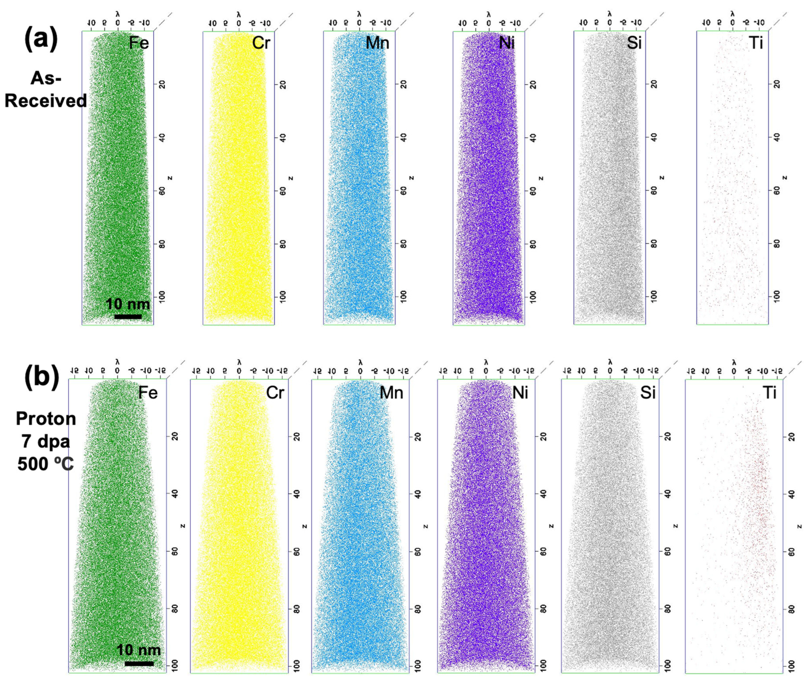
3.1. As-Received and Proton-Irradiated
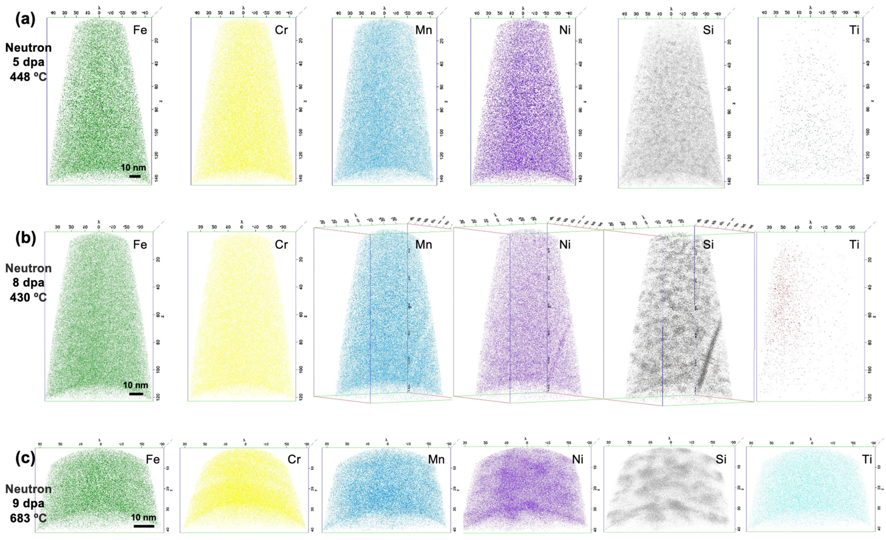
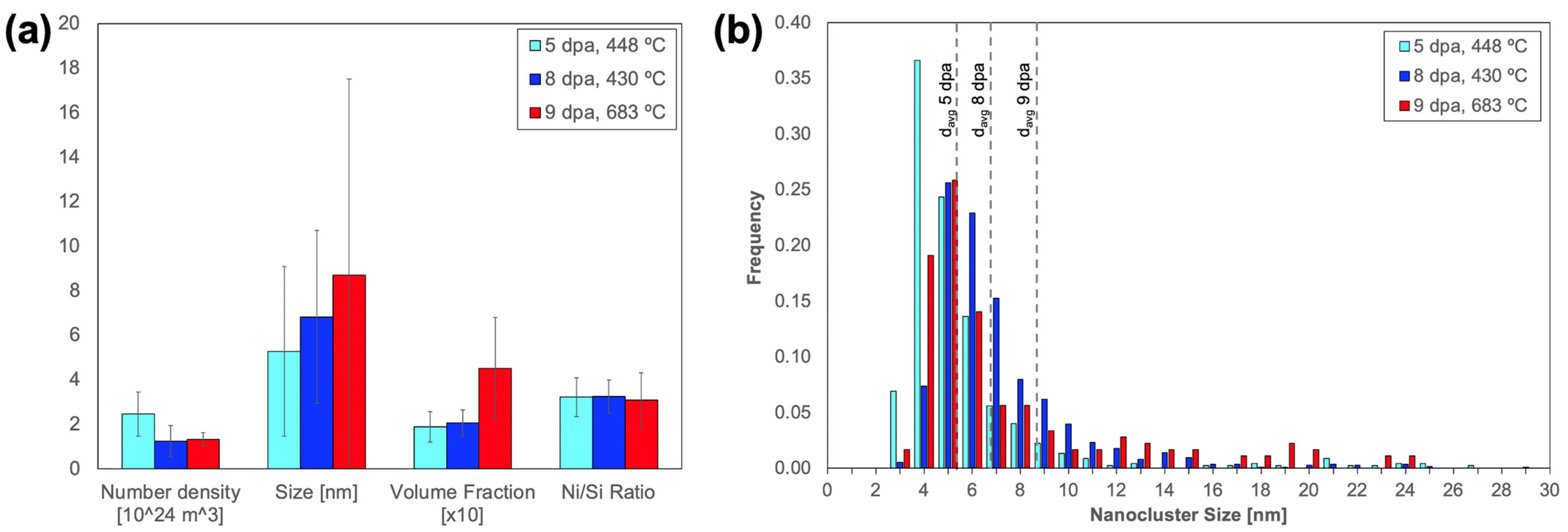
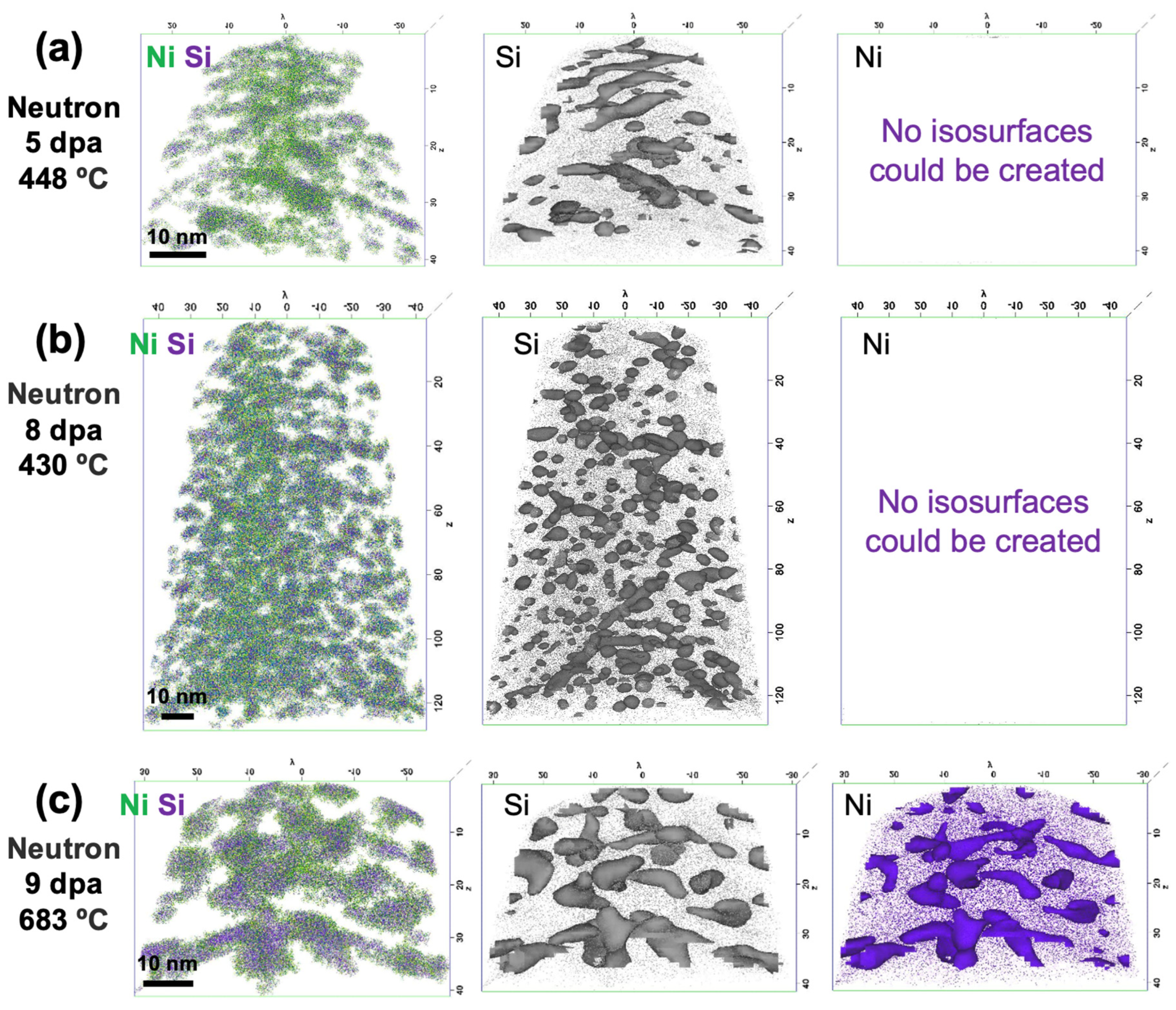
3.2. Neutron-Irradiated
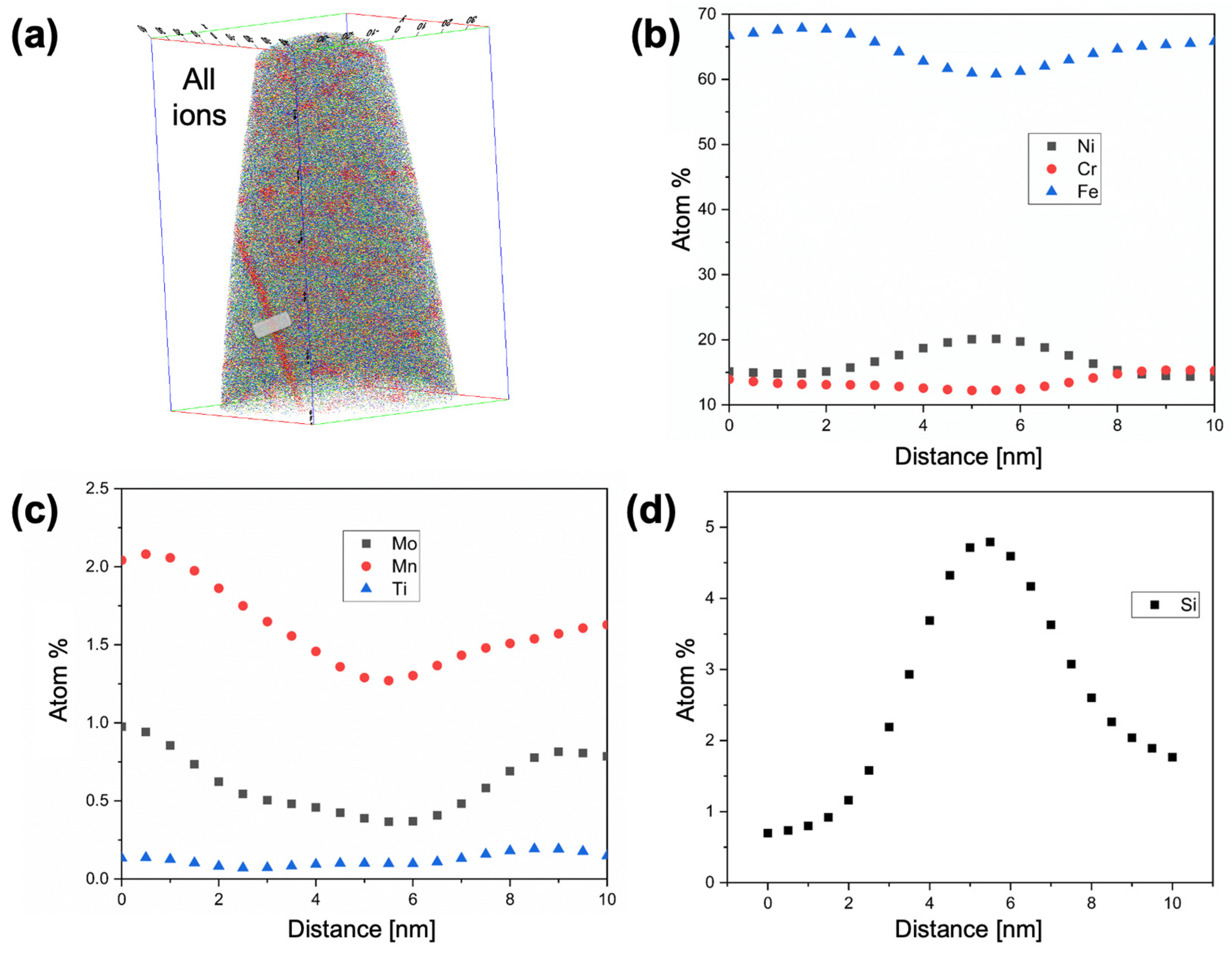
3.3. Radiation Induced Segregation
4. Discussion
5. Conclusions
Author Contributions
Funding
Institutional Review Board Statement
Informed Consent Statement
Data Availability Statement
Acknowledgments
Conflicts of Interest
References
- Zinkle, S.J.; Was, G.S. Materials challenges in nuclear energy. Acta Mater. 2013, 61, 735–758. [Google Scholar] [CrossRef]
- Zinkle, S.J.; Busby, J.T. Structural materials for fission and fusion energy. Mater. Today 2009, 12, 12–19. [Google Scholar] [CrossRef]
- Huang, Y.; Wiezorek, J.M.K.; Garner, F.A.; Freyer, P.D.; Okita, T.; Sagisaka, M.; Isobe, Y.; Allen, T.R. Microstructural characterization and density change of 304 stainless steel reflector blocks after long-term irradiation in EBR-II. J. Nucl. Mater. 2015, 465, 516–530. [Google Scholar] [CrossRef]
- Van Renterghem, W.; Al Mazouzi, A.; Van Dyck, S. Influence of post irradiation annealing on the mechanical properties and defect structure of AISI 304 steel. J. Nucl. Mater. 2011, 413, 95–102. [Google Scholar] [CrossRef]
- Pokor, C.; Brechet, Y.; Dubuisson, P.; Massoud, J.-P.; Averty, X. Irradiation damage in 304 and 316 stainless steels: Experimental investigation and modeling. Part II: Irradiation induced hardening. J. Nucl. Mater. 2004, 326, 30–37. [Google Scholar] [CrossRef]
- Cole, J.I.; Bruemmer, S.M. Post-irradiation deformation characteristics of heavy-ion irradiated 304L SS. J. Nucl. Mater. 1995, 225, 53–58. [Google Scholar] [CrossRef] [Green Version]
- Toyama, T.; Nozawa, Y.; Van Renterghem, W.; Matsukawa, Y.; Hatakeyama, M.; Nagai, Y.; Al Mazouzi, A.; Van Dyck, S. Irradiation-induced precipitates in a neutron irradiated 304 stainless steel studied by three-dimensional atom probe. J. Nucl. Mater. 2011, 418, 62–68. [Google Scholar] [CrossRef]
- Wiezorek, J.M.K.; Huang, Y.; Garner, F.A.; Freyer, P.D.; Sagisaka, M.; Isobe, Y.; Okita, T. Transmission electron microscopy of 304-type stainless steel after exposure to neutron flux and irradiation temperature gradients. Microsc. Microanal. 2014, 20, 1822–1823. [Google Scholar] [CrossRef] [Green Version]
- Mao, K.S.; French, A.J.; Liu, X.; Wu, Y.; Giannuzzi, L.A.; Sun, C.; Dubey, M.; Freyer, P.D.; Tatman, J.K.; Garner, F.A.; et al. Microstructure and microchemistry of laser welds of irradiated austenitic steels. Mater. Des. 2021, 206, 109764. [Google Scholar] [CrossRef]
- Mao, K.S.; Sun, C.; Shiau, C.-H.; Yano, K.H.; Freyer, P.D.; El-Azab, A.A.; Garner, F.A.; French, A.; Shao, L.; Wharry, J.P. Role of cavities on deformation-induced martensitic transformation pathways in a laser-welded, neutron irradiated austenitic stainless steel. Scr. Mater. 2020, 178, 1–6. [Google Scholar] [CrossRef]
- Jayakumar, T.; Mathew, M.D.; Laha, K.; Sandhya, R. Materials development for fast reactor applications. Nucl. Eng. Des. 2013, 265, 1175–1180. [Google Scholar] [CrossRef]
- Mansur, L.K.; Rowcliffe, A.F.; Nanstad, R.K.; Zinkle, S.J.; Corwin, W.R.; Stoller, R.E. Materials needs for fusion, Generation IV fission reactors and spallation neutron sources—Similarities and differences. J. Nucl. Mater. 2004, 329–333, 166–172. [Google Scholar] [CrossRef]
- Crawford, D.C.; Porter, D.L.; Hayes, S.L. Fuels for sodium-cooled fast reactors: US perspective. J. Nucl. Mater. 2007, 371, 202–231. [Google Scholar] [CrossRef]
- Karthik, V.; Murugan, S.; Parameswaran, P.; Venkiteswaran, C.N.; Gopal, K.A.; Muralidharan, N.G.; Saroja, S.; Kasiviswanathan, K.V. Austenitic Stainless Steels for Fast Reactors -Irradiation Experiments. Prop. Eval. Microstruct. Stud. Energy Procedia 2011, 7, 257–263. [Google Scholar] [CrossRef] [Green Version]
- Maziasz, P.J. Overview of microstructural evolution in neutron-irradiated austenitic stainless steels. J. Nucl. Mater. 1993, 205, 118–145. [Google Scholar] [CrossRef]
- Maziasz, P.J. Formation and stability of radiation-induced phases in neutron-irradiated austenitic and ferritic steels. J. Nucl. Mater. 1989, 169, 95–115. [Google Scholar] [CrossRef]
- Garner, F.A.; Toloczko, M.B.; Sencer, B.H. Comparison of swelling and irradiation creep behavior of fcc-austenitic and bcc-ferritic/martensitic alloys at high neutron exposure. J. Nucl. Mater. 2000, 276, 123–142. [Google Scholar] [CrossRef]
- Huang, F.H. Comparison of fracture behavior for low-swelling ferritic and austenitic alloys irradiated in the Fast Flux Test Facility (FFTF) to 180 DPA. Eng. Fract. Mech. 1992, 43, 733–748. [Google Scholar] [CrossRef] [Green Version]
- Pitner, A.L.; Gneiting, B.C.; Bard, F.E. Irradiation Performance of Fast Flux Test Facility Drivers Using D9 Alloy. Nucl. Technol. 1995, 112, 194–203. [Google Scholar] [CrossRef]
- Latha, S.; Mathew, M.D.; Parameswaran, P.; Rao, K.B.S.; Mannan, S.L. Thermal creep properties of alloy D9 stainless steel and 316 stainless steel fuel clad tubes. Int. J. Press. Vessel. Pip. 2008, 85, 866–870. [Google Scholar] [CrossRef]
- Cannon, N.; Huang, F.; Hamilton, M. Transient and Static Mechanical Properties of D9 Fuel Pin Cladding and Duct Material Irradiated to High Fluence, in: Effects of Radiation on Materials. In Proceedings of the 15th Symposium on Effects of Radiation on Materials, Nashville, TN, USA, 17–21 June 1990; pp. 1071–1082. [Google Scholar] [CrossRef]
- Maziasz, P.J. Void swelling resistance of phosphorus-modified austenitic stainless steels during HFIR irradiation at 300–500C to 57 dpa. J. Nucl. Mater. 1993, 200, 90–107. [Google Scholar] [CrossRef]
- Lee, E.H.; Mansur, L.K. Relationships between phase stability and void swelling in Fe-Cr-Ni alloys during irradiation. Metall. Trans. A 1992, 23, 1977–1986. [Google Scholar] [CrossRef]
- Mansur, L.K.; Grossbeck, M.L. Mechanical property changes induced in structural alloys by neutron irradiations with different helium to displacement ratios. J. Nucl. Mater. 1988, 155–157, 130–147. [Google Scholar] [CrossRef]
- MacLean, H.J.; Sridharan, K.; Hyde, T.A. Irradiation Test Plan for the ATR National Scientific User Facility; University of Wisconsin Pilot Project; Idaho National Laboratory: Idaho Falls, ID, USA, 2008. [Google Scholar] [CrossRef] [Green Version]
- Chen, T.; He, L.; Cullison, M.H.; Hay, C.; Burns, J.; Wu, Y.; Tan, L. The correlation between microstructure and nanoindentation property of neutron-irradiated austenitic alloy D9. Acta Mater. 2020, 195, 433–445. [Google Scholar] [CrossRef]
- Wilson, S. As-Run Thermal Analysis for the University of Wisconsin Experiment (ECAR-3186); Idaho National Laboratory: Idaho Falls, ID, USA, 2012.
- Guillen, D.P.; Wharry, J.P.; Housley, G.; Hale, C.D.; Brookman, J.; Gandy, D.W. Irradiation Experiment Design for the Evaluation of PM-HIP Alloys for Nuclear Reactors. Nucl. Eng. Des. 2023, 402, 112114. [Google Scholar] [CrossRef]
- Penisten, J. The Mechanism of Radiation-Induced Segregation in Ferritic-Martensitic Steels. Ph.D. Dissertation, University of Michigan, Ann Arbor, MI, USA, 2012. [Google Scholar]
- Wharry, J.P.; Was, G.S. A systematic study of radiation-induced segregation in ferritic-martensitic alloys. J. Nucl. Mater. 2013, 442, 7–16. [Google Scholar] [CrossRef] [Green Version]
- Wharry, J.P.; Jiao, Z.; Was, G.S. Application of the inverse Kirkendall model of radiation-induced segregation to ferritic–martensitic alloys. J. Nucl. Mater. 2012, 425, 117–124. [Google Scholar] [CrossRef]
- Wharry, J.P.; Jiao, Z.; Shankar, V.; Busby, J.T.; Was, G.S. Radiation-induced segregation and phase stability in ferritic–martensitic alloy T91. J. Nucl. Mater. 2011, 417, 140–144. [Google Scholar] [CrossRef]
- Miller, M.K.; Russell, K.F.; Thompson, K.; Alvis, R.; Larson, D.J. Review of Atom Probe FIB-Based Specimen Preparation Methods. Microsc. Microanal. 2007, 13, 428–436. [Google Scholar] [CrossRef]
- London, A.J. Quantifying Uncertainty from Mass-Peak Overlaps in Atom Probe Microscopy. Microsc. Microanal. 2019, 25, 378–388. [Google Scholar] [CrossRef] [Green Version]
- Hyde, J.M.; Marquis, E.A.; Wilford, K.B.; Williams, T.J. A sensitivity analysis of the maximum separation method for the characterisation of solute clusters. Ultramicroscopy 2011, 111, 440–447. [Google Scholar] [CrossRef] [PubMed]
- Kolli, R.P.; Seidman, D.N. Comparison of Compositional and Morphological Atom-Probe Tomography Analyses for a Multicomponent Fe-Cu Steel. Microsc. Microanal. 2007, 13, 272–284. [Google Scholar] [CrossRef] [Green Version]
- Swenson, M.J.; Wharry, J.P. Collected data set size considerations for atom probe cluster analysis. Microsc. Microanal. 2016, 22, 690–691. [Google Scholar] [CrossRef] [Green Version]
- Swenson, M.J. The Mechanism of Radiation-Induced Nanocluster Evolution in Oxide Dispersion Strengthened and Ferritic-Martensitic Alloys. Ph.D. Dissertation, Boise State University, Boise, ID, USA, 2017. [Google Scholar]
- Williams, C.A.; Haley, D.; Marquis, E.A.; Smith, G.D.W.; Moody, M.P. Defining clusters in APT reconstructions of ODS steels. Ultramicroscopy 2013, 132, 271–278. [Google Scholar] [CrossRef]
- Pareige, P.; Miller, M.K.; Stoller, R.E.; Hoelzer, D.T.; Cadel, E.; Radiguet, B. Stability of nanometer-sized oxide clusters in mechanically-alloyed steel under ion-induced displacement cascade damage conditions. J. Nucl. Mater. 2007, 360, 136–142. [Google Scholar] [CrossRef]
- Miller, M.K.; Forbes, R.G. Atom-Probe Tomography; Springer: Boston, MA, USA, 2014. [Google Scholar] [CrossRef]
- Duh, T.S.; Kai, J.J.; Chen, F.R. Effects of grain boundary misorientation on solute segregation in thermally sensitized and proton-irradiated 304 stainless steel. J. Nucl. Mater. 2000, 283–287, 198–204. [Google Scholar] [CrossRef]
- Hu, R.; Smith, G.D.W.; Marquis, E.A. Effect of grain boundary orientation on radiation-induced segregation in a Fe-15.2 at.% Cr alloy. Acta Mater. 2013, 61, 3490–3498. [Google Scholar] [CrossRef]
- Hu, C.; Berbenni, S.; Medlin, D.L.; Dingreville, R. Discontinuous segregation patterning across disconnections. Acta Mater. 2023, 246, 118724. [Google Scholar] [CrossRef]
- Liu, X.; He, L.; Yan, H.; Bachhav, M.; Stubbins, J.F. A transmission electron microscopy study of EBR-II neutron-irradiated austenitic stainless steel 304 and nickel-base alloy X-750. J. Nucl. Mater. 2020, 528, 151851. [Google Scholar] [CrossRef]
- Lach, T.G.; Olszta, M.J.; Taylor, S.D.; Yano, K.H.; Edwards, D.J.; Byun, T.S.; Chou, P.H.; Schreiber, D.K. Correlative STEM-APT characterization of radiation-induced segregation and precipitation of in-service BWR 304 stainless steel. J. Nucl. Mater. 2021, 549, 152894. [Google Scholar] [CrossRef]
- Jiao, Z.; Hesterberg, J.; Was, G.S. Effect of post-irradiation annealing on the irradiated microstructure of neutron-irradiated 304L stainless steel. J. Nucl. Mater. 2018, 500, 220–234. [Google Scholar] [CrossRef]
- Van Renterghem, W.; Konstantinović, M.J.; Vankeerberghen, M. Evolution of the radiation-induced defect structure in 316 type stainless steel after post-irradiation annealing. J. Nucl. Mater. 2014, 452, 158–165. [Google Scholar] [CrossRef]
- Edwards, D.J.; Simonen, E.P.; Garner, F.A.; Greenwood, L.R.; Oliver, B.M.; Bruemmer, S.M. Influence of irradiation temperature and dose gradients on the microstructural evolution in neutron-irradiated 316SS. J. Nucl. Mater. 2003, 317, 32–45. [Google Scholar] [CrossRef]
- Maziasz, P.J.; McHargue, C.J. Microstructural evolution in annealed austenitic steels during neutron irradiation. Int. Mater. Rev. 1987, 32, 190–219. [Google Scholar] [CrossRef]
- Cawthorne, C.; Brown, C. The occurrence of an ordered FCC phase in neutron irradiated M316 stainless steel. J. Nucl. Mater. 1977, 66, 201–202. [Google Scholar] [CrossRef]
- Brager, H.R.; Garner, F.A. Swelling as a consequence of gamma prime (γ’) and M23(C, Si)6 formation in neutron irradiated 316 stainless steel. J. Nucl. Mater. 1978, 73, 9–19. [Google Scholar] [CrossRef]
- Fukumoto, K.-I.; Mabuchi, T.; Yabuuchi, K.; Fujii, K. Irradiation hardening of stainless steel model alloy after Fe-ion irradiation and post-irradiation annealing treatment. J. Nucl. Mater. 2021, 557, 153296. [Google Scholar] [CrossRef]
- Fukuya, K.; Nakano, M.; Fujii, K.; Torimaru, T.; Kitsunai, Y. Separation of Microstructural and Microchemical Effects in Irradiation Assisted Stress Corrosion Cracking using Post-irradiation Annealing. J. Nucl. Sci. Technol. 2004, 41, 1218–1227. [Google Scholar] [CrossRef]
- Zinkle, S.J.; Maziasz, P.J.; Stoller, R.E. Dose dependence of the microstructural evolution in neutron-irradiated austenitic stainless steel. J. Nucl. Mater. 1993, 206, 266–286. [Google Scholar] [CrossRef]
- Wang-Koh, Y.M. Understanding the yield behaviour of L12-ordered alloys. Mater. Sci. Technol. 2017, 33, 934–943. [Google Scholar] [CrossRef]
- Edwards, D.J.; Simonen, E.P.; Bruemmer, S.M. Evolution of fine-scale defects in stainless steels neutron-irradiated at 275 °C. J. Nucl. Mater. 2003, 317, 13–31. [Google Scholar] [CrossRef]
- Kenik, E.A. Radiation-induced segregation in irradiated Type 304 stainless steels. J. Nucl. Mater. 1992, 187, 239–246. [Google Scholar] [CrossRef]
- Porter, D.L.; Wood, E.L. In-reactor precipitation and ferritic transformation in neutron-irradiated stainless steels. J. Nucl. Mater. 1979, 83, 90–97. [Google Scholar] [CrossRef] [Green Version]
- Kenik, E.A.; Hojou, K. Radiation-induced segregation in FFTF-irradiated austenitic stainless steels. J. Nucl. Mater. 1992, 191–194, 1331–1335. [Google Scholar] [CrossRef]
- Was, G.S.; Wharry, J.P.; Frisbie, B.; Wirth, B.D.; Morgan, D.; Tucker, J.D.; Allen, T.R. Assessment of radiation-induced segregation mechanisms in austenitic and ferritic–martensitic alloys. J. Nucl. Mater. 2011, 411, 41–50. [Google Scholar] [CrossRef]
- Allen, T.R.; Busby, J.T.; Was, G.S.; Kenik, E.A. On the mechanism of radiation-induced segregation in austenitic Fe–Cr–Ni alloys. J. Nucl. Mater. 1998, 255, 44–58. [Google Scholar] [CrossRef]
- Averback, R.S.; Rehn, L.E.; Wagner, W.; Wiedersich, H.; Okamoto, P.R. Kinetics of radiation-induced segregation in Ni-12.7 at.% Si. Phys. Rev. B 1983, 28, 3100–3109. [Google Scholar] [CrossRef]
- Batz, W.; Mead, H.W.; Birchenall, C.E. Diffusion of Silicon in Iron. JOM 1952, 4, 1070. [Google Scholar] [CrossRef] [Green Version]
- Perkins, R.A. Tracer diffusion of63Ni in Fe-17 wt pct Cr-12 wt pct Ni. Metall. Trans. 1973, 4, 1665–1669. [Google Scholar] [CrossRef]
- Arioka, K.; Iijima, Y.; Miyamoto, T. Rapid nickel diffusion in cold-worked type 316 austenitic steel at 360–500 °C. Int. J. Mater. Res. 2017, 108, 791–797. [Google Scholar] [CrossRef]
- Anento, N.; Serra, A.; Osetsky, Y. Effect of nickel on point defects diffusion in Fe–Ni alloys. Acta Mater. 2017, 132, 367–373. [Google Scholar] [CrossRef]
- Gupta, A.; Kumar, D.; Phatak, V. Asymmetric diffusion at the interfaces in Fe/Si multilayers. Phys. Rev. B 2010, 81, 155402. [Google Scholar] [CrossRef] [Green Version]
- Pachaury, Y.; Kumagai, T.; Wharry, J.P.; El-Azab, A. A data science approach for analysis and reconstruction of spinodal-like composition fields in irradiated FeCrAl alloys. Acta Mater. 2022, 234, 118019. [Google Scholar] [CrossRef]
- Sen, A.; Bachhav, M.; Pu, X.; Teng, F.; Yao, T.; Wharry, J.P. Irradiation Effects on Stability of δ-UZr2 phase in U-50 wt% Zr Alloy. J. Nucl. Mater. 2023, 576, 154251. [Google Scholar] [CrossRef]
- Yao, T.; Sen, A.; Wagner, A.; Teng, F.; Bachhav, M.; EI-Azab, A.; Murray, D.; Gan, J.; Hurley, D.H.; Wharry, J.P.; et al. Understanding spinodal and binodal phase transformations in U-50Zr. Materialia 2021, 16, 101092. [Google Scholar] [CrossRef]
- Ke, H.; Wells, P.; Edmondson, P.D.; Almirall, N.; Barnard, L.; Odette, G.R.; Morgan, D. Thermodynamic and kinetic modeling of Mn-Ni-Si precipitates in low-Cu reactor pressure vessel steels. Acta Mater. 2017, 138, 10–26. [Google Scholar] [CrossRef] [Green Version]
- Pareige, C.; Kuksenko, V.; Pareige, P. Behaviour of P, Ni impurities and Cr in self ion irradiated Fe–Cr alloys—Comparison to neutron irradiation. J. Nucl. Mater. 2015, 456, 471–476. [Google Scholar] [CrossRef]
- Pareige, C.; Etienne, A.; Gueye, P.-M.; Medvedev, A.; Kaden, C.; Konstantinovic, M.J.; Malerba, L. Solute rich cluster formation and Cr precipitation in irradiated Fe-Cr-(Ni,Si,P) alloys: Ion and neutron irradiation. J. Nucl. Mater. 2022, 572, 154060. [Google Scholar] [CrossRef]
- Swenson, M.J.; Wharry, J.P. Nanocluster irradiation evolution in Fe-9%Cr ODS and ferritic-martensitic alloys. J. Nucl. Mater. 2017, 496, 24–40. [Google Scholar] [CrossRef]
- Swenson, M.J.; Wharry, J.P. The comparison of microstructure and nanocluster evolution in proton and neutron irradiated Fe-9%Cr ODS steel to 3 dpa at 500C. J. Nucl. Mater. 2015, 467, 97–112. [Google Scholar] [CrossRef] [Green Version]
- Swenson, M.J.; Wharry, J.P. Rate Theory Model of Irradiation-Induced Solute Clustering in b.c.c. Fe-Based Alloys. JOM 2020, 72, 4017–4027. [Google Scholar] [CrossRef]
- Patki, P.V.; Pownell, T.J.; Bazarbayev, Y.; Zhang, D.; Field, K.G.; Wharry, J.P. Systematic study of radiation-induced segregation in neutron-irradiated FeCrAl alloys. J. Nucl. Mater. 2022, 574, 154205. [Google Scholar] [CrossRef]
- Tissot, O.; Pareige, C.; Meslin, E.; Décamps, B.; Henry, J. Influence of injected interstitials on α′ precipitation in Fe–Cr alloys under self-ion irradiation. Mater. Res. Lett. 2017, 5, 117–123. [Google Scholar] [CrossRef] [Green Version]

| Method | Fe | Ni | Cr | Mo | Mn | Si |
| Nominal [25] | Bal. | 16.27 | 13.35 | 1.55 | 1.70 | – |
| EDS [26] | Bal. | 17.3 ± 0.3 | 12.86 ± 0.07 | 1.27 ± 0.06 | 1.87 ± 0.03 | 1.25 ± 0.05 |
| APT | 65.6 | 15.2 | 13.95 | 2.20 | 2.03 | 0.66 |
| Method | Ti | Nb | V | Al | Cu | Co |
| Nominal [25] | – | – | 0.02 | 0.02 | 0.01 | – |
| EDS [26] | 0.11 ± 0.03 | 0.01 ± 0.01 | – | 0.09 ± 0.02 | – | – |
| APT | 0.26 | 0.02 | 0.02 | 0.014 | 0.005 | 0.014 |
| Irradiating Particle | Dose [dpa] | Temperature [°C] |
|---|---|---|
| Fast neutrons | 5 | 448 ± 20 |
| Fast neutrons | 8 | 430 ± 20 |
| Fast neutrons | 9 | 683 ± 20 |
| 2 MeV protons | 7 | 498 ± 7.5 |
| Irradiation Condition | RHIT File Name | Laser Energy [NJ] | No. of Ions [million] | Pulse Rate [kHz] | Detection Rate [%] | Base Temp. [K] | dmax [nm] | Nmin [ions] | No. of Clusters |
|---|---|---|---|---|---|---|---|---|---|
| As Received | 07660 | 0.1 | 2 | 200 | 0.5 | 40.3 | n/a | n/a | n/a |
| 07731 | 0.1 | 9.5 | 250 | 0.5 | 60.5 | n/a | n/a | n/a | |
| 07596 | 0.1 | 1.2 | 200 | 0.5 | 55 | n/a | n/a | n/a | |
| Neutron, 5 dpa, 448 °C | 07273 | 0.08 | 5 | 200 | 0.5 | 49.7 | 0.56 | 34 | 141 |
| 07274 | 0.08 | 1.5 | 200 | 0.5 | 49.7 | 0.54 | 31 | 39 | |
| 07279 | 0.1 | 2 | 200 | 1.0 | 49.7 | 0.58 | 39 | 69 | |
| 07597 | 0.1 | 2.2 | 200 | 0.5 | 55 | 0.60 | 32 | 70 | |
| 07662 | 0.1 | 6.5 | 200 | 0.5 | 39.5 | 0.60 | 41 | 129 | |
| 07598 | 0.14 | 50 | 200 | 1.0 | 55.1 | 0.60 | 40 | 271 | |
| Neutron, 8 dpa, 430 °C | 07605 | 0.14 | 35 | 200 | 0.8 | 54.9 | 0.63 | 81 | 351 |
| 07606 | 0.14 | 48 | 200 | 0.5 | 55 | 0.61 | 68 | 706 | |
| Neutron, 9 dpa, 683 °C | 07267 | 0.07 | 2.25 | 200 | 0.5 | 56.1 | 0.49 | 27 | 66 |
| 07269 | 0.1 | 9 | 200 | 0.5 | 48.5 | 0.52 | 29 | 59 | |
| 07270 | 0.1 | 1.3 | 200 | 0.7 | 49.5 | 0.53 | 22 | 20 | |
| 07271 | 0.1 | 1.5 | 200 | 0.5 | 49.6 | 0.41 | 25 | 18 | |
| 07272 | 0.08 | 8 | 200 | 0.5 | 49.6 | 0.50 | 22 | 15 | |
| Proton, 7 dpa, 500 °C | 07055 | 0.1 | 6 | 200 | 0.5 | 49.7 | n/a | n/a | n/a |
| 07056 | 0.1 | 5.5 | 200 | 0.5 | 49.7 | n/a | n/a | n/a | |
| 07057 | 0.1 | 6 | 200 | 0.5 | 49.6 | n/a | n/a | n/a | |
| 07058 | 0.06 | 0.8 | 200 | 0.5 | 49 | n/a | n/a | n/a | |
| 07068 | 0.07 | 3.75 | 200 | 0.5 | 49.7 | n/a | n/a | n/a |
| Irradiation | Number Density [1024 m3] | Size [nm] | Volume Fraction | Average Composition [at%] | ||||||||
|---|---|---|---|---|---|---|---|---|---|---|---|---|
| Fe | Ni | Cr | Mo | Mn | Si | Ti | C | Ni/Si Ratio | ||||
| 5 dpa 448 °C | 2.45 ± 0.99 | 5.3 ± 3.8 | 0.19 ± 0.07 | 56.58 | 22.53 | 11.36 | 0.60 | 1.48 | 7.00 | 0.22 | 0.03 | 3.22 |
| 8 dpa 430 °C | 1.24 ± 0.71 | 6.8 ± 3.9 | 0.21 ± 0.06 | 52.84 | 25.52 | 10.91 | 0.84 | 1.45 | 7.85 | 0.28 | 0.02 | 3.25 |
| 9 dpa 683 °C | 1.31 ± 0.31 | 8.7 ± 8.8 | 0.45 ± 0.23 | 45.71 | 32.74 | 8.61 | 0.48 | 1.04 | 10.64 | 0.47 | 0.02 | 3.08 |
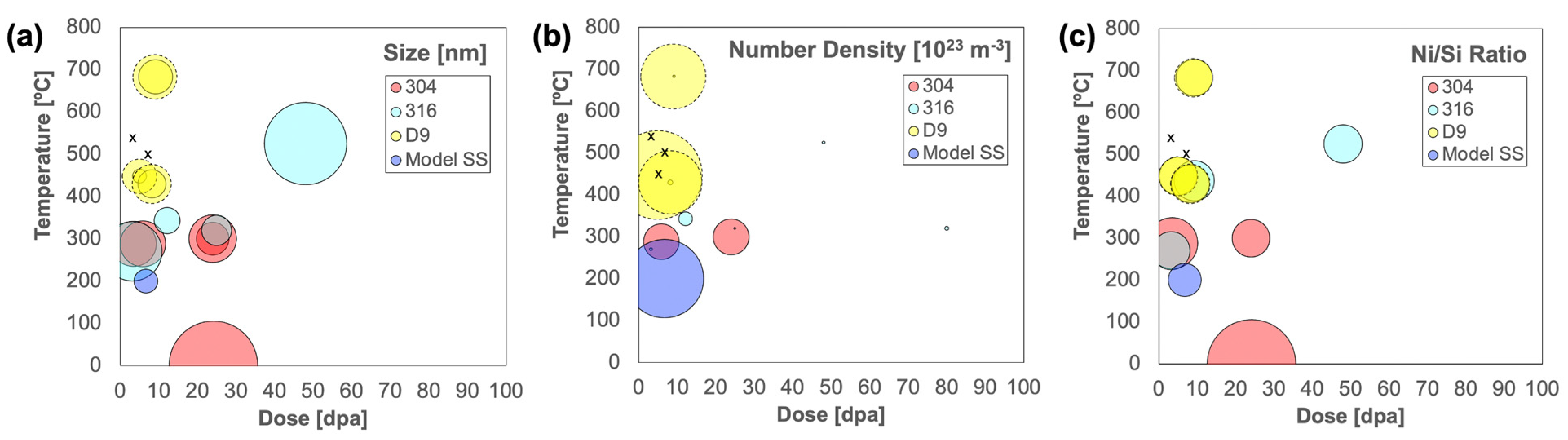
| Alloy | Si Conc. [wt%] | Irradiating Particle | Dose [dpa] | Temp. [°C] | Average Size [nm] | No. Density [1024 m−3] | Ni/Si Ratio | Technique | Ref. |
|---|---|---|---|---|---|---|---|---|---|
| 304 | 0.57 | n (EBR-II) | 24.15 | 371–389 | ~35 | – | 16.5 | TEM | [45] |
| 304 | 0.45 | n | 3.5 | 288 | ~8–10 | – | 5.5 | APT | [46] |
| 304 | 0.30 | n (Barsebäck) | 5.9 | 288 | 9.2 ± 0.7 | 0.39 ± 0.06 | – | APT | [47] |
| 304 | 0.78 | n (Chooz A) | 24 | 300 | 4.7 | – | – | TEM | [4] |
| 304 | 0.78 | n | 24 | 300 | 10 | 0.4 | 3 | TEM | [7] |
| 316 | 0.53 | n (Tihange) | 80 | 320 | – | 0.005 | – | TEM | [48] |
| 316 | 0.72 | n (Tihange) | 12.2 | 343 | 3 | 0.06 | – | TEM | [49] |
| 316 | n.s. | n | 9.2 | 425–450 | – | – | 3.4 | TEM | [50] |
| 316 | n.s. | n (Dounreay) | 3.2 | 270–540 | 8–23 | 0.0008–0.0042 | ~3 | TEM | [51] |
| 316 | n.s. | n (Dounreay) | 3.2 | ≥540 | n.o. | n.o. | n.o. | TEM | [51] |
| 316 | 0.51 | n (EBR-II) | 48 | 525 | 30 | ~0.001–0.003 | 3.14 | TEM | [52] |
| 316 | 0.62 | n | 25 | 320 | 4 | 0.001 | – | TEM | [54] |
| Fe-17Cr-12Ni-1Si | 0.96 | 6.4 MeV Fe3+ | 2–6.7 | 200 | 2.5 | 1.8–2 | 2.1–2.5 | APT | [53] |
| D9 | 0.66 | Neutron | 5 | 448 | <1 | – | 3 | TEM | [26] |
| D9 | 0.66 | Neutron | 8.2 | 430 | 3.5 ± 0.1 | 0.008 ± 0.003 | 3 | TEM | [26] |
| D9 | 0.66 | Neutron | 9.2 | 683 | 5.1 ± 0.3 | 0.002 ± 0.001 | <3 | TEM | [26] |
| D9 | 0.66 | Neutron | 5 | 448 | 5.27 ± 3.81 | 2.45 ± 0.99 | 3.22 ± 0.87 | APT | This study |
| D9 | 0.66 | Neutron | 8 | 430 | 6.82 ± 3.88 | 1.24 ± 0.71 | 3.25 ± 0.74 | APT | This study |
| D9 | 0.66 | Neutron | 9 | 683 | 8.70 ± 8.81 | 1.31 ± 0.31 | 3.08 ± 1.24 | APT | This study |
| D9 | 0.66 | Proton | 7 | 500 | n.o. | n.o. | n.o. | APT | This study |
Disclaimer/Publisher’s Note: The statements, opinions and data contained in all publications are solely those of the individual author(s) and contributor(s) and not of MDPI and/or the editor(s). MDPI and/or the editor(s) disclaim responsibility for any injury to people or property resulting from any ideas, methods, instructions or products referred to in the content. |
© 2023 by the authors. Licensee MDPI, Basel, Switzerland. This article is an open access article distributed under the terms and conditions of the Creative Commons Attribution (CC BY) license (https://creativecommons.org/licenses/by/4.0/).
Share and Cite
Mullurkara, S.V.; Bejawada, A.; Sen, A.; Sun, C.; Bachhav, M.; Wharry, J.P. Nanocluster Evolution in D9 Austenitic Steel under Neutron and Proton Irradiation. Materials 2023, 16, 4852. https://doi.org/10.3390/ma16134852
Mullurkara SV, Bejawada A, Sen A, Sun C, Bachhav M, Wharry JP. Nanocluster Evolution in D9 Austenitic Steel under Neutron and Proton Irradiation. Materials. 2023; 16(13):4852. https://doi.org/10.3390/ma16134852
Chicago/Turabian StyleMullurkara, Suraj Venkateshwaran, Akshara Bejawada, Amrita Sen, Cheng Sun, Mukesh Bachhav, and Janelle P. Wharry. 2023. "Nanocluster Evolution in D9 Austenitic Steel under Neutron and Proton Irradiation" Materials 16, no. 13: 4852. https://doi.org/10.3390/ma16134852